


Glenohumeral joint
- 2. • DEFINITION; The glenohumeral joint is a balland-socket type of synovial joint that permits a wide range of movement; however the mobility makes the joint relatively unstable. • Joint stability is provided, instead, by the rotator cuff muscles, the long head of the biceps brachii muscle, related bony processes, and extra-capsular ligaments. 2/10/2014 2
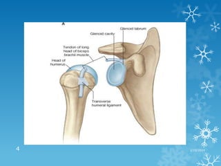
- 3. • ARTICULATION; The articular surfaces of the glenohumeral joint are the large spherical head of the humerus and the small glenoid cavity of the scapula. • Each of the surfaces is covered by hyaline cartilage. • The glenoid cavity is deepened and expanded peripherally by a fibro-cartilaginous collar (the glenoid labrum), which attaches to the margin of the fossa. • Superiorly, this labrum is continuous with the tendon of the long head of the biceps brachii muscle, which attaches to the supraglenoid tubercle and passes through the articular cavity superior to the head of the humerus. 2/10/2014 3
- 4. 4 2/10/2014
- 5. • CAPSULE; a loose fibrous capsule surrounds the glenohumeral joint and is attached; • Medially to the margin of the glenoid cavity. • Laterally to the anatomical neck of the humerus. • Superiorly, the capsule encroaches on the root of the coracoid process so that the fibrous capsule encloses the proximal attachment of the long head of biceps brachiisupraglenoid tubercle of scapula. 2/10/2014 5
- 6. 2/10/2014 6
- 7. • LIGAMENTS; the fibrous membrane of the joint capsule is thickened and strengthened by ligaments. a. The glenohumeral ligaments are three fibrous bands. Anterosuperiorly found in three locations to form superior, middle, and inferior glenohumeral ligaments, which pass between the superomedial margin of the glenoid cavity to the lesser tubercle and inferiorly related anatomical neck of the humerus. 2/10/2014 7
- 8. b. The coracohumeral ligament; found superiorly between the base of the coracoid process and the greater tubercle of the humerus. c. The transverse humeral ligament; a broad fibrous band that runs more or less obliquely from the greater to the lesser tubercle of the humerus, bridging over the intertubercular groove. The ligament converts the groove into a canal that holds the synovial sheath and tendon of the biceps brachii in place during movements of the glenohumeral joint. 2/10/2014 8
- 10. INNERVATION • • • • • The Glenohumeral Joint is innervated by; The suprascapular, Axillary, Lateral pectoral and Branches from the posterior cord of brachial plexus. 2/10/2014 10
- 11. MOVEMENTS OF THE GLENOHUMERAL JOINT • The glenohumeral joint has more freedom of movement than any other joint in the body. • The glenohumeral joint allows movements around three axes and permits: • Flexion-extension • Abduction-adduction • Rotation (medial and lateral) of the humerus • Circumduction 2/10/2014 11
- 12. MUSCLES INVOLED IN THE MOVEMENTS • Flexion; clavicular part of the pectorialis major. • Anterior fibre of deltoid muscle. • Coracobrachialis. • Biceps brachii. Both are synergist. • Extenson; posterior part of the deltoid muscle assisted by the teres major muscle. 2/10/2014 12
- 13. • Abduction; the central part of the deltoid muscle, • Synergized by the supraspinatus muscle. • Adduction; pectorialis major and latissimus dorsi, • Assisted by the subscapularis, infraspinatus and teres minor muscles. 2/10/2014 13
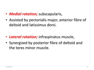
- 14. • Medial rotation; subscapularis, • Assisted by pectorialis major, anterior fibre of deltoid and latissimus dorsi. • Lateral rotation; infraspinatus muscle, • Synergized by posterior fibre of deltoid and the teres minor muscle. 2/10/2014 14
- 15. Note; • Joint stability is provided by surrounding muscle tendons and a skeletal arch formed superiorly by the coracoid process, acromion and the coraco-acromial ligament. • Tendons of the rotator cuff muscles (the supraspinatus, infraspinatus, teres minor, and subscapularis muscles) blend with the joint capsule and form a musculotendinous collar that surrounds the posterior, superior, and anterior aspects of the glenohumeral joint . 2/10/2014 15
- 16. • This cuff of muscles stabilizes and holds the head of the humerus in the glenoid cavity of the scapula without compromising the arm's flexibility and range of motion. • The tendon of the long head of the biceps brachii muscle passes superiorly through the joint and restricts upward movement of the humeral head on the glenoid cavity. 2/10/2014 16
- 17. 17 2/10/2014
- 18. BURSAE AROUND THE GLENOHUMERAL JOINT • Several bursae containing capillary films of synovial fluid are found within the vicinity of the glenohumeral joint. • Bursae are located where tendons rub against bone, ligaments, or other tendons and where skin moves over a bony prominence. • Subscapular Bursa; is located between the tendon of the subscapularis and the neck of the scapula. • The bursa protects the tendon where it passes inferior to the root of the coracoid process and over the neck of the scapula. • It usually communicates with the cavity of the shoulder joint through an opening in the fibrous capsule; thus, it is really an extension of the glenohumeral joint cavity. 2/10/2014 18
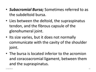
- 19. • Subacromial Bursa; Sometimes referred to as the subdeltoid bursa. • Lies between the deltoid, the supraspinatus tendon, and the fibrous capsule of the glenohumeral joint. • Its size varies, but it does not normally communicate with the cavity of the shoulder joint. • The bursa is located inferior to the acromion and coracoacromial ligament, between them and the supraspinatus. 2/10/2014 19
- 20. BLOOD SUPPLY • Vascular supply to the glenohumeral joint is predominantly through branches of the anterior and posterior circumflex humeral and suprascapular arteries through the scapula anastomosis. 2/10/2014 20
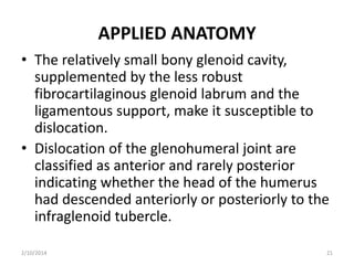
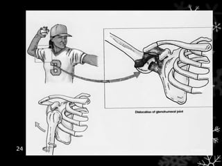
- 21. APPLIED ANATOMY • The relatively small bony glenoid cavity, supplemented by the less robust fibrocartilaginous glenoid labrum and the ligamentous support, make it susceptible to dislocation. • Dislocation of the glenohumeral joint are classified as anterior and rarely posterior indicating whether the head of the humerus had descended anteriorly or posteriorly to the infraglenoid tubercle. 2/10/2014 21
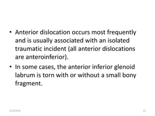
- 22. • Anterior dislocation occurs most frequently and is usually associated with an isolated traumatic incident (all anterior dislocations are anteroinferior). • In some cases, the anterior inferior glenoid labrum is torn with or without a small bony fragment. 2/10/2014 22
- 23. • Once the joint capsule and cartilage are disrupted, the joint is susceptible to further (recurrent) dislocations. • When an anteroinferior dislocation occurs, the axillary nerve may be injured by direct compression of the humeral head on the nerve inferiorly as it passes through the quadrangular space. • Axillary nerve injury is indicated by paralysis of the deltoid and loss of sensation in a small area of skin covering the central part of the deltoid. 2/10/2014 23
- 24. 24 2/10/2014
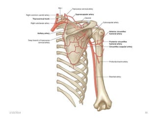
- 26. • The anastomosis around the scapula is an arterial anastomosis around both surfaces of scapula bone. • Between the branches of subclavian and axillary arteries. • Arteries taking part in this anastomosis include; 2/10/2014 26
- 27. 1. The suprascapular artery from the thyrocervical trunk of the first part of the subclavian artery. The artery reaches the upper border of the scapula and passes above suprascapular ligament to reach the supraspinatus fossa, then it curves around the spinoglenoid notch to reach infraspinatus fossa. 2/10/2014 27
- 28. 2. The deep branch of transverse cervical artery which is also a branch of the thyrocervical trunk. The artery descends along medial border of scapula deep to the levator scapulae and rhomboids (sometimes the artery may arise from the third part of the subclavian artery and is known as dorsal scapular artery. 2/10/2014 28
- 29. 3. The subscapular artery from third part of axillary artery. Its circumflex scapular branches passes between the two origins of teres minor muscle, enters infraspinous fossa. 2/10/2014 29
- 30. 2/10/2014 30
- 31. 2/10/2014 31
- 32. • FUNCTIONAL IMPORTANCE a. the anastomosis provides sufficient amount of blood to the scapula muscles and upper extremity during movement of the shoulder joint from lateral border of scapula on its dorsal surface. b. In the case of blockage of main arterial trunk distal to the origin of the thyrocervical trunk and proximal to the origin of subscapular artery, this anastomoses provides alternative route for the supply of blood to the upper extremity. 2/10/2014 32