Haematopoiesis
•
9 likes•5,272 views

The document discusses hematopoiesis, the production and development of blood cells. It begins in the fetal liver and spleen and later continues in the bone marrow. There are two types of hematopoiesis - medullary, which occurs in the bone marrow, and extramedullary, which can occur in other tissues like the liver and spleen. The process is regulated by hematopoietic growth factors and involves stem cell differentiation down myeloid or lymphoid lineages to produce the various mature blood cell types.
Report
Share


Haematopoiesis
- 3. Hemo: Referring to blood cells Poiesis: “The development or production of” The word Hematopoiesis refers to the production & development of all the blood cells: ◦ Erythrocytes: Erythropoiesis ◦ Leucocytes: Leucopoiesis ◦ Thrombocytes: Thrombopoiesis. Begins in the 20th week of life in the fetal liver & spleen, continues in the bone marrow till young adulthood & beyond!
- 4. Medullary ◦ Origin of blood cells and sequential sites of normal blood production within the bone marrow Extramedullary ◦ Blood cell production in hematopoietic tissue other than bone marrow Liver Spleen ◦ Compensatory mechanism to provide blood cells in times of need
- 5. AGE SITE Fetus: 0-2 months Yolk sac 2-7 months Liver, spleen 5-9 months Bone marrow Infants Bone marrow, practically all bones Adults Vertebrae, ribs, sternum, sacrum and pelvis, proximal ends of femur
- 8. Suitable environment for SC growth & development. Composed of stromal cells + microvascular network. Stromal cells: • Adipocytes • Fibroblast • Reticular cells • Endothelial cells • Macrophages Extracellular molecules: • Collagen • Glycoprotein (fibronectin, thrombospondin) • Glycosaminoglycans (hyaluronic acid & chondroitin derivates) • Growth factors for cell survival
- 9. Glycoprotein hormones regulate proliferation & differentiation of HPC & function of mature blood cells. Biological effects of HGF mediated through specific receptors on target cells. Act: ◦ Locally at the site where they are produce by cell-cell contact. ◦ Circulate in plasma
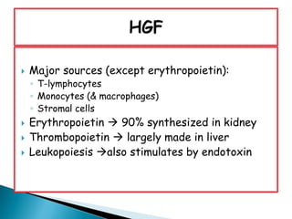
- 10. Major sources (except erythropoietin): ◦ T-lymphocytes ◦ Monocytes (& macrophages) ◦ Stromal cells Erythropoietin 90% synthesized in kidney Thrombopoietin largely made in liver Leukopoiesis also stimulates by endotoxin
- 11. Synthesized by peritubular cells of kidney in response to hypoxemia Present in minute amounts in urine Liver secretes 10% of endogenous erythropoietin. Responsible for low level erythroid activity. Half life of 6-9 hrs. in anemic patient
- 12. 1. Impaired O2 delivery to the kidney resulting to decreased red cell mass 2. Impaired O2 loading of the hemoglobin molecule 3. Impaired blood flow to the kidney
- 13. It is a glycoprotein hormone produced mainly by liver and kidney that regulates the production of platelets in bone marrow. It stimulates the production and differentiation of Megakaryocytes
- 14. 3. GM-CSF: Produced by fibroblasts, stromal cells,T.lymphocytes and endothelial cells. Stimulate progenitors for granulocytes, monocytes and erythrocytes
- 15. 4. G-CSF: LMW glycoprotein Stimulates proliferation and maturation of granulocyte precursors. Produced by stromal cells, monocytes, macrophages, and endothelial cells.
- 16. 5.M-CSF: Secreted by stromal cells, macrophages and fibroblasts. Heavily glycosylated glycoprotein Potent stimulator of macrophage function and activation as it increases the expression of MHC.II antigen on macrophages.
- 17. Site of action HGF Stromal cell IL-1, TNF Pluripotential stem cell Stem cell factor (SCF), Flt ligand (Flt-L) Multipotential progenitor cell IL-3, GM-CSF, IL-6, G-CSF, thrombopoietin Committed progenitor cell G-CSF, M-CSF, IL-5 (eosinophil- CSF), erythropoietin, thrombopoietin
- 18. Binding of GF to its receptor activates JAKs then phosphorylate STATs which translocate to the nucleus and activate transcription of specific genes
- 20. Homeostasis: - cell production ↔ cell destruction Regulated process of physiological cell death triggered to activate intracellular proteins lead to cell death. Important proses for maintaining tissue homeostasis in hematopoiesis & lymphocytes development
- 21. Apoptosis results from the action of intracellular cysteine proteases called CASPASES which are activated following cleavage and lead to endonuclease digestion of DNA and disintegration of the cell skeleton.
- 22. There are two major pathways by which CASPASES can be activated: 1. By signaling through membrane proteins such as Fas or TNF receptor via their intracellular death domain. 2. Via the release of cytochrome c from mitochondria. Cytochrome c binds to APAF‐1 which then activates caspases.
- 24. Apoptosis have the Following steps: 1) Cell shrinkage 2) Organelle reduction 3) Mitochondrial leakage 4) Chromatin condensation 5) Nuclear fragmentation 6) Membrane blebbing & changes
- 26. Differentiate into multiple cell lines. Proliferation is under influence of hematopoietic growth factors present in reticuloendothelial system. Morphologically they resemble large immature lymphocytes Exact phenotype unkown immunological testing: CD34+, CD38- 1 SC capable of producing + 106 mature blood cells after 20 cell divisions.
- 27. Pluripotent Stem cells undergo several divisions and become committed unipotent cells(CFU) to either myeloid or lymphoid lineage Committed stem cells lose self-renewal capability but retain the potential to differentiate They acquire distinguishing cell surface receptors and respond to specific signals and growth factors
- 28. Myeloid lineage stem cells mature in bone marrow to become : Erythrocytes/red blood cells Granulocytes including segmented neutrophils, eosinophils, and basophils Monocytes/macrophages Platelets/thrombocytes
- 29. Lymphoid lineage stem cells will become either a pre- Bcell or a prothymocyte(pre-Tcell) and will travel to the Thymus and lymph nodes for further maturation into lymphocytes
- 30. Myeloid and lymphoid cells are progenitor cells. Both cell types originate from hematopoietic stem cells. Both cell types are produced in bone marrows. Both cell types produce different types of daughter cells.
- 31. LymphoidMyeloid Lymphoid cells are daughter cells of hematopoietic stem cells which produce lymphocytes. Myeloid cells are daughter cells of hematopoietic stem cells which give rise to several other types of blood cells. Definition T cells, B cells, and natural killer cells (NK). Monocytes, macrophages, neutrophils, basophils, eosinophils, erythrocytes, dendritic cells, megakaryocyte, and platelets. Daughter cells
- 32. Changes in cytoplasm Changes in nucleus Reduction in cell size Gradual transformation Changes are simultaneous and parallel
- 33. Loss of basophilia: ◦ Romanowsky staining (Giemsa) ◦ The more basophilic cytoplasma, the more immature cell Cytoplasmic granules in granulocytes ◦ Azurophil (primary granules) ◦ Specific (secondary granules): Eosinophilic Basophilic Neutrophilic
- 34. Elaboration of hemoglobin: blue cytoplasm in pronormoblast and red cytoplasm in erythrocyte in pronormoblast
- 35. Nuclear Maturation: 1. Shape ◦ Round or oval in blasts ◦ Striking changes in granulocytes 2. Structure delicate netlike or sponge like ◦ chromatin in blasts ◦ Chromatin strands become more coarse and clumped as the cells matures ◦ Reduction in the number of nucleoli
- 36. A feature of all cells, except in the megakaryocytic series. N/C ratio is high in young cells and low in mature cells.
- 37. Asynchronous maturation of cytoplasma and nucleus atypical cells. Granulocytes: - A granular - Persistent primary granules - Abnormal inclusions - Large nucleus - Hypersegmentation/hyposegmentation Erythrocytes: - persistent basophilia