heart anatomy pdf.pdf
- 1. ANATOMY AND PHYSIOLOGY OF HEART ANATOMY AND PHYSIOLOGY OF HEART
- 2. THE HEART The heart is a chambered muscular organ that pumps blood received from the veins into the arteries, and maintains flow of blood through the entire circulatory system. It has roughly the size of closed fist. It lies in the mediastinum behind sternum between 4th and 8th thoracic vertebrae. The weight of the heart is 225 to 300 gms in adults. Heart is transverse in shape for children but it has normal pyramidal shape in adults by 25 years.
- 6. LOCATION OF HEART The human heart is located within the thoracic cavity, medially between the lungs in the space known as the mediastinum. Within the mediastinum, the heart lies in its own space called the pericardial cavity. The base of the heart is located at the level of the third costal cartilage. The inferior tip of the heart, the apex, lies just to the left of the sternum between the junction of the fourth and fifth ribs.
- 7. Cont.. The slight deviation of the apex to the left is reflected in a depression in the medial surface of the inferior lobe of the left lung, called the cardiac notch.
- 8. COVERINGS OF HEART The heart is surrounded by membrane called Pericardium. The pericardium lies within the middle mediastinum. Its function is to restrict excessive movements of the heart as a whole and to serve as a lubricated container in which the different parts of the heart can contract.
- 9. STRUCTURE OF HEART WALL
- 11. LAYERS OF HEART WALL Epicardium: The outer layer of the heart. The term epicardium means “on the heart”. This is the visceral layer of the pericardium called as serous pericardium.
- 12. Cont.. Myocardium: a. This is the thick, contractile, middle layer of the heart. b. They possess interconnected muscle cell junctions called syncytium for the electrical connections to work. It can pass an action potential from fiber to fiber. c. The interconnected muscle fibers also helps to hold the high pressured blood strongly inside the heart. d. Myocardium is damaged in case of MI and cardiac arrest.
- 13. Cont.. Endocardium: a. It is innermost layer of heart and made of endothelial tissues. b. It also cover the beam like projections of myocardial tissue. These projections are called trabeculae carneae and it helps to add force to the contraction of heart. c. Inward folds or pockets formed by the endocardium and connective tissues are called valves (AV & SL) which prevents the return flow of the blood. There are tricuspid valves and bicuspid valves.
- 15. CHAMBERS OF HEART The heart consists of four chambers. 1. Right atrium: Two large veins deliver de- oxygenated blood to the right atrium. The superior vena cava carries blood from the upper body. The inferior vena cava brings blood from the lower body. Then the right atrium pumps the blood to right ventricle. 2. Right ventricle: The lower right chamber pumps the de-oxygenated blood to the lungs through the pulmonary artery. The lungs reload blood with oxygen.
- 16. Cont.. 3. Left atrium: After the lungs fill blood with oxygen, the pulmonary veins carry the blood to the left atrium. This upper chamber pumps the blood to left ventricle. 4. Left ventricle: The left ventricle is slightly larger than the right. It pumps oxygen-rich blood to the rest of body.
- 17. THE RIGHT ATRIUM Right border of the heart. It receives blood from three veins; superior vena cava, inferior vena cava and coronary sinus. The valve between right atrium and right ventricle is called tricuspid valve/right atrioventricular valve. It is made of dense cusps of connective tissue.
- 19. THE RIGHT VENTRICLE Receives blood from the right atrium through the tricuspid valve . The edges of the valve cusps are attached to chordae tendineae which are, in turn, attached below to papillary muscles. The wall of the right ventricle is thicker than that of the atria and contains a mass of muscular bundles known as trabeculae carneae. Blood flows through the valve and into the pulmonary arteries via the pulmonary trunk to be oxygenated in the lungs.
- 21. The Left Atrium Receives oxygenated blood from four pulmonary veins. The walls are same thick as right atrium. The mitral (bicuspid) / left atrioventricular valve guards the passage of blood from the left atrium to the left ventricle.
- 22. The Left Atrium
- 23. The Left Ventricle The wall of the left ventricle is thicker than the right ventricle but the structure is similar. This is the thickest chamber of the heart. It forms the apex of the heart. Like the right ventricle it also contains Trabeculae carneae and chordae tendineae that anchor the cusps to papillary muscles. Blood pass from the aortic valve/semilunar valve into the ascending aorta. A branch of the ascending aorta called coronary artery provides blood supply to the cardiac tissues.
- 25. The Heart Valves Atrio-ventricular valves Right AV (Tricuspid): Separates the right atrium from the right ventricle. Prevents backflow into atrium. Left AV (Bicuspid): Separates the left atrium from the left ventricle. Prevents backflow into atrium. Semi-lunar valves Pulmonary valve: Separates the right ventricle from the pulmonary arteries. Prevents backflow after ventricular contraction. Aortic valve: Separates the left ventricle from the aorta. Prevents backflow after ventricular contraction.
- 28. Blood flow through the heart The blood flow starts from right atrium. Then it flows to right ventricle through atrio-ventricular / tricuspid valve. From right ventricle it flows through pulmonary semilunar valves into the pulmonary artery and then to pulmonary trunk. The pulmonary trunk branches to form right and left pulmonary artery for gas exchange at left and right lungs. From lungs the oxygenated blood comes to left atrium through pulmonary veins. From left atrium blood flows through mitral valve/left atrio-ventricular/bicuspid valve to left ventricle.
- 29. Cont.. From left ventricle blood is pumped to aorta through aortic valve then the supply goes to different branches to different parts of the body. The venous drainage from different parts of the body brings deoxygenated blood back to right atrium through inferior vena cava and superior vena cava.
- 32. Arterial Supply of the Heart Myocardial cells receive blood supply from coronary arteries both right and left. The closure of aortic valve during ventricular relaxation prevents the backflow of the blood and fills the coronary artery. The arterial supply of the heart is provided by the right and left coronary arteries, which arise from the ascending aorta immediately above the aortic valve.
- 33. Branches of Coronary Arteries 1. Right Coronary artery: Branches Right marginal arteries. Posterior descending artery 2. Left Coronary artery: Branches Circumflex artery. Left Marginal artery. Left anterior descending artery Diagonal branches
- 35. Venous Drainage of the Heart Most venous blood from the coronary capillaries drain into the coronary sinus ,which lies in the posterior part of the atrio- ventricular groove . Some veins do not enter the sinus rather it drain the venous blood directly into right atrium with inferior vena cava.
- 36. Nerve Supply of the Heart
- 37. CARDIAC CONDUCTION SYSTEM The cardiac conduction system is a network of specialized cardiac muscle cells that initiate and transmit the electrical impulses responsible for the coordinated contractions of each cardiac cycle.
- 38. Cont.. Two types of cells control heartbeat: Conducting cells carry the electric signals. Muscle cells control heart’s contractions.
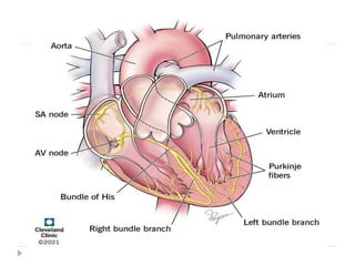
- 39. Components of the Cardiac Conduction System 1. Sinoatrial node 2. Atrioventricular node 3. Atrioventricular bundle (bundle of His) 4. Purkinje fibres
- 41. 1. Sinoatrial Node Sinoatrial node is sometimes called as heart’s natural pacemaker. It sends the electrical impulses that start the heartbeat. The SA node is in the upper part of heart’s right atrium. It is at the edge of atrium near superior vena cava.
- 42. 2. Atrioventricular Node The atrio-ventricular node delays the SA node’s electrical signal. It delays the signal by a consistent amount of time (a fraction of a second) each time. The AV node is located in an area known as the triangle of Koch. This is near the central area of the heart.
- 44. 3. Bundle of His The bundle of His is also called the atrioventricular bundle. It is a branch of fibers (nerve cells) that extends from AV node. This fiber bundle receives the electrical signal from the AV node and carries it to the Purkinje fibers. The bundle of His runs down the length of the interventricular septum.
- 46. Cont.. The bundle of His has two branches: Left bundle branch sends electrical signals through the Purkinje fibers to left ventricle. Right bundle branch sends electrical signals through the Purkinje fibers to right ventricle.
- 47. 4. Purkinje Fibers The Purkinje fibers are branches of specialized nerve cells. They send electrical signals very quickly to the right and left heart ventricles. When the Purkinje fibers deliver electrical signals to the ventricles, the ventricles contract.
- 48. CONDUCTING SYSTEM OF THE HEART
- 49. Conduction of Impulse in the Heart Initiation of impulse: Impulse is generated in SA node at a rate of 70-80/minute. Therefore SA node is called as pacemaker of heart.
- 50. Cont.. Spread of impulse: The wave of impulse spreads to both atria through muscle tissues simultaneously causing them to contract. From SA node impulse passes to ventricles through AV node. Upon reaching the atrioventricular (AV) node, the signal is delayed by 0.13 seconds. This delay causes ventricles to contract after atrial contraction is over.
- 51. Cont.. It is then conducted into the bundle of his, down the interventricular septum. The bundle branches and the Purkinje fibres spread the wave impulses at fastest rate along the ventricles, causing them to contract. Impulse generation and transmission is an electrical event whereas contraction and relaxation of heart muscle are mechanical events. Mechanical events always follow electrical events.
- 52. Conduction of Impulse in the Heart


