Hip joint
•
11 likes•1,158 views

The hip joint is a ball and socket joint that connects the femur to the pelvis. It is the body's largest weight bearing joint. The rounded head of the femur fits into the cup-shaped acetabulum of the pelvis. Strong ligaments and muscles provide stability to the joint. Damage to any of the hip joint components can negatively affect its range of motion and weight bearing ability, and may require hip replacement surgery. The hip allows for flexion, extension, abduction, adduction, internal and external rotation.
Report
Share


Hip joint
- 1. KINESOLOGY OF HIP JOINT PRESENTED BY : DR.ASER MOHAMED KAMAL PHYSICAL THERAPIST
- 2. Hip joint The hip is the body’s second largest weight-bearing joint (after the knee). It is a ball and socket joint at the juncture of the leg and pelvis. The rounded head of the femur (thighbone) forms the ball, which fits into the acetabulum (a cup-shaped socket in the pelvis). Ligaments connect the ball to the socket and usually provide tremendous stability to the joint. The hip joint is normally very sturdy because of the fit between the femoral head and acetabulum as well as strong ligaments and muscles at the joint. All of the various components of the hip mechanism assist in the mobility of the joint. Damage to any single component can negatively affect range of motion and ability to bear weight on the joint. Orthopedic degeneration or trauma – those conditions affecting the bones in the hip joint – can necessitate total hip replacement, partial hip replacement orhip resurfacing. Bones of the hip joint The femur is the upper leg bone or thigh. It is the largest bone in the body. At the top of the femur is a rounded protrusion which articulates with the pelvis. This portion is referred to as the head of the femur, or femoral head. There are two other protrusions near the top of the femur, known as the greater and lesser trochanters. The muscles involved in hip motion are attached to the joint at these trochanters.
- 3. The acetabulum is a concave area in the pelvis, into which the femoral head fits. The pelvis is a girdle of bones, connected at the front by cartilage pad, called the pubis, and at the back by the lowest four fused vertebrae (the sacrum). The sacro-iliac joints are located where the sacrum meets the pelvis. The bone surfaces of the femoral head and acetabulum have a smooth durable layer of articular cartilage that cushions the ends of the bones and allows for smooth movement. Pelvis o 1/5 pubis o 2/5 ischium o 2/5 ilium o Pelvis full ossification = 15-25 y.o. Acetabulum o Concave socket o Lateral, inferior, anterior (LIA) o Roundness ↓ w/ age o Inferior = acetabular notch o Central/deepest part = acetabular fossa o Anteversion = anterior orientation of acetabulum Men = 18.50 Women = 21.5 0 Pathologic ↑ = ↓ jt. stability, risk for anterior dislocation of femoral head Femur o Circular o Smaller in women o Fovea Inferior to medial pt. of femoral head Attachment of ligament of femoral head
- 4. o Medially, superiorly, anteriorly (SAM) o Neck = 5cm long o Angulation Angle of inclination (medial) Frontal plane bet. Femoral neck & shaft Early infancy = 1500 Adult = 1250 Elderly = 1200 ↓ in women due to width of female pelvis Pathologic ↑ = coxa valga Pathologic ↓ = coxa vara Angle of torsion Transverse plane bet. Femoral neck & condyles Anterior torsion =relative lateral rotation ↓ w/age Newborn = 400 Adult = 150 Anteversion o Pathologic ↑ o Internal femoral torsion o ↓ ER o ↑ IR Retroversion o Pathologic ↓ o External femoral torsion o Frog-leg position FABER True physiologic position of hip o A congruent fit under low load would lead to incongruence under high load o Periphery of acetabulum in contact, fossa is non-articular
- 5. Bony landmarks: Femur: 1. Greater trochanger – lateral aspect of thigh just distal to hip joint. 2. Medial and lateral condyles– on distal end of femur 3. Medial and lateral epicondyles (Epicondyles) Patella: “Knee cap” – anterior aspect of knee (a sesamoid bone located in quadriceps tendon). HIP JOINT › ROM o Flex 0-1200 o Hypertext 0-100 o Abd 0-450 o Add across30-400 o ER 0-450 o IR 0-350 › Close-packed o Full ext, IR, Abd › Open-packed o 300 flex o 300 abd o Slight ER › Capsular pattern o Flex, abd, IR › End feel o Flex soft/firm o Ext firm o Abd soft/firm o Add soft/firm o IR firm o ER firm › End Feels › Normal: o Flexion & Adduction Elastic or Tissue Approximation
- 6. o SLR Elastic o Extension & Abduction Elastic/Firm › IR & ER Elastic/Firm › Tonic labyrinthine & optical righting reflexes o Head effectively behaves as if it’s fixed in a vertical position o Maintains head over BOS › When hip flexor ms. Is tight, keep LOG w/in BOS o Open-chain response = displacement of head from vertical (Fig. A) o Closed-chain response = maintain head in upright position (Fig. B) › Acetabulum of pelvis + head of femur › Diarthrodial, ball-and-socket jt. w/ 30 freedom: o flex/ext in sagittal plane o abd/add in frontal plane o IR/ER in transverse plane › 10 function of hip o To support wt. of head, arms & trunk (HAT) › Also provides pathway for transmission of forces bet. Pelvis & LEs › Hip tends to operate in a closed kinematic chain o Proximal end = head o Distal end = foot Hip joint capsule or socket You may hear your hip surgeon refer to the capsule or socket, when describing the structure of the hip joint. The joint capsule is a thick ligamentous structure surrounding the entire joint. Inside the capsule, the surfaces of the hip joint are covered by a thin tissue called the synovial membrane. This membrane nourishes and lubricates the joint.
- 7. o capsule has major contribution to stability o femoral neck = intracapsular o greater & lesser trochanters = extracapsular o thickened anterosuperiorly o thin & loose posteroinferiorly Ligaments As noted above, the stability of the hip joint is directly related to its muscles and ligaments. The most notable ligaments in the hip joint are: Iliofemoral ligament, which connects the pelvis to the femur at the front of the joint. It keeps the hip from hyper-extension o Iliofemoral ligament Y ligament of Bigelow Origin = AIIS 2 arms fan out to insert = intertrochanteric line of femur Strongest ligament of hip Taut in hyperextension Superior fibers taut in adduction Inferior tense during abduction Pubofemoral ligament, which attaches the most forward part of the pelvis known as the pubis to the femur Origin = anterior pubic ramus Insertion = anterior intertrochanteric fossa Taut in hip abd & ext
- 8. Ischiofemoral ligament, which attaches to the ischium (the lowest part of the pelvis) and between the two trochanters of the femur. Origin = posterior acetabular rim, acetabulum labrum Insertion = spiral around femoral neck Spiral fibers taut during ext, loosen in flex o Position of stability Full extension of hip o Position of vulnerability Flex & add (such as sitting w/thighs crossed) Ligamentum teres Triangular Ligament of head of femur Labrum The labrum is a circular layer of cartilage which surrounds the outer part of the acetabulum effectively making the socket deeper to provide more stability for the joint. Labrum tears are not an uncommon hip injury. o Acetabular labrum Fibrocartilage rimming entire periphery o Transverse acetabular ligament Roof of tunnel passage for blood vessels & nerves entering hip o Has Center Edge angle (CE) or angle of Wiberg Men = 380 Women = 350 Smaller CE angle (more vertical) = ↓ coverage of head of femur, ↑ risk superior dislocation of femoral head ↑ w/age
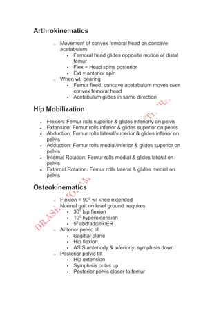
- 9. Arthrokinematics o Movement of convex femoral head on concave acetabulum Femoral head glides opposite motion of distal femur Flex = Head spins posterior Ext = anterior spin o When wt. bearing Femur fixed, concave acetabulum moves over convex femoral head Acetabulum glides in same direction Hip Mobilization Flexion: Femur rolls superior & glides inferiorly on pelvis Extension: Femur rolls inferior & glides superior on pelvis Abduction: Femur rolls lateral/superior & glides inferior on pelvis Adduction: Femur rolls medial/inferior & glides superior on pelvis Internal Rotation: Femur rolls medial & glides lateral on pelvis External Rotation: Femur rolls lateral & glides medial on pelvis Osteokinematics o Flexion = 900 w/ knee extended o Normal gait on level ground requires 300 hip flexion 100 hyperextension 50 abd/add/IR/ER o Anterior pelvic tilt Sagittal plane Hip flexion ASIS anteriorly & inferiorly, symphisis down o Posterior pelvic tilt Hip extension Symphisis pubis up Posterior pelvis closer to femur
- 10. o Lateral pelvic tilt Frontal plane One hip joint serves as pivot/axis Opposite iliac crest elevates (hip hike) or drop (pelvic drop) Reference is side farthest from supporting hip o Pelvic rotation Transverse plane Occurs in single-limb support around axis of supporting hip jt. Forward rotation Side opposite supporting hip moves anteriorly Backward rotation Side opposite supporting hip moves posteriorly o Lumbar-Pelvic Rhythm Open-chain E.g. reaching the floor Hip flexion up to 900 only Anterior tilt of pelvis on femurs Flexion of lumbar spine adds 450 E.g. side-lying abduction Lateral tilt of pelvis & lumbar spine adds 450 o Closed chain response to motions of pelvis Keeps one or both feet on the ground Maintain head upright & vertical Anterior pelvic tilt during hip flexion = head & trunk displaced forward + lumbar extension Posterior pelvic tilt + lumbar flexion to keep head forward over sacrum
- 11. pelvic motion co-hip motion compensatory lumbar anterior tilt hip flex lumbar ext posterior tilt hip ext lumbar flex lateral tilt (drop) right hip add right lateral flex lateral tilt (hike) right hip abd left lateral flex forward rot right hip IR rotation to left backward rot right hip ER rotation to right Muscle Groups The muscles of the hip consist of four main groups 1. Gluteal group: The gluteals are the muscles in your buttocks. The gluteal muscles include the gluteus maximus, gluteus medius, gluteus minimus, and tensor fasciae latae. They cover the lateral surface of the ilium.
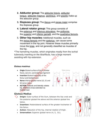
- 12. 2. Adductor group: The adductor brevis, adductor longus, adductor magnus, pectineus, and gracilis make up the adductor group. 3. Iliopsoas group: The iliacus and psoas major comprise the iliopsoas group. 4. Lateral rotator group: This group consists of the externus and internus obturators, the piriformis, the superior and inferior gemelli, and the quadratus femoris. 5. Other hip muscles: Additional muscles, such as the rectus femoris and the sartorius, can cause some movement in the hip joint. However these muscles primarily move the knee, and not generally classified as muscles of the hip. *The hamstring muscles, which originates mostly from the ischial tuberosity inserting on the tibia/fibula, has a large moment assisting with hip extension. Gluteus maximus Origin Gluteal surface of ilium, lumbar fascia, sacrum, sacrotuberous ligament Insertion Gluteal tuberosity of the femur and iliotibial tract Nerve Inferior gluteal nerve (L5, S1 and S2 nerve roots) ACTION Extends and laterally rotates hip. Maintains knee extended via iliotibial tract Gluteus Medius Origin: Outer surface of the ilium, between the iliac crest and the posterior gluteal line above and the anterior gluteal line below. Insertion: Posterolateral surface of the greater trochanter of the femur. Action: Abduction of the hip, internal rotation of thigh. Innervation: Superior gluteal nerve L4, 5, S1).
- 13. Gluteus Minimus Origin: Outer surface of the ilium, between the anterior and inferior gluteal lines, and the edge of the greater sciatic notch. Insertion: Anterior surface of the greater trochanter of the femur. Action: Abduction of the thigh, internal rotation of thigh. Innervation: Superior gluteal nerve L4, 5, S1). Tensor Fasciae Latae Origin: Outer surface of the anterior iliac crest, between the tubercle of the iliac spine. A thick fascia covers the outer surface of the muscle, making it appear to be sandwiched between the layers of fasciae latae. Insertion: By the iliotibial band anterior surface of the lateral condyle of the tibia. Action: Assists with flexion of the thigh at the hip, assists with adduction of the thigh at the hip Innervation: Superior gluteal nerve (4 -5, S1) Adductor Magnus Muscle Origin: Anterior: Inferior pubic ramus and the ramus of the ischium Posterior: Inferolateral aspect of the ischial tuberosity Insertion: Anterior: Medial margin of the gluteal tuberosity of the femur, medial to gluteus maximus. Posterior: By a broad attachment into the linea aspera and the proximal part of the medial supracondylar line and by a small tendon to the adductor tubercle. Action: Adduction of the thigh at the hip, extension of the thigh at the hip Innervation: Posterior division of the obturator nerve (L2 – 4) Adductor Longus Origin: Anterior surface of the pubis, in the angle between the crest and pubic symphysis. Insertion: Lower two-thirds of the medial lip of the linea aspera on the posterior surface. Action: Adduction of the thigh at the hip, assists with internal rotation of the thigh at the hip, assists with flexion of the thigh at the hip
- 14. Innervation: Anterior division of the obturator nerve (L2 -3) Adductor Brevis Origin: Inferior ramus and body of the pubis, between gracilis and obturator externus. Insertion: Along a line from the lesser trochanter to the linea aspera, the upper third of the linea aspera, downward along the upper third of the linea aspera, immediately behind the pectineus and the upper part of adductor longus Action: Adduction of the thigh at the hip, assists with internal rotation of the thigh at the hip. Innervation: Anterior division of the obturator nerve (L2 -3) Pectineus Origin: Pectineal line of the pubis and a narrow area of the superior pubic ramus below it. Insertion: A vertical line from the lesser trochanter to the linea aspera Action: Assists with flexion of the thigh at the hip, assists with adduction of the thigh at the hip Innervation: Anterior division of the femoral nerve (L2 – 3) Gracilis Origin: Lower half of the body of the pubis, the inferior pubic ramus, and the adjoining part of the ischial ramus. Insertion: Upper part of the medial flare of the tibia, just below the medial condyle, proximal and slightly anterior to the attachment of the semitendinosus, and posterior and somewhat inferior to the attachment of sartorius. Action: Adduction of the thigh at the hip, assists with internal rotation of the thigh at the hip, assists with flexion of the thigh at the hip Innervation: Anterior division of the obturator nerve (L2 – 3)
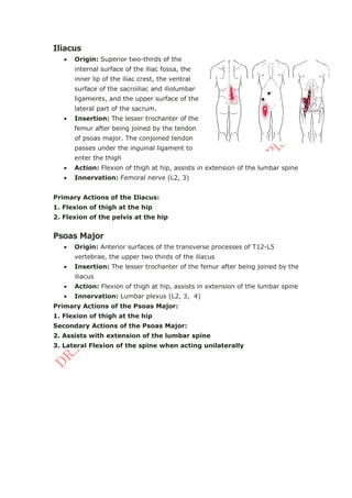
- 15. Iliacus Origin: Superior two-thirds of the internal surface of the iliac fossa, the inner lip of the iliac crest, the ventral surface of the sacroiliac and iliolumbar ligaments, and the upper surface of the lateral part of the sacrum. Insertion: The lesser trochanter of the femur after being joined by the tendon of psoas major. The conjoined tendon passes under the inguinal ligament to enter the thigh Action: Flexion of thigh at hip, assists in extension of the lumbar spine Innervation: Femoral nerve (L2, 3) Primary Actions of the Iliacus: 1. Flexion of thigh at the hip 2. Flexion of the pelvis at the hip Psoas Major Origin: Anterior surfaces of the transverse processes of T12-L5 vertebrae, the upper two thirds of the iliacus Insertion: The lesser trochanter of the femur after being joined by the iliacus Action: Flexion of thigh at hip, assists in extension of the lumbar spine Innervation: Lumbar plexus (L2, 3, 4) Primary Actions of the Psoas Major: 1. Flexion of thigh at the hip Secondary Actions of the Psoas Major: 2. Assists with extension of the lumbar spine 3. Lateral Flexion of the spine when acting unilaterally
- 16. Piriformis Origin: Anterior surfaces of the sacrum by three or four slips off the portions of bone between the foramina of the sacrum, the ilium near the posterior inferior iliac spine, the capsule of the sacro-iliac joint, and occasionally the upper part of the sacrotuberous ligament. Insertion: By a rounded tendon to the upper part of the medial surface of the greater trochanter, occasionally blending with the common tendon of obturator internus, gemellus superior, and gemellus inferior. Action: Assists with lateral rotation and abduction of the thigh Innervation: Nerve to piriformis (S1, 2) Sartorius Origin: Inferior portion of the anterior superior iliac spine Insertion: Upper medial surface of tibial shaft at the tibial flare Action: Assists with hip flexion, knee flexion, medial rotation of the knee, lateral rotation of the hip Innervation: Anterior division of the femoral nerve (L3- 4)