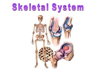

Human skeletal system - Movement and Locomotion
- 2. Function of the Skeletal System Support - framework that supports body and cradles its soft organs Protection - for delicate organs, heart, lungs, brain Movement - bones act as levers for muscles Mineral storage - calcium & phosphate Blood cell formation - hematopoiesis
- 3. Long Bones - metacarples, metatarsals, phelangies, humerus, ulna, radius, tibia, fibula Short Bones - carpals, tarsals Flat Bones - rib, scapula, skull, sternum Irregular Bones - vertebrae, some facial bones Sesamoid - patella Types of Bones
- 5. Distal epiphysis Proximal epiphysis diaphysis yellow marrow epiphyseal line periosteum compact bone spongy bone Endosteum hyaline cartilage Anatomy of a Long Bone Sharpey’s fibers
- 8. Cranium Facial Bones Anterior View Axial Skeleton
- 9. Cranium Facial Bones Lateral View Axial Skeleton
- 10. Posterior View Axial Skeleton
- 11. Axial Skeleton Inferior View
- 12. Sinal Cavities Warm and moisten air Lighten the skull Enhance voice resonance Frontal Sinus Ethmoid Sinus Sphenoid Sinus Maxillary Sinus
- 13. Cervical Vertebrae (7) Thoracic Vertebrae (12) Lumbar Vertberae (5) Sacrum (5) Coccyx (4) The Vertebral Column Axial Skeleton
- 17. Sternum True Ribs (7) False Ribs (3) Floating Ribs (2) The Thoracic Cage Axial Skeleton
- 18. Sacrum & Coccyx Axial Skeleton
- 19. Bones of the Pectoral Girdle Appendicular Skeleton
- 20. Humerus Ulna Radius Carpals Metacarpals Phalanges The Upper Limb (Forelimb) Appendicular Skeleton
- 21. Humerus
- 22. Ulna & Radius
- 23. Hand Bones
- 25. Ischium Ilium Acetabulum Pubis Ischium Obturator foramen Appendicular Skeleton Pelvis (lateral view)
- 27. Femur Patella Tibia Fibula Tarsals Metatarsals Phalanges The Lower Limb (Legs) Appendicular Skeleton
- 28. Femur
- 29. Patella
- 30. Tibia & Fibula
- 31. metatarsals phalanges tarsals metatarsals phalanges tarsals Foot
- 32. Fetal Skull
- 33. Immovable Joints (synarthrosis) Fibrous Joints suture pubis symphisis
- 34. Slightly Movable Joint (ampharthrosis) Cartilagenous Joints
- 35. Synovial Joints (diarthrosis)- freely moveable femur ligaments pelvis
- 36. Synovial Joints femur pelvis hyaline cartilage synovial cavity joint capsule
- 37. Knee Joint
- 38. Abduction Extension Rotation Flexion Adduction Synovial Joint Movement
- 39. 275 bones 12 weeks (6-9 inches long) Fetal Skeleton
- 40. cartilage calcified cartilage bone epiphyseal plate epiphyseal line Endochondral Ossification 2 o ossification center Fetus: 1 st 2 months Adult Childhood Just before birth
- 41. Osteoblast Osteocyte Osteoclast Eats bone Builds new bone Mature bone cell Bone cells that aid in remodeling
- 42. Bone Repair : Electrical stimulation of the fracture site: Increases speed and completeness of healing The e- stimulation inhibits PTH and slow osteoclasts down from reabsorbing bone 2. Ultrasound treatment: Daily treatments reduce healing time of broken bones by 25-35% 3. Free vascular fibular graft technique: Transplant fibula in arm Gives good blood supply not available in other treatments 4. Bone substitutes: Crushed bone from cadaver- but risk of HIV and hepatitis Sea bone- coral Artificial bone- ceramic
- 44. hematoma callus bony callus Repair of Fractures bone remodeling
- 45. Diseases of the Skeletal System: Osteoporosis - bone reabsorption outpaces bone deposit; bones become lighter and fracture easier Factors: age, gender (more in women) estrogen and testosterone decrease insufficient exercise (or too much) diet poor in Ca ++ and protein abnormal vitamin D receptors smoking
- 46. Osteoporosis 29 40 84 92
- 47. Rickets - vitamin D deficiency Osteomalacia - soft bones, inadequate mineralization in bones, lack of vitamin D Pagets Disease - spotty weakening in the bones, excessive and abnormal bone remodeling Rheumatoid arthritis - autoimmune reaction Diseases of the Skeletal System:
- 48. INQUIRY http://www.youtube.com/watch?v=DSHoonPWwXQ What is a fontanel? How many bones in the adult skeleton? What is the difference between the appendicular and axial skeleton? What is a meniscus? Demonstrate adduction. Weight bearing vertebrae are called? What does an osteoclast do/ Extra Credit: 1-page reaction paper on bipedalism and problems associated with our human frame. Attach article. Turn in 1-week from today.
Editor's Notes
- Osteoblasts Osteoblasts are responsible for building new bone and lie at the centre of bone physiology. Their functions include the synthesis of collagen and the control of mineralisation. Osteoclasts Osteoclasts are specialised cells that resorb bone. They work by sealing off an area of bone surface then, when activated, they pump out hydrogen ions to produce a very acid environment, which dissolves the hydroxyapatite. Osteocytes Bone adapts to applied forces by growing stronger in order to withstand them; it is known that exercise can help to improve bone strength. Osteocytes are thought to be part of the cellular feed-back mechanism which directs bone to form in the places where it is most needed. They lie within mineralised bone and it is thought that they may detect mechanical deformation and mediate the response of the osteoblasts.