


Muscle Microanatomy
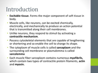
- 2. Introduction • Excitable tissue. Forms the major component of soft tissue in body. • Muscle cells, like neurons, can be excited chemically, electrically, and mechanically to produce an action potential that is transmitted along their cell membranes. • Unlike neurons, they respond to stimuli by activating a contractile mechanism. • Possess cytoskeletal elements that are capable of lengthening or shortening and so enable the cell to change its shape. • The cytoplasm of muscle cells is called sarcoplasm and the surrounding cell membrane or plasmalemma is called sarcolemma • Each muscle fiber sarcoplasm contains numerous myofibrils, which contain two types of contractile protein filaments, actin and myosin.
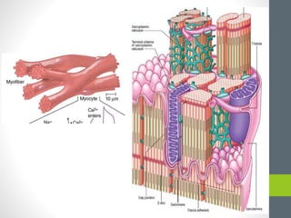
- 3. Terminology Muscle fibre: Individual Celll Myofibril: Long chains of contractile proteins
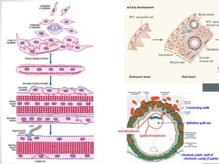
- 5. Skeletal muscle found throughout the body is derived from the paraxial mesenchyme, which is formed from ingression at the streak and subsequently segmented into somites. Skeletal muscle originates from a pool of premyoblastic cells which arise in the dermatomyotome of the maturing somite and begin to differentiate into myoblasts at 4–5 weeks of gestation. Smooth muscle cells developed in situ from the splanchnopleuric mesenchyme in the walls of the viscera. Cardiac myocytes differentiate from the splanchnic coelomic cells of the pericardium initially subjacent to the endoderm.
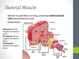
- 7. Skeletal Muscle • Skeletal muscle fibers are long, cylindrical,multinucleated cells,with peripheral nuclei. • Organization: Epimysium: dense, irregular connective tissue Perimysium: dense, irregular connective tissue Endomysium: reticular connective tissue
- 13. Striations and Sarcomere • Differences in the refractive indexes of the various parts of the muscle fiber are responsible for the characteristic cross- striations seen in skeletal muscle when viewed under the microscope. • The parts of the cross-striations are frequently identified by letters. The light I band is divided by the dark Z line, and the dark A band has the lighter H band in its center. • A transverse M line is seen in the middle of the H band, and this line plus the narrow light areas on either side of it are sometimes called the pseudo-H zone. • The area between two adjacent Z lines is called a sarcomere. • The orderly arrangement of actin, myosin, and related proteins that produces this pattern as shown
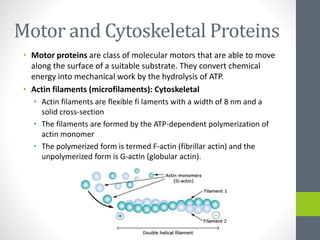
- 14. Motor and Cytoskeletal Proteins • Motor proteins are class of molecular motors that are able to move along the surface of a suitable substrate. They convert chemical energy into mechanical work by the hydrolysis of ATP. • Actin filaments (microfilaments): Cytoskeletal • Actin filaments are flexible fi laments with a width of 8 nm and a solid cross-section • The filaments are formed by the ATP-dependent polymerization of actin monomer • The polymerized form is termed F-actin (fibrillar actin) and the unpolymerized form is G-actin (globular actin).
- 15. • A wide variety of Actin-binding Proteins are capable of modulating the form of actin within the cell. • Types: • Bundling proteins: Tie actin filaments together in longitudinal arrays to form cables or core structures. E.g. fimbrin, villin, myosin • Gel-forming proteins: Interconnect adjacent actin filaments to produce loose filamentous meshworks (gels) composed of randomly orientated F-actin. • Filament severing proteins: Severing proteins, such as gelsolin and severin, bind to F-actin fi laments and sever them, which produces profound changes within the actin cytoskeleton and in its coupling to the cell surface. • Membrane-associated proteins
- 16. Myosins – The Motor Proteins • Myosin proteins have a globular head region consisting of a heavy and a light chain. The heavy chain bears an α-helical tail of varying length. The head has an ATPase activity and can bind to and move along actin fi laments – the basis for myosin function as a motor protein. • The best-known class is myosin II
- 18. Sarcotubular System • The muscle fibrils are surrounded by structures made up of membranes • These structures form the sarcotubular system, which is made up of a T system and a sarcoplasmic reticulum. • The T system of transverse tubules, which is continuous with the sarcolemma of the muscle fiber, forms a grid perforated by the individual muscle fibrils. The sarcoplasmic reticulum, which forms an irregular curtain around each of the fibrils, has enlarged terminal cisterns in close contact with the T system
- 20. Motor End Plate and Muscle Spindle
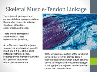
- 21. Skeletal Muscle-Tendon Linkage The epimysial, perimysial and endomysial sheaths coalesce where the muscles connect to adjacent structures at tendons, aponeuroses, and fasciae. There are no desmosomal attachments at these myotendinous junctions, Actin filaments from the adjacent sarcomeres, which would normally insert into a Z-disc at this point, instead penetrate a dense, subsarcolemmal filamentous matrix that provides attachment to the plasma membrane. At the extracellular surface of the junctional sarcolemma, integrins provide contact with the basal lamina which in turn adheres closely to collagen and reticular fibres (type III collagen) of the adjacent tendon or other connective tissue structure.
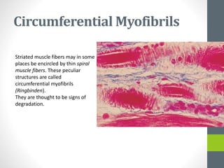
- 22. Circumferential Myofibrils Striated muscle fibers may in some places be encircled by thin spiral muscle fibers. These peculiar structures are called circumferential myofibrils (Ringbinden). They are thought to be signs of degradation.
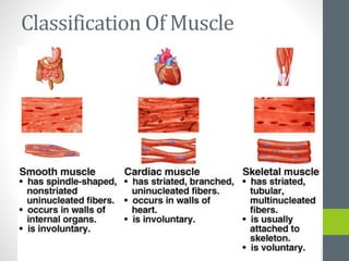
- 23. Smooth Muscles • In smooth muscle tissue the contractile proteins actin and myosin are not organized into regular sarcomeres, visible as transverse striations. • Involuntary • Much smaller in size • The nucleus is single, located at the midpoint, and often twisted into a corkscrew shape by the contraction of the cell. • Typically found in the walls of tubular structures and hollow viscera • Smooth muscle has no attachment structures equivalent to the fasciae, tendons and aponeuroses associated with skeletal muscle. • Each cell is covered almost entirely by a prominent basal lamina which merges with a reticular layer consisting of a network of fine elastin, reticular fibres (collagen type III) and type I collagen fibres.
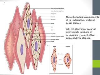
- 24. The cell attaches to components of this extracellular matrix at dense plaques cell–cell attachment occurs at intermediate junctions or desmosomes, formed of two adjacent dense plaques.
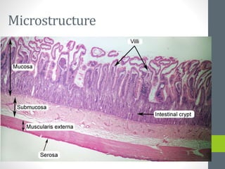
- 26. Microstructure
- 32. Cardiac Muscle Cell • In cardiac muscle, as in skeletal muscle, the contractile proteins are organized structurally into sarcomeres. • fine cross-striations that are visible in the light microscope.