Location via proxy:   [ UP ]  
[Report a bug]   [Manage cookies]                
SlideShare a Scribd company logo
Assessment of the Chest and Lungs
Functions of the Respiratory
                  System

 Ventilation
 Diffusion and Perfusion
 Control of Breathing
Functions

 Ventilation
     Movement of air into and out of the lungs
     Inspiratory phase
     Expiratory phase
Functions

 Hypoventilation
     Slow, shallow breathing
     Causes CO2 to build up in the blood
         Acidosis
 Hyperventilation
     Rapid, deep breathing
     Causes CO2 to be blown off
         Alkalosis
Functions

 Diffusion and Perfusion
     Gas exchange across the alveolar-pulmonary
      capillary membranes
 Control of breathing
     Influenced by neural and chemical factors
     Pons, medulla, chemoreceptors in the carotid
      body
     Stimulus for breathing
         Increased carbon dioxide - PRIMARY
Anatomical Structures

 Reference points for pinpointing findings from
  the physical examination
     Topographical Landmarks
     Reference Lines
Topographical Landmarks

 Nipples
 Manubriosternal junction (Angle of Louis)
      Point at which the 2nd rib articulates with the sternum
 Suprasternal notch
 Costal Angle
      Usually no more than 90 degrees
      Ribs insert at approximately 45 degree angles
 Clavicles
Manubrium


Manubriosternal junction
(Angle of Louis)


      Nipple


   Costal Angle
Reference Lines

 Anterior Chest
      Midsternal line
      Anterior axillary lines
      Midclavicular lines
 Posterior Chest
      Vertebral line
      Midscapular lines
 Axilla
      Anterior axillary lines
      Midaxillary lines
      Posterior axillary lines
Anterior Chest
Posterior Chest
Axilla
Anatomy
Anatomy Points to Remember

 Lungs are symmetric
 Lungs are divided into lobes
     Right lung = 3 lobes
     Left lung = 2 lobes
 Primary muscles of respiration
     Diaphragm – divides chest from abdomen
     External intercostal muscles
     Accessory muscles
Anatomy Points to Remember

 Upper Airway
     Nose, pharynx, larynx, intrathoracic trachea
     Functions in respiration
         Conduct air to lower airway
         Filter to protect lower airway
         Warm and humidify inspired air
Anatomy Points to Remember

 Lower Airway
     Trachea, bronchi, bronchioles
         Functions in respiration
           Conduct air to alveoli
           Clear mucociliary structures
     Alveoli
         Functional unit
           Gas exchange
           Production of surfactant
Anatomy Points to Remember

 Lower Airway
     Trachea splits into left and right mainstem
      bronchi which are further subdivided into
      bronchioles
         Right bronchus is shorted, wider and more
          upright than the left
         Functions in respiration
           Conduct air to alveoli
           Clear mucociliary structures
Respiratory assessment
Chest Anatomy

 Web Anatomy:
  http://www.gen.umn.edu/faculty_staff/jensen
  /1135/webanatomy/
History

 Chief Complaint and HPI
     Cough
     Shortness of breath/Dyspnea
Cough

   Onset – sudden, gradual
   Duration
   Nature – dry, moist, hacking, barking
   Sputum – amount, color, odor
   Severity – disrupts activities
   Associated symptoms – sneezing, dyspnea, fever, chills,
    congestion, gagging
   What brings it on? – anxiety, talking, activity
   What makes it better?
   What has been tried? – medications, treatments
   Anything similar in the past?
Shortness of Breath (SOB) /
                     Dyspnea
   Onset – sudden, gradual
   Duration
   Severity – disrupts activities
   Associated symptoms – night sweats, pain, chest
    pressure, discomfort, ankle edema, diaphoresis, cyanosis
   What brings it on? – position, time of day, exercise,
    allergens, emotions
   What makes it better?
   What has been tried? – medications, inhalers, oxygen
   Anything similar in the past?
History

 Past Health History
     Lung disease or breathing problems
         Frequent severe colds, asthma, emphysema,
          bronchitis, pneumonia, tuberculosis
     Last PPD and/or chest x-ray
     Allergies
     Medication use
 Family History
History

 Personal and Social History
     Tobacco
     Alcohol
     Drugs
     Home environment
     Occupational environment
     Travel
 Health Promotional Activities
Physical Examination
Equipment and Techniques

 Equipment
     Stethoscope
 Techniques
     Inspection
     Palpation
     Percussion
     Auscultation
Inspection

 General
     Appearance
     Posturing
     Breathing effort
     Trachea position
         Midline
Inspection

 Chest Wall Configuration
     Form
     Symmetry
     Muscle development
     Anterior-Posterior (AP) diameter
         Approximately ½ the transverse diameter
         Transverse: Anterior-Posterior = 2:1
     Costal angle
         90 degrees or less
Inspection

 Oxygenation: cyanosis
     Nails
     Skin
     Lips
 Respiratory Effort
     Respiratory rate and depth
     Breathing pattern
     Chest expansion
Palpation

 Trachea – for position
 Chest wall – for symmetry
Palpation

 Thoracic Expansion (Excursion)
      Place both thumbs at about 7th rib
       posteriorly along the spinal process
                                               Click on the pictures to view video
      Extend the fingers of both hands
       outward over the posterior chest wall
      Have the person take a deep breath
       and observe for bilateral outward
       movement of thumbs
          Normal: bilateral, symmetric
           expansion
          Abnormal: unilateral or unequal
Palpation

 Vocal (Tactile) Fremitus
     Use palmar or ulnar surfaces of hands
     Systematically position hands over both sides of
      posterior chest
     Have person repeat “1 – 2 – 3” or “99” as you
      move from the apices to the bases
         Normal: bilaterally symmetrical vibrations
         Decreased or absent: obstruction of transmission
          0bronchitis, emphysema)
         Increased: consolidation (compression) of lung
          tissue (pneumonia)
Auscultation

 Auscultate in a systematic manner
 Compare one side to the other
 Listen one full respiration at each spot
 Displace breast tissue to listen directly over
  chest wall
 DO NOT listen through gowns, clothes, etc.
       Place your stethoscope over bare skin
Auscultation

 Evaluate posterior, lateral, and anterior chest
 Instruct person to sit upright and breathe in
  and out slowly through the mouth
     This makes it easier to hear the air movement
 Use the diaphragm of the stethoscope
 Move from the apices to the bases
Auscultation

 Evaluate for normal sounds
  Sound              Pitch      Intensity Quality             I:E   Location

  Bronchial          High       Loud       Blowing/           I<E   Trachea
                                           hollow


  Bronchovesicular   Moderate   Moderate   Combination        I=E   Between scapulae,
                                                                    1st & 2nd ICS lateral to
                                                                    sternum

  Vesicular          Low        Soft       Gentle rustling/   I>E   Peripheral lung
                                           breezy
Auscultation

 Evaluate for adventitious sounds
 Sound          Intensity/ Pitch     I/E   Quality                     Clear with Cough

 Crackles/      Soft (fine)/ High      I   Discontinuous,              Possibly
 Rales          Loud (coarse)/ Low         nonmusical, brief
 Wheeze         High                   E   Continuous musical          Possibly
                                           sounds


 Ronchi         Low                    E   Continuous snoring          Possibly
                                           sounds
 Pleural                             I&E   Continuous or               Never
 Friction Rub                              discontinuous creaking or
                                           brushing sounds
 Stridor                               I   Continuous, crowing         Never
Auscultation


Copy this URL into your Web browser to hear normal and abnormal lung sounds :
http://medocs.ucdavis.edu/IMD/420C/sounds/lngsound.htm
Developmental Variations

 Neonates
     Measure the chest circumference
         Usually 2-3 cm smaller than head circumference
         Chest is round (i.e. AP diameter = transverse)
     Obligate nose breathers
     Periodic breathing is common
         Sequence of vigorous breathing followed by apnea
          for 10-15 seconds
         Only concern if it is prolonged or baby becomes
          cyanotic
Developmental Variations

 Neonates
     Breathing is diaphragmatic and abdominal
     Signs of compromise
         Stridor (“crowing”)
         Grunting
         Central cyanosis
         Flaring nares
Developmental Variations

 Infants and Young Children
     Roundness of the chest persist for first 2 years
     Chest walls are thinner than the adult’s

         Breath sounds may sound louder, and more
          bronchial than the adult
           Bronchovesicular sounds may be heard
            throughout the chest
Developmental Variations

 Pregnancy
     Costal angle increases to about 105 degrees in
      the third trimester
     Dyspnea and orthopnea are common
     Breathes more deeply
Developmental Variations

 Older Adult
     Chest expansion is often decreased
     Bony prominences are marked
     AP diameter is increased with respect to
      transverse (but not 1:1)
Videos of Thorax and Lung
                  Assessment

 Copy these URLs into your Web browser
     http://www.conntutorials.com/chapter5.html
                         OR
     http://medinfo.ufl.edu/other/opeta/chest/CH_main

More Related Content

Respiratory assessment

  • 1. Assessment of the Chest and Lungs
  • 2. Functions of the Respiratory System  Ventilation  Diffusion and Perfusion  Control of Breathing
  • 3. Functions  Ventilation  Movement of air into and out of the lungs  Inspiratory phase  Expiratory phase
  • 4. Functions  Hypoventilation  Slow, shallow breathing  Causes CO2 to build up in the blood  Acidosis  Hyperventilation  Rapid, deep breathing  Causes CO2 to be blown off  Alkalosis
  • 5. Functions  Diffusion and Perfusion  Gas exchange across the alveolar-pulmonary capillary membranes  Control of breathing  Influenced by neural and chemical factors  Pons, medulla, chemoreceptors in the carotid body  Stimulus for breathing  Increased carbon dioxide - PRIMARY
  • 6. Anatomical Structures  Reference points for pinpointing findings from the physical examination  Topographical Landmarks  Reference Lines
  • 7. Topographical Landmarks  Nipples  Manubriosternal junction (Angle of Louis)  Point at which the 2nd rib articulates with the sternum  Suprasternal notch  Costal Angle  Usually no more than 90 degrees  Ribs insert at approximately 45 degree angles  Clavicles
  • 8. Manubrium Manubriosternal junction (Angle of Louis) Nipple Costal Angle
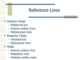
  • 9. Reference Lines  Anterior Chest  Midsternal line  Anterior axillary lines  Midclavicular lines  Posterior Chest  Vertebral line  Midscapular lines  Axilla  Anterior axillary lines  Midaxillary lines  Posterior axillary lines
  • 14. Anatomy Points to Remember  Lungs are symmetric  Lungs are divided into lobes  Right lung = 3 lobes  Left lung = 2 lobes  Primary muscles of respiration  Diaphragm – divides chest from abdomen  External intercostal muscles  Accessory muscles
  • 15. Anatomy Points to Remember  Upper Airway  Nose, pharynx, larynx, intrathoracic trachea  Functions in respiration  Conduct air to lower airway  Filter to protect lower airway  Warm and humidify inspired air
  • 16. Anatomy Points to Remember  Lower Airway  Trachea, bronchi, bronchioles  Functions in respiration  Conduct air to alveoli  Clear mucociliary structures  Alveoli  Functional unit  Gas exchange  Production of surfactant
  • 17. Anatomy Points to Remember  Lower Airway  Trachea splits into left and right mainstem bronchi which are further subdivided into bronchioles  Right bronchus is shorted, wider and more upright than the left  Functions in respiration  Conduct air to alveoli  Clear mucociliary structures
  • 22. Chest Anatomy  Web Anatomy: http://www.gen.umn.edu/faculty_staff/jensen /1135/webanatomy/
  • 23. History  Chief Complaint and HPI  Cough  Shortness of breath/Dyspnea
  • 24. Cough  Onset – sudden, gradual  Duration  Nature – dry, moist, hacking, barking  Sputum – amount, color, odor  Severity – disrupts activities  Associated symptoms – sneezing, dyspnea, fever, chills, congestion, gagging  What brings it on? – anxiety, talking, activity  What makes it better?  What has been tried? – medications, treatments  Anything similar in the past?
  • 25. Shortness of Breath (SOB) / Dyspnea  Onset – sudden, gradual  Duration  Severity – disrupts activities  Associated symptoms – night sweats, pain, chest pressure, discomfort, ankle edema, diaphoresis, cyanosis  What brings it on? – position, time of day, exercise, allergens, emotions  What makes it better?  What has been tried? – medications, inhalers, oxygen  Anything similar in the past?
  • 26. History  Past Health History  Lung disease or breathing problems  Frequent severe colds, asthma, emphysema, bronchitis, pneumonia, tuberculosis  Last PPD and/or chest x-ray  Allergies  Medication use  Family History
  • 27. History  Personal and Social History  Tobacco  Alcohol  Drugs  Home environment  Occupational environment  Travel  Health Promotional Activities
  • 29. Equipment and Techniques  Equipment  Stethoscope  Techniques  Inspection  Palpation  Percussion  Auscultation
  • 30. Inspection  General  Appearance  Posturing  Breathing effort  Trachea position  Midline
  • 31. Inspection  Chest Wall Configuration  Form  Symmetry  Muscle development  Anterior-Posterior (AP) diameter  Approximately ½ the transverse diameter  Transverse: Anterior-Posterior = 2:1  Costal angle  90 degrees or less
  • 32. Inspection  Oxygenation: cyanosis  Nails  Skin  Lips  Respiratory Effort  Respiratory rate and depth  Breathing pattern  Chest expansion
  • 33. Palpation  Trachea – for position  Chest wall – for symmetry
  • 34. Palpation  Thoracic Expansion (Excursion)  Place both thumbs at about 7th rib posteriorly along the spinal process Click on the pictures to view video  Extend the fingers of both hands outward over the posterior chest wall  Have the person take a deep breath and observe for bilateral outward movement of thumbs  Normal: bilateral, symmetric expansion  Abnormal: unilateral or unequal
  • 35. Palpation  Vocal (Tactile) Fremitus  Use palmar or ulnar surfaces of hands  Systematically position hands over both sides of posterior chest  Have person repeat “1 – 2 – 3” or “99” as you move from the apices to the bases  Normal: bilaterally symmetrical vibrations  Decreased or absent: obstruction of transmission 0bronchitis, emphysema)  Increased: consolidation (compression) of lung tissue (pneumonia)
  • 36. Auscultation  Auscultate in a systematic manner  Compare one side to the other  Listen one full respiration at each spot  Displace breast tissue to listen directly over chest wall  DO NOT listen through gowns, clothes, etc.  Place your stethoscope over bare skin
  • 37. Auscultation  Evaluate posterior, lateral, and anterior chest  Instruct person to sit upright and breathe in and out slowly through the mouth  This makes it easier to hear the air movement  Use the diaphragm of the stethoscope  Move from the apices to the bases
  • 38. Auscultation  Evaluate for normal sounds Sound Pitch Intensity Quality I:E Location Bronchial High Loud Blowing/ I<E Trachea hollow Bronchovesicular Moderate Moderate Combination I=E Between scapulae, 1st & 2nd ICS lateral to sternum Vesicular Low Soft Gentle rustling/ I>E Peripheral lung breezy
  • 39. Auscultation  Evaluate for adventitious sounds Sound Intensity/ Pitch I/E Quality Clear with Cough Crackles/ Soft (fine)/ High I Discontinuous, Possibly Rales Loud (coarse)/ Low nonmusical, brief Wheeze High E Continuous musical Possibly sounds Ronchi Low E Continuous snoring Possibly sounds Pleural I&E Continuous or Never Friction Rub discontinuous creaking or brushing sounds Stridor I Continuous, crowing Never
  • 40. Auscultation Copy this URL into your Web browser to hear normal and abnormal lung sounds : http://medocs.ucdavis.edu/IMD/420C/sounds/lngsound.htm
  • 41. Developmental Variations  Neonates  Measure the chest circumference  Usually 2-3 cm smaller than head circumference  Chest is round (i.e. AP diameter = transverse)  Obligate nose breathers  Periodic breathing is common  Sequence of vigorous breathing followed by apnea for 10-15 seconds  Only concern if it is prolonged or baby becomes cyanotic
  • 42. Developmental Variations  Neonates  Breathing is diaphragmatic and abdominal  Signs of compromise  Stridor (“crowing”)  Grunting  Central cyanosis  Flaring nares
  • 43. Developmental Variations  Infants and Young Children  Roundness of the chest persist for first 2 years  Chest walls are thinner than the adult’s  Breath sounds may sound louder, and more bronchial than the adult  Bronchovesicular sounds may be heard throughout the chest
  • 44. Developmental Variations  Pregnancy  Costal angle increases to about 105 degrees in the third trimester  Dyspnea and orthopnea are common  Breathes more deeply
  • 45. Developmental Variations  Older Adult  Chest expansion is often decreased  Bony prominences are marked  AP diameter is increased with respect to transverse (but not 1:1)
  • 46. Videos of Thorax and Lung Assessment  Copy these URLs into your Web browser  http://www.conntutorials.com/chapter5.html OR  http://medinfo.ufl.edu/other/opeta/chest/CH_main