


Types of embryosac
- 1. Picture representing the title of the Topic University of Kashmir Department of Bioresources By Shariqa Aisha
- 2. To get an idea about an embryo sac. To know the concept regarding the formation of Embryo sac. To learn the different types of the embryo sacs.
- 3. The embryo sac or female gametophyte is an oval structure present in the ovule of flowering plants. The female gametophyte, also named as megagametophyte or embryo sac, is developed within the carpel, which consists of three elements: the stigma, the style and the ovary. The Polygonum-type ( common type) embryo sac has one egg cell, two synergids, three antipodal cells, and a central cell that contains two nuclei.
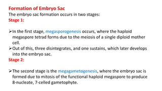
- 4. Formation of Embryo Sac The embryo sac formation occurs in two stages: Stage 1: In the first stage, megasporogenesis occurs, where the haploid megaspore tetrad forms due to the meiosis of a single diploid mother cell. Out of this, three disintegrates, and one sustains, which later develops into the embryo sac. Stage 2: The second stage is the megagametogenesis, where the embryo sac is formed due to mitosis of the functional haploid megaspore to produce 8-nucleate, 7-celled gametophyte.
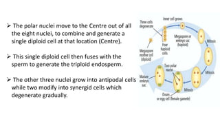
- 5. The polar nuclei move to the Centre out of all the eight nuclei, to combine and generate a single diploid cell at that location (Centre). This single diploid cell then fuses with the sperm to generate the triploid endosperm. The other three nuclei grow into antipodal cells while two modify into synergid cells which degenerate gradually.
- 6. Basis for classification The number of megaspores taking part in the development of embryo sac The number of divisions occurring in the nucleus of the functional megaspore Organization of nuclei in the mature embryo sac
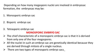
- 7. Depending on how many megaspore nuclei are involved in embryosac formation, the embryosac may be: 1. Monosporic embryo sac 2. Bisporic embryo sac 3. Tetrasporic embryo sac MONOSPORIC EMBRYO SAC The chief characteristic of a monosporic embryo sac is that it is derived from only one of the four megaspores. All the nuclei in such an embryo sac are genetically identical because they are derived through mitosis of a single nucleus. There are two types of monosporic embryo sacs_
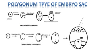
- 8. i) POLYGONUM TYPE: • It is the most common type (81% families). It was first time described in Polygonum divaricatum by Strasburger (1879) . • The embryo sac is formed by the chalazal megaspore of the tetrad and is eight nucleate. • The mature embryo sac comprises a 3- celled egg apparatus, three antipodal cells, and a binucleate central cell.
- 9. j
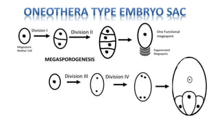
- 10. ii) ONEOTHERA TYPE: • This type of embryo sac is derived from the micropylar megaspore of the tetrad and is four nucleate. • The organization of the mature embryo sac is_ an egg apparatus and a uninucleate central cell. It does not have antipodals. • This type of embryo sac is characteristic of the family Onagraceae. • Schisandra chinensis, however, is the only example outside this family where such a type of embryo sac occurs.
- 12. BISPORIC EMBRYO SAC In plants bearing bisporic embryo sacs the first meiotic division is accomplished by wall formation, so that a dyad is formed. Only one of the dyad cells undergoes the second meiotic division whereas the other one degenerates. In the functional dyad cell wall formation does not occur after the second division, and both the megaspore nuclei contribute to the formation of the embryo sac. Each megaspore nucleus undergoes two mitotic divisions forming eight nuclei.
- 14. On the basis of the position of functional dyad bisporic embryo sacs are of two types: i) ALLIUM TYPE: The embryo sac is derived from the chalazal dyad cell. ii) ENDYMION TYPE: The embryo sac is formed by the micropylar dyad cell. TETRASPORIC EMBRYO SAC In this type of embryo sac, meiotic division of the megaspore mother cell is not accompanied by cytokinesis and hence all the four haploid nuclei lie in a single cell called Coenomegaspore. All four nuclei of coenomegaspore participate in the formation of embryo sac.
- 15. Genetically, it is more heterogeneous than bisporic type of embryo sac because the four products of meiosis involved in its formation are genetically different. The tetrasporic embryo sacs are further divided into many types. TYPES OF TETRASPORIC EMBRYO SAC I- No nuclear fusion occurs • AdoxaType • Plumbago Type • PenaeaType • PepromiaType • DrusaType
- 16. ADOXA TYPE • It has 8 nuclei which are formed by the mitotic division of the four haploid nuclei of the coenomegaspore. • The arrangement of the 8 nuclei in the embryo sac is the same as in Polygonum type. • Example –Adoxa, Sambucus, Ulmus, Tulipa, Erythronium etc
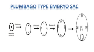
- 17. PLUMBAGO TYPE • This type of embryo sac is characterized by the absence of synergids and antipodals. • Out of four haploid coenomegaspore one migrates to the micropylar end, one at chalazal end and two at the lateral sides. • Each nuclei divides again and formed four groups of two nuclei. • One of the nucleus from each group moves to the center of the cell and form four polar nuclei. • The remaining nucleus at the micropylar is cut off by a membrane and form the egg. There are no synergids. • The other three nuclei usually disappear but occasionally they too may be cut off by membranes and appear as accessory egg cells. • Example –Plumbaginaceae family
- 19. PENAEA TYPE • The four haploid nuclei of the coenomegaspore undergo two successive mitotic divisions forming 16 nuclei. • These nuclei arrange themselves in four groups of four each, one at the micropylar end, one at chalazal end and one each on the two lateral sides. • Now one nucleus from each groups migrates to the centre, and these four nuclei in the centre form polar nuclei.. • The three nuclei at the micropylar end are cut off by membranes and form the egg apparatus. • The remaining three groups of nuclei (one chalazal and two lateral) degenerate at maturity. • Example –Family Penaeaceae, Malpighiaceae and Euphorbiaceae.
- 21. PEPEROMIA TYPE • The egg apparatus of Peperomia type is characterized by a single synergid. • The four haploid nuclei of coenomegaspore undergo two successive mitotic divisions forming 16 nuclei. • Two nuclei at the micropylar end form egg and a synergid, eight fuse in the centre of the cell to form a polar nucleus and the remaining six at the chalazal end formed antipodals. • Example-Peperomia and Gunnera
- 23. DRUSA TYPE • 16 nucleate embryosac. • This type of embryo sac is characterized by large number of antipodals. • In the mature embryo sac three nuclei form egg apparatus. Two act as polar nuclei and the remaining 11 nuclei are cut off by membrane and form antipodal cells. • The number and organization of nuclei may vary due to irregularity in the divisions. • Example– Drusa, Rubia, Chrysanthemum,Ulmus etc.
- 25. After the second meiotic division three megaspore nuclei fuse to form triploid nucleus at the chalazal end of the coenomegaspore, The fourth nucleus at micropylar end remains haploid • Fritillaria Type • Plumbagella Type FRITILARIA TYPE • The four haploid nuclei of the coenomegaspore arrange themselves in two groups –three at the chalazal end in the form of a triploid nucleus and one haploid at the micropylar end. • The triploid chalazal as well as the haploid micropylar nucleus undergo two mitotic divisions and as a result four triploid nuclei are formed at the chalazal end and four haploid at the micropylar end.
- 26. • In mature embryo sac three haploid nuclei organize into egg apparatus, three triploid into antipodal and remaining one haploid and one triploid nuclei move to the centre where they fuse to form a tetraploid polar nucleus. • Example –Fritillaria, Lilium, Piper and Gaillardia .
- 27. PLUMBAGELLA TYPE • The initial development is similar to Fritillaria type and a triploid nucleus is formed at the chalazal end and a haploid at the micropylar end. • Each of these nuclei undergoes a single mitotic division and form two groups of two nuclei each. • One triploid nuclei from chalazal end and one haploid nucleus from the micropylar end fuse at the centre and form tetraploid polar nucleus. • One haploid nucleus at the micropylar end forms the egg and one triploid nucleus at the chalazal end the single antipodal. • There is no synergids.
- 28. THE EMBROYOLOGY OF ANGIOSPERMS _ S S BHOJWANI & S P BHATNAGAR ARIHANT_ LIFE SCIENCES_ ASHISH NAGESH & PRASHANT KUMAR http://www.sciencedirect.com