Location via proxy:   [ UP ]  
[Report a bug]   [Manage cookies]                
SlideShare a Scribd company logo

1

Water vascular system in
Echinodermata: structure and
functions
By
Shivani R Bhomle
M.Sc 1st year
section zoology

2

Echinodermata
• Jacob Klein (1734) first used the term
Echinodermata for sea urchins.
• The Echinoderms are spiny skinned animals.
• They are exclusively marine animals. They
do not show distinct head.
• They show peculiar water vascular system of
coelomic origin.
• They are coelomate animals with
pentaramous radial symmetry, that is the
body can be divided into five parts arranged
around a central axis, but the larva is
bilaterally symmetrical.
• They have an endoskeleton of calcareous

3

Water vascular system in
Echinodermata
• It is a system of canals and appendages of body wall .Water
vascular System is a sort of hydraulic pressure mechanism which
is also known as the ambulocral system.
• Water vascular system functions as a means of locomotion.
• It consist of
• Madreporite or sieve plate
• Stone canal
• Water ring of ring canal
• Radial canals
• Lateral canal
• Polian vesicles
• Tiedemann’s bodies
• Podia or tube feets

4

madreporite

5

• Internal canal of the system connect with
sea water outside through button shaped
madreporite also called sieve plate
• Located on aboral surface
• It is calcareous, having about 200 pores
each lead into pore canal
• madreporite- pores-pore canal-to form
common canal-open into ampulla beneath
madreporite

6

Stone canal
stone canal is a vertical s-shaped canal that starts
from the madreporite and extends orally and joins
the ring canal.
It is named stone canal because its walls are
strengthened by hard calcareous rings.
Its cavity is lined with flagellated or ciliated cell

7

water ring or Ring canal
• The stone canal opens into a
circular canal which is known as
the ring canal.
• It is situated just internal to the
peristomial ring of ossicles.
• Wall of ring canal are folded,
dividing its lumen into more or less
separate channels.

8

Radial canal
• Ring canal gives off five radial canals, one in each arm.
• They are long and ciliated.
• Each radial canal terminates in the terminal tentacle present at
the tip of the arm.
• They are located on oral side of arms in the ambulacral
grooves and are covered by the ambulacral spines

9

Lateral canal
• In each arm the radial canal gives off two rows
of numerous short narrow branches which are
known as lateral canals.
• Lateral canal passes between the osicles on
each side to enter the coelom.
• Each lateral canal is attached to the base of a
tube foot and is provided with a valve and
terminates into a bulb or ampulla .
• which prevents the backflow of the fluid.

10

Polian vesicals
• The ring canal gives off inter radically on
its outer side five elongated pear shaped
muscular sacs with long necks are known
as the polian vesicles.
• These store water which is utilized when
the star fish comes out of water.

11

Tiedemann’s
Bodies
• The ring canal on its inner
side gives off small
irregularly shaped bodies
which are known as the
tiedemann’s bodies.
• They are of five pairs.
• The function of these bodies
is to produce coelomic
corpuscles which are
released into the ring canal
and so the system is known
as water vascular system.

12

Tube feets
• Tube feet is hollow, elastic, thin walled closed
cylinder.
• The tip of podium is flattered forming a sucker
for attachment.
• The basal end of the tube feet expands to
form a little rounded bulb or bladder called
ampullae.
• The ampullae have the same arrangement as
the tube feet.
• Muscle fibers are present on walls of the
ampullae and the tube feet.
• The lumens of the ampullae and tube feet are
the continuation of the lateral canal.

13

Mechanism of water
vascular system
• Each tube foot-ampulla unit may be cut off from the rest of
the water vascular system by a valve, so arranged as to
maintain the pressure developed within the unit. The
functioning of the unit depends upon differences between
the musculature of the ampulla and the tube foot.
• In the ampulla the muscles consist mainly of rings of
smooth muscles which are set vertically and lie parallel to
the long axis of the arm. Contraction of these muscles
brings about protraction of the tube foot and drives the
fluid out of the ampulla into the foot.
• The increase in pressure is wholly translated into
elongation of the foot that subsequently comes in contact
with the substratum. The musculature of the tube foot, in
contrast to that of the ampulla, consists of longitudinal
muscles, which are bounded on the inner side of the
coelom.

14

water vascular system by shivani bhomle

15

.Locomotion:
• The main function of the water vascular
system is to help in locomotion.
Echinoderms having suctorial podia (tube-
feet) can adhere to the substratum
temporarily. The mechanism of locomotion
has discussed in detail under the water
vascular system of Asterias and Echinus.

More Related Content

water vascular system by shivani bhomle

  • 1. Water vascular system in Echinodermata: structure and functions By Shivani R Bhomle M.Sc 1st year section zoology
  • 2. Echinodermata • Jacob Klein (1734) first used the term Echinodermata for sea urchins. • The Echinoderms are spiny skinned animals. • They are exclusively marine animals. They do not show distinct head. • They show peculiar water vascular system of coelomic origin. • They are coelomate animals with pentaramous radial symmetry, that is the body can be divided into five parts arranged around a central axis, but the larva is bilaterally symmetrical. • They have an endoskeleton of calcareous
  • 3. Water vascular system in Echinodermata • It is a system of canals and appendages of body wall .Water vascular System is a sort of hydraulic pressure mechanism which is also known as the ambulocral system. • Water vascular system functions as a means of locomotion. • It consist of • Madreporite or sieve plate • Stone canal • Water ring of ring canal • Radial canals • Lateral canal • Polian vesicles • Tiedemann’s bodies • Podia or tube feets
  • 5. • Internal canal of the system connect with sea water outside through button shaped madreporite also called sieve plate • Located on aboral surface • It is calcareous, having about 200 pores each lead into pore canal • madreporite- pores-pore canal-to form common canal-open into ampulla beneath madreporite
  • 6. Stone canal stone canal is a vertical s-shaped canal that starts from the madreporite and extends orally and joins the ring canal. It is named stone canal because its walls are strengthened by hard calcareous rings. Its cavity is lined with flagellated or ciliated cell
  • 7. water ring or Ring canal • The stone canal opens into a circular canal which is known as the ring canal. • It is situated just internal to the peristomial ring of ossicles. • Wall of ring canal are folded, dividing its lumen into more or less separate channels.
  • 8. Radial canal • Ring canal gives off five radial canals, one in each arm. • They are long and ciliated. • Each radial canal terminates in the terminal tentacle present at the tip of the arm. • They are located on oral side of arms in the ambulacral grooves and are covered by the ambulacral spines
  • 9. Lateral canal • In each arm the radial canal gives off two rows of numerous short narrow branches which are known as lateral canals. • Lateral canal passes between the osicles on each side to enter the coelom. • Each lateral canal is attached to the base of a tube foot and is provided with a valve and terminates into a bulb or ampulla . • which prevents the backflow of the fluid.
  • 10. Polian vesicals • The ring canal gives off inter radically on its outer side five elongated pear shaped muscular sacs with long necks are known as the polian vesicles. • These store water which is utilized when the star fish comes out of water.
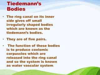
  • 11. Tiedemann’s Bodies • The ring canal on its inner side gives off small irregularly shaped bodies which are known as the tiedemann’s bodies. • They are of five pairs. • The function of these bodies is to produce coelomic corpuscles which are released into the ring canal and so the system is known as water vascular system.
  • 12. Tube feets • Tube feet is hollow, elastic, thin walled closed cylinder. • The tip of podium is flattered forming a sucker for attachment. • The basal end of the tube feet expands to form a little rounded bulb or bladder called ampullae. • The ampullae have the same arrangement as the tube feet. • Muscle fibers are present on walls of the ampullae and the tube feet. • The lumens of the ampullae and tube feet are the continuation of the lateral canal.
  • 13. Mechanism of water vascular system • Each tube foot-ampulla unit may be cut off from the rest of the water vascular system by a valve, so arranged as to maintain the pressure developed within the unit. The functioning of the unit depends upon differences between the musculature of the ampulla and the tube foot. • In the ampulla the muscles consist mainly of rings of smooth muscles which are set vertically and lie parallel to the long axis of the arm. Contraction of these muscles brings about protraction of the tube foot and drives the fluid out of the ampulla into the foot. • The increase in pressure is wholly translated into elongation of the foot that subsequently comes in contact with the substratum. The musculature of the tube foot, in contrast to that of the ampulla, consists of longitudinal muscles, which are bounded on the inner side of the coelom.
  • 15. .Locomotion: • The main function of the water vascular system is to help in locomotion. Echinoderms having suctorial podia (tube- feet) can adhere to the substratum temporarily. The mechanism of locomotion has discussed in detail under the water vascular system of Asterias and Echinus.