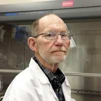
D. Lindsay
Virginia Tech, Biomedical Sciences and Pathobiology, Faculty Member
- Dr. David S. Lindsay is a Parasitologist and has been conducting animal disease research since 1978. The focus of his... moreDr. David S. Lindsay is a Parasitologist and has been conducting animal disease research since 1978. The focus of his research has been on Apicomplexan parasites that cause coccidiosis (Eimeria, Cystoisospora, and Caryospora), cryptosporidiosis, sarcocystosis, neosporosis, and toxoplasmosis in man and domestic animals. He also has studied zoonotic flagellates (Leishmania infantum and Trypanosoma cruzi) that cause leishmaniasis and Chagas’ disease. His major contributions science so far include (1). Describing the life cycle and epidemiology of Cystoisospora suis (syn Isospora suis) an important cause of diarrhea in neonatal pigs (1978-1990). (2.) Defining the life cycles and host specificity of Cryptosporidium spp. in chickens and demonstrating that they were of minimal zoonotic threat. (3.) He pioneered the study of Toxoplasma gondii tissue cysts in mammalian cell culture. (4.) Made major contributions to biology of Neospora caninum and Sarcocystis neurona economically important parasites of cattle and horses, respectively. And (5) Isolated of Leishmania infantum and Trypanosoma cruzi from dogs that never left the United States. His group also demonstrated that L. infantum could be maternally transmitted in dogs helping explain it presence in the absence of appropriate sand fly vectors.edit
Sarcocystis neurona was isolated from the brain of a juvenile, male southern sea otter (Enhydra lutris nereis) suering from CNS disease. Schizonts and merozoites in tissue sections of the otter's brain reacted with anti-S. neurona... more
Sarcocystis neurona was isolated from the brain of a juvenile, male southern sea otter (Enhydra lutris nereis) suering from CNS disease. Schizonts and merozoites in tissue sections of the otter's brain reacted with anti-S. neurona antiserum immunohistochemically. Development in cell culture was by endopolyogeny and mature schizonts were ®rst observed at 3 days postinoculation. PCR of merozoite DNA using primer pairs JNB33/JNB54 and restriction enzyme digestion of the 1100 bp product with Dra I indicated the organism was S. neurona. Four of four interferon-g gene knockout mice inoculated with merozoites developed S. neurona-associated encephalitis. Antibodies to S. neurona but not Sarcocystis falcatula, Toxoplasma gondii, or Neospora caninum were present in the serum of inoculated mice. This is the ®rst isolation of S. neurona from the brain of a non-equine host. 7
Research Interests:
Toxoplasma gondii was isolated from brain or heart tissue from 15 southern sea otters (Enhydra lutris nereis) in cell cultures. These strains were used to infect mice that developed antibodies to T. gondii as detected in the modified... more
Toxoplasma gondii was isolated from brain or heart tissue from 15 southern sea otters (Enhydra lutris nereis) in cell cultures. These strains were used to infect mice that developed antibodies to T. gondii as detected in the modified direct agglu-tination test and had T. gondii tissue cysts in their brains at necropsy. Mouse brains containing tissue cysts from 4 of the strains were fed to 4 cats. Two of the cats excreted T. gondii oocysts in their feces that were infectious for mice. Molecular analyses of 13 strains indicated that they were all type II strains, but that they were genetically distinct from one another.
Research Interests:
Tissue cyst stages are an intriguing aspect of the developmental cycle and transmission of species of Sarcocystidae. Tissue-cyst stages of Toxoplasma, Hammondia, Neospora, Besnoitia, and Sarcocystis contain many infectious stages... more
Tissue cyst stages are an intriguing aspect of the developmental cycle and transmission of species of Sarcocystidae. Tissue-cyst stages of Toxoplasma, Hammondia, Neospora, Besnoitia, and Sarcocystis contain many infectious stages (bradyzoites). The tissue cyst stage of Cystoisospora (syn. Isospora) possesses only 1 infectious stage (zoite), and is therefore referred to as a monozoic tissue cyst (MZTC). No tissue cyst stages are presently known for members of Nephroisospora. The present report examines the developmental biology of MZTC stages of Cystoisospora Frenkel, 1977. These parasites cause intestinal coccidiosis in cats, dogs, pigs, and humans. The MZTC stages of C. belli are believed to be associated with reoccurrence of clinical disease in humans.
Research Interests: Coccidia and Coccidiosis
Equine protozoal myeloencephalitis (EPM) is a serious neurological disease of horses in the Americas. The protozoan most commonly associated with EPM is Sarcocystis neurona. The complete life cycle of S. neurona is unknown, including its... more
Equine protozoal myeloencephalitis (EPM) is a serious neurological disease of horses in the Americas. The protozoan most commonly associated with EPM is Sarcocystis neurona. The complete life cycle of S. neurona is unknown, including its natural intermediate host that harbors its sarcocyst. Opossums (Didelphis virginiana, Didelphis albiventris) are its definitive hosts. Horses are considered its aberrant hosts because only schizonts and merozoites (no sarcocysts) are found in horses. EPM-like disease occurs in a variety of mammals including cats, mink, raccoons, skunks, Pacific harbor seals, ponies, and Southern sea otters. Cats can act as an experimental intermediate host harboring the sarcocyst stage after ingesting sporocysts. This paper reviews information on the history, structure, life cycle, biology, pathogenesis, induction of disease in animals, clinical signs, 0304-4017/01/$ – see front matter Published by Elsevier Science B.V. PII: S 0 3 0 4-4 0 1 7 (0 0) 0 0 3 8 4-8
Research Interests:
Raptors serve as the definitive host for several Sarcocystis species. The complete life cycles of only a few of these Sarcocystis species that use birds of prey as definitive hosts have been described. In the present study, Sarcocystis... more
Raptors serve as the definitive host for several Sarcocystis species. The complete life cycles of only a few of these Sarcocystis species that use birds of prey as definitive hosts have been described. In the present study, Sarcocystis species sporocysts were obtained from the intestine of a Cooper's hawk (Accipiter cooperii) and were used to infect cell cultures of African green monkey kidney cells to isolate a continuous culture and describe asexual stages of the parasite. Two clones of the parasite were obtained by limiting dilution. Asexual stages were used to obtain DNA for molecular classification and identification. PCR amplification and sequencing were done at three nuclear ribosomal DNA loci; 18S rRNA, 28S rRNA, and ITS-1, and the mitochondrial cytochrome c oxidase subunit 1 (cox1) locus. Examination of clonal isolates of the parasite indicated a single species related to S. columbae (termed Sarcocystis sp. ex Accipiter cooperii) was present in the Cooper's hawk. Our results document for the first time Sarcocystis sp. ex A. cooperii occurs naturally in an unknown intermediate host in North America and that Cooper's hawks (A. cooperii) are a natural definitive host.
