Academia.edu no longer supports Internet Explorer.
To browse Academia.edu and the wider internet faster and more securely, please take a few seconds to upgrade your browser.
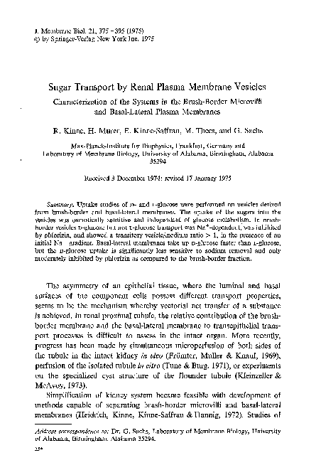
Sugar transport by renal plasma membrane vesicles
Related Papers
Proceedings of the National …
Sugar uptake into brush border vesicles from normal human kidney1977 •
Uptake studies of simple sugars were performed on a membrane fractions containing osmotically active vesicles prepared from normal human kidney cortex. The uptake of D-glucose was found to be sodium-dependent and phlorizin-sensitive. The ...
Biochimica et Biophysica Acta (BBA) - Biomembranes
Sugar uptake into brush border vesicles from dog kidney. II. Kinetics1978 •
Annals of the New York Academy of Sciences
Small Proteins Affect Na + Gradient-Dependent d-Glucose Transport in Isolated Renal Brush-Border Membrane Vesicles1985 •
Biochimica et Biophysica Acta (BBA) - Biomembranes
Interactions between Na+-dependent uptake of d-glucose, phosphate and l-alanine in rat renal brush border membrane vesicles1981 •
Biochimica et Biophysica Acta (BBA) - Biomembranes
Proton/solute cotransport in rat kidney brush-border membrane vesicles: relative importance to both d-glucose and peptide transport1996 •
Biochimica et Biophysica Acta (BBA) - Molecular Cell Research
Kinetic studies of d-glucose transport in renal brush-border membrane vesicles of streptozotocin-induced diabetic rats1985 •
Biochimica et Biophysica Acta (BBA) - Biomembranes
Enzyme activities and sodium-dependent active d-glucose transport in apical membrane vesicles isolated from kidney epithelial cell line (LLC-PK1)1984 •
Biochimica et Biophysica Acta (BBA) - Biomembranes
Studies of the kinetics of Na+ gradient-coupled glucose transport as found in brush-border membrane vesicles from rabbit jejunum1984 •
Pfl�gers Archiv European Journal of Physiology
Co-expression of an anion conductance pathway with Na+-glucose cotransport in rat renal brush-border membrane vesicles1993 •
Biochimica et Biophysica Acta (BBA) - Biomembranes
In vitro effect of ethanol on sodium and glucose transport in rabbit renal brush border membrane vesicles1991 •
RELATED PAPERS
Academia Letters
Conflict, famine, outbreaks, and a pandemic: a deadly mix2022 •
The Ramanujan Journal
New hypergeometric connection formulae between Fibonacci and Chebyshev polynomials2015 •
Feminisms: An Anthology of Literary Theory and …
Visual pleasure and narrative cinema1975 •
2023 •
XIII. ULUSLARARASI SİNAN SEMPOZYUMU ''MİMARLIK VE SORUMLULUK''
Dijital Çağın Gereklilikleri ve Şehrin Sorumlulukları Üzerine Mahalle Medeniyeti2023 •
2015 •
2021 •
Buletin Ekonomi Moneter dan Perbankan
COVID-19, Policy Responses, and Industrial Productivity Around the Globe2021 •
2013 •
2022 •
Journal of Macroeconomics
Striking a balance: Optimal tax policy with labor market duality2020 •
Journal of Islamic Finance
The Role of Islamic Micro-Finance in Enhancing Human Development in Muslim Countries2016 •
cetak notes dengan logo, cetak notes dengan nama, cetak notes dengan gambar, cetak notes dengan tulisan, cetak notes personalisasi, cetak notes untuk souvenir, cetak notes untuk perusahaan, cetak notes untuk acara, cetak notes untuk hadiah, cetak notes untuk promosi
cetak notes dengan logo, cetak notes dengan nama, cetak notes dengan gambar, cetak notes dengan tulisan, cetak notes personalisasi, cetak notes untuk souvenir, cetak notes untuk perusahaan, cetak notes untuk acara, cetak notes untuk hadiah, cetak notes untuk promosi2024 •
RELATED TOPICS
- Find new research papers in:
- Physics
- Chemistry
- Biology
- Health Sciences
- Ecology
- Earth Sciences
- Cognitive Science
- Mathematics
- Computer Science

 Rolf Kinne