Review
For reprint orders, please contact reprints@future-science.com
Inhaled therapies for tuberculosis and the
relevance of activation of lung macrophages by
particulate drug-delivery systems
Pathogenic strains of Mycobacterium tuberculosis (Mtb) induce ‘alternative activation’ of lung macrophages that they
colonize, in order to create conditions that promote the establishment and progression of infection. There is some
evidence to indicate that such macrophages may be rescued from alternative activation by inhalable microparticles
containing a variety of drugs. This review summarizes the experience of various groups of researchers, relating to
observations of induction of a number of classical macrophage activation pathways. Restoration of a ‘respiratory
burst’ and upregulation of reactive oxygen species and nitrogen intermediates through the phagocyte oxidase and
nitric oxide synthetase enzyme systems; induction of proinlammatory macrophage cytokines; and inally induction
of apoptosis rather than necrosis of the infected macrophage are discussed. It is suggested that there is scope to
co-opt host responses in the management of tuberculosis, through the route of pulmonary drug delivery.
Inhalation therapy by means of particulate
drug-delivery systems shows promise as an
approach to combat tuberculosis (TB). Recent
research authenticates the applicability of various inhalable drug-delivery systems such as
microparticles [1–3] , nanoparticles [4] and liposomes [5,6] apart from drug powders by themselves (compounded with inhalable excipients) [7–9] , for TB therapy. Inhalations deliver
therapeutic or prophylactic agents directly to
the site of infection, specifically lung (alveolar) macrophages (AM). This noninvasive
route of delivery also offers additional advantages: reduction in dose, frequency and duration of treatment, lower systemic toxicity, and
improved patient compliance. Table 1 summarizes the drugs and delivery systems investigated
for use as inhaled therapies for TB.
Some research groups have experience with
pulmonary delivery of drugs or drug-containing particulates or vesicles by nebulization of
a suspension of the delivery system in a liquid
medium, while others favor dry-powder inhalations as insufflations or ambient-pressure inhalations [1,7,10,11] . Even though nebulization is
more familiar as a method of aerosol delivery for
lung diseases (asthma, hay fever, COPD), drypowder inhalations can deliver larger amounts
of payload to the deep lung. The optimum
size range of particles for inhalation is usually
considered to lie within 1–5 µm, but the key
measurement of suitability for lung delivery is a
particle’s median mass aerodynamic diameter.
10.4155/TDE.11.34 © 2011 Future Science Ltd
Generally, particles below approximately 0.5 µm
are exhaled undeposited as they do not have
enough inertia to go through impaction or sedimentation in the lung, while particles larger
than approximately 5 µm get entangled in the
oropharyngeal and upper airway regions of the
respiratory tree. Microparticles in the respirable size range deposit in the deep lung and are
readily taken up by AM [12,13] .
In recent years, evidence has been presented
to demonstrate that AM infected with Mtb
display markers of classical macrophage activation when they take up inhalable microparticles [14–15,201] . Some researchers have prepared
microparticles incorporating a single anti-TB
drug. Monotherapy, however, is not recommended in TB, so microparticles incorporating multiple drugs may be better suited to the
task. Biodegradable microparticles composed
of poly(lactide) and incorporating a high payload of isoniazid and rifabutin have been investigated in some detail, including their in vitro/
in vivo efficacy in experimental animals,
pharmacokinetics and biodistribution of the
incorporated drugs, ana lysis of innate effectors
or inflammatory mediators induced as a result
of inhalation or phagocytosis by macrophages
in culture, induction of caspase-dependent and
-independent apoptosis, autophagy, purinergic receptor activity, mitochondrial membrane
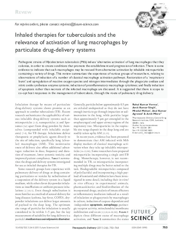
stabilization and so on [1,3,10,16–18,201] . FiguRe 1
depicts three different states of macrophage
activation, and Table 2 summarizes the status
Therapeutic Delivery (2011) 2(6), 753–768
Rahul Kumar Verma1,
Amit Kumar Singh1,
Mradul Mohan1, Atul Kumar
Agrawal1 & Amit Misra†1
1
Pharmaceutics Division, Central Drug
Research Institute, CSIR, Lucknow,
226001, India
†
Author for correspondence:
Tel.: +91 522 261 2411
Fax: +91 522 262 3405
E-mail: amit_misra@cdri.res.in
ISSN 2041-5990
753
�Review | Verma, Singh, Mohan, Agrawal & Misra
Table 1. Drugs and delivery systems investigated as inhalation therapies for tuberculosis.
Drug-delivery system
Drug(s)/peptide
Mode of administration Animal model
Microparticles
Rifampicin
Rifampicin
Rifampicin
P-aminosalicylic acid
Isoniazid + rifabutin
Isoxyl
Suspension nebulization
Insufflation
Intra-tracheal
Insufflation
Ambient pressure inhalation
Insufflation
Guinea pig
Guinea pig
Rat
Rat
Guinea pig, mouse
Recombinant antigen-85
Capreomycin sulfate
Capreomycin oleate
Insufflation
Inhalation
Insufflation
Guinea pig
Guinea pig
Isoniazid + rifampicin + pyrazinamide
Antigen Rv1733c
Rifampicin
Capreomycin sulfate
Nebulization
Nebulization
Nebulization
Nebulization
Guinea pig
Mouse
Guinea pig, mouse, rat
Isoniazid + rifampicin
Isoniazid + rifampicin + pyrazinamide
Capreomycin sulfate
Live-attenuated BCG + L-leucine
P-824
Nebulization
Nebulization
Insufflation
Insufflation
Insufflation
Guinea pig
Guinea pig
Guinea pig
Mouse
Guinea pig
Nanoparticles
Liposomes
Solid-lipid particles
Dry-powder inhalation
Key Terms
Mass median aerodynamic
diameter: Median of the
distribution of inhalable particle
mass with respect to the
aerodynamic diameter. A better
indicator of lung and airway
deposition and distribution than
just the physical dimensions,
since it takes relative density,
aerosolization conditions and so
on, into account.
Apoptosis: Programmed cell
death by which cells undergo an
ordered sequence of events
which lead to death of the cell.
Autophagy: Extensive
recycling of cytosolic contents
through generation of Golgi
vesicles in a section of the
cytoplasm accomplished
through self-digestion.
Classically activated
macrophages: Exhibit a
Th1-like phenotype, promoting
inlammation, extracellular
matrix destruction
and apoptosis.
Alternative activated
macrophages: display a
Th2-like phenotype, promoting
ECM construction, cell
proliferation and angiogenesis.
754
of various markers associated with each of these
activation states. The intention of this article is
to adduce scientific rationale for incorporating
into the standard treatment of TB, the objective
of activating lung macrophages (e.g., by inhalable particles), so that host–defense responses
may also be co-opted to combat infection.
Induction of alternative activation of
host macrophages by Mtb
Mtb spreads through inhalation of moist
droplets containing small numbers of bacilli,
expelled in the cough of infected persons.
Inhaled droplet nuclei lodge in the pulmonary alveoli and Mtb invades resident AM
through a variety of cell-surface receptors.
Other environ mental microorganisms or
inert particles of similar size and composition
are also phagocytosed by AM in the natural
course of events. Particle phagocytosis is sufficient to induce a classical activation response
in the AM, which mobilizes intracellular and
extracellular calcium [19] , undergoes a respiratory burst, generates free radicals [14,20,201] and
Ref.
[114]
[11]
[115]
[116]
[1,3]
[117]
[118]
[119]
[120]
[121]
[122]
[123]
[124]
[16]
[4]
[8,125]
[126]
[127,128]
thereby destroy and digest the foreign particle.
These responses have collectively been referred
to as the ‘activation’ response [21,22] . Mtb has
co-evolved with the human host to effect entry
into the AM. Mtb enters macrophages through
a variety of surface receptors including mannose receptors, complement receptors, Fc and
scavenger receptors, which results in the delivery of mycobacterium into phagosomes [23] .
Within the phagosome formed, as a result of
phagocytosis of Mtb by AM of ‘susceptible’
individuals, the pathogen is able to evade or
counteract the biochemical events that lead to
destruction of phagocytosed particles. In general terms, the pathogen is able to subvert the
activation response of the infected macrophage
into a phenotype that is best described as alternative activation [21,24] . In individuals that are
‘resistant’ to TB, classical activation of the AM
evidently ensues, resulting in containment or
elimination of the infection. However, there is
of course no clear dichotomy between ‘resistant’ and ‘susceptible’ individuals – nor is there
any reason to expect that exclusively classical
Figure 1. Classical, alternative and microparticle-mediated activation of macrophages (on
facing page). Classical activation is initiated by IFN-g, subsequent to recognition of a ‘pathogenassociated molecular pattern’ by pattern-recognition receptors , which in turn induce the
production of proinflammatory cytokines, such as TNF, IL-12, IFNs, IL-6 on the one hand, and
direct-effector molecules such as ROS and nitric oxide. Alternative activation is initiated by IL-4 and
IL-13 secreted as a consequence of mycobacterial infection, which enhance anti-inflammatory
cytokines and inhibition of proinflammatory mediators. Microparticle-induced activation tends to
reprogam alternatively activated macrophages to display markers of classical activation.
RNI: Reactive nitrogen intermediate; ROS: Reactive oxygen species; TLR: Toll-like receptor.
Therapeutic Delivery (2011) 2(6)
future science group
�Inhaled therapies for TB
Classical activation
Ap
opt
osi
s
IL-
Inflammasome
activation
Proteasome
IFN-γ, TNF,
LPS-TLR ligands
1β
IL-1β
Pro-IL-1β
O2–
TNF, IL-12,
IFN, IL-6, MIP-1
p65 p50
Endoplasmic
reticulum
IκB
p65 p50
AT
ST
TC
R
C-I
I
MHC-I
ONOO
TNF, IL-6, IL-12,
IL-1β
NO
RNI, ROS
iNOS
n
tio
a
z
eri
dim JAK
TLR
MH
Proinflammatory
cytokines
NADPH
oxidases
Caspase-1
NF-κB
| Review
Activation of
inflammasome
Apoptosis
IFN-γ
TNF
TCR
C4+ T cells
C8+ T cells
Alternative activation
Mannose receptor
upregulation
Scavenger receptor
upregulation
Ap
opt
osi
Inhibition of
inflammasome
activation
s
Anti-inflammatory
cytokines
Arginase
IL-10, AMAC-1
IL-4, IL-13
IL-10, IL-4,
AMAC-1
NF-κB
NO
STAT6
Endoplasmic
reticulum
K
JA
JAK STAT3
iNOS
T6
A
ST
Microparticle-mediated
activation
Scavenger
receptor
upregulation
MHC-I
Mannose
receptor
upregulation
IL-4
IL-13
Ap
Caspase
independent
opt
osi
1β
Pro-IL-1β
Golgi
bodies
IL-1β
O2–
TNF, IL-12,
IFN, IL-6, MIP-1
NF-κB
p65 p50
Endoplasmic
reticulum
IκB
p65 p50
AT
ST
MHC-I
ONOO
TNF, IL-6, IL-12,
MIP-1α
NO
RNI, ROS
iNOS
n
tio
a
z
eri
m
i
JAK
d
TLR
TCR
MHC-II
upregulation
Proinflammatory
cytokines
NADPH
oxidases
Caspase-1
Autophagy?
s
IL-
Inflammasome
activation
Microparticles
Humoral
immunity
IL-4Rα
IL-13Rα
MHC-II
downregulation
Arginase-1
Activation of
inflammasome
Apoptosis
IFN-γ
TNF
Mycobacterium
Microparticle
Mycobacterium antigen
C4+ T cells
Therapeutic Delivery © Future Science Group (2011)
future science group
www.future-science.com
755
�Review | Verma, Singh, Mohan, Agrawal & Misra
Table 2. Molecular mediators in classical, alternative and microparticle-induced activation of macrophages.
Classical activation
Alternate activation
Microparticle-induced activation
Free radicals
Inflammasome
↑ •NO, ↑ O2•, ↑ iNOS [136]
Activation [47]
IL-4, IL-13 [21,24]
↑ IL-10, ↑ IL-4, ↓ TNF, ↓ IL-6,
↓ IL-12 [64]
↑ AMAC-1 [131]
↓ MHC-II [135] , ↑ Mannose
receptor [56] , ↑ CD86, ↓ CD18 [134]
↑ Arginase, ↓ •NO [30,60]
Inhibition [51]
Microparticles [14,201]
↑ TNF [3] , ↑ IL-6 [94] , ↑ IL-12 [16]
Chemokines
Marker molecules
IFN-g [28]
↑ TNF [40] , ↑ IL-1b [47] , ↑ IL-6,
↑ IL-12 [46]
↑ MCP-1 [129] , ↑ MIP-1a [130]
↑ MHC-II [30] , ↑ CD86 [134]
Key activation signal
Cytokines
or alternative activation phenotypes would be
discernible in all the AM present in a single
individual at any time. Instead there is evidence
to indicate that in TB-infected mice, alternatively and classically activated AM are present
in close proximity and in kinetically varying
proportions at sites that are proximal or distal
to granulomatous lesions [25] .
Key Terms
Th1 phenotype: Derived
from the phenotype of T helper
1 lymphocytes that secrete
signals (cytokines, chemokines,
growth factors) favoring the
development of inlammatory
immune responses. These
responses can kill intracellular
pathogens or cancerous cells.
Inlammasome:
A multiprotein complex that is
responsible for inlammatory
diseases via activation of
caspases.
Th2 phenotype: deined by
the set of cytokines/
chemokines/factors secreted by
T helper 2 lymphocytes that
promote the development of an
immune response directed
against ‘dangerous material’ in
the systemic circulation.
Phagosome maturation:
Phagosomes steadily acidify and
sequentially acquire markers of
endosomes and lysosomes to
kill engulfed microbe.
756
Classical bactericidal
macrophage activation
Classical activation of macrophages to combat foreign entities is extensively studied and
well characterized [26] . Macrophage activation
needs two steps: preliminary priming and a
subsequent, secondary stimulus [27] . IFN-g and
TNF are recognized as the foremost mediators
of classical macrophage activation [28] . IFN-g
produced by activated natural killer cells, macrophages, T helper cells and cytotoxic T cells,
is usually the major stimulus which primes
macrophage through the JAK-STAT pathway
of transcriptional activation [29,30] . The second
stimulus for classical activation is often provided
by pathogen-associated molecular patterns or
‘dangerous material’ such as microbial lipopolysaccharide or peptidoglycans. Various surface
receptors present on macrophages – Toll-like
receptors (TLRs), mannose receptors, complement receptors, Fc receptors transduce the
second signal. Stimulation of these receptors
induces high levels of proinflammatory mediators such as Th1 cytokines, generation of reactive nitrogen intermediate (RNI) and reactive
oxygen species (ROS) [31] . Classically activated
macrophages with high levels of inflammatory
mediators possess improved capacity of killing
intracellular bacteria, such as Mtb [32] . With
reference to TB, it is of interest to note that
AM are a significant source of Th1 cytokines
[33] and immunocompetent individuals may be
expected to initiate their first encounter with
Mtb by expressing some or all of these cytokines. Cytokine responses will be examined
Therapeutic Delivery (2011) 2(6)
↑ MCP-1 [132] , ↑ MIP-1a [133]
Unknown
↑ •NO, ↑ O2•, ↑ iNOS [102]
Activation [137]
in greater detail after the following sub-sections address other important components of
classical activation.
Effector molecules
(ROS, RNI, host antimicrobial peptides)
Reactive oxygen species and RNI generated as
a consequence of phagocytosis are important
components of the antimicrobial defense mechanism within macrophages or other phagocytic
cells [34] . These are involved in direct killing of
invading pathogens. Phagocytosis and internalization of Mtb into phagosomes induces
macrophages to sharply increase their oxygen
consumption. This phenomenon is called a respiratory burst, and is important in killing intracellular Mtb [35] . O2 acquired in the respiratory
burst participates in a nonmitochondrial reaction
in which it is reduced by an electron to superoxide radical (O2• ) through the NADPH oxidase
enzyme complex [34] . O2• serves as precursor for
highly reactive oxygen and halogen metabolites
that are lethal to engulfed microbes. RNI are
generated through downstream reactions with
O2• following production of nitric oxide (NO),
principally through the involvement of inducible NO synthetase (iNOS). Both NO and RNI
are clearly mycobactericidal in mice [36] , but this
activity is yet to be confirmed in human macrophages [37] . The mechanism of action of free radicals is related to applying oxidative stress on Mtb,
which is a facultative anaerobe, and oxidizing key
surface and cytosolic molecules of the bacillus.
Several antimicrobial molecules produced
by macrophages and other cells of the respiratory tract have been demonstrated to possess
antimycobactericidal activity. These include
host defense peptides suchas defensins a and b,
cathelicidins, histatins hepcidin, collectins and
other molecules such as lysozyme, complement,
immunoglobulins, fibronectin, lactoferrin, transferrin, and so on [38] . These possess direct antimicrobial activity or facilitate the elimination of
infectious pathogens by phagocytes [39] .
future science group
�Inhaled therapies for TB
Signaling molecules (cytokines, chemokines)
Infected macrophages, and lymphocytes that
interact with them, initially produce inflammatory cytokines that signal the presence of an intracellular pathogen. Th1 cells amplify phagocytemediated defense mechanisms against infection
by secreting macrophage-activating cytokines
such as IFN-g, TNF, IL-2, IL-12, and chemokines such as IL-18 [40] . These cytokines promote
the ability of macrophages to phagocytose and
destroy intracellular microbes. IFN-g and TNF
are considered to play a central role in the activation of macrophages, with multiple effects on
T-cell recruitment and phagocyte-mediated clearance of pathogens [41] . These cytokines potentiate the respiratory burst and NO generation [42] ,
activate professional phagocytes and upregulate MHC-II expression on surface phagocytic
cells [43] . Aerosolized IFN-g as adjunct therapy
in TB is under investigation [44] . TNF also plays
a role as modulator of macrophage activation
and contributes to the elimination of mycobacteria [45] . Another Th1 cytokine, IL-12 primarily produced by macrophages and dendritic cells,
induces production of IFN-g and TNF which
further enhance microbial killing [46] .
Cell biological processes
(inlammasome, autophagy, apoptosis)
Recent evidence shows that stimulation of
macrophages by cytokines such as IFN-g and
TNF induces activation of inlammasomes [47] ,
autophagy [48] and apoptosis of the infected macrophage [49] . All three processes, by themselves
or in coordination with each other, are extremely
important in the killing and clearance of intracellular Mtb. Assembly of the inflammasome (a
multiple protein complex) is linked to maturation of the Mtb-containing phagosome [50] ,
and is actively inhibited by a specific Mtb gene
product [51] . If inflammasome assembly, activation and maturation proceeds as expected in
classical activation of macrophages, IL-1b and
caspase-1 are induced. Enhanced production of
IL-1b directs the generation of ROS and RNI
and secretion of proinflammatory cytokines,
which results in a strong host response to the
microorganism [47] . Autophagy is another phenomenon which plays an important role in the
human innate defense mechanism in which
a cell degrades its own intracellular compartments and removes unnecessary proteins and
organelles by sequestering them into a vacuole which ultimately fuses with lysosomes and
degrades [48] . Gutierrez et al. demonstrated that
future science group
| Review
IFN-g induces autophagy in murine macrophages which is essential for antimicrobial action
against Mtb [52] . In another study, Alonso et al.
demonstrated mycobacterial killing by ubiquitin-peptide induced autophagy in infected
macrophages [53] . Another potential mechanism involved in macrophage defense against
mycobacterium is apoptosis. Host cell apoptosis is a last resort defense strategy to limit the
growth of certain intracellular pathogens such
as mycobacterium. It has been shown that this
response sequesters bacilli within apoptotic cells
and restricts their replication directly [54] , and is
also thought to result in ‘appropriate’ presentation of Mtb antigens when such apoptotic bodies
are picked up by bystander antigen-presenting
cells (APCs).
Alternative activation induced by bacteria
A macrophage that has recently been exposed
to IL-4 and IL-13 embarks on a program of
activation that is markedly different from classical activation even if the stimuli provided are
identical [21,24] . Consequently, alternatively activated macrophages are more susceptible to intracellular infections [55] . IL-4 [56] and IL-13 [57]
directly induce alternative activation, while
other Th2 cytokines such as IL-21, IL-25 and
IL-33 further augment it [58,59] . Kaufmann and
colleagues described critical disparity between
transcription responses of alternatively versus
classically activated macrophages. They demonstrated in mouse macrophages, that in contrast
to macrophages activated ‘classically’ using
IFN-g, alternatively activated macrophages
(treated with IL-4) show differential regulation
of genes relating to cell-surface expression of
pathogen recognition receptors, production of
cytokines and chemokines, proteins important
for phagosome maturation and intracellular
matrix remodeling, and so forth. Several key
enzyme systems are also differentially regulated
in alternatively activated macrophages. For
instance, alternatively activated macrophages
upregulate arginase activity which reduces
NO production. It was concluded that alternatively activated macrophages support rather
than inhibit growth of Mtb inside macrophages [60] . The Mtb-infected macrophage
exhibits reduced generation of RNI and ROS,
displays weaker antigen processing, decreased
sensitivity to IFN-g and reduced secretion of
proinflammatory cytokines [61,62] . Virulent Mtb
also stimulates upregulation of the expression
of anti-inflammatory cytokines, manipulates
www.future-science.com
757
�Review | Verma, Singh, Mohan, Agrawal & Misra
signal transduction pathways and suppresses
macrophage apoptosis [54] . In summary, Mtb
can switch ‘classical activation’ of macrophages
into ‘alternative activation’ in susceptible individuals. The ability of mycobacteria to persists
in the host depends on evading or minimizing the induction of protective macrophages
responses, as follows.
Th2 rather than Th1 cytokines
& anti-inlammatory chemokines
Studies on immune responses and cytokine
production by peripheral blood mononuclear
cells isolated from TB patients illustrated elevated levels of IL-4 and reduced IFN-g [63] .
Intracellular Mtb in macrophages downregulates
proinflammatory modulators such as IL-12,
TNF, IL-6 and so on and evokes antinflammatory cytokines such as IL-10, IL-13, IL-4 and
IL-10 [64] . Furthermore, virulent strains seem
to manipulate the activation of the eukaryotic
superfamily of MAPKs to impair cytokine
production or to stimulate anti-inflammatory
response. Mtb inhibits IFN-signaling pathways in human macrophages and it directly or
indirectly interrupts the association of STAT-1
with transcription co-activator CREB binding
protein that are essential for the transcriptional
response to IFN-g [29,30] . It is open to debate
whether reactivation of latent TB is supported
by a switch of Th1-polarized macrophages to the
Th2 phenotype.
Preferential antigen presentation on MHC
class II rather than class I
Mtb infection is intracellular in the strictest
sense, but located in a unique, maturationarrested phagosome. It therefore typically
presents an array of nonessential epitopes on
MHC class II, for recognition by CD4 + T cells.
Peptides derived from Mtb secretory proteins
are loaded on MHC class I via proteasomal processing in the cytosol, but at much lower efficiency [65,66] . In susceptible individuals, therefore, recognition of Mtb-infected macrophages
by CD4 + T cells predominates, typically leading
to secretion of Th2 cytokines, which intensify
alternative activation. Macrophages infected
with Mtb, however, usually show a markedly
reduced expression of MHC class II and hence
decreased antigen presentation. Normally,
expression of MHC class II on macrophages
is unregulated by IFN-g in classical activation,
but chronic TB subdues antigen presentation.
Recent findings suggest that this may be related
758
Therapeutic Delivery (2011) 2(6)
to attenuation of MHC gene transcription by a
19 kDa lipoprotein of Mtb, which could work by
downregulating MHC class II synthesis, inhibiting vesicular trafficking or reducing antigen
processing [67] .
MHC class I-restricted CD8 + cytotoxic T lymphocytes play a crucial role against intracellular
infections in general, and Mtb in particular
[43,68–70] . Flynn demonstrated that mice lacking b-2-microglobulin (a component of MHC
class I molecules) display greater vulnerability to
TB, implying that MHC class I-restricted cells
have a important function in protecting immunity against TB [43] . CD8 + T cells release IFN-g,
lyse infected cells, and exert a direct antimicrobial activity through release of peptides, such as
perforin and granulysin.
Neutralization of free radicals
& antimicrobial peptides
Mtb produces antioxidant enzymes such as
catalase-peroxidase systems and superoxide dismutase to scavenge or degrade ROS produced by
macrophages as a defense mechanism. A variety
of bacterial components are evolved to neutralize
ROS, including sulfatides, lipoarabinomannan
and PGL-1 [62,65,66] . Mtb-encoded methionine
sulfoxide reductase confers resistance to the toxic
effect of peroxynitrite [67] . Upregulation of arginase-1 which inhibits nitric oxide synthesis has
been referred to earlier. Recent studies show that
NoxR1 and NoxR3 expression confer resistance
to the otherwise susceptible M. smegmatis [68,69] .
Mtb-expressed peroxiredoxin alkyl hydroperoxide reductase subunit C (AhpC) protects bacteria
from RNI toxicity [70] .
Inlammasomes, phagosome maturation,
autophagy & apoptosis
Although numerous proinflammatory cytokines
are initially secreted by Mtb-infected macrophages, IL-1b, an important multifunctional proinflammatory cytokine, is found at unexpectedly
low levels. Generally, IL-1b requires proteolytic
cleavage for its activation and processing which
is carried by activation of inflammasomes (a caspase activating protein complex). Master et al.
provided evidence that Mtb inhibits inflammasome assembly and activation and hence IL-1b
processing. Several other reports indicate that
Mtb infection induces assembly of inflammasomes, and macrophages infected in vitro with
mycobacteria continue to release IL-1b, through
an exocytosis pathway involving the pathogenencoded proteins of the ESX–ESAT system [71,72] .
future science group
�Inhaled therapies for TB
Furthermore, excessive inflammasome activation
may be detrimental to the host, both in terms
of immunopathology, as well as spread of disease [73] . Microparticle design aimed at modulating inflammasome assembly has not yet been
reported in the public domain to our knowledge.
Autophagy is also inhibited by Mtb, but
the mechanism whereby this occurs is not yet
known. Normally autophagy involves phagosome–lysosome fusion which depends on production of membrane lipids, phosphatidylinositol 3-phosphate (PI3P) [52] . Evidence indicates
that Mtb inhibits PI3P to prevent autophagy [74] .
Another mechanism by which Mtb evades
autophagy is its translocation to cytoplasm from
the phagosome using ESAT6 protein.
Mtb infection is reported to prime human
macrophages for TNF-mediated apoptosis. The
virulent strains, however, induce significantly less
apoptosis than avirulant or attenuated strains.
Avirulant or attenuated strains of Mtb (H37Ra)
induce apoptosis at even moderate multiplicities
| Review
of infection (MOI), while a closely-related virulent strain (H37Rv) inhibits apoptosis of host
macrophages even at high MOI [54,75,76] . These
observations suggest that apoptosis works as a
host-defense mechanism which is suppressed
by virulent mycobacterium [54] . Placido and
colleagues found that using the virulent strain
H37Rv, apoptosis was induced in a dosedependent fashion in BAL cells recovered from
patients with TB, particularly in macrophages
from HIV-infected patients [77] .
Dual action of inhalable
microparticles: drug delivery
& macrophage activation
Inhaled microparticles are recognized as alien
by AM, which phagocytose them and initiate
innate immune responses. Geiser et al. demonstrated an increase in numbers of airway/lung
macrophages by approximately three times,
immediately after inhalation of polystyrene
microparticles by hamsters [78,79] . This was
Table 3. Factors relevant to inhaled particles and their influence on
macrophage activation.
Factors affecting
macrophage activation
Outcome
Particle-related factors
1–6 µm ↑ phagocytosis and macrophage activation
>1–6 < ↓ phagocytosis and macrophage activation
Rough surface, ↑ phagocytosis,
↑ inflammasome activation
Smooth surface, ↓ phagocytosis
Phagocytosis and macrophage activation:
cationic > anionic > nonanionic†
↑ phagocytosis†, ↑ ROS inflammatory cytokines TNF,
IL-1b, IL-6, IL-12
Phagocytosis and macrophage activation:
opsonized particles > polysaccharide particles >
polymeric > mineral†
↑ crystallinity, ↓ ROS
↑ phagocytosis and macrophage activation, ↑ ROS
Size
Surface topology
Surface charge
Surface coating/
opsonization
Material of construction
Crystallinity
Hydrophobicity
Cell-related factors
Receptor interaction
Cytoskeleton movement
Macrophage status
Ref.
↑ complement receptor
↑ mannose receptor
↑ scavenger receptor
↑ Fc receptor
↑ phagocytosis and macrophage activation
Actin remodeling status
Relaxed and thicker, ↓ phagocytosis
Contracted and thinner, ↑ phagocytosis and activation
Preclassically activated cell, ↑ phagocytosis
and activation
[12,14,138,139]
[140]
[12,141]
[12,14,89,142]
[14,139]
[113]
[12]
[20,143–145]
[146]
[147]
†
These facts are not universal generalizations and not necessarily pertinent to all types of particles. Variations will be
observed with composition of the particles, status of the macrophage and similar related conditions.
ROS: Reactive oxygen species.
future science group
www.future-science.com
759
�Review | Verma, Singh, Mohan, Agrawal & Misra
attributed to chemotaxis following signaling
by macrophages that had ingested the inhaled
particles. The cell biological processes involved
in microparticle-induced macrophage activation
include interactions of the microparticle with
the macrophage cell-membrane molecules, signal transduction down a variety of pathways
and thus activation of gene transcription [80] .
As the outcome of activation, macrophages
release potent substances, proinflammatory
mediators, growth factors and reactive oxygen
or nitrogen intermediates and, failing all else,
undergo programmed cell death. Cellular and
molecular response after microparticle-induced
macrophage activation depends upon the type of
microparticles. Artusson et al. demonstrated that
phagocytosis of polysaccharide microspheres
induces macrophage activation: the production
of cytokines and ROS, which are involved in
host–defense against pathogenic micro-organisms [20] . Similar results were shown by Tabata
and Ikada with polymeric microspheres [81] .
Poly(lactide) (PLA) and poly(lactide-co-glycolide) (PLGA) microspheres have been extensively studied as polymers for encapsulation of
a variety of drugs and vaccines for controlled
delivery [1,2,7,11] . Prior and colleagues studied
microparticles made of PLA and PLGA of different monomer ratios. Microparticle-induced
macrophage activation as reflected in increased
oxidative burst, correlated with increase in
hydrophobicity of the polymer [14] . We too have
observed that inhalable microparticles induce
proinflammatory responses in macrophages
[3,201] . Thus, in addition to selective drug delivery, the ingestion of microparticles may result in
the activation of macrophages and, subsequently,
enhance host–defense functions of the immune
system. Terada et al., however, report that PLGA
microparticles containing rifampicin do not
induce responses such as TNF, NO, IL-10 and
TGF-b1 in a rat macrophage line [2] .
Induction of a proinflammatory macrophage
response, however, has important implications
for immunopathology. Generalized inflammation of lung and airway tissue as a consequence
of inhaled material is unquestionably dangerous
for the patient. Especially in the case of TB, a
patient with already compromised lung function
might be severely stressed if induction of inflammatory responses in lung macrophages progresses
to full-blown inflammation. Thus, it is necessary to arrive at an optimal formulation that
could work at or below a threshold of inducing
proinflammatory responses; promoting innate
760
Therapeutic Delivery (2011) 2(6)
bactericidal activity of infected macrophages, but
not extensive inflammation involving mast cells
or granulocytes and so on.
Surface interaction & phagocytosis
Phagocytic uptake of inhaled microparticles
depends on particle geometry (size, shape, surface coating, hydrophilicity, stiffness), macrophage status (classical or alternative activation,
synchronized cytoskeleton movements, phagosome position) and interaction of particles with
macrophage membrane (nonspecifically by
electrostatic contact or specifically by surface
molecules such as the scavenger receptor, Fc
receptor, complement receptor, mannose receptor, and so on (Table 3) . Some receptors also
facilitate the phagocytosis of inert particles, for
example scavenger receptors enhance uptake of
polystyrene latex [82] , titanium dioxide, silica and
iron oxide microparticles [83,84] and Fc receptors
facilitate uptake of quartz particles [85] . Physical
dimension-wise, there is significantly lower
phagocytic uptake of large (>5 µm) polymer
particles compared with particles of <5 µm of
same aerodynamic diameter [13,86] . Coating the
microparticle surface significantly influences
the uptake of microparticles by macrophages.
Stringer and colleagues demonstrated that a
coating of surfactant protein over microparticles
significantly enhances the uptake of polystyrene
and TiO2 particles [87,88] whereas Evora and
colleagues showed modestly decreased phagocytosis of 1,2-dipalmitoylphosphatidylcholine
(surfactant phospholipid)-coated PLGA microparticles by rat lung macrophages [89] . Prior
et al. demonstrated that phagocytosis was more
efficient with increasing polymer hydrophobicity
(PLA:PLGA [75:25] PLA:PLGA [50:50]) [14] .
Disease is also an important factor for phagocytic
uptake of inhaled particles. For example, Bennett
et al. demonstrated improved particle uptake by
macrophages in mild asthmatic conditions [90] .
Release of cytokines, chemokines
& growth factors
Exposure of lung macrophages to microparticles
can lead to the secretion of cytokine and chemokines which contribute to inflammation in
lungs [91] . Among the important proinflammatory cytokines, TNF, IFN-g and IL-12 play
an important role in the host-defense mechanism against TB. Due to the ability of TNF-a,
IL-1b, and IL-6 to induce varied cellular events,
these cytokines may play a role in inflammatory
processes elicited by inhaled particulates. We
future science group
�Inhaled therapies for TB
demonstrated enhanced secretion of TNF by
Mtb-infected macrophages when treated with
microparticles [3,18,201] . Sharma et al. showed that
TNF-a and IL-12 were significantly induced in
response to treatment of macrophages infected
with Mtb with inhalable microparticles in two
experimental systems – cultured J774 cells and
primary AM isolated from Swiss mice [3] . Yadav
et al. demonstrated similar results with human
monocyte-derived macrophages obtained by subjecting THP-1 cells to phorbol-induced differentiation, and also by peripheral blood monocytederived macrophages from five healthy volunteers.
However, there was very wide variation in the
extent of cytokine secretion by macrophages
obtained from different individuals, and IL-12
upregulation was not observed in human macrophages [201] . An interesting observation that we
consistently encounter is the simultaneous secretion of both Th1 and Th2 cytokines by murine,
as well as human, macrophages infected with
Mtb. Contrary to the well-known fact of crossregulation of Th1 and Th2 lymphocytes, macrophages evidently secrete both IL-10 and TNF/
IFN-g even under controlled culture conditions
and using a clonal cell population. To some
extent, inhalable microparticles apparently induce
and sustain higher secretion of Th1 cytokines by
infected macrophages in comparison to drugs in
solution [3,92,201] .
Some research groups have shown that micronsized asbestos and silica particles augment the
release of TNF from macrophages in vitro and in
vivo but IL-1b and IL-6 release was inconsistent
[93,94] . Lemaire and co-workers demonstrated
an enhanced release of IL-1b and IL-6 by lung
macrophages upon exposure to asbestos particles
[95,96] whereas Driscoll and colleagues did not
observe augmentation of either cytokine [97] .
Howard et al. showed that cells exposed to
micron-sized mineral particles released chemokines such as MIP-la or MIP-2 which are monocyte and macrophage chemoattractants and can
activate the NF-kB transcription factor [98] .
The release of proinflammatory cytokines
results in an array of events including recruitment of other inflammatory cells, stimulation of
cell activation and proliferation and eicosanoid
biosynthesis [97] . All of these are extremely
important in macrophage activation and the
elimination of intracellular pathogens. However,
at this time, there is no clear consensus on the
nature of cytokine and chemokine responses that
can be evoked from Mtb-infected AM as a result
of exposure to microparticles of diverse nature.
future science group
| Review
Oxidative burst-generation RNI & ROS
Upon phagocytosis, microparticles induce
macrophages to generate ROS by stimulating
NADPH oxidase, responsible for the production
of superoxide anions. Prior et al. [14] and Sharma
et al. [3] have demonstrated generation of oxidative
radicals in response to biodegradable polyester
microparticles, and numerous others report that
AM release large amounts of superoxide anions
upon exposure to micron-sized particulates such
as silica [19] or asbestos [99] . Kagan and colleagues
illustrated that exposure to micron-sized mineral particles such as crocidolite and chrysotile
stimulates iNOS, which in turn generates NO
and RNI, both in vivo and in vitro [100] . Primary
macrophages exposed to inhalable microparticles
containing isoniazid and rifabutin, upregulate
NO production, regardless of whether or not
they are infected with Mtb in vitro. Infection
does, however, delay the peak NO response by
approximately 9 h. Drug-containing microparticles also induced significant O2• production by
infected macrophages in the same period [3] . As in
the case of proinflammatory cytokines, enhanced
production and release of ROS and RNI by AM
upon exposure to various particulates may have
both positive and negative effects. It may cause
lung tissue damage, or damage the pathogen, or
both. Indirectly elevated ROS and RNI activate
signal-transduction pathways via kinases and
transcriptions factors, processes that result in a
complex cascade of events that may contribute to
the eradication of pulmonary infection.
Signal-transduction cascades
Microparticles stimulate macrophages and activate signal-transduction cascades, but the specific
receptors and pathways are inadequately understood. Microparticulates including biodegradable particles such as PLGA, polystyrene microparticles and inorganic micron-sized particles
such as silica, asbestos, iron oxide, alum, monosodium urate, and so on, significantly augment
the secretion of IL-1b and activation of caspase-1
in phagocytic cells [17,19,201] . The body of evidence
suggests that this phenomenon is mediated by
activation of inflammasomes (via lysosomal damage) by microparticles [101] . There are evidences
that stimulation of macrophages with micronsized monosodium urate particles induces activation of TLR2 and TLR4 [102] , induces MyD88dependent IL-1 receptor signaling [102,103] , directs
the activation of MAPK pathway (ERK1/2) [104]
and chemokine secretion [105] . Several studies
illustrate that mineral microparticles (asbestos,
www.future-science.com
761
�Review | Verma, Singh, Mohan, Agrawal & Misra
quartz) enhanced the intracellular calcium concentration of macrophage in a dose-dependent
manner, which in turn mediates a number of
calcium-dependent cellular events that may
affect cellular physiological functions including
activation of tyrosine kinases, protein kinase and
phospholipase [19,103] . Several studies illustrate
that enhanced gene expression of inflammatory
mediators upon exposure of particles is associated
with an increased activity of transcription factors
NF-kB and AP-1 [106] .
Apoptosis
Inhalable microparticles containing standard
antituberculosis drugs, enhance both caspase-8
and -9-dependent apoptosis, while blank polymer microparticles (without drug) induced
apoptosis through a caspase-independent pathway [17] . Crystalline mineral microparticles
(mylonite, gabbro, basalt, feldspar, quart and
hornfels) induce similar extents of macrophage
apoptosis [107] . Kang et al. demonstrated that
PLA–PLGA microparticles prepared by supercritical fluid technology were able to induce
apoptosis in a lung cancer cell line A549 [108] ,
while Powell and colleagues demonstrated that
dietary calcium phosphate and titanium oxide
microparticles enhance apoptosis of intestinal
macrophages [109] . As argued earlier, apoptosis is
a last-resort response of the AM to Mtb infection.
Apoptosis of the infected AM denies the pathogen
its sanctuary, and also packages it in apoptotic
bodies for APC uptake through pathways that a
naked bacillus would not employ. It is speculated
that processing and antigen presentation of Mtb
taken up in an apoptotic body would result in
presentation of appropriate, protective epitopes,
rather than establishment of the pathogen in a
maturation-arrested phagosome [201] .
Conclusion
Pathogenic strains of Mtb induce alternative
activation of AM that they colonize, in order to
create conditions that promote the establishment
and progression of infection. A growing body of
evidence suggests that such macrophages may
be rescued from alternative activation by inhalable microparticles containing a variety of drugs.
Biodegradable microparticles can be designed to
mimic the surface properties of pathogens so that
they can be easily phagocytized by AM. Besides
providing efficient drug targeting and high chemotherapeutic potential, inhalable polymeric
particles offer an additional potential to enlist
the innate responses of the macrophage towards
762
Therapeutic Delivery (2011) 2(6)
clearing the infection. Inhaled microparticles have
exhibited the potential to reprogram the activation status of TB-infected macrophages [201] .
Other researchers have reported observations of
activation of a number of classical macrophageactivation pathways as a consequence of exposure
to microparticles [14] . Our own findings point to
restoration of a respiratory burst and upregulation
of ROS and RNI through the phagocyte oxidase
and NOS enzyme systems; induction of proinflammatory macrophage cytokines; and finally
induction of apoptosis rather than necrosis of the
infected macrophage [3,17,18,201] . There is evidence
that particle-induced activation of macrophages
depends upon particle size, surface properties and
chemical composition of the particles. In an early
study, Atwood et al. showed that carbon particles
phagocytosed by Mycobacterium-infected macrophages resulted in a decrease of superoxide and
hydrogen peroxide production and hence reduced
killing of M. bovis by AM [110] . By contrast, our
results illustrate that PLA microparticles enhance
superoxide release and enhance in vitro killing of
intracellular Mtb [3,201] . Prior et al. have indicated
that physicochemical properties of the polymers
used to prepare polyester microparticles had
differential effects on macrophage uptake and
activation [14] . By contrast, Terada et al. showed
that molecular weight and monomer ratio of
PLGA did not influence the phagocytosis activity [15] . Upon stimulation or during phagocytosis,
monocytes produce reactive oxygen metabolites
and other oxygen-independent products [111] .
Terada and colleagues investigated the effect
of mycobacterial infection on phagocytic activity of infected macrophages towards polymeric
microparticles. Results showed that phagocytic
ability of the macrophages is enhanced 1.3–1.5
times compared with uninfected macrophages
up to 24 h [112] . This is an extremely encouraging finding in light of the suspicion that Mtbinfected AMs may lose their phagocytic ability.
Biggs and colleagues investigated the cellular
activation by PLA microparticle in vitro (in the
rat AM cell line NR8383) and in vivo (by pulmonary delivery to guinea pigs) in terms of ROS
and RNI production, and ascertained that as microparticle crystallinity increases, the activation
level of the macrophage cells decreases [113] . This
result suggests that immunomodulatory activity
of the microparticles can be tailored by choice
of polymer.
Finally, inhaled particles containing anti-TB
agents demonstrate surprisingly high efficacy
against experimental animal TB [7,202] . This level
future science group
�Inhaled therapies for TB
of efficacy is not satisfactorily explained by arguments suggesting that high intra-cytosolic drug
concentrations are sufficient to kill this recalcitrant pathogen. The evidence of macrophage
activation offers room to formulate an alternative view: that of enlisting innate macrophage
responses against Mtb infection. Given that a
very small proportion of humans exposed to Mtb
infection progress to active disease, it is likely that
most of us are capable of eliminating or containing the infection through innate (macrophage)
and acquired (lymphocyte) immune responses.
It is also likely that augmentation of host macrophage responses may assist in clearing intracellular Mtb. It is, therefore, suggested that there is
scope to co-opt host responses in the management of TB through the use of appropriately
formulated inhalable microparticles.
Future perspective
There is every likelihood that the possibility
of pharmacological intervention in the hostpathogen dialectic in TB will gain notice of
the research community in the near future.
Pulmonary delivery of the causative agent is a
familiar technique for laboratory researchers
addressing TB. Sufficient expertise and motivation is therefore available to investigate whether
pharmacological or immunomodulatory agents
| Review
can play a role in managing experimental animal TB. Also, several groups have reached an
advanced stage of preclinical testing of inhaled
formulations prior to conducting clinical trials.
The next 5 years should see publication of clinical
data on the safety and efficacy of inhaled therapies for TB. It would be doubly exciting to see
whether speculations on host macrophage activation can be borne out by clinical findings, (and
the present authors are of the view that they definitely will), if only clinical studies also address
host AM parameters in human subjects. Finally, a
novel body of literature is likely to emerge on the
use of agents that possess macrophage-activating
activity for pulmonary delivery in TB.
Financial & competing interests disclosure
Work in our laboratory is funded by CSIR NWP0035.
RK Verma, AK Singh and AK Agrawal acknowledge
Research Fellowships from CSIR, M Mohan from ICMR.
This is CDRI Communication Number 8082. All authors
are employed by the Council of Scientific and Industrial
Research, co assignee of patent US20070154408. The
authors have no other relevant affiliations or financial
involvement with any organization or entity with a financial interest in or financial conflict with the subject matter
or materials discussed in the manuscript apart from those
disclosed. No writing assistance was utilized in the
production of this manuscript.
Executive summary
Mycobacterium tuberculosis induces alternative activation of macrophages that it colonizes.
Phagocytosis of particles is a signal for classical macrophage activation.
Inhaled particles induce markers of classical activation in Mtb-infected macrophages, and possess extraordinarily high efficacy against TB
in mice and guinea pigs.
The physicochemical and pharmaceutical properties of inhalable particles strongly influence their ability to activate macrophages, and it
is possible to engineer particles of ‘desired’ macrophage-activating capability.
In general, hydrophobic, amorphous, multicomponent particles evoke higher levels of macrophage activation.
There is no clear consensus on what degree of macrophage activation is necessary or sufficient to kill intracellular Mtb without
inducing immunopathology.
Nor is there sufficient understanding of inter-species or -individual differences in the amounts and kinetics of effector or signaling
molecules that are produced following macrophage activation.
Nevertheless, there is scope to co-opt innate host immune responses to eliminate Mtb from alveolar macrophages.
Bibliography
2
Papers of special note have been highlighted as:
of interest
of considerable interest
1
Muttil P, Kaur J, Kumar K, Yadav AB,
Sharma R, Misra A. Inhalable microparticles
containing large payload of antituberculosis
drugs. Eur. J. Pharm. Sci. 32(2), 140–150
(2007).
future science group
Hirota K, Hasegawa T, Nakajima T
et al. Delivery of rifampicin-PLGA
microspheres into alveolar macrophages
is promising for treatment of tuberculosis.
J. Control. Release 142(3), 339–346
(2010).
No activation of macrophages observed
despite extensive phagocytic uptake
of microparticles.
www.future-science.com
3
Sharma R, Muttil P, Yadav AB et al. Uptake
of inhalable microparticles affects defense
responses of macrophages infected with
Mycobacterium tuberculosis H37Ra.
J. Antimicrob. Chemother. 59(3), 499–506
(2007).
First report of markers of classical activation
being displayed by macrophages infected
with Mycobacterium tuberculosis (Mtb).
763
�Review | Verma, Singh, Mohan, Agrawal & Misra
4
Pandey R, Zahoor A, Sharma S, Khuller GK.
Nanoparticle encapsulated antitubercular
drugs as a potential oral drug delivery system
against murine tuberculosis. Tuberculosis
83(6), 373–378 (2003).
5
Chimote G, Banerjee R. In vitro evaluation of
inhalable isoniazid-loaded surfactant
liposomes as an adjunct therapy in pulmonary
tuberculosis. J. Biomed. Mater. Res. B Appl.
Biomater. 94(1), 1–10 (2010).
6
7
8
9
10
11
12
13
Deol P, Khuller GK, Joshi K. Therapeutic
efficacies of isoniazid and rifampin
encapsulated in lung-specific stealth
liposomes against Mycobacterium tuberculosis
infection induced in mice. Antimicrob. Agents
Chemother. 41(6), 1211–1214 (1997).
Suarez S, O’Hara P, Kazantseva M et al.
Respirable PLGA microspheres containing
rifampicin for the treatment of tuberculosis:
screening in an infectious disease model.
Pharm. Res. 18(9), 1315–1319 (2001).
First demonstration of high efficacy
of inhaled microparticles in the guinea
pig model.
Garcia-Contreras L, Fiegel J, Telko MJ et al.
Inhaled large porous particles of capreomycin
for treatment of tuberculosis in a guinea pig
model. Antimicrob. Agents Chemother. 51(8),
2830–2836 (2007).
Second-line anti-TB agent as a stand-alone
inhaled therapy.
Garcia-Contreras L, Sung JC, Muttil P
et al. Dry powder PA-824 aerosols for
treatment of tuberculosis in guinea pigs.
Antimicrob. Agents Chemother. 54(4),
1436–1442 (2010).
Verma RK, Kaur J, Kumar K, Yadav AB,
Misra A. Intracellular time course,
pharmacokinetics, and biodistribution of
isoniazid and rifabutin following pulmonary
delivery of inhalable microparticles to mice.
Antimicrob. Agents Chemother. 52(9),
3195–3201 (2008).
Suarez S, O’Hara P, Kazantseva M et al.
Airway delivery of rifampicin microparticles
for the treatment of tuberculosis.
J. Antimicrob. Chemother. 48(3), 431–434
(2001).
14
15
Prior S, Gander B, Blarer N et al. In vitro
phagocytosis and monocyte–macrophage
activation with poly(lactide) and poly(lactideco-glycolide) microspheres. Eur. J. Pharm.
Sci. 15(2), 197–207 (2002).
Illustrated activation of macrophages
by varied ratio of sterile
polymeric microspheres.
Yadav AB, Muttil P, Singh AK et al.
Microparticles induce variable levels of
activation in macrophages infected with
Mycobacterium tuberculosis. Tuberculosis
(Edinb.) 90(3), 188–196 (2010).
Sharma R, Saxena D, Dwivedi AK, Misra A.
Inhalable microparticles containing drug
combinations to target alveolar macrophages
for treatment of pulmonary tuberculosis.
Pharm. Res. 18(10), 1405–1410 (2001).
17
Yadav AB, Misra A. Enhancement of
apoptosis of THP-1 cells infected with
Mycobacterium tuberculosis by inhalable
microparticles and relevance to bactericidal
activity. Antimicrob. Agents Chemother.
51(10), 3740–3742 (2007).
18
Yadav AB, Sharma R, Muttil P et al. Inhalable
microparticles containing isoniazid and
rifabutin target macrophages and ‘stimulate
the phagocyte’ to achieve high efficacy. Indian
J. Exp. Biol. 47(6), 469–474 (2009).
Holian A, Kelley K, Hamilton RF Jr.
Mechanisms associated with human alveolar
macrophage stimulation by particulates.
Environ. Health Perspect. 102(Suppl. 10),
69–74 (1994).
20
Artursson P, Arro E, Edman P, Ericsson JL,
Sjoholm I. Biodegradable microspheres.
V: stimulation of macrophages with
microparticles made of various
polysaccharides. J. Pharm. Sci. 76(2),
127–133 (1987).
Nathan C. Mechanisms and modulation of
macrophage activation. Behring Inst Mitt (88),
200–207 (1991).
27
Meldrum DR, Cleveland JC Jr, Moore EE,
Partrick DA, Banerjee A, Harken AH.
Adaptive and maladaptive mechanisms of
cellular priming. Ann. Surg. 226(5), 587–598
(1997).
28
Van Ginderachter JA, Movahedi K,
Hassanzadeh Ghassabeh G et al. Classical and
alternative activation of mononuclear
phagocytes: picking the best of both worlds
for tumor promotion. Immunobiology
211(6–8), 487–501 (2006).
29
Stark GR, Kerr IM, Williams BR,
Silverman RH, Schreiber RD. How cells
respond to interferons. Annu. Rev. Biochem.
67, 227–264 (1998).
30
Ehrt S, Schnappinger D, Bekiranov S et al.
Reprogramming of the macrophage
transcriptome in response to interferon-g and
Mycobacterium tuberculosis: signaling roles of
nitric oxide synthase-2 and phagocyte oxidase.
J. Exp. Med. 194(8), 1123–1140 (2001).
31
Mosser DM. The many faces of macrophage
activation. J. Leukoc. Biol. 73(2), 209–212
(2003).
32
Day J, Friedman A, Schlesinger LS. Modeling
the immune rheostat of macrophages in the
lung in response to infection. Proc. Natl Acad.
Sci. USA 106(27), 11246–11251 (2009).
33
Wang J, Wakeham J, Harkness R, Xing Z.
Macrophages are a significant source of type 1
cytokines during mycobacterial infection.
J. Clin. Invest. 103(7), 1023–1029 (1999).
34
Hansen K, Mossman BT. Generation of
superoxide (O2-.) from alveolar macrophages
exposed to asbestiform and nonfibrous
particles. Cancer Res. 47(6), 1681–1686
(1987).
35
Nau GJ, Richmond JF, Schlesinger A,
Jennings EG, Lander ES, Young RA. Human
macrophage activation programs induced by
bacterial pathogens. Proc. Natl Acad. Sci. USA
99(3), 1503–1508 (2002).
36
Scanga CA, Mohan VP, Tanaka K, Alland D,
Flynn JL, Chan J. The inducible nitric oxide
synthase locus confers protection against
aerogenic challenge of both clinical and
laboratory strains of Mycobacterium
tuberculosis in mice. Infect. Immun. 69(12),
7711–7717 (2001).
First described activation of alveolar
macrophges by microparticles.
21
Tabata Y, Ikada Y. Effect of the size and
surface charge of polymer microspheres on
their phagocytosis by macrophage.
Biomaterials 9(4), 356–362 (1988).
Gordon S, Martinez FO. Alternative
activation of macrophages: mechanism and
functions. Immunity 32(5), 593–604 (2010).
22
Illustrated effect of size and surface
properties on mactophage activation.
Cohn ZA. Activation of mononuclear
phagocytes: fact, fancy, and future.
J. Immunol. 121(3), 813–816 (1978).
23
Ayhan H, Tuncel A, Bor N, Piskin E.
Phagocytosis of monosize polystyrene-based
microspheres having different size and surface
properties. J. Biomater. Sci. Polym. Ed. 7(4),
329–342 (1995).
Ernst JD. Macrophage receptors for
Mycobacterium tuberculosis. Infect. Immun.
66(4), 1277–1281 (1998).
24
Gordon S. Alternative activation of
macrophages. Nat. Rev. Immunol. 3(1), 23–35
(2003).
764
26
Demonstrated reversal of alternative
activation induced by microparticles.
19
Redente EF, Higgins DM, Dwyer-Nield LD,
Orme IM, Gonzalez-Juarrero M, Malkinson
AM. Differential polarization of alveolar
macrophages and bone marrow-derived
monocytes following chemically and
pathogen-induced chronic lung inflammation.
J. Leukoc. Biol. 88(1), 159–168 (2010).
Illustrated diverse level of macrophage
activation by microparticles.
16
25
Therapeutic Delivery (2011) 2(6)
future science group
�Inhaled therapies for TB
37
Kwon OJ. The role of nitric oxide in the
immune response of tuberculosis. J. Korean
Med. Sci. 12(6), 481–487 (1997).
38
Stenger S, Hanson DA, Teitelbaum R et al.
An antimicrobial activity of cytolytic T cells
mediated by granulysin. Science 282(5386),
121–125 (1998).
39
40
41
42
43
44
45
46
Bowdish DM, Davidson DJ, Hancock RE.
Immunomodulatory properties of defensins
and cathelicidins. Curr. Top. Microbiol.
Immunol. 306, 27–66 (2006).
Raveh D, Kruskal BA, Farland J,
Ezekowitz RA. Th1 and Th2 cytokines
cooperate to stimulate mannose-receptormediated phagocytosis. J. Leukoc. Biol. 64(1),
108–113 (1998).
Flynn JL, Chan J, Triebold KJ, Dalton DK,
Stewart TA, Bloom BR. An essential role for
interferon g in resistance to Mycobacterium
tuberculosis infection. J. Exp. Med. 178(6),
2249–2254 (1993).
Chan J, Xing Y, Magliozzo RS, Bloom BR.
Killing of virulent Mycobacterium
tuberculosis by reactive nitrogen
intermediates produced by activated murine
macrophages. J. Exp. Med. 175(4),
1111–1122 (1992).
Flynn JL, Goldstein MM, Triebold KJ,
Koller B, Bloom BR. Major
histocompatibility complex class I-restricted
T cells are required for resistance to
Mycobacterium tuberculosis infection. Proc.
Natl Acad. Sci. USA 89(24), 12013–12017
(1992).
Condos R, Rom WN, Schluger NW.
Treatment of multidrug-resistant pulmonary
tuberculosis with interferon-g via aerosol.
Lancet 349(9064), 1513–1515 (1997).
Engele M, Stossel E, Castiglione K et al.
Induction of TNF in human alveolar
macrophages as a potential evasion
mechanism of virulent Mycobacterium
tuberculosis. J. Immunol. 168(3), 1328–1337
(2002).
Feng CG, Jankovic D, Kullberg M et al.
Maintenance of pulmonary Th1 effector
function in chronic tuberculosis requires
persistent IL-12 production. J. Immunol.
174(7), 4185–4192 (2005).
47
Lazarevic V, Martinon F. Linking
inflammasome activation and phagosome
maturation. Cell Host Microbe 3(4), 199–200
(2008).
48
Deretic V, Delgado M, Vergne I et al.
Autophagy in immunity against
mycobacterium tuberculosis: a model system
to dissect immunological roles of autophagy.
Curr. Top. Microbiol. Immunol. 335, 169–188
(2009).
future science group
49
Kelly DM, ten Bokum AM, O’Leary SM,
O’Sullivan MP, Keane J. Bystander
macrophage apoptosis after Mycobacterium
tuberculosis H37Ra infection. Infect. Immun.
76(1), 351–360 (2008).
50
Lazarevic V, Martinon F. Linking
inflammasome activation and phagosome
maturation. Cell Host Microbe 3(4), 199–200
(2008).
51
52
53
54
55
56
Master SS, Rampini SK, Davis AS et al.
Mycobacterium tuberculosis prevents
inflammasome activation. Cell Host Microbe
3(4), 224–232 (2008).
Gutierrez MG, Master SS, Singh SB,
Taylor GA, Colombo MI, Deretic V.
Autophagy is a defense mechanism inhibiting
BCG and Mycobacterium tuberculosis survival
in infected macrophages. Cell 119(6),
753–766 (2004).
Alonso S, Pethe K, Russell DG, Purdy GE.
Lysosomal killing of Mycobacterium
mediated by ubiquitin-derived peptides is
enhanced by autophagy. Proc. Natl Acad. Sci.
USA 104(14), 6031–6036 (2007).
Keane J, Remold HG, Kornfeld H. Virulent
Mycobacterium tuberculosis strains evade
apoptosis of infected alveolar macrophages.
J. Immunol. 164(4), 2016–2020 (2000).
Holscher C, Arendse B, Schwegmann A,
Myburgh E, Brombacher F. Impairment of
alternative macrophage activation delays
cutaneous leishmaniasis in nonhealing
BALB/c mice. J. Immunol. 176(2), 1115–1121
(2006).
Stein M, Keshav S, Harris N, Gordon S.
Interleukin 4 potently enhances murine
macrophage mannose receptor activity:
a marker of alternative immunologic
macrophage activation. J. Exp. Med. 176(1),
287–292 (1992).
| Review
61
Moreno C, Mehlert A, Lamb J. The
inhibitory effects of mycobacterial
lipoarabinomannan and polysaccharides upon
polyclonal and monoclonal human T cell
proliferation. Clin. Exp. Immunol. 74(2),
206–210 (1988).
62
Chan J, Fan XD, Hunter SW, Brennan PJ,
Bloom BR. Lipoarabinomannan, a possible
virulence factor involved in persistence of
Mycobacterium tuberculosis within macrophages.
Infect. Immun. 59(5), 1755–1761 (1991).
63
Surcel HM, Troye-Blomberg M, Paulie S
et al. Th1/Th2 profiles in tuberculosis, based
on the proliferation and cytokine response of
blood lymphocytes to mycobacterial antigens.
Immunology 81(2), 171–176 (1994).
64
Friedland JS, Hartley JC, Hartley CG,
Shattock RJ, Griffin GE. Inhibition of ex vivo
proinflammatory cytokine secretion in fatal
Mycobacterium tuberculosis infection. Clin.
Exp. Immunol. 100(2), 233–238 (1995).
65
Pabst MJ, Gross JM, Brozna JP, Goren MB.
Inhibition of macrophage priming by
sulfatide from Mycobacterium tuberculosis.
J. Immunol. 140(2), 634–640 (1988).
66
Launois P, Blum L, Dieye A, Millan J,
Sarthou JL, Bach MA. Phenolic glycolipid-1
from M. leprae inhibits oxygen free radical
production by human mononuclear cells. Res.
Immunol. 140(9), 847–855 (1989).
67
Lee WL, Gold B, Darby C et al. Mycobacterium
tuberculosis expresses methionine sulphoxide
reductases A and B that protect from killing by
nitrite and hypochlorite. Mol. Microbiol. 71(3),
583–593 (2009).
68
Ehrt S, Shiloh MU, Ruan J et al. A novel
antioxidant gene from Mycobacterium
tuberculosis. J. Exp. Med. 186(11), 1885–1896
(1997).
69
Ruan J, St John G, Ehrt S, Riley L, Nathan C.
noxR3, a novel gene from Mycobacterium
tuberculosis, protects Salmonella typhimurium
from nitrosative and oxidative stress. Infect.
Immun. 67(7), 3276–3283 (1999).
70
Hillas PJ, del Alba FS, Oyarzabal J, Wilks A,
Ortiz De Montellano PR. The AhpC and
AhpD antioxidant defense system of
Mycobacterium tuberculosis. J. Biol. Chem.
275(25), 18801–18809 (2000).
57
Doyle AG, Herbein G, Montaner LJ et al.
Interleukin-13 alters the activation state of
murine macrophages in vitro: comparison
with interleukin-4 and interferon-g.
Eur. J. Immunol. 24(6), 1441–1445 (1994).
58
O’Shea JJ, Paul WE. Mechanisms underlying
lineage commitment and plasticity of helper
CD4 + T cells. Science 327(5969), 1098–1102
(2010).
59
Pesce J, Kaviratne M, Ramalingam TR et al.
The IL-21 receptor augments Th2 effector
function and alternative macrophage
activation. J. Clin. Invest. 116(7), 2044–2055
(2006).
71
Koo IC, Wang C, Raghavan S, Morisaki JH,
Cox JS, Brown EJ. ESX-1-dependent cytolysis
in lysosome secretion and inflammasome
activation during mycobacterial infection.
Cell Microbiol. 10(9), 1866–1878 (2008).
60
Kahnert A, Seiler P, Stein M et al. Alternative
activation deprives macrophages of a
coordinated defense program to
Mycobacterium tuberculosis. Eur. J. Immunol.
36(3), 631–647 (2006).
72
Mishra BB, Moura-Alves P, Sonawane A et al.
Mycobacterium tuberculosis protein ESAT-6 is
a potent activator of the NLRP3/ASC
inflammasome. Cell Microbiol. 12(8),
1046–1063 (2010).
www.future-science.com
765
�Review | Verma, Singh, Mohan, Agrawal & Misra
73
Carlsson F, Kim J, Dumitru C et al.
Host-detrimental role of Esx-1-mediated
inflammasome activation in mycobacterial
infection. PLoS Pathog. 6(5), e1000895
(2010).
84
Thakur SA, Hamilton RF Jr, Holian A.
Role of scavenger receptor a family in lung
inflammation from exposure to
environmental particles. J. Immunotoxicol.
5(2), 151–157 (2008).
95
Lemaire I, Ouellet S. Distinctive profile of
alveolar macrophage-derived cytokine release
induced by fibrogenic and nonfibrogenic
mineral dusts. J. Toxicol. Environ. Health
47(5), 465–478 (1996).
74
Deretic V, Singh S, Master S et al.
Mycobacterium tuberculosis inhibition of
phagolysosome biogenesis and autophagy as a
host defense mechanism. Cell Microbiol. 8(5),
719–727 (2006).
85
96
75
Riendeau CJ, Kornfeld H. THP-1 cell
apoptosis in response to mycobacterial
infection. Infect. Immun. 71(1), 254–259
(2003).
Haberzettl P, Schins RP, Hohr D,
Wilhelmi V, Borm PJ, Albrecht C. Impact of
the FcgII-receptor on quartz uptake and
inflammatory response by alveolar
macrophages. Am. J. Physiol. Lung Cell Mol.
Physiol. 294(6), L1137–L1148 (2008).
Lemaire I, Ouellet S. Role of
transforming growth factor-b(1) in
down-regulating TNF production by alveolar
macrophages during asbestos-induced
pulmonary fibrosis. Mediators Inflamm. 5(1),
37–42 (1996).
86
Ayhan H, Piskin E. Interaction of
activated leukocytes with polymeric
microspheres. Int. J. Artif. Organs 20(12),
704–707 (1997).
97
87
Stringer B, Imrich A, Kobzik L. Lung
epithelial cell (A549) interaction with
unopsonized environmental particulates:
quantitation of particle-specific binding and
IL-8 production. Exp. Lung Res. 22(5),
495–508 (1996).
Driscoll KE, Higgins JM, Leytart MJ,
Crosby LL. Differential effects of mineral
dusts on the in vitro activation of alveolar
macrophage eicosanoid and cytokine
release. Toxicol. In vitro 4(4–5), 284–288
(1990).
98
Driscoll KE, Howard BW, Carter JM
et al. a-quartz-induced chemokine
expression by rat lung epithelial cells:
effects of in vivo and in vitro particle
exposure. Am J. Pathol. 149(5), 1627–1637
(1996).
76
77
78
79
80
81
Balcewicz-Sablinska MK, Keane J,
Kornfeld H, Remold HG. Pathogenic
Mycobacterium tuberculosis evades apoptosis of
host macrophages by release of TNF-R2,
resulting in inactivation of TNF-a.
J. Immunol. 161(5), 2636–2641 (1998).
Demonstrated one of the important
mechanisms by which mycobacterium
evade hostile envionments inside
activated macrophages.
Placido R, Mancino G, Amendola A et al.
Apoptosis of human monocytes/macrophages
in Mycobacterium tuberculosis infection.
J. Pathol. 181(1), 31–38 (1997).
Geiser M, Cruz-Orive LM, Im Hof V,
Gehr P. Assessment of particle retention and
clearance in the intrapulmonary conducting
airways of hamster lungs with the
fractionator. J. Microsc. 160(Pt 1), 75–88
(1990).
Geiser M, Baumann M, Cruz-Orive LM,
Im Hof V, Waber U, Gehr P. The effect of
particle inhalation on macrophage number
and phagocytic activity in the
intrapulmonary conducting airways of
hamsters. Am. J. Respir. Cell Mol. Biol. 10(6),
594–603 (1994).
88
89
Evora C, Soriano I, Rogers RA,
Shakesheff KN, Hanes J, Langer R.
Relating the phagocytosis of microparticles by
alveolar macrophages to surface chemistry:
the effect of 1,2-dipalmitoylphosphatidylcholine. J. Control. Release
51(2–3), 143–152 (1998).
90
Lay JC, Alexis NE, Zeman KL, Peden DB,
Bennett WD. In vivo uptake of inhaled
particles by airway phagocytes is enhanced in
patients with mild asthma compared with
normal volunteers. Thorax 64(4), 313–320
(2009).
91
Dorger M, Krombach F. Interaction of
alveolar macrophages with inhaled mineral
particulates. J. Aerosol. Med. 13(4), 369–380
(2000).
92
Tabata Y, Inoue Y, Ikada Y. Size effect on
systemic and mucosal immune responses
induced by oral administration of
biodegradable microspheres. Vaccine
14(17–18), 1677–1685 (1996).
93
82
Nagao G, Ishii K, Hirota K, Makino K,
Terada H. Role of lipid rafts in phagocytic
uptake of polystyrene latex microspheres by
macrophages. Anticancer Res. 30(8),
3167–3176 (2010).
83
Thakur SA, Hamilton R Jr, Pikkarainen T,
Holian A. Differential binding of inorganic
particles to MARCO. Toxicol. Sci. 107(1),
238–246 (2009).
766
Stringer B, Kobzik L. Alveolar macrophage
uptake of the environmental particulate
titanium dioxide: role of surfactant
components. Am. J. Respir. Cell Mol. Biol.
14(2), 155–160 (1996).
94
99
Illustrated variable level intensity of
alveolar macrophage activation induced
by inhalable microparticles.
Donaldson K, Slight J, Hannant D,
Bolton RE. Increased release of hydrogen
peroxide and superoxide anion from
asbestos-primed macrophages. Effect of
hydrogen peroxide on the functional activity
of a1-protease inhibitor. Inflammation 9(2),
139–147 (1985).
100 Choe N, Tanaka S, Xia W, Hemenway DR,
Roggli VL, Kagan E. Pleural macrophage
recruitment and activation in asbestosinduced pleural injury. Environ. Health
Perspect. 105(Suppl. 5), 1257–1260 (1997).
Saldiva PH, Clarke RW, Coull BA et al.
Lung inflammation induced by concentrated
ambient air particles is related to particle
composition. Am. J. Respir. Crit. Care Med.
165(12), 1610–1617 (2002).
101 Sharp FA, Ruane D, Claass B et al.
Sharma R, Yadav AB, Muttil P, Kajal H,
Misra A. Inhalable microparticles modify
cytokine secretion by lung macrophages of
infected mice. Tuberculosis (Edinb) 91(1),
107–110 (2010).
102 Liu-Bryan R, Pritzker K, Firestein GS,
Cantin A, Dubois F, Begin R. Lung
exposure to mineral dusts enhances the
capacity of lung inflammatory cells to release
superoxide. J. Leukoc. Biol. 43(4), 299–303
(1988).
Dubois CM, Bissonnette E,
Rola-Pleszczynski M. Asbestos fibers and
silica particles stimulate rat alveolar
macrophages to release tumor necrosis factor.
Autoregulatory role of leukotriene B4. Am.
Rev. Respir. Dis. 139(5), 1257–1264 (1989).
Therapeutic Delivery (2011) 2(6)
Uptake of particulate vaccine adjuvants by
dendritic cells activates the NALP3
inflammasome. Proc. Natl Acad. Sci. USA
106(3), 870–875 (2009).
Terkeltaub R. TLR2 signaling in
chondrocytes drives calcium pyrophosphate
dihydrate and monosodium urate crystalinduced nitric oxide generation. J. Immunol.
174(8), 5016–5023 (2005).
103 Chen CJ, Shi Y, Hearn A et al. MyD88-
dependent IL-1 receptor signaling is essential
for gouty inflammation stimulated by
monosodium urate crystals. J. Clin. Invest.
116(8), 2262–2271 (2006).
104 Popa-Nita O, Marois L, Pare G,
Naccache PH. Crystal-induced neutrophil
activation: X. Proinflammatory role of the
tyrosine kinase Tec. Arthritis Rheum. 58(6),
1866–1876 (2008).
future science group
�Inhaled therapies for TB
105 Jaramillo M, Godbout M, Naccache PH,
Olivier M. Signaling events involved in
macrophage chemokine expression in response
to monosodium urate crystals. J. Biol. Chem.
279(50), 52797–52805 (2004).
106 Drumm K, Oettinger R, Smolarski R,
Bay M, Kienast K. In vitro study of human
alveolar macrophages inflammatory mediator
transcriptions and releases induced by soot
FR 101, Printex 90, titandioxide and
Chrysotile B. Eur. J. Med. Res. 3(9), 432–438
(1998).
107 Refsnes M, Hetland RB, Ovrevik J, Sundfor I,
Schwarze PE, Lag M. Different particle
determinants induce apoptosis and cytokine
release in primary alveolar macrophage
cultures. Part. Fibre Toxicol. 3, 10 (2006).
108 Kang Y, Wu J, Yin G et al. Preparation,
characterization and in vitro cytotoxicity of
indomethacin-loaded PLLA/PLGA
microparticles using supercritical CO2
technique. Eur. J. Pharm. Biopharm. 70(1),
85–97 (2008).
109 Evans SM, Ashwood P, Warley A, Berisha F,
Thompson RP, Powell JJ. The role of dietary
microparticles and calcium in apoptosis and
interleukin-1b release of intestinal
macrophages. Gastroenterology 123(5),
1543–1553 (2002).
110 Attwood EM, Weich DJ, Oosthuizen JM.
Influence of carbon particles on superoxide
and hydrogen peroxide radical release during
the killing of Mycobacterium bovis by alveolar
macrophages. Tuber. Lung Dis. 77(5),
462–467 (1996).
111 Robinson JM, Badwey JA. Production of
active oxygen species by phagocytic
leukocytes. Immunol. Ser. 60, 159–178
(1994).
112 Hirota K, Tomoda K, Inagawa H et al.
Stimulation of phagocytic activity of alveolar
macrophages toward artificial microspheres by
infection with mycobacteria. Pharm. Res.
25(6), 1420–1430 (2008).
113 Biggs DL, Lengsfeld CS, Hybertson BM,
Ng KY, Manning MC, Randolph TW.
In vitro and in vivo evaluation of the effects of
PLA microparticle crystallinity on cellular
response. J. Control. Release 92(1–2), 147–161
(2003).
114 Garcia-Contreras L, Sethuraman V,
Kazantseva M, Godfrey V, Hickey AJ.
Evaluation of dosing regimen of respirable
rifampicin biodegradable microspheres in the
treatment of tuberculosis in the guinea pig.
J. Antimicrob. Chemother. 58(5), 980–986
(2006).
115 Yoshida A, Matumoto M, Hshizume H et al.
Selective delivery of rifampicin incorporated
into poly(dl-lactic-co-glycolic) acid
future science group
microspheres after phagocytotic uptake by
alveolar macrophages, and the killing effect
against intracellular Mycobacterium bovis
Calmette-Guerin. Microbes Infect. 8(9–10),
2484–2491 (2006).
116 Tsapis N, Bennett D, O’Driscoll K et al.
Direct lung delivery of para-aminosalicylic
acid by aerosol particles. Tuberculosis (Edinb.)
83(6), 379–385 (2003).
117 Wang C, Hickey AJ. Isoxyl aerosols for
tuberculosis treatment: preparation and
characterization of particles. AAPS
PharmSciTech 11(2), 538–549 (2010).
118 Lu D, Garcia-Contreras L, Muttil P et al.
Pulmonary immunization using antigen 85-B
polymeric microparticles to boost tuberculosis
immunity. AAPS J. 12(3), 338–347 (2010).
119 Giovagnoli S, Blasi P, Schoubben A, Rossi C,
Ricci M. Preparation of large porous
biodegradable microspheres by using a simple
double-emulsion method for capreomycin
sulfate pulmonary delivery. Int. J. Pharm.
333(1–2), 103–111 (2007).
120 Schoubben A, Blasi P, Giovagnoli S, Ricci M,
Rossi C. Simple and scalable method for
peptide inhalable powder production. Eur.
J. Pharm. Sci. 39(1–3), 53–58 (2010).
121 Sharma A, Pandey R, Sharma S, Khuller GK.
Chemotherapeutic efficacy of poly (dl-lactideco-glycolide) nanoparticle encapsulated
antitubercular drugs at sub-therapeutic dose
against experimental tuberculosis. Int.
J. Antimicrob. Agents 24(6), 599–604 (2004).
122 Bivas-Benita M, Lin MY, Bal SM et al.
Pulmonary delivery of DNA encoding
Mycobacterium tuberculosis latency antigen
Rv1733c associated to PLGA-PEI nanoparticles
enhances T cell responses in a DNA prime/
protein boost vaccination regimen in mice.
Vaccine 27(30), 4010–4017 (2009).
123 Vyas SP, Kannan ME, Jain S, Mishra V,
Singh P. Design of liposomal aerosols for
improved delivery of rifampicin to alveolar
macrophages. Int. J. Pharm. 269(1), 37–49
(2004).
124 Ricci M, Giovagnoli S, Blasi P, Schoubben A,
Perioli L, Rossi C. Development of liposomal
capreomycin sulfate formulations: effects of
formulation variables on peptide
encapsulation. Int. J. Pharm. 311(1–2),
172–181 (2006).
125 Fiegel J, Garcia-Contreras L, Thomas M et al.
Preparation and in vivo evaluation of a dry
powder for inhalation of capreomycin. Pharm.
Res. 25(4), 805–811 (2008).
126 Morello M, Krone CL, Dickerson S et al.
Dry-powder pulmonary insufflation in the
mouse for application to vaccine or drug
studies. Tuberculosis (Edinb.) 89(5), 371–377
(2009).
www.future-science.com
| Review
127 Tyagi S, Nuermberger E, Yoshimatsu T et al.
Bactericidal activity of the nitroimidazopyran
PA-824 in a murine model of tuberculosis.
Antimicrob. Agents Chemother. 49(6),
2289–2293 (2005).
128 Sung JC, Garcia-Contreras L,
Verberkmoes JL et al. Dry powder
nitroimidazopyran antibiotic PA-824 aerosol
for inhalation. Antimicrob. Agents Chemother.
53(4), 1338–1343 (2009).
129 Hasan Z, Zaidi I, Jamil B, Khan MA,
Kanji A, Hussain R. Elevated ex vivo
monocyte chemotactic protein-1 (CCL2) in
pulmonary as compared with extrapulmonary tuberculosis. BMC Immunol. 6, 14
(2005).
130 Saukkonen JJ, Bazydlo B, Thomas M,
Strieter RM, Keane J, Kornfeld H.
b-chemokines are induced by Mycobacterium
tuberculosis and inhibit its growth. Infect.
Immun. 70(4), 1684–1693 (2002).
131 Kodelja V, Muller C, Politz O, Hakij N,
Orfanos CE, Goerdt S. Alternative
macrophage activation-associated CCchemokine-1, a novel structural homologue of
macrophage inflammatory protein-1 a with a
Th2-associated expression pattern.
J. Immunol. 160(3), 1411–1418 (1998).
132 Roursgaard M, Poulsen SS, Poulsen LK et al.
Time-response relationship of nano and micro
particle induced lung inflammation. Quartz
as reference compound. Hum. Exp. Toxicol.
29(11), 915–933 (2010).
133 Wilson DW, Aung HH, Lame MW et al.
Exposure of mice to concentrated ambient
particulate matter results in platelet and
systemic cytokine activation. Inhal. Toxicol.
22(4), 267–276 (2010).
134 Bonato VL, Medeiros AI, Lima VM,
Dias AR, Faccioliti LH, Silva CL.
Downmodulation of CD18 and CD86 on
macrophages and VLA-4 on lymphocytes in
experimental tuberculosis. Scand. J. Immunol.
54(6), 564–573 (2001).
135 DesJardin LE, Kaufman TM, Potts B,
Kutzbach B, Yi H, Schlesinger LS.
Mycobacterium tuberculosis-infected human
macrophages exhibit enhanced cellular
adhesion with increased expression of LFA-1
and ICAM-1 and reduced expression and/or
function of complement receptors, FcgRII
and the mannose receptor. Microbiology
148(Pt 10), 3161–3171 (2002).
136 Hibbs JB Jr. Infection and nitric oxide.
J. Infect. Dis. 185(Suppl. 1), S9–S17 (2002).
137 Dostert C, Petrilli V, Van Bruggen R,
Steele C, Mossman BT, Tschopp J. Innate
immune activation through Nalp3
inflammasome sensing of asbestos and silica.
Science 320(5876), 674–677 (2008).
767
�Review | Verma, Singh, Mohan, Agrawal & Misra
138 Hirota K, Hasegawa T, Hinata H et al.
Optimum conditions for efficient
phagocytosis of rifampicin-loaded PLGA
microspheres by alveolar macrophages.
J. Control. Release 119(1), 69–76 (2007).
139 Shanbhag AS, Jacobs JJ, Black J, Galante JO,
Glant TT. Macrophage/particle interactions:
effect of size, composition and surface area.
J. Biomed. Mater. Res. 28(1), 81–90 (1994).
140 St Pierre C, Zhu J, Hayward J, Kurt-Jones E.
Size and shape matter: role of surface
curvature in inflammasome activation by
synthetic microparticles J. Immunol.
2010(184), 136–139 (2010).
141 Roser M, Fischer D, Kissel T. Surface-
modified biodegradable albumin nano- and
microspheres. II: effect of surface charges on
in vitro phagocytosis and biodistribution in
rats. Eur. J. Pharm. Biopharm. 46(3),
255–263 (1998).
768
142 Tabata Y, Ikada Y. Macrophage activation
146 Moller W, Hofer T, Ziesenis A, Karg E,
through phagocytosis of muramyl dipeptide
encapsulated in gelatin microspheres.
J. Pharm. Pharmacol. 39(9), 698–704
(1987).
143 Bianco C, Griffin FM Jr, Silverstein SC.
Heyder J. Ultrafine particles cause
cytoskeletal dysfunctions in macrophages.
Toxicol. Appl. Pharmacol. 182(3), 197–207
(2002).
147 Rieger AM, Hall BE, Barreda DR.
Studies of the macrophage complement
receptor. Alteration of receptor function upon
macrophage activation. J. Exp. Med. 141(6),
1278–1290 (1975).
Macrophage activation differentially
modulates particle binding, phagocytosis and
downstream antimicrobial mechanisms.
Dev. Comp. Immunol. 34(11), 1144–1159
(2010).
144 Wright SD. Cellular strategies in receptor-
mediated phagocytosis. Rev. Infect. Dis. 7(3),
395–397 (1985).
145 Artursson P, Johansson D, Sjoholm I.
Receptor-mediated uptake of starch and
mannan microparticles by macrophages:
relative contribution of receptors for
complement, immunoglobulins and
carbohydrates. Biomaterials 9(3), 241–246
(1988).
Therapeutic Delivery (2011) 2(6)
Patents
201 Terada H. US2010/0178354A1 (2010).
202 Council of Scientific and Industrial Research;
Lupin limited. US20070154408 (2005).
future science group
�