Academia.edu no longer supports Internet Explorer.
To browse Academia.edu and the wider internet faster and more securely, please take a few seconds to upgrade your browser.
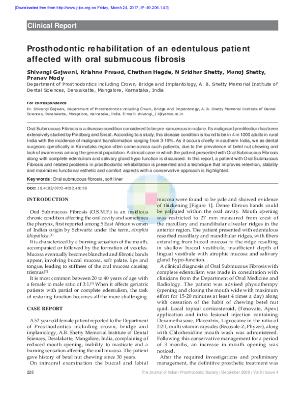
Prosthodontic rehabilitation of an edentulous patient affected with oral submucous fibrosis
Prosthodontic rehabilitation of an edentulous patient affected with oral submucous fibrosis
2008, The Journal of Indian Prosthodontic Society
Related Papers
Late Medieval and Renaissance Illuminated Manuscripts, 1350-1525, in the Houghton Library, Cambridge (MA), 1983.
Late Medieval and Renaissance Illuminated Manuscripts, 1350-1525, in the Houghton Library, Cambridge (MA), 1983.1983 •
2023 •
This is the final installment of a three-part article that I've been invited to write on the question of how Christians might engage our contemporary postmodern culture, especially the toxic polarization that characterizes so much of our world today.
2021 •
Ante la crítica del Management como pensamiento dominante en las llamadas “Ciencias Administrativas”, falta un abordaje claro y profundo con las relaciones de poder que se da en la gestión. Las relaciones de poder en la administración tienen varias aristas que en consonancia se presentan de manera omnímoda, instrumental o en “corrientes exteriores” como fuerzas exógenas de coacción en el sistema capitalista mundial en las organizaciones. Por ello, es necesario, abordar el análisis de las relaciones de poder en la administración a un nivel superior, integrador, transdisciplinario, político, social, económico, cultural, ético y sostenible, para poder romper con la hegemonía del Management, y no quedar atrapado en el pensamiento único que él rige. El presente es un ensayo epistemológico crítico que aborda algunas aristas, ejes y tópicos de las relaciones de poder en cuanto a los temas sobre: la administración y la censura; la administración del poder y sus estructuras.
Accademia dell'Arcadia / Fondazione Camillo Caetani, 26 ottobre 2023, ore 17:30
Söylem 3. Uluslararası Filoloji Sempozyumu Bildiri Özetleri
Nail Vahdeti Çakırhan ve Şiiri2024 •
1910 yılında Muğla’nın Ula ilçesinde doğan Nail Vahdeti Çakırhan gençlik yıllarını şairliğe, yetişkinlik yıllarını ise mimarlığa adamış velut bir şahsiyettir. Kaleme aldığı muhtelif metinlerde “Nail V.”, “Nail Vahdeti” ve “Nail Çakırhan” imzalarını kullanan şairin ilk şiirleri, o yıllarda Ahmet Hamdi Tanpınar’ın da görev yapmakta olduğu Konya Lisesi’nde çıkan Kervan ve Halka Doğru dergilerinde yayımlanır. Sonraki yıllarda yazdığı şiirleri ise Resimli Ay, Resimli Hafta, Çınaraltı, Ses, Görüşler, Yeni Edebiyat ve Gerçek adlı dergilerde neşredilir. Öte yandan şairin yazdığı şiirler henüz lise yıllarındayken başına bela olmaya başlar. Şair, lise son sınıftayken arkadaşlarıyla beraber çıkardığı Halka Doğru dergisinde yayımlanan “Alev Yağmuru” adlı şiiri nedeniyle ilkin Konya’da, bu şiirin Hareket dergisinde yayımlanmasının ardından ise İstanbul’da yargılanır. Söz konusu şiir, aynı zamanda onun Nâzım Hikmet’le tanışmasına da vesile olur. Çakırhan ve Nâzım’ın dostlukları ilerleyen yıllarda gittikçe pekişir ve ikili, 1930’da 1+1=Bir adlı ortak bir şiir kitabı çıkarırlar. Bu kitapta “Nail V.” imzasını kullanan şairin heyecanı ve şiire ilgisi hat safhadadır. Ancak 1940’lı yıllardan sonra şairin şiirle kurduğu bağ zayıflamaya başlar. Nitekim son şiirinin yayımlanma tarihi 1945’tir. Bu tarihten itibaren Çakırhan’ın, yaşamına şair kimliğinden ziyade mimar kimliğiyle devam etme kararı aldığını söylemek mümkündür. Şairin biyografisine bütüncül olarak bakıldığında, ona asıl şöhretini kazandıranın da bu “alaylı mimar” kimliğinin olduğu görülür. Bununla birlikte bu çalışmada Çakırhan’ın ihmal edilen diğer kimliğine, şairliğine odaklanılmış ve onun bütün şiirlerini topladığı Daha Çok Onlar Yaşamalıydı kitabına yönelik etraflı bir incelemeye girişilmiştir.
African Affairs
Encyclopedia of Precolonial Africa: Archaeology, history, languages, cultures, and environments1999 •
ΜΕΤΑΠΤΥΧΙΑΚΟ ΔΙΠΛΩΜΑ ΕΙΔΙΚΕΥΣΗΣ
ΟΙ ΑΠΑΡΧΕΣ ΤΗΣ ΒΥΖΑΝΤΙΝΗΣ ΑΡΧΑΙΟΛΟΓΙΑΣ ΣΤΗΝ ΕΛΛΑΔΑ: ΤΟ ΠΑΡΑΔΕΙΓΜΑ ΤΗΣ ΚΑΤΑΓΡΑΦΙΚΗΣ ΔΙΑΣΩΣΗΣ ΚΑΙ ΤΕΚΜΗΡΙΩΣΗΣ ΤΩΝ ΜΝΗΜΕΙΩΝ ΑΠΟ ΤΟΝ ΓΕΩΡΓΙΟ ΛΑΜΠΑΚΗ2020 •
Στη μελέτη εξετάζεται η ιστορία της Χριστιανικής Αρχαιολογικής Εταιρείας κατά την πρώτη περίοδο λειτουργίας της (1884-1923), καθώς και η δράση του πρωτεργάτη της Γεωργίου Λαμπάκη (1854-1914) για τη διάσωση των χριστιανικών μνημείων, σε μια προσπάθεια να ανιχνευθούν οι απαρχές της βυζαντινής αρχαιολογίας στην Ελλάδα. Παράλληλα, προσεγγίζονται ποικίλα θέματα που σχετίζονται με την πρόσληψη, την επιστημονική θεώρηση και τη διαχείριση των καταλοίπων του Βυζαντίου στην Ελλάδα, από την ίδρυση του ελληνικού κράτους έως και τη θεσμοθέτηση των βυζαντινών σπουδών κατά τις πρώτες δεκαετίες του 20ού αιώνα.
RELATED PAPERS
Journal of Environmental Management
Brazilian Environmental-Economic Accounting for Water: A structural decomposition analysis2020 •
Asean Journal Of Science And Engineering
Hypocholesterolemic Effect of Mature Leaf Extract of Sugarcane, Saccharum officinarum (Linnaeus, 1753), in Induced Rats2021 •
2014 •
Procedia - Social and Behavioral Sciences
Optimizing the stacking of the Intermodal Transport Units in an inland terminal: an heuristic procedure2011 •
Turkish Journal of Forestry
Erzurum Kent Halkinin Süs Bi̇tki̇leri̇ne Olan Talebi̇ni̇n Beli̇rlenmesi̇2009 •