International Journal of Osteoarchaeology
Int. J. Osteoarchaeol. (2015)
Published online in Wiley Online Library
(wileyonlinelibrary.com) DOI: 10.1002/oa.2483
An Osteobiography of a 19th-Century Dog
from Toronto, Canada
E. TOURIGNY,a* R. THOMAS,a E. GUIRY,b R. EARP,c A. ALLEN,d
J. L. ROTHENBURGER,e D. LAWLERf AND M. NUSSBAUMERg
a
School of Archaeology and Ancient History, University of Leicester, Leicester, LE1 7RH, UK
Department of Anthropology, University of British Columbia, Vancouver, V6T 1Z1, Canada
c
Department of Engineering, University of Leicester, Leicester, LE1 7RH, UK
d
Department of Veterinary Pathology, Western College of Veterinary Medicine, University of Saskatchewan,
Saskatoon, SK S7N 5B4, Canada
e
Department of Pathobiology, Ontario Veterinary College, University of Guelph, Guelph, N1G 2W1, Canada
f
Illinois State Museum Research and Collections Center, Springfield, IL 62703, USA
g
Natural History Museum Bern, Bernastrasse 15, CH-3005 Bern, Switzerland
b
ABSTRACT
A 19th-century dog burial uncovered from a historical homelot in Toronto, Canada, provided a unique
opportunity to reconstruct the individual’s osteobiography. Of particular interest are the dog’s very large
size and a suite of skeletal pathologies. Recovery of a nearly complete skeleton combined with the use
of X-rays and micro-computed tomography (micro-CT) allowed for a discriminating differential diagnoses.
Stable isotope analyses were applied to investigate questions of diet. Results reveal an individual who
suffered greatly from disease towards the end of his life and hint at its owners attitudes towards dogs.
The interdisciplinary approach applied to this case study highlights the potential information obtainable
from pet burials. We argue that better analyses and reporting of pet burials will help address research
questions targeting broader themes related to human–animal relationships. Copyright © 2015 John Wiley
& Sons, Ltd.
Key words: historical archaeology; human–animal relationships; micro-CT; Ontario archaeology; paleopathology;
stable isotope analyses; zooarchaeology
Introduction
Traditionally, historical period zooarchaeological research has focussed on themes of diet and economy;
however, recent trends in zooarchaeological theory
and practice, influenced by ideas emanating through
an engagement with the field of animal studies, are demanding alternative perspectives (Thomas & Fothergill,
2014). The significance of animal agency and the complexity of human–animal relationships are being recognized increasingly (Russell, 2012; Overton & Hamilakis,
2013; Sykes, 2014). Following Kopytoff (1986) and
Morris (2011), this paper adopts a biographical
approach to the study of archaeological remains as a
way of exploring these relationships through the con* Correspondence to: Eric Tourigny, School of Archaeology and Ancient
History, University of Leicester, University Road, Leicester LE1 7RH, UK.
e-mail: edt6@le.ac.uk
Copyright © 2015 John Wiley & Sons, Ltd.
sideration of the life and death histories of individual
animals. Careful skeletal analysis including investigation of diseases and injuries experienced by an animal,
combined with archaeological context and historical
documents, informs upon the interaction between
individual people and animals. This in turn permits
reflection on the contingency and complexity of
human–animal relationships.
A dog burial recovered from a mid-19th-century,
Euro-Canadian homelot in Toronto, Ontario, provided
an ideal opportunity to employ an osteobiographical
approach and investigate human–dog relationships. The
temporal context is important here because the 19th century marked a period of transformation in human–animal
relationships (Ritvo, 1994; Thomas, 2005; Thomas &
Fothergill, 2014). Through the Victorian period in
Britain, dogs were regularly considered as part of the
domestic realm, forming deep bonds with their owners.
By the late Victorian period, some pet dogs were cared
Received 23 February 2015
Revised 23 June 2015
Accepted 29 June 2015
E. Tourigny et al.
for in life and death in a way that better befitted one’s
best friend (Howell, 2002). Concurrently, designated
pet cemeteries first appeared in English cities, marking
a departure from previous practice in which the deposition of animals, including pets, likely reflected functional necessities rather than spiritual beliefs. Despite
the significance of this change within the context of
contemporary attitudes towards pet animals, the archaeological study of this phenomenon has been sorely
neglected (Thomas, 2005, 2009). The purpose of this
case study is to: (i) draw upon archaeological and biomedical information in order to construct a biography
of an individual dog; and (ii) consider how detailed
analyses of pet burials, commonly identified on archaeological sites from this period, would significantly
contribute to our understanding of human–dog relationships in the mid-19th century.
Materials
Located at the intersection of King Street West and
Bathurst Street in the city of Toronto, the Bell Site
(AjGu-68) represents a homelot developed on land
originally intended as a military reserve. The reserve
was deemed redundant after the War of 1812 and
earmarked for subdivision and sale. This particular
homelot was first sold in 1840 to Thomas Bell Jr., a local
land agent and accountant who would later become an
alderman serving on the Toronto city council. Thomas
was a member of one of the earliest families to settle in
York/Toronto and owned a few vacant lots and houses
throughout the city. By 1842, he and his wife Katherine
had built a small, timber-frame house on the homelot. In
1858, a larger house was erected immediately west of the
original one. Thomas died in 1857 but Katherine lived
here until her death in 1864. The buildings were then
briefly occupied by a succession of tenants (William
McCune, a driver, and M. Octavius Miller, Captain of
the military store staff) and vacated by 1869. The original house and outbuildings were demolished in 1870
upon sale of the property (ASI, 2012: 1–2).
Archaeological excavations in 2011 identified the
remains of the original homelot, which lay buried
beneath a car park. Planned development led to salvage
excavations of the initial residential structure and
other features related to the 1840–1870 occupation.
Associated artefacts and historical documents suggest
the Bells were fairly affluent members of society (ASI,
2012: 44).
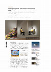
Dog burial
Identified in the backyard of the homelot was a single
dog burial. The dog was fully articulated and buried
in a flexed position, with the right side of the body
on the ground and its head facing west (Figure 1).
The shallow burial (~17 cm below surface) was
Figure 1. Dog burial. Scale bars display 10-cm intervals, trowel points north. Image is courtesy of Archaeological Services Inc. This figure is available
in colour online at wileyonlinelibrary.com/journal/oa
Copyright © 2015 John Wiley & Sons, Ltd.
Int. J. Osteoarchaeol. (2015)
Osteobiography of a 19th-Century Dog from Toronto
excavated by hand, and all soil was screened through a
6-mm wire mesh. Most of the bones were recovered
whole, while more fragile elements such as the skull
and scapulae were somewhat fragmented by excavation and post-depositional taphonomic processes. Elements of the left forelimb featured post-depositional
fragmentation as the result of a pathology leading to
increased fragility of the bones. The dog’s pelvic area
was slightly raised in its burial position and was consequently vulnerable to disturbance. Bones missing from
this area include lumbar vertebrae three to five, the
sacrum, all caudal vertebrae, innominate bones, the left
femur and the right proximal femur. Most of the left hind
paw and a few of the left rib shafts were also absent.
These bones may have disappeared as a result of 1870s
demolition activities or, more likely, taken during
mechanical removal of top soil prior to archaeological
excavations. Nine indeterminate pieces of rusted metal,
presumed to be iron, were recovered from the neck area.
Similar metal concretions were identified on the axis, left
parietal and left mandible. Energy dispersive X-ray
fluorescence (EDXRF) analysis of the concretion that
appears on the left, lateral surface of the axis identified
it as iron oxide. These are likely the remains of a dog
collar or iron chain placed around his neck.
Methods
Species identification was achieved through macroscopic examination of the specimen and comparison
with materials held in the Howard G. Savage
Zooarchaeological Reference Collection at the University of Toronto. Distinctive morphological characteristics were used to differentiate between members of
the genus and identify this individual as a dog (Canis
familiaris) (Krantz, 1959; Lawrence & Bossert, 1967;
Benecke, 1987; Morey & Wiant, 1992; Lupo & Janetski,
1994; Yates, 2000). The dog’s age at death was determined based on the state of epiphyseal fusion (Seoudi,
1948; Hare, 1959; Smith & Allcock, 1960; SumnerSmith, 1966; Silver, 1969) combined with dental eruption and wear patterns (Stiner, 1994; Horard-Herbin,
2000). Pathology was recorded macroscopically using
a descriptive recording protocol modelled after Vann
& Thomas (2006).
Biometric analysis of the axial and appendicular
skeleton was undertaken using the standards set forth
in von den Driesch (1976). Withers height (height at
the dorsal aspect of the shoulders) was calculated based
on regression formulae developed by Harcourt (1974)
and applied to all complete long bones. To investigate
phenotypic resemblances to modern breeds, skull
Copyright © 2015 John Wiley & Sons, Ltd.
measurements were taken following Phillips et al.
(2009) and subjected to discriminant function analysis
(using SPSS 22 for Windows) along with data from
over 300 reference specimens from 64 different large
breeds housed at the Albert-Heim-Foundation for canine research, Natural History Museum in Bern. Results
are used to suggest similarities to modern breeds rather
than claim direct breed assignation.
The radii, ulnae, humerii and right tibia were radiographed using a mobile X-ray unit (60 KvP; 64 mAs;
0.025 s; Xograph Dragon, University of Leicester,
School of Archaeology and Ancient History, Leicester,
UK). Bones exhibiting pathology were radiographed
alongside the contralateral bone to exclude the possibility that radiolucencies and radiodensities were
caused by taphonomic factors (Mays, 2012).
High resolution micro-computed tomography
(micro-CT) images were taken of the left humerus, left
radius and skull (Nikon Metrology XTH 225 micro-CT
scanner, with a Paxscan detector, University of
Leicester, Department of Engineering, Leicester, UK).
Micro-CT offers greater spatial resolution than possible
with clinical computed tomography; however, the
exact resolution is difficult to quantify as it is dependent
on a large number of factors (Rutty et al., 2013). The
X-ray data were reconstructed using CT-Pro 2.0 (Nikon
Metrology UK Ltd), and all 3D rendering and subsequent analysis was performed in VGStudio MAX
2.1 (Volume Graphics GmbH, Germany), and Drishti
v2.5.1 (The Australian National University, Australia).
We initially conducted our scans with the X-ray voltage
and current set at 95 KV and 224 μA, using a 0.5-mmthick copper filter. This combination was judged to provide the best X-ray penetration and image contrast
when viewed as a live radiograph. However, the images
showed some signs of saturation after completion of the
scan, and so further scans were conducted at 110 kV and
129 μA with the same filter, to improve image quality.
Metadata obtained following the scans provides information on voxel size (3D pixels) which is calculated
based on the distance between the sample and the
X-ray source. In this case, the voxel sizes ranged
between 78.6 and 99 microns.
For the stable isotope analysis, collagen extractions
followed well-established methods (Brown et al., 1988;
Richards & Hedges, 1999). Samples were soaked in a
0.5 M hydrochloric acid (HCl) solution (refreshed
every 48 h) at 4 °C until fully demineralized. Collagen
pseudomorphs were then placed in 0.1 M Sodium
Hydroxide (NaOH) overnight to remove potential
humic acid contaminants (Jørkov et al., 2007; UCI
KCCAMS Facility, 2011). Afterwards, samples were
gelatinized in water adjusted to a pH of 3 with 0.5 M
Int. J. Osteoarchaeol. (2015)
E. Tourigny et al.
HCl on a heating block set to 75 °C for 48 h. Gelatins
were then Ezee Filtered (5–8 μm mesh), Ultra Filtered
(30 kDa), frozen for 24 h and lyophilized over 48 h in
a freeze dryer. Replicate isotopic measurements were
made using 0.5 mg of collagen on an Elementar vario
MICRO elemental analyser coupled to an IsoPrime
mass spectrometer (University of British Columbia,
Department of Anthropology, Vancouver, Canada).
Stable carbon and nitrogen isotope values were calibrated to VPDB and AIR, respectively, with USGS40
and USGS41. International and internal reference standards (NIST 1577c bovine liver, SIGMA-ALDRICH
methionine and seal collagen) are also routinely analysed to monitor precision and accuracy. Standard
deviations for δ13C and δ15N values of standard materials were better than 0.1‰ and 0.2‰, respectively.
Collagen integrity was deemed adequate if samples
produced a collagen yield above 1%, a carbon to nitrogen ratio (C:N) between 2.9 and 3.6, and carbon and
nitrogen concentrations above 18% and 6%, respectively (DeNiro, 1985; van Klinken, 1999).
Results
Age and sex
All long bones were fused indicating that this animal
was an adult (Seoudi, 1948; Hare, 1959; Smith &
Allcock, 1960; Sumner-Smith, 1966; Silver, 1969). The
adult dentition was fully erupted which occurs by
approximately 28–30 weeks of age in the dog (Hillson,
2005). Macroscopic observations indicate that both
lower first premolars were lost ante-mortem allowing
enough time for the alveolar bone to begin remodelling.
Wear patterns were identified on the teeth and following
Stiner’s (1994) and Horard-Herbin’s (2000) criteria,
this individual’s teeth fall under Stage V and Stage
E-F respectively, which are consistent with a ‘prime’
aged dog.
Evidence suggests that, after reaching skeletal maturity, this dog lived long enough to develop significant
dental wear, lose permanent dentition and begin significant alveolar remodelling. Furthermore, sites of
epiphyseal fusion have undergone extensive remodelling and epiphyseal fusion lines are not visible. This
evidence presented alongside the degenerative joint
disease identified in the skeleton (discussed later) suggests that this dog was as a mature to elderly individual
when it died.
A baculum was recovered identifying the individual
as male.
Copyright © 2015 John Wiley & Sons, Ltd.
Size and breed affiliation
The height of the dog at the withers was approximately 73 to 75 cm (Table 1). Withers height is comparable to giant dog breeds such as the Great Dane,
Newfoundland and St. Bernard dogs among others
(The Kennel Club, 2003).
Discriminant function analysis shows 89.4% variance
explained by the first two functions and plots the
archaeological specimen alongside large breeds belonging to the working group of dogs (The Kennel Club,
2003) (Figure 2). The Bell dog plots closest to the
group centroids and individual cases for Great Danes,
Landseers, Leonbergers, St. Bernards, Newfoundlands
and Greater Swiss Mountain dogs, suggesting a closer
affinity to these breeds.
Pathologies
A full description of the lesions identified on the skeleton
is presented in supplementary materials available with
the online version of this article, what follows here is a
digest of the primary forms of the pathologies observed.
Degenerative joint disease
Multiple bones on this dog’s skeleton display osteophytes and lipping along articular margins. This is
especially marked in the spine where every vertebra
exhibited evidence of osteophytosis and/or lipping
along the margins of vertebral bodies and/or along
the margins of the articular facets and tubercles for
rib articulation. The majority of recovered rib heads
also display slight to moderate osteophytes and/or evidence of lipping. Osteophytes affecting ventrolateral
vertebral margins are evidence of a condition known
as spondylosis deformans (Thompson, 2007: 157).
Multiple joints exhibit degenerative disease. These
include lipping of the left glenoid fossa and both humeral heads, and osteophytosis of the left second
cuneiform and the distal end of a proximal phalanx.
Degenerative joint disease (mild) and spondylosis
Table 1. Estimated height at the withers, from Harcourt (1974)
Data source
Humerus
Radius
Ulna
Humerus
and radius
Tibia
Regression
formula*
Estimated height
at withers (cm)
((3.43 × GL) 26.54) / 10
((3.18 × GL) + 19.51) / 10
((2.78 × GL) + 6.21) / 10
((1.65 × GL) 4.32) / 10
74.92
73.51
73.23
74.01
((2.92 × GL) + 9.41) / 10
75.87
*GL = greatest length, after von den Driesch (1976).
Int. J. Osteoarchaeol. (2015)
Osteobiography of a 19th-Century Dog from Toronto
Figure 2. Scatter plot of the Bell Site cranium and group centroid values of selected dog breeds on the two first canonical discriminant functions. This
figure is available in colour online at wileyonlinelibrary.com/journal/oa
deformans are common age-related ‘wear and tear’
lesions although their onset and progression are
influenced by a range of factors including behaviour,
genetic predisposition, environment and body mass.
Enthesophytes (ossifications at ligament/tendon
attachment sites) are present throughout the body
(on mandibles, humeri and the right ulna), further
supporting our assessment of advanced age.
Periodontal disease
The maxillae and mandibles exhibit extensive periodontal disease signalled by the heavy porosity along
the entirety of both upper and lower dental arcades.
This is interpreted as advanced gingivitis and probable
periostitis–periodontitis. Alveolar pockets are also evident behind the mandibular second premolar. The
upper left fourth premolar features a chipped enamel surface that exhibits use wear, indicating the chip occurred
ante-mortem. A cloaca has formed in the maxilla, just
above this tooth suggesting a bacterial infection (possibly
a result of the chipped tooth) that was quite advanced
(Figure 3). Silver (1969) notes that the upper fourth
pre-molar of domestic dogs is often subjected to abscesses of the root and that the maxillary bone over the
roots can become so eroded that a sinus forms between
the alveolus and the exterior surface, as is the case here.
Infection of tympanic bulla
Macroscopic examinations and micro-CT scans reveal
significant remodelling of the tympanic bulla and
its cavity (Figure 4). Reactive bone formation and destruction of the left tympanic bulla and external
Figure 3. Lateral surface of left maxilla, premolars and molars. Note the porosity observed along the alveolar margin. Arrow points to the cloaca that
formed above the fourth, upper premolar. This figure is available in colour online at wileyonlinelibrary.com/journal/oa
Copyright © 2015 John Wiley & Sons, Ltd.
Int. J. Osteoarchaeol. (2015)
E. Tourigny et al.
Figure 4. Radiograph of cranium. Arrow points to advanced reactive bone formation and destruction of the left tympanic bulla.
acoustic meatus are consistent with chronic osteitis.
This lesion may have arisen secondary to bacterial
otitis media and/or otitis externa, possibly initiated
from chronic yeast infections or foreign bodies in the
external ear canal. Inflammation of the external ear or
ear canal is common in domestic dogs (Wilcock, 2007).
Periosteal and endosteal reactive bone
Prolific, reactive periosteal new bone formation was evident on the left radius, ulna and carpal accessory (Os
carpi accessorium) with articular surfaces remaining unaffected (Figures 5(a) and 6(a)). Small foci of periosteal
reactive bone were also observed on the right ulna
Figure 5. (a) Medial surface of left ulna with active periosteal reactive bone formation along the diaphysis; (b) lateral surface of right ulna, arrow points
to the small locus of active periosteal reactive bone formation. This figure is available in colour online at wileyonlinelibrary.com/journal/oa
Copyright © 2015 John Wiley & Sons, Ltd.
Int. J. Osteoarchaeol. (2015)
Osteobiography of a 19th-Century Dog from Toronto
Figure 6. (a) 3D rendering of anterior surface of left radius exhibiting periosteal reactive bone formation. (b) Transverse section of left radius showing
periosteal and endosteal reactive bone formation. This figure is available in colour online at wileyonlinelibrary.com/journal/oa
(Figure 5(b)) and on metacarpals of both forelimbs.
Endosteal reactive bone formation filled the medullary cavity of the diaphysis of the left radius fragment. Examination of radiographs indicates that the
observed endosteal growth extends approximately
3 cm within the diaphysis towards the proximal end
of the bone (Figure 6(b)). Imaging indicates the remainder of the marrow cavity in the proximal radius
was unaffected.
Periosteal reactive bone formation can be attributed
to metabolic disorders (metaphyseal osteopathy, canine
panosteitis and hypertrophic osteopathy), neoplasms,
bacterial infections or ‘non-specific’ infections (Weston,
2012). Here, we conduct a differential diagnosis to
narrow down the potential cause.
Metaphyseal osteopathy (hypertrophic osteodystrophy) produces bone lesions in larger dogs, resembling those observed in this specimen. However, this
condition only affects growing individuals with open
physes (Lenehan & Fetter, 1985; Woodard, 1997;
Thompson, 2007). Metaphyseal osteopathy is unlikely
in this case as this dog was skeletally mature at time of
death and the diaphysis of the left radius is extensively
involved.
Copyright © 2015 John Wiley & Sons, Ltd.
Canine panosteitis, an inflammatory bone disease of
unknown cause, is another condition stimulating periosteal new bone formation. However, the related bone
surface proliferation is typically much less pronounced
than that observed in this specimen. An important
radiographic hallmark of panosteitis is patchy increased
ill-defined densities in long bone marrow cavities,
which was not observed. Panosteitis commonly affects
younger individuals in certain family lines of modern
large breeds of dog, but can be observed at any age
(Johnson & Watson, 2000). The pathologies observed
on this specimen are inconsistent with this disease.
A third metabolic bone disorder to consider is
hypertrophic osteopathy (HO). This disease affects
the bones of the lower limbs, such as the radius, ulna,
tibia and metapodials, and is characterized by diffuse
periosteal new bone formation oriented in a roughly
perpendicular direction to the diaphysis with coarse
osseous exostoses which have a wart- or cauliflower-like
appearance, much like those observed in Figures 5(a)
and 6(a). The proliferations generally respect the synovial joints and articular surfaces of the bone. The
pathogenesis of this disease remains poorly understood,
but it is known to relate to lesions in the pleural cavity
Int. J. Osteoarchaeol. (2015)
E. Tourigny et al.
caused by tumours, bacterial or fungal infections as
well as genitourinary tumours (Johnson & Watson,
2000).
The macroscopic appearance of the reactive bone
formation and the similarity to previous published cases
(Bathurst & Barta, 2004; von Hunnius, 2009) led the
archaeologists who first recorded these bones to suggest
a possible diagnosis of HO (ASI, 2012). Upon further
scrutiny of the skeleton and relevant clinical literature,
we identified an issue with this possible diagnosis as
some of the symptoms do not fit with the typical presentation of the disease. HO is a symmetrically bilateral
disease, and similar lesions are not present in the contralateral limb of this dog. Small patches of periosteal
reactive bone were identified on metacarpals of both
forelimbs and on the ulna of the right forelimb; however, these are very small compared to the extensive
lesions observed in the left forelimb.
Neoplasms or bone tumours were carefully considered. However, these tend to include cortical bone
destruction resulting in unclear boundaries between
the original cortex and the newly deposited bone
(Rothschild & Martin, 1992: 167). Metastasis of soft
tissue cancers onto the bone results in either lysis or
in distinctive periosteal reactions having a sun burst
like appearance, neither of which is seen here. Furthermore, primary bone tumours rarely cross joints proximal or distal to an affected bone.
Given the localised extent of the lesions in the left
forelimb, an alternative diagnosis is a chronic bacterial
or fungal infection secondary to one or multiple
penetrating wound(s) centred on the mid-left forearm.
It is possible that the overlying soft tissue infections
led to hyperactive new bone formation. Further, the
presence of the endosteal new or reactive bone is consistent with an insult injury (i.e. penetrating wound(s),
through the cortex of the radius or entry through a
vascular channel). Therefore, our suggested diagnosis
regarding the left forelimb include: (i) chronic-active
and extensive, periostitis of the left radius, ulna and Os
carpi accessorium, with new (reactive) bone formation;
and (ii) chronic-active and focal osteomyelitis with
moderate endosteal new (reactive) bone formation in
the diaphysis of the left radius.
Diet and nutrition
To determine whether this individual had a distinctive diet, we submitted two samples for stable isotope
analyses. Biological tissues such as bone are constructed and maintained using materials derived from
foods consumed by the individual. Foods can have
distinctive isotope signatures which can be identified
in analyses, affording the opportunity to assess if this
dog’s diet was consistent with other animals living in
the same region or, rather, if it was afforded a diet
that was in some way distinctive (Katzenberg, 2008).
Guiry (2012, 2013) reviews the role of dogs in stable
isotope bone chemistry and paleodietary reconstruction.
We analyzed the stable carbon and nitrogen isotope
composition of collagen from a bone (left os carpi ulnare)
and tooth (right lower canine). Because of different
remodelling characteristics between these tissues, the
isotope values from the bone and tooth should reflect
the dog’s diet in later and earlier (post weaning) life, respectively. To help contextualize these data, we also
analysed an additional loose dog bone (an isolated
bone recovered from the original A-horizon associated
with the 1840–1870 occupation and without evidence
for special treatment by its owners) and bones from
three individual cattle (Bos taurus) from the site. The
loose dog bone was intended to provide a comparison
for the interred dog’s values. The cattle were analysed
to provide a rough isotopic baseline for herbivore diets
in the area. Six analyses were performed in total and
the results are presented in Table 2 and Figure 7. These
results suggest the individual recovered in the burial
did not have a diet that was atypical of 19th-century
dogs living in urban areas based on comparisons with
the other dog specimen from this site, to others from
urban sites (e.g. Guiry et al., 2014) and to early results
of ongoing work in southern Ontario by one of this
paper’s co-authors (Guiry). Moreover, isotopic values
from early and late forming tissues are similar
Table 2. Stable isotope values for dog burial and other specimens from the Bell site
Taxon
Bos taurus
Bos taurus
Bos taurus
Canis familiaris
Canis familiaris
Canis familiaris
Element
% Collagen
δ13C‰
δ15N‰
%C
%N
C:N
Metacarpal III and IV
Metacarpal III and IV
Metacarpal III and IV
Humerus
Lower canine
Carpal ulnar
11.6
9.5
7.8
3.6
15.8
6.3
22.4
22.8
23.9
20.5
19.6
19.9
5.8
7.2
7.5
10.4
11.2
10.1
42.9
42.2
41.7
41.3
42.4
42.6
15.2
14.8
12.7
14.1
14.9
14.7
3.3
3.3
3.8
3.4
3.3
3.4
Copyright © 2015 John Wiley & Sons, Ltd.
Int. J. Osteoarchaeol. (2015)
Osteobiography of a 19th-Century Dog from Toronto
Figure 7. Stable isotope values for dog burial and other specimens from
the Bell site.
suggesting that this individual’s diet was (in terms of
stable isotopes) consistent throughout his life.
Discussion and conclusions
Taking an interdisciplinary approach to the construction of an osteobiography permits us to understand
the activities that took place to create the burial and
postulate what this dog meant to those who interacted
with him (Morris, 2011). Cranial morphology suggests
that he belonged to one or a mixture of the working
class group of giant breeds. Little is known on the
subject of dog breeds from this time period in Toronto,
and archaeological analyses of pet burials rarely go
beyond species identification. Newfoundland dogs
represent one of the few breeds mentioned in early
19th-century documents, although these likely reference large dogs rather than the breed standards we
associate with the name today (Grier, 2006: 35). They
were favoured for hunting deer and managing cattle
and pigs according to personal letters penned by the
Cooper brothers in nearby Adelaide township in
1832, 1833 and 1838 (Cameron et al., 2000). The Bell
Site dog is unlikely to have served as a farm animal
given its burial in an urban homelot. Fox and deer
hunting with dogs was a popular pastime among the
city’s political and entrepreneurial elite, many of whom
were members of the Toronto Hunt Club (Joyce, 1997:
94–95). Other pursuits, such as bull-baiting and dog
fighting, were popular among the working class until
city by-laws in 1864 and 1876 aimed to stop these
activities (Joyce, 1997: 94). The osteobiography of this
individual does not support life or death as a fighting
Copyright © 2015 John Wiley & Sons, Ltd.
dog, but Thomas Bell Jr.’s affluence and political position is typical of a member of the Toronto Hunt
Club, and this dog may have served for hunting
purposes. Other possible functions for a dog of this size
living in the city include hauling/pulling carts, security
and/or companionship. Such a large dog would have
been a rather imposing figure to anyone who came
across his path, regardless of his demeanour. It is unlikely the dog was ever left to roam free about the
neighbourhood as legislation was put in place in 1834
to ‘prevent and regulate the running at large of dogs’.1
In 1836, a new dog licensing system was introduced
requiring city residents to tag and collar their dogs
(City of Toronto Archives, By-law 23 in Kheraj, 2013).
Large breeds of modern dogs live relatively short
lives (as short as 6 years) (Galis et al., 2007) and are particularly susceptible to a number of diseases, including
those identified here. Multiple degenerative joint diseases likely resulted in pain and difficulty or limitations
in movement. Severe periodontal disease, a chipped
tooth and associated bacterial infection probably resulted in a painful eating experience. An advanced ear
infection likely caused deafness in the left ear, and he
may have walked around with his head tilted to that
side and exhibited behaviours such as head shaking
and scratching. His ear infection would have carried a
foul odour and periodontal disease likely gave him
bad breath, rendering him unpleasant to be around. A
penetrating wound to his left forelimb could have been
the result of a number of causes (e.g. a dog bite or
being stuck with a nail), which led to a chronic infection and increasing discomfort as the body reacted to
the infection. Inflammation of the left forelimb likely
resulted in this dog walking with a limp. This dog
would have been very sick over a period of perhaps
12–16 weeks as a result of a sub-chronic stage infection
and degenerative process and therefore could have
experienced other soft tissue problems not observable
in archaeological remains.
The inter-disciplinary approach used here provided
us with a wider range of information to re-construct
the osteobiography, while advanced radiographic
imaging techniques provided better information for
the differential diagnosis. This case study highlights
the potential information obtainable from 19th-century
pet burials and the need for more detailed analyses of
such deposits. A greater sample of pet burials from
across the city and province would help address
1
‘An act to extend the Limits of the Town of York; to erect the said Town
into a City; and to Incorporate it under the name of the City of Toronto,’
6 March 1834, Statutes of His Majesty’s province of Upper Canada (Toronto: G.
Tiffany, 1834) 73; 81. Cited in Kheraj (2013: 129).
Int. J. Osteoarchaeol. (2015)
E. Tourigny et al.
research questions targeting broader themes, such as
the treatment of pets (in life and after death) and
human–animal relationships in general. In late Victorian
England, the practice of pet burials is reminiscent of
the increasingly popular belief that heaven was a
reflection of one’s home. Pet animals who served as
part of the family home on earth would also be
reunited with their owners in the afterlife. For this to
happen, these creatures must have souls; consequently,
pet burials came to hold significantly greater meaning
and functioned as more than just good hygiene practice
(Howell, 2002).
Acknowledgements
The authors would like to thank Eva MacDonald and
Archaeological Services Inc. for access to the dog
skeleton and to Eva for valuable comments on the manuscript. Thanks to Ian Whitbread of the University of
Leicester’s School of Archaeology and Ancient History
for performing the EDXRF analysis of the iron. Special
thanks to Prof Sarah Hainsworth for access to the
micro-CT equipment and to Graham Clark for assistance with this work. We would also like to thank
Michael Richards for financial support and Paul Szpak
and Reba Macdonald, for technical advice regarding
the stable isotope analysis. Thanks to T. Max Friesen
for access to the faunal reference collection. We also
thank the two anonymous reviewers for their helpful
comments on this manuscript. This work was made
possible by a fellowship to the lead author from the
Social Sciences and Humanities Research Council of
Canada.
References
ASI (Archaeological Services Inc.). 2012. Stage 4 salvage
excavation of the Bell Site (AjGu-68) 621 King Street
West, Lots 11 and 12, South Side of King Street.
Registered plan D-82 and part of Lots 4 and 5, Section
M, Military Reserve, City of Toronto, Ontario. Unpublished report on file at Archaeological Services Inc.,
Toronto.
Bathurst RR, Barta JL. 2004. Molecular evidence of tuberculosis induced hypertrophic osteopathy in a 16th-century
Iroquoian dog. Journal of Archaeological Science 31: 917–925.
DOI: 10.1016/j.jas.2003.12.006
Benecke N. 1987. Studies on early dog remains from northern Europe. Journal of Archaeological Science 14: 31–49.
Brown TA, Nelson DE, Vogel JS, Southon JR. 1988. Improved collagen extraction by modified Longin method.
Radiocarbon 30: 171–177.
Copyright © 2015 John Wiley & Sons, Ltd.
Cameron W, Haines S, Maude MM. 2000. English Immigrant
Voices: Labourer’s Letters from Upper Canada in the 1830s.
McGill-Queen’s University Press: Montreal and Kingston.
DeNiro MJ. 1985. Postmortem preservation and alteration
of in vivo bone collagen isotope ratios in relation to
paleodietary reconstruction. Nature 317: 806–809.
Galis F, Sluijs IVD, Dooren TJMV, Metz JAJ, Nussbaumer
M. 2007. Do large dogs die young? Journal of Experimental
Zoology Part B: Molecular and Developmental Evolution 308B:
119–126. DOI: 10.1002/jez.b.21116
Guiry EJ. 2012. Dog as analogs in human stable isotope based
paleodietary reconstructions: A review and consideration
for future use. Journal of Archaeological Method and Theory
19(3): 351–376. DOI: 10.1007/s10816-011-9118-z
Guiry EJ. 2013. A canine surrogacy approach to human
paleodietary bone chemistry: Past development and future
directions. Archaeological and Anthropological Sciences 5:
275–286. DOI: 10.1007/s12520-013-0133-8
Guiry EJ, Harpley B, Jones Z, Smith C. 2014. Integrating
stable isotope and zooarchaeological analyses in historical
archaeology: A case study from the urban nineteenthcentury commonwealth block site, Melbourne, Australia.
International Journal of Historical Archaeology 18: 415–440.
DOI: 10.1007/s10761-014-0264-3
Grier KC. 2006. Pets in America. University of North Carolina
Press: Chapel Hill, NC.
Harcourt RA. 1974. The dog in prehistoric and early historic
Britain. Journal of Archaeological Science 1: 151–175.
Hare WCD. 1959. Radiographic anatomy of the canine
pectoral limb. Journal of the American Veterinary Medical Association 135: 264–271, 305–310.
Hillson S. 2005. Teeth. Cambridge University Press:
Cambridge.
Horard-Herbin M-P. 2000. Dog management and use in the
Late Iron Age: The evidence from the Gallic Site of
Levroux (France). Dogs Through Time: An Archaeological
Perspective, SJ Crockford. BAR International Series 889. BAR:
Oxford; 115–121.
Howell P. 2002. A place for the animal dead: Pets, pet
cemeteries and animal ethics in late Victorian Britain.
Ethics, Place & Environment 5: 5–22. DOI: 10.1080/
13668790220146401
Johnson KA, Watson ADJ. 2000. Skeletal diseases. Textbook of
Veterinary Internal Medicine, 5th edn, SJ Ettinger, EC Feldman.
Saunders: Philadelphia; 1887–1916.
Jørkov MLS, Heinemeier J, Lynnerup N. 2007. Evaluating
bone collagen extraction methods for stable isotope
analysis in dietary studies. Journal of Archaeological Science
34: 1824–1829. DOI: 10.1016/j.jas.2006.12.020
Joyce CA. 1997. From left field: Sport and class in
Toronto, 1845–1886. Unpublished Ph.D thesis. Queen’s
University: Kingston, ON, Canada.
Katzenberg MA. 2008. Stable isotope analysis: A tool
for studying past diet, demography, and life history. Biological Anthropology of the Human Skeleton, MA Katzenberg,
SR Saunders. Wiley-Liss: New York; 387–410.
Kheraj S. 2013. Living and working with domestic
animals in nineteenth-century Toronto. Urban Explorations:
Int. J. Osteoarchaeol. (2015)
Osteobiography of a 19th-Century Dog from Toronto
Environmental Histories of the Toronto Region, LA Sandberg, S
Bocking, C Coates, K Cruikshank. L.R. Wilson Institute
for Canadian History: Hamilton, ON; 120–140.
Krantz GS. 1959. Distinctions between the skulls of coyotes
and dogs. Kroeber Archaeological Society Papers 21: 40–45.
Kopytoff I. 1986. The cultural biography of things: Commoditization as process. The Social Life of Things:
Commodities in Cultural Perspective, A Appadurai. Cambridge
University Press: Cambridge; 64–91.
Lawrence B, Bossert WH. 1967. Multiple character analysis
of Canis lupus, latrans, and familiaris, with a discussion of
the relationships of Canis niger. American Zoologist 7:
223–232.
Lenehan TM, Fetter AW. 1985. Hypertrophic osteodystrophy.
Textbook of Small Animal Orthopaedics, CD Newton, DM
Nunmaker. J.B. Lippincott Company: Philadelphia;
597–602.
Lupo KD, Janetski JC. 1994. Evidence of domesticated dogs
and some related canids in the eastern Great Basin. Journal
of California and Great Basin Anthropology 16: 199–220.
Mays S. 2012. The relationship between paleopathology
and the clinical sciences. A Companion to Paleopathology,
AL Grauer. Wiley Blackwell: Chichester, West Sussex,
UK; 285–309.
Morey DF, Wiant MD. 1992. Early holocene domestic dog
burials from the North American Midwest. Current Anthropology 33: 224–229.
Morris J. 2011. Investigating Animal Burials: Ritual, Mundane and
Beyond. BAR British Series 535. BAR: Oxford.
Overton N, Hamilakis Y. 2013. A manifesto for a social
zooarchaeology. Swans and other beings in the
Mesolithic. Archaeological Dialogues 20: 111–136.
Phillips C, Baxter IL, Nussbaumer M. 2009. The application
of discriminant function analysis to archaeological dog remains as an aid to the elucidation of possible affinities with
modern breeds. Archaeofauna 18: 49–62.
Richards MP, Hedges REM. 1999. Stable isotope evidence
for similarities in the types of marine foods used by Late
Mesolithic humans at sites along the Atlantic coast of
Europe. Journal of Archaeological Science 26: 717–722.
DOI: 10.1006/jasc.1998.0387
Ritvo H. 1994. Animals in nineteenth-century Britain: Complicated attitudes and competing categories. Animals and
Human Society: Changing Perspectives, A Manning, J Serpell.
Routledge: New York & London; 106–126.
Rothschild BM, Martin L. 1992. Paleopathology: Disease in the
Fossil Record. CRC Press: Boca Raton.
Russell N. 2012. Social Zooarchaeology. Cambridge University
Press: Cambridge.
Rutty GN, Brough A, Biggs MJP, Robinson C, Lawes SDA,
Hainsworth SV. 2013. The role of micro-computed
tomography in forensic investigations. Forensic Science
International 225: 60–66. DOI: 10.1016/j.forsciint.2012.
10.030
Seoudi K. 1948. X-ray examination of “epiphyseal union” as
an aid to the estimation of the age in dogs. The British
Veterinary Journal 104: 150–155.
Copyright © 2015 John Wiley & Sons, Ltd.
Silver IA. 1969. The aging of domestic animals. Science in
Archaeology, 2nd edn, D Brothwell, E Higgs. Praeger
Publishers: New York; 283–302.
Smith RN, Allcock J. 1960. Epiphysial fusion in the
greyhound. The Veterinary Record 72: 75–79.
Sumner-Smith G. 1966. Observations on epiphyseal fusion
of the canine appendicular skeleton. Journal of Small Animal
Practice 7: 303–311.
Stiner MC. 1994. Honor Among Thieves: A Zooarchaeological Study of
Neanderthal Ecology. Princeton University Press: Princeton, NJ.
Sykes N. 2014. Beastly Questions: Animal Answers to Archaeological Questions. Bloomsbury: London.
The Kennel Club. 2003. The Kennel Club’s Illustrated Breeds Standards. (The Official Guide to Registered Breeds). Edbury Press:
London.
Thomas R. 2005. Perceptions versus reality: Changing
attitudes towards pets in medieval and post-medieval
England. Just Skin and Bones? New Perspectives on
Human–Animal Relations in the Historic Past, A Pluskowski.
BAR International Series 1410. Archaeopress: Oxford;
95–105.
Thomas R. 2009. Bones of contention: Why later postmedieval assemblages of animal bones matter. Crossing
Paths or Sharing Tracks: Future Directions in the Archaeological
Study of Post-1550 Britain and Ireland, A Horning, M Palmer.
Boydell and Brewer Ltd.: Woodbridge; 133–148.
Thomas R, Fothergill B. 2014. Animals, and their bones, in
the ‘modern’ world: A multi-scalar zooarchaeology.
Anthropozoologica 49: 1–9. DOI: 10.5252/az2014n1a01
Thompson K. 2007. Bones and joints. Jubb, Kennedy and
Palmer’s Pathology of Domestic Animals, 5th edn, MG Maxie.
Elsevier Saunders: Edinburgh; 1–184.
UCI KCCAMS Facility. 2011. Chemical pretreatment of
bone. Electronic document, http://www.ess.uci.edu/group/
ams/files/bone_protocol.pdf. Department of Earth System
Science, University of California. [Accessed 9 January 2015]
van Klinken GJ. 1999. Bone collagen quality indicators
for paleodietary and radiocarbon measurements. Journal
of Archaeological Science 26: 687–695. DOI: 10.1006/
jasc.1998.0385
Vann S, Thomas R. 2006. Humans, other animals and
disease: A comparative approach towards the development of a standardised recording protocol for animal
palaeopathology. Internet Archaeology 20. DOI: 10.11141/
ia.20.5
von den Driesch A. 1976. A Guide to Measurement of Animal
Bones from Archaeological Sites. Peabody Museum of Archaeology and Ethnology, bulletin I: Cambridge, MA.
von Hunnius T. 2009. Using microscopy to improve a
diagnosis: An isolated case of tuberculosis-induced
hypertrophic osteopathy in archaeological dog remains.
International Journal of Osteoarchaeology 19: 397–495. DOI:
10.1002/oa.989
Weston DA. 2012. Nonspecific infection in paleopathology:
Interpreting periosteal reactions. A Companion to Paleopathology, AL Grauer. Wiley Blackwell: Chichester, West
Sussex, UK; 492–512.
Int. J. Osteoarchaeol. (2015)
E. Tourigny et al.
Wilcock BP. 2007. Eye and ear. Jubb, Kennedy, and Palmer’s Pathology of Domestic Animals, 5th Edn, MG Maxie. Elsevier
Saunders: Edinburgh; 459–553.
Woodard JC. 1997. Skeletal system. Veterinary Pathology, 6th
edn, TC Jones, RD Hunt, NW King. Lippincott, Williams
and Wilkins: Philadelphia; 899–946.
Yates BC. 2000. Use of the mastoid region of the crania
of canids to distinguish wolves, dogs, and wolf/dog
hybrids. Dogs through Time: An Archaeological Perspective, SJ
Copyright © 2015 John Wiley & Sons, Ltd.
Crockford. BAR International Series 889. BAR: Oxford;
269–270.
Supporting information
Additional supporting information may be found in the
online version of this article at the publisher’s web site.
Int. J. Osteoarchaeol. (2015)
An osteobiography of a 19th-century dog from Toronto, Canada
International Journal of Osteoarchaeology
A 19th-century dog burial uncovered from a historical homelot in Toronto, Canada, provided a unique opportunity to reconstruct the individual's osteobiography. Of particular interest are the dog's very large size and a suite of skeletal pathologies. Recovery of a nearly complete skeleton combined with the use of x-rays and micro-computed tomography (micro-CT) allowed for a discriminating differential diagnoses. Stable isotope analyses were applied to investigate questions of diet. Results reveal an individual who suffered greatly from disease towards the end of his life and hint at its owners attitudes towards dogs. The interdisciplinary approach applied to this case study highlights the potential information obtainable from pet burials. We argue that better analyses and reporting of pet burials will help address research questions targeting broader themes related to human-animal relationships....Read more