Immunology and Cell Biology (2011) 89, 437–446
& 2011 Australasian Society for Immunology Inc. All rights reserved 0818-9641/11
www.nature.com/icb
ORIGINAL ARTICLE
Bystander inhibition of dendritic cell differentiation
by Mycobacterium tuberculosis-induced IL-10
Maria Elena Remoli1,3, Elena Giacomini1,3, Elisa Petruccioli1, Valerie Gafa1, Martina Severa1,
Maria Cristina Gagliardi1, Elisabetta Iona1, Richard Pine2, Roberto Nisini1 and Eliana Marina Coccia1
Mycobacterium tuberculosis (Mtb) evades the immune response by impairing the functions of different antigen-presenting
cells. We have recently shown that Mtb hijacks differentiation of monocytes into dendritic cells (DCs). To further characterize
the mechanisms underlying this process, we investigated the consequences of inducing dendritic cell differentiation using
interferon-a and granulocyte-macrophage colony-stimulating factor in the presence of supernatants (SNs) obtained from
monocyte cultures treated with or without heat-inactivated Mtb. Although the SNs from control cultures do not interfere with
the generation of fully differentiated DCs, monocytes stimulated with SNs from Mtb-stimulated cells (SN Mtb) remained CD14+
and poorly differentiated into CD1a+ cells. Among cytokines known to affect dendritic cell differentiation, we observed a robust
production of interleukin-1b, interleukin-6, interleukin-10 and tumor necrosis factor-a upon Mtb stimulation. However, only
interleukin-10 neutralization through the addition of soluble interleukin-10 receptor reversed the inhibitory activity of SN Mtb.
Accordingly, the addition of recombinant interleukin-10 was able to significantly reduce CD1a expression. The interaction
of Mtb with differentiating monocytes rapidly activates p38 mitogen-activated protein kinase, signal transducer and activator
of transcription pathways, which are likely involved in interleukin-10 gene expression. Taken together, our results suggest
that Mtb may inhibit the differentiation of bystander non-infected monocytes into DCs through the release of interleukin-10.
These results shed light on new aspects of the host–pathogen interaction, which might help to identify innovative immunological
strategies to limit Mtb virulence.
Immunology and Cell Biology (2011) 89, 437–446; doi:10.1038/icb.2010.106; published online 31 August 2010
Keywords: dendritic cell differentiation; immunoevasion strategy; IL-10; monocyte; Mycobacterium tuberculosis
Mycobacterium tuberculosis (Mtb) is one of the most successful human
pathogens and tuberculosis still remains one of the major health
threats to mankind.1,2 The success of Mtb as a highly adapted human
pathogen also relies on the ability to evade the immune response and,
in turn, to persist in an immunocompetent host. This phenomenon is
based on the capacity of Mtb to switch to a dormant status of latency,
as well as on the ability to survive within macrophages by arresting
phagosomal maturation.3,4 Besides the well-characterized immunoevasion strategies occurring in Mtb-infected macrophages, more
recently, a new mechanism of Mtb immune evasion that relies
on the capacity of Mtb to interfere with monocyte differentiation
into dendritic cells (DCs) has been characterized.5,6
The importance of DCs in regulating the immune response against
pathogens, including Mtb, has been largely demonstrated7,8 given their
capacity to act as antigen-presenting cells for naı̈ve T lymphocytes and,
hence, to play a crucial role in the induction of adaptive immunity.
Thus, by limiting the generation of functionally active DCs, Mtb could
block the afferent limb of specific adaptive immunity. Previous findings
1Department
showed that despite the presence of interferon (IFN)-a and granulocyte
macrophage colony-stimulating factor (GM-CSF), Mtb-infected
monocytes do not differentiate into DCs, rather they develop into
macrophage-like cells, which retain CD14 without acquiring CD1a,
partially express CD86 and do not upregulate CD80 and HLA-DR.6
Moreover, Mtb promotes the differentiation of subverted CD83+ DCs
characterized by a selective failure in the expression of CD1 molecules
and no upregulation of CD80 and HLA-DR molecules in the presence
of interleukin (IL)-4 and GM-CSF.5 As a consequence T lymphocytes
stimulated by macrophage-like cells or subverted DCs showed a
reduced ability to proliferate and to produce IFN-g. Thus, the expansion of T-lymphocytes lacking effector function would reduce the T-cell
help provided to infected alveolar macrophages for killing the intracellular pathogen. Furthermore, given their low expression of stimulatory and co-stimulatory molecules, Mtb-infected macrophage-like cells
could turn into immunoprivileged host cells for pathogen replication.
Cytokines represent key molecules involved in the regulation of the
immune response directed against Mtb.9 Indeed, cross-talk between
of Infectious, Parasitic and Immune-mediated Diseases, Istituto Superiore di Sanità, Rome, Italy and 2UMDNJ-New Jersey Medical School, Public Health Research
Institute, Newark, NJ, USA
3These two authors contributed equally to this work.
Correspondence: Dr EM Coccia, Department of Infectious, Parasitic and Immune-mediated Diseases, Istituto Superiore di Sanità, Rome 00161, Italy.
E-mail: eliana.coccia@iss.it
Received 13 February 2010; revised 3 July 2010; accepted 4 July 2010; published online 31 August 2010
�Mtb bystander inhibition of DC differentiation
ME Remoli et al
438
immune cells is mainly based on the release of immune mediators,
such as IFN-g and tumor necrosis factor (TNF)-a, which activate
macrophages and promote bacterial killing.10 However, a broader
panel of regulatory and inflammatory cytokines are released from
Mtb-infected macrophages and DCs, from innate cells (mainly natural
killer cells) and specific T cells. Their influence on the inflammatory
environment and the impact on the killing capacity of macrophages
have been well elucidated.9 On the contrary, the influence of the
inflammatory milieu on the capacity of recruited monocytes to
differentiate into DCs has been poorly investigated so far despite
the importance of DCs in controlling the immune response against
Mtb. To this aim, we sought to investigate whether, besides the
direct interaction with monocytes,6 Mtb would affect DC differentiation through the secretion of a variety of cytokines that would
act in a paracrine manner on the surrounding uninfected monocytes.
Within the cytokine storm released by Mtb-infected cells,11,12 type I
IFN has a key role given the importance of these cytokines in
promoting monocyte differentiation into DCs13,14 and in enhancing
a protective Th1 immune response against Mtb by acting on DC
immunological functions.15 In addition to that, our previous
studies showing the release of type I IFN from Mtb-infected DCs11and
the capacity of Mtb to inhibit type I IFN responses in infected
monocytes and macrophages16,17 suggested that Mtb has established
a complex interplay with the type I IFN system. Based on this
evidence, we investigated the impact of the inflammatory milieu
induced locally by Mtb on DC differentiation promoted by IFN-a
and GM-CSF. Having found that supernatants (SNs) obtained from
monocyte cultures stimulated for 5 days with IFN-a and GM-CSF
in the presence of heat-inactivated Mtb (SN Mtb) inhibited DC
differentiation, a set of experiments were conducted to identify
the cytokine(s) responsible for this phenomenon. Interestingly,
we found that only IL-10 was able to inhibit significantly CD1a
expression. In addition, the molecular mechanisms underlying
IL-10 gene expression in Mtb-challenged monocytes were investigated
to further characterize the immunoevasion strategies of Mtb through
IL-10.
RESULTS
Inhibition of DC differentiation by SNs obtained from
IFN-a-differentiating monocytes upon Mtb stimulation
To investigate whether cytokines released from cells interacting
with Mtb could affect DC differentiation, monocyte cultures were
stimulated for 5 days with IFN-a (1000 U ml�1) and GM-CSF
(50 ng ml�1) in the absence or presence of heat-killed Mtb (Mtb:cell
ratio 5:1). Supernatants from control cultures (SN Ctr) or Mtb-treated
cultures (SN Mtb) were harvested and filtered to remove extracellular
bacteria (Figure 1). As both live and heat-killed Mtb are able to inhibit
DC differentiation,6 we chose to treat monocyte cultures with heatkilled Mtb to avoid the possible cytopathic effects induced by live
bacteria that could alter the composition of SN. Preliminary experiments were performed to find out the optimal dilution of SN required
to investigate their effect on DC differentiation (data not shown).
Having found that 50% of heterologous SN exhibited the best effect,
the viability of DC cultures was evaluated to exclude any possible
artifact due to the addition of SN. A comparative analysis between
monocytes induced to differentiate into DCs with IFN-a and GM-CSF
alone or in the presence of 50% of heterologous SN was performed.
Both non-stimulated cultures (Ctr) and cultures treated either with
SN Ctr or with SN Mtb displayed a similar viability as evaluated by
propidium iodide staining (data not shown), indicating that the
replacement of 50% cell medium with heterologous SN did not affect
the viability of these cultures. Similarly, the addition of heat-inactivated Mtb to monocyte-differentiating cultures did not affect the
percentage of live cells in the cultures (data not shown). The effect of
SN Ctr and SN Mtb addition to cultures of monocytes induced to
differentiate into DCs with IFN-a and GM-CSF was then investigated
(Figure 2). No effect on monocyte differentiation into DCs was
observed when the heterologous SN Ctr was tested, as the monocytes
fully differentiated into CD14� and CD1a+ cells as occurred in control
cultures (Figure 2a, left panel). Conversely, a strong reduction of
CD1a+ cell frequency was observed after 5 days of culture in the
presence of 50% SN Mtb or when monocytes were treated with heatkilled Mtb (Figure 2a, right panel). Accordingly, the majority of
Mtb-stimulated cells maintained the CD14 expression whereas the
addition of SN Mtb only partially reduced the expression of CD14 on
differentiating monocytes. The effect exerted by the SN derived from
Mtb-stimulated cultures was specific for the differentiation into DCs
because no modulation of costimulatory and MHC molecule expression (CD80, CD86, HLA-DR and HLA-ABC) was observed on monocytes stimulated with SN Ctr and SN Mtb (Supplementary Figure 1).
Furthermore, a higher percentage of double-negative CD1a and CD14
cells was observed in SN Mtb-stimulated cultures compared with
Mtb-treated cells (37.6 vs 18.6%, respectively), whose phenotype
resulted similar to CD1a+ cells as shown by fluorescence-activated
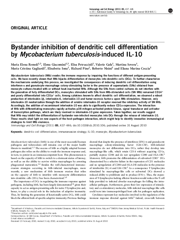
Figure 1 Schematic diagram of the experimental design. The cartoon represents the experimental plan used to investigate the effect of cytokines released
from Mtb-stimulated cultures on the differentiation of monocytes into DCs induced by IFN-a and GM-CSF.
Immunology and Cell Biology
�Mtb bystander inhibition of DC differentiation
ME Remoli et al
439
Figure 2 Inhibition of monocyte differentiation into DCs in response to treatment with SN Ctr and SN Mtb. Monocytes were cultured for 5 days with GM-CSF
and IFN-a, in the presence or not of heat-inactivated Mtb (Mtb:cell ratio 5:1) or 50% of SN Ctr or SN Mtb. The expression of CD1a and CD14 was then
analyzed by flow cytometry. (a) Dot plots of CD1a and CD14-stained cells from a representative experiment are shown. (b) The mean values ± s.e. of the
percentage of CD1a+ and CD14+ cells obtained from different donors are shown (n¼10). The SNs used to treat GM-CSF and IFN-a differentiating monocytes
were derived from seven different donors. CD1a expression: *P¼0.0003 Mtb vs Ctr and **P¼0.002 SN Mtb vs SN Ctr. CD14 expression: *P¼0.011 Ctr vs
Mtb and **P¼0.007 SN Ctr vs SN Mtb. (c) Kinetics of CD1a expression. The percentage of CD1a+ cells was analyzed after 1, 2, 3, 4 or 5 days of culture
with GM-CSF and IFN-a in the presence or not of heat-inactivated Mtb or 50% of SN Ctr or SN Mtb. Each value is the mean ± s.e. of the percentage of
CD1a+ cells obtained from three experiments (n¼3) performed with different donors treated with SNs collected from two different cultures.
cell sorter analysis of CD86, CD80, HLA-DR and CD38 (data not
shown). Given these results, it is likely that the SN Mtb blocks the final
differentiation of CD14+ monocytes into CD1a+ DC, leading to the
development of an intermediate population of CD1a� and CD14� cells.
The same effect was observed using monocytes isolated from 10
different donors (Figure 2b). Indeed, the percentage of CD1a+ cells
was significantly reduced from 78 to 27% in SN Mtb- or Mtbstimulated cells, while roughly 30% of cells remained CD14�.
A kinetic study was also performed to follow the effect of Mtb and
the cytokines released from Mtb-stimulated cultures on the time
course of monocyte differentiation into DCs (Figure 2c). In both
cases CD1a expression did not arise after 1 day of culture and the
cultures remained CD1a� compared with those stimulated with SN
Ctr, in which the percentage of CD1a+ cells reached nearly 90%.
Real-time reverse transcriptase (RT)-PCR performed on RNA
obtained from three different DC cultures showed that, in line with
the surface expression of CD1a and CD14, SN Mtb also reduced CD1a
mRNA content and had little effect on the level of CD14 mRNA as
compared with the expression in monocytes (Figure 3). A similar
profile of CD1a and CD14 mRNA expression was observed in cells
interacting with heat-killed Mtb, suggesting that the expression of
mRNA encoding cell surface antigens associated with DC differentiation represents an important target of mycobacterial immune evasion
strategies.
Cytokine production from differentiating monocytes upon
Mtb stimulation
It has been previously demonstrated by several groups that pro- and
anti-inflammatory cytokines, such as IL-1b, IL-6, IL-10 and TNF-a,
can inhibit the differentiation of monocytes into DC.18–22 To investigate whether Mtb may affect DC differentiation through the release
of these factors, SNs from Ctr and Mtb-stimulated cultures were
Immunology and Cell Biology
�Mtb bystander inhibition of DC differentiation
ME Remoli et al
440
Figure 3 Expression of CD1a and CD14 mRNA in IFN-a and GM-CSF differentiating monocytes stimulated with heat-inactivated Mtb, SN Ctr and SN Mtb.
Total RNA was extracted after 5 days of treatment with heat-inactivated Mtb or 50% SN Ctr or SN Mtb. Freshly isolated monocytes were used as internal
control. CD1a and CD14 mRNA expression was analyzed by real-time RT-PCR in three different donors (a–c). All quantification data are presented as a ratio
to the GAPDH level and represent the mean±s.e. of triplicate values. The standard errors (95% confidence limits) were calculated using the Student’s
t test.
Figure 4 Production of IL-10, IL-6, IL-1b and TNF-a from IFN-a and GM-CSF differentiating monocytes upon Mtb stimulation. The concentration of
cytokines present in SN Ctr and SN Mtb was determined by cytometric bead array. The values represent the means±s.e. of the cytokine concentrations
detected in the SN of cultures collected from nine independent experiments. IL-10 production: *P¼0.011; IL-6 production: *P¼0.002; IL-1b production:
*P¼0.0015; TNF-a production: *P¼0.035.
harvested at day 5 and cytometric bead array analysis was performed.
A robust production of IL-1b, IL-6, IL-10 and TNF-a was induced,
although to different extents, by Mtb stimulation (Figure 4), supporting our working hypothesis on the inhibitory role of Mtb-stimulated
cytokines in DC differentiation.
Immunology and Cell Biology
Definition of the inhibitory role of IL-10 in DC differentiation
To identify the cytokines that could be responsible for the inhibition
of DC differentiation, recombinant IL-1b, IL-6, IL-10 and TNF-a
cytokines were added to the monocytes cultured in differentiation
medium and the expression of CD1a and CD14 was evaluated after
�Mtb bystander inhibition of DC differentiation
ME Remoli et al
441
Figure 5 Effects of recombinant cytokines on DC differentiation. Monocytes were stimulated to differentiate with IFN-a and GM-CSF in the presence of IL-6
(20 ng ml�1), IL-10 (20 ng ml�1), TNF-a (50 ng ml�1) and IL-1b (10 ng ml�1). CD1a and CD14 expression was evaluated by flow cytometry after 5 days.
(a) Dot plots of CD1a- and CD14-stained cells from a representative donor are shown. (b) The mean value ± s.e. of the percentage of CD1a+ and CD14+
cells obtained with eight different donors are shown (n¼8). CD1a expression: *P¼0.0006 Ctr vs IL-10. CD14 expression: *P¼0.011 Ctr vs IL-10.
5 days of culture (Figures 5a and b). We found that only IL-10
reproduced the effect of SN Mtb, limiting the induction of CD1a
(to about 50%) and preserving the level of CD14 (at about 40%).
Dose–response experiments showed that the inhibitory effect of IL-10
on CD1a expression was not observed when 0.1 and 1 ng ml�1 doses
were used, while it started with 2 ng ml�1 and was stronger at the dose
of 10 ng ml�1 as evaluated by immunofluorescence at day 5 (data not
shown). This apparent discrepancy between the dose of recombinant
IL-10 required to inhibit monocyte differentiation and the amount of
IL-10 found in the SN Mtb could be ascribed to the higher bioactivity
of natural cytokines compared with the recombinant counterpart
produced in a prokaryotic expression system. Moreover, the capacity
of live and heat-inactivated Mtb to inhibit the differentiation of
monocytes into DCs correlates well with the observation that IL-10
production from differentiating monocytes was independent of the
vitality of Mtb6 (and data not shown).
To further define the role of IL-10 in DC differentiation, both SN
Ctr and SN Mtb were incubated with soluble IL-10 receptor
(5 mg ml�1) to neutralize IL-10 activity (Figure 6). The addition of
soluble IL-10 receptor significantly reversed the inhibitory activity of
Immunology and Cell Biology
�Mtb bystander inhibition of DC differentiation
ME Remoli et al
442
Figure 6 Recovery of CD1a expression upon the neutralization of IL-10. SN
Ctr and SN Mtb were incubated for 1 h with sIL10r (5 ng ml�1) to neutralize
the released IL-10 and, then, added to monocytes cultured with GM-CSF
and IFN-a. The percentage of cells expressing CD1a and CD14 was
evaluated by flow cytometry after 5 days of culture. The mean values±s.e.
of the percentage of CD1a+ and CD14+ cells obtained from four different
donors, each stimulated with SN collected from four different donors, are
shown (n¼4). CD1a expression: *P¼0.005 SN Mtb vs SN Mtb+sIL-10r.
CD14 expression: *P¼0.003 SN Mtb vs SN Mtb+sIL-10r.
SN Mtb on CD1a expression, although it partially affected the
expression of CD14 as evaluated by fluorescence-activated cell sorter.
Thus, IL-10 is necessary and largely sufficient to mediate the effect of
Mtb and SN Mtb on differentiation of monocytes to DCs evidenced
by monitoring CD1a and CD14 cell surface expression.
Contribution of the p38 mitogen-activated protein kinase (MAPK)
pathway and STAT-3 activation to IL-10 gene expression in
differentiating monocytes upon Mtb stimulation
Having found that IL-10 is an important player in CD1a inhibition
observed in differentiating monocytes, we focused our studies on the
regulation of IL-10 gene expression in IFN-a and GM-CSF cultured
monocytes upon Mtb stimulation. To study whether the release of
IL-10 correlated with an increased expression of the IL-10 gene, RNA
was extracted from differentiating monocytes at different times
following Mtb stimulation and the IL-10 mRNA expression was
then analyzed by real-time RT-PCR (Figure 7a). An increased
mRNA level was observed as early as 4 h post-Mtb treatment and
was maximally detected at 16 h.
Immunology and Cell Biology
Based on this finding suggesting a transcriptional control of IL-10
expression by Mtb, we sought to investigate STAT-3 and p38 MAPK
pathways, the involvement of which in the transcription of human
IL-10 gene was previously demonstrated.23–25 The contribution of p38
MAPK was evaluated in IFN-a and GM-CSF differentiating monocytes stimulated with Mtb. A kinetic experiment was performed
to characterize the consequences of IFN-a- and GM-CSF-induced
differentiation for the previously characterized activation of p38 by
Mtb in human monocytes.26 p38 MAPK was rapidly phosphorylated
1 h after Mtb stimulation and declined after 2 h (Figure 7b), confirming our previous data.26 To determine the role of MAPK in the
Mtb-driven induction of IL-10, we performed a dose–response experiment with p38 inhibitor SB203580 and Erk inhibitor PD98059, and
the production of IL-10 was measured by enzyme-linked immunosorbent assay upon Mtb stimulation (Supplementary Figure 2). A clear
reduction of IL-10 expression started when 3 mM SB203580 was used
to treat the cultures, and a stronger effect was found with higher doses.
Conversely, IL-10 release was not reduced by Erk inhibitor PD98059.
However, a specific effect on CD1a expression was observed when high
doses of SB203580 (5 and 10 mM) were used to treat the differentiating
cultures (Supplementary Figure 3). Based on these results, the
SB203580 dose of 3 mM was chosen to inhibit p38 MAPK in our
experimental setting. We found that p38 was likely involved in
Mtb-induced IL-10 expression because a significant reduction of
IL-10 production was observed in cultures treated with p38 inhibitor
SB203580, whereas the Erk inhibitor PD98059 even reinforced
Mtb-stimulated IL-10 production (Figure 7c). Specificity in the effect
of these inhibitors on IL-10 production is shown by the lack of effect
on production of IL-6 elicited by Mtb (Figure 7c).
The analysis of STAT-3 activation showed a weak tyrosine
phosphorylation detected as early as 4 h of stimulation, which
increased at 8 h and remained sustained until 24 h following Mtb
stimulation as evaluated by western blot analysis performed with
antibodies raised against the phosphorylated isoform of STAT-3
(data not shown). The kinetics of STAT-3 activation paralleled the
data obtained with electro-mobility shift assay performed with nuclear
cell extracts prepared at different times (4, 16 and 24 h) after Mtb
treatment and an oligo containing the STAT sequence present within
the IL-10 promoter. A STAT-3 DNA-binding complex with the STAT
binding site from the IL-10 promoter was faintly detected 4 h after
Mtb stimulation and reached a maximal level at 16–24 h (Figure 7d).
The identity of the complex was confirmed by supershift experiments
using an antibody raised against STAT-3. Collectively, these results
suggest that STAT-3 might cooperate with p38 MAPK in IL-10 gene
regulation in response to Mtb challenge.
DISCUSSION
Our study provides evidence for a novel mechanism of Mtb immunoevasion involving the inhibitory effect of IL-10 on the differentiation of monocytes into DC. The exploitation of IL-10 as a common
mechanism of immunosuppression by a heterogeneous group of
intracellular pathogens that can infect macrophages, including Mtb,
has been largely demonstrated.27 In particular, the capacity of IL-10 to
modulate the anti-mycobacterial immune response has been established in in vitro and animal models.27 The central molecular effect of
IL-10 is to dampen the expression of inflammatory cytokines, chemokines and cell surface molecules crucial for the induction of inflammation.28 Indeed, IL-10 is known to inhibit the production of
IL-12 and the antigen-presenting cell functions of monocytes and
macrophages, hence suppressing the development of Th1-mediated
immunity.29–31 More recently, a report addressed the question of how
�Mtb bystander inhibition of DC differentiation
ME Remoli et al
443
Figure 7 Regulation of IL-10 gene transcription in IFN-a and GM-CSF differentiating monocytes upon Mtb stimulation. (a) The kinetics of IL-10 mRNA
expression was investigated in IFN-a and GM-CSF differentiating monocytes stimulated with heat-inactivated Mtb. At the indicated time points, RNA was
extracted and the expression of IL-10 was measured by real-time RT-PCR. All quantification data are presented as a ratio to the GAPDH level. The results
shown are representative of one out of three experiments performed with RNA extracted from three different donors that yielded similar results. (b) Activation
of p38 MAPK was studied at the indicated time points following Mtb challenge by western blotting using antibodies directed against the phosphorylated
(p p38) and total p38 (p38). The results shown are from one out of three experiments that yielded similar results. (c) p38 inhibitor SB203580 (Mtb+SB) or
the ERK inhibitor PD98059 (Mtb+PD) were added 30 min before Mtb stimulation. The SNs were collected after 5 days and the production of IL-10 and
IL-6 was analyzed by enzyme-linked immunosorbent assay. The values represent the means ± s.e. of the cytokine concentration detected in the SN
of cultures collected in four independent experiments. *P¼0.018 Ctr vs Mtb: **P¼0.03 Mtb vs Mtb+SB. (d) Cells were treated with heat-inactivated Mtb
for the indicated time points. Nuclear extracts were prepared and analyzed by EMSA using a specific radiolabeled oligonucleotide corresponding to the
STAT-binding site present within the IL-10 promoter. The supershift assay was performed using an anti-STAT-3 or unrelated (Unr) antibodies, where
indicated. The supershifted complex is indicated with the SS arrow. This is a representative EMSA experiment, which was repeated two additional times with
cell extracts from different monocyte cultures stimulated with heat-inactivated Mtb.
an increased production of IL-10 by murine macrophages allows Mtb
replication.32 in spite of an active Th1 response. An elegant demonstration has been provided indicating the importance of this cytokine
in the control of the chronic phase and reactivation of infection.
Indeed, macrophages conditioned in vivo by high levels of IL-10
exhibited a reduced killing capacity despite a vigorous specific Th1
response. These data correlate well with the causal link from both
elevated levels of IL-10 in the sera of tuberculosis patients and the
polymorphism of IL-10 to tuberculosis susceptibility.33–38
Although macrophages are the main producers of IL-10 and, at the
same time, the targets of its immunosuppressive effects, DCs also
represent key participants in the IL-10-mediated strategies induced to
evade surveillance against tumors and pathogens.39 Indeed, IL-10,
which is commonly found in the microenvironment of tumors, is a
potent inhibitor of DC maturation and differentiation.40,41 In line
with these observations, we found that, in addition to a direct effect on
the infected cells,5,6 Mtb might also inhibit monocyte differentiation into DCs by inducing a bystander effect on the surrounding
Immunology and Cell Biology
�Mtb bystander inhibition of DC differentiation
ME Remoli et al
444
non-infected monocytes through the release of IL-10. Indeed,
IL-10-conditioned monocytes undergo a partial differentiation leading
to the development of a heterogenous population of cells expressing
CD14 and displaying variable levels of CD1a.
The impact of IL-10 on DC functions was previously demonstrated
by different groups, which found that the exogenous addition of IL-10
to immature DCs downregulated the expression of CD1a and their
capacity to stimulate an alloreactive response.20,42 These results are
consistent with our observation that CD1a induction is inhibited by
the IL-10 induced with Mtb or SN Mtb stimulation of monocyte
cultures during IFN-a and GM-CSF treatment. A different conclusion
on the capacity of IL-10 to modulate CD1 expression was drawn by
Stenger et al.,43 who demonstrated that the inhibition of CD1a
expression on antigen-presenting cells by Mtb was independent of
IL-10. This apparent discrepancy between these data and the results
published by our group (this paper and Mariotti et al.6) could depend
on the experimental model used to investigate the effect of IL-10.
Indeed, although Stenger analyzed the effect of Mtb infection on
differentiated antigen-presenting cells obtained from the adherent
fraction of peripheral blood mononuclear cells cultivated for 5 days
with GM-CSF and IL-4, in our experimental setting we studied
a purified CD14+/CD1a� population induced to differentiate by
GM-CSF and IFN-a and stimulated at day 0 with Mtb or SN Mtb.
In addition, the stronger production of IL-10 observed in cultures
stimulated with IFN-a and GM-CSF in the presence of heat-inactivated Mtb compared with those induced with IL-4 and GM-CSF
(roughly 20–30-fold higher; data not shown) could also explain the
differential outcome of IL-10 neutralization shown in this paper and
in the report by Stenger et al.,43 in which the production of IL-10 was
not determined at all. Moreover, another possible reason for the
differences in the observations of Stenger et al.43 and of this paper
could come from the stimulation of monocyte cultures with live
Mycobacteria at MOI 10 (10 Mtb for 1 cell), compared with our use of
five heat-inactivated Mtb to stimulate one monocyte. Thus, the MOI
and the timing of Mtb stimulation, the procedure used to isolate
monocytes as well as the composition of the cytokine cocktail used to
promote monocyte differentiation could account for the contrasting
conclusions on the role of IL-10 in the Mtb-driven impairment of
DC generation.
Collectively, our results shed light on a new aspect of Mtb
immunoevasion mechanism suggesting that at the site of a local
immune response, such as a tuberculous granuloma, the release of
IL-10 from infected monocytes/macrophages and its diffusion in
the lung microenvironment might counteract the differentiation of
recruited monocytes promoted by type I IFN released by infected
DCs.6 These data confirmed our previous observations showing
distinct roles for DCs and macrophages in driving the immune
response against Mtb on the basis of the differential profile of
cytokines and chemokines released following Mtb infection.12,44
Indeed, although macrophages control the granulomatous inflammatory response through the release of pro- and anti-inflammatory
cytokines, DCs are primarily involved in inducing anti-mycobacterial
T-cell immune responses.6
Based on these findings, understanding of how Mtb initiates
the production of IL-10 in differentiating monocytes would be
instrumental in revealing important targets for therapeutic intervention in tuberculosis. To this aim, the regulation of IL-10 gene
expression was investigated by having as background the knowledge
accumulated over the past years showing the capacity of mycobacterial
antigen or mycobacteria to activate different intracellular pathways
leading to IL-10 gene expression.45–52 Although our results confirmed
Immunology and Cell Biology
the involvement of p38 MAPK, we also observed activation of
STAT-3 associated with the expression of the IL-10 gene. Indeed,
upon Mtb challenge a transcription complex composed of STAT-3
binding to a functional STAT site characterized within the IL-10
promoter25 was observed in differentiating monocytes. Thus, in line
with previous data describing the capacity of hyperactivated STAT-3 to
promote IL-10 production,23,53 we can envisage that Mtb-activated
STAT-3 likely cooperates with p38 MAPK in inducing the expression
of this inhibitory cytokine. In addition to their capacity to modulate
IL-10 gene transcription, the combined effect of p38 and STAT-3
pathways could directly affect the differentiation of infected monocytes, which occurs in cancer, wherein the activation of these two
molecules leads to an abnormal development of myeloid cells.18,40,53–56
In line with this evidence, the capacity of mycobacteria to exploit the
p38 MAPK pathway to dampen different anti-microbial mechanisms
has been demonstrated by several groups.57,58 In particular, we recently
showed that CR3 engagement on monocytes by mycobacteria leads to
p38 and ATF-2 phosphorylation and that antibody-dependent CR3
blockade or treatment with a specific p38 inhibitor caused a notable
increase in CD1 molecule expression in DCs derived from mycobacteria-infected cells.26
In conclusion, it is tempting to speculate that the Mtb-induced
activation of p38 MAPK and STAT-3 may disarm DCs by inhibiting
their differentiation and, in turn, subvert a protective immune
surveillance. Indeed, Mtb may control the differentiation of the
infected monocytes into DC both through the activation of p38
and STAT-3-mediated signaling and the release of IL-10, which acts
indirectly on differentiation of uninfected monocytes.
Pharmacological interventions and innovative immunological
approaches aimed at counteracting the immunoevasion strategies of
Mtb should carefully evaluate p38 and STAT-3 pathways to replenish
DCs with full antigen presentation capacity.
METHODS
Antibodies and other reagents
Monoclonal antibodies specific for CD1a (FITC), CD14 (PE), CD80 (PE),
CD86 (FITC), HLA-ABC (FITC), HLA-DR (PE), IgG1 and IgG2a (BD
Pharmingen, San Diego, CA, USA) were used. Recombinant human soluble
IL-10 receptor was purchased from R&D Systems (Abingdom, UK) and used at
5 mg ml�1 for 1 h preincubation at 37 1C with the SNs. Recombinant TNF-a
(50 ng ml�1), IL-1b (10 ng ml�1), IL-6 (20 ng ml�1) and IL-10 (20 ng ml�1)
(PeproTech EC Ltd, London, UK) were used to treat monocytes. Preliminary
experiments were conducted to define the optimal dose of each cytokine (data
not shown). The p38 MAPK inhibitor SB203580 and the ERK inhibitor
PD98059, at a concentration of 3 mM, were added 30 min before Mtb stimulation. The inhibitors were not replenished during the 5-day incubation period.
Propidium iodide (Sigma Aldrich, St Louis, MO, USA) was used to test cell
viability by fluorescence.
Bacterial strain, media and growth conditions
All the experiments were performed with Mtb H37Rv (ATCC27294; American
Type Culture Collection, Manassas, VA, USA). Bacteria were prepared as
previously described.12 Mtb was heat killed at 80 1C for 30 min and used at
an Mtb:cell ratio of 5:1 (MOI 5). All preparations were analyzed for LPS
contamination by the Limulus lysate assay (BioWhittaker Europe, Verviers,
Belgium) and H37Rv contained o1 EU ml�1 LPS.
DC preparation and stimulation
DCs were prepared as previously described.6 Briefly, peripheral blood mononuclear cells were isolated from freshly collected buffy coats obtained from
healthy voluntary blood donors (Blood Bank of University ‘La Sapienza’, Rome,
Italy) by density gradient centrifugation using Lympholyte-H (Cedarlane,
Hornby, Ontario, Canada). Monocytes were purified by positive sorting using
�Mtb bystander inhibition of DC differentiation
ME Remoli et al
445
anti-CD14-conjugated magnetic microbeads (Miltenyi, Bergisch Gladbech,
Germany). The recovered cells were 499% CD14+ as determined by flow
cytometry with the anti-CD14 antibody. DCs were generated by culturing
monocytes in six-well tissue culture plates (Costar Corporation, Cambridge,
MA, USA) with 50 ng ml�1 GM-CSF (Schering-Plough, Levallois Perret, France)
and 1000 U ml�1 IFN-a (Roche, Nutley, NJ, USA) for 5 days at 0.5�106
cells ml�1 in RPMI 1640 (BioWhittaker Europe) supplemented with 2 mM Lglutamine and 15% fetal bovine serum (BioWhittaker Europe) and antibiotics.
To prepare SN Ctr and SN Mtb, freshly isolated monocytes were cultured in the
presence of IFN-a (1000 U ml�1) and GM-CSF (50 ng ml�1) for 5 days with or
without heat-killed Mtb (Mtb:cell ratio 5:1). The SN Ctr and SN Mtb were
harvested, filtered to remove the extracellular bacteria and then used to treat
new heterologous monocyte cultures obtained from different donors. Cell
viability was assessed by propidium iodide staining (Sigma Aldrich). Cells were
washed with PBS and then incubated with propidium iodide (final concentration, 50 mg ml�1) for 10 min at 4 1C. The percentage of live cells (propidium
iodide negative cells) was evaluated by flow-cytometric analysis.
Fluorescence-activated cell sorter analysis
Approximately 1–2�105 cells were aliquoted into tubes and washed once in
PBS containing 2% fetal bovine serum. The cells were incubated with purified
mAbs at 4 1C for 30 min. The cells were then washed and fixed with
2% paraformaldehyde before analysis on a fluorescence-activated cell sorter
scan using CellQuest software (Becton Dickinson, Mountain View, CA, USA).
A total of 5000 cells were analyzed per sample. The percentage of positive cells
for a specific antigen was obtained by subtracting the percentage obtained with
the matched isotype control antibody.
Cytokine determinations
SNs from cultures were harvested 5 days after treatment and stored at �80 1C.
IL-6, TNF-a, IL-10 and IL-1b were measured with the human inflammation
cytometric bead array (BD Pharmingen). IL-10 and IL-6 levels were also
determined by Instant enzyme-linked immunosorbent assay Kit (Bender
MedSystems, Burlingame, CA, USA). The assays were conducted according
to the manufacturer’s instructions.
Western blot analysis
Western blots were performed as previously described.26 Monocytes (2�106)
were stimulated as indicated and washed twice with cold phosphate-buffered
saline. The pellet was resuspended in 200 ml of 2� SDS sample buffer (20 mM
dithiothreitol, 6% SDS, 0.25 M Tris, pH 6.8, 10% glycerol, 10 mM NaF and
bromophenyl blue) and boiled for 5 min. Proteins were separated by SDS/PAGE
and blotted onto nitrocellulose membranes (Hybond C-Extra; GE Healthcare,
Uppsala, Sweden). Blots were incubated with phospho- and total-p38 (R&D
System), reacted with anti-rabbit HRP-coupled secondary antibody (GE
Healthcare) and developed using an ECL system.
Electro-mobility shift assay
Nuclear cell extracts (15 mg) were prepared and used in electro-mobility shift
assay as previously described.11 Synthetic double-stranded oligonucleotide
containing the STAT-binding sequence of the IL-10 promoter was end-labeled
with [g32P]-ATP using T4 polynucleotide kinase. The sequence binding STAT-3
within the IL-10 promoter was described by Unterberger et al.:25 LS4 forward
5¢-ATCCTGTGCCGGGAAACC-3¢; LS4 reverse 5¢-GGTTTCCCGGCACAGG
AT-3¢. For supershift analysis, 1 mg of control antibody or anti-STAT-3 antibody
(Santa Cruz Biotechnology, Santa Cruz, CA, USA) was added to the reaction.
RNA isolation and real-time RT-PCR quantification
RNA was extracted with RNeasy Mini kit (Qiagen Inc., Valencia, CA, USA)
according to the manufacturer’s instructions. Reverse transcriptions were
primed with oligo (dT) and performed using the Murine Leukemia Virus
Reverse Transcriptase (Invitrogen Life Technologies, Carlsbad, CA, USA).
Quantitative RT-PCR assays were done in triplicate using the Platinum Taq
DNA Polymerase (Invitrogen Life Technologies) and the SYBR Green I
(Biowhittaker Molecular Applications, Rockland, ME, USA) on a LightCycler
(Roche Diagnostics). A calibration curve of a purified positive control RT-PCR
product, to which arbitrary values were assigned, was used to calculate the
value of a target gene. The quantification standard curves were obtained using
dilutions (4-log range) of the purified positive control RT-PCR product in
10 mg ml�1 sonicated salmon sperm DNA. All quantification data of CD1a,
CD14 and IL-10 transcripts are shown as a ratio to the GAPDH level present in
the same sample and represent the mean + s.e. of triplicate values. The standard
errors (95% confidence limits) were calculated using the Student’s t-test. The
sequences of the primer pairs used for the quantification of GAPDH59
and for IL-1060 have been previously described. The primers used for CD1a
mRNA quantification were: 5¢-TATCACCGCCAAGATGATGA-3¢ (forward)
and 5¢-TGTGTGCCATGTCTCAGGAT-3¢ (reverse).
Statistical analysis
Statistical analysis was calculated using a two-tailed Student’s t-test for paired
data. A P value o0.05 was considered statistically significant.
ACKNOWLEDGEMENTS
This work was supported by ISS-NIH Program (#5303) and European
Community 71 PQ NewTBVAC grants. We also thank Eugenio Morassi for
preparing the drawings.
1 WHO. Tuberculosis Factsheet. World Health Organization: Geneva, 2009. http://www.
who.int/tb/publications/factsheets/en/index.html.
2 Lauzardo M, Ashkin D. Phthisiology at the dawn of the new century. Chest 2000; 117:
1455–1473.
3 Ehrt S, Schnappinger D. Mycobacterial survival strategies in the phagosome: defence
against host stresses. Cell Microbiol 2009; 11: 1170–1178.
4 Flannagan RS, Cosio G, Grinstein S. Antimicrobial mechanisms of phagocytes and
bacterial evasion strategies. Nat Rev Microbiol 2009; 7: 355–366.
5 Mariotti S, Teloni R, Iona E, Fattorini L, Giannoni F, Romagnoli G et al. Mycobacterium
tuberculosis subverts the differentiation of human monocytes into dendritic cells.
Eur J Immunol 2002; 32: 3050–3058.
6 Mariotti S, Teloni R, Iona E, Fattorini L, Romagnoli G, Gagliardi MC et al. Mycobacterium tuberculosis diverts alpha interferon-induced monocyte differentiation from
dendritic cells into immunoprivileged macrophage-like host cells. Infect Immun 2004;
72: 4385–4392.
7 Sinha A, Salam N, Gupta S, Natarajan K. Mycobacterium tuberculosis and dendritic
cells: recognition, activation and functional implications. Indian J Biochem Biophys
2007; 44: 279–288.
8 Herrmann JL, Lagrange PH. Dendritic cells and Mycobacterium tuberculosis: which is
the Trojan horse? Pathol Biol (Paris) 2005; 53: 35–40.
9 Cooper AM, Khader SA. The role of cytokines in the initiation, expansion, and control of
cellular immunity to tuberculosis. Immunol Rev 2008; 226: 191–204.
10 Salgame P. Host innate and Th1 responses and the bacterial factors that control
Mycobacterium tuberculosis infection. Curr Opin Immunol 2005; 17: 374–380.
11 Remoli ME, Giacomini E, Lutfalla G, Dondi E, Orefici G, Battistini A et al. Selective
expression of type I IFN genes in human dendritic cells infected with Mycobacterium
tuberculosis. J Immunol 2002; 169: 366–374.
12 Giacomini E, Iona E, Ferroni L, Miettinen M, Fattorini L, Orefici G et al. Infection of
human macrophages and dendritic cells with Mycobacterium tuberculosis induces a
differential cytokine gene expression that modulates T cell response. J Immunol 2001;
166: 7033–7041.
13 Montoya M, Schiavoni G, Mattei F, Gresser I, Belardelli F, Borrow P et al. Type I
interferons produced by dendritic cells promote their phenotypic and functional
activation. Blood 2002; 99: 3263–3271.
14 Santini SM, Lapenta C, Logozzi M, Parlato S, Spada M, Di Pucchio T et al. Type I
interferon as a powerful adjuvant for monocyte-derived dendritic cell development and
activity in vitro and in Hu-PBL-SCID mice. J Exp Med 2000; 191: 1777–1788.
15 Giacomini E, Remoli ME, Gafa V, Pardini M, Fattorini L, Coccia EM. IFN-beta improves
BCG immunogenicity by acting on DC maturation. J Leukoc Biol 2009; 85: 462–468.
16 Prabhakar S, Qiao Y, Hoshino Y, Weiden M, Canova A, Giacomini E et al. Inhibition of
response to alpha interferon by Mycobacterium tuberculosis. Infect Immun 2003; 71:
2487–2497.
17 Prabhakar S, Qiao Y, Canova A, Tse DB, Pine R. IFN-alpha beta secreted during
infection is necessary but not sufficient for negative feedback regulation of IFN-alpha
beta signaling by Mycobacterium tuberculosis. J Immunol 2005; 174: 1003–1012.
18 Wang S, Hong S, Yang J, Qian J, Zhang X, Shpall E et al. Optimizing immunotherapy
in multiple myeloma: restoring the function of patients’ monocyte-derived dendritic
cells by inhibiting p38 or activating MEK/ERK MAPK and neutralizing interleukin-6 in
progenitor cells. Blood 2006; 108: 4071–4077.
19 Makino M, Maeda Y, Mukai T, Kaufmann SH. Impaired maturation and function
of dendritic cells by mycobacteria through IL-1beta. Eur J Immunol 2006; 36:
1443–1452.
Immunology and Cell Biology
�Mtb bystander inhibition of DC differentiation
ME Remoli et al
446
20 Fortsch D, Rollinghoff M, Stenger S. IL-10 converts human dendritic cells into
macrophage-like cells with increased antibacterial activity against virulent Mycobacterium tuberculosis. J Immunol 2000; 165: 978–987.
21 Chomarat P, Banchereau J, Davoust J, Palucka AK. IL-6 switches the differentiation of
monocytes from dendritic cells to macrophages. Nat Immunol 2000; 1: 510–514.
22 Allavena P, Piemonti L, Longoni D, Bernasconi S, Stoppacciaro A, Ruco L et al. IL-10
prevents the generation of dendritic cells from CD14+ blood monocytes, promotes
the differentiation to mature macrophages and stimulates endocytosis of FITC-dextran.
Adv Exp Med Biol 1997; 417: 323–327.
23 Staples KJ, Smallie T, Williams LM, Foey A, Burke B, Foxwell BM et al. IL-10 induces
IL-10 in primary human monocyte-derived macrophages via the transcription factor
Stat3. J Immunol 2007; 178: 4779–4785.
24 Ma W, Lim W, Gee K, Aucoin S, Nandan D, Kozlowski M et al. The p38 mitogenactivated kinase pathway regulates the human interleukin-10 promoter via the activation of Sp1 transcription factor in lipopolysaccharide-stimulated human macrophages.
J Biol Chem 2001; 276: 13664–13674.
25 Unterberger C, Staples KJ, Smallie T, Williams L, Foxwell B, Schaefer A et al. Role of
STAT3 in glucocorticoid-induced expression of the human IL-10 gene. Mol Immunol
2008; 45: 3230–3237.
26 Gagliardi MC, Teloni R, Giannoni F, Mariotti S, Remoli ME, Sargentini V et al.
Mycobacteria exploit p38 signaling to affect CD1 expression and lipid antigen
presentation by human dendritic cells. Infect Immun 2009; 77: 4947–4952.
27 Redpath S, Ghazal P, Gascoigne NR. Hijacking and exploitation of IL-10 by intracellular pathogens. Trends Microbiol 2001; 9: 86–92.
28 Murray PJ. Understanding and exploiting the endogenous interleukin-10/STAT3mediated anti-inflammatory response. Curr Opin Pharmacol 2006; 6: 379–386.
29 Bogdan C, Vodovotz Y, Nathan C. Macrophage deactivation by interleukin 10. J Exp
Med 1991; 174: 1549–1555.
30 Fiorentino DF, Zlotnik A, Mosmann TR, Howard M, O’Garra A. IL-10 inhibits cytokine
production by activated macrophages. J Immunol 1991; 147: 3815–3822.
31 Aste-Amezaga M, Ma X, Sartori A, Trinchieri G. Molecular mechanisms of the induction
of IL-12 and its inhibition by IL-10. J Immunol 1998; 160: 5936–5944.
32 Schreiber T, Ehlers S, Heitmann L, Rausch A, Mages J, Murray PJ et al. Autocrine IL-10
induces hallmarks of alternative activation in macrophages and suppresses antituberculosis effector mechanisms without compromising T cell immunity. J Immunol 2009;
183: 1301–1312.
33 Olobo JO, Geletu M, Demissie A, Eguale T, Hiwot K, Aderaye G et al. Circulating
TNF-alpha TGF-beta, and IL-10 in tuberculosis patients and healthy contacts. Scand J
Immunol 2001; 53: 85–91.
34 Boussiotis VA, Tsai EY, Yunis EJ, Thim S, Delgado JC, Dascher CC et al. IL-10producing T cells suppress immune responses in anergic tuberculosis patients.
J Clin Invest 2000; 105: 1317–1325.
35 Verbon A, Juffermans N, Van Deventer SJ, Speelman P, Van Deutekom H, Van Der Poll
T. Serum concentrations of cytokines in patients with active tuberculosis (TB) and after
treatment. Clin Exp Immunol 1999; 115: 110–113.
36 Eum SY, Jeon BY, Min JH, Kim SC, Cho S, Park SK et al. Tumor necrosis factor-alpha
and interleukin-10 in whole blood is associated with disease progression in pulmonary
multidrug-resistant tuberculosis patients. Respiration 2008; 76: 331–337.
37 Ates O, Musellim B, Ongen G, Topal-Sarikaya A. Interleukin-10 and tumor necrosis
factor-alpha gene polymorphisms in tuberculosis. J Clin Immunol 2008; 28: 232–236.
38 Oral HB, Budak F, Uzaslan EK, Basturk B, Bekar A, Akalin H et al. Interleukin-10
(IL-10) gene polymorphism as a potential host susceptibility factor in tuberculosis.
Cytokine 2006; 35: 143–147.
39 Moore KW, de Waal Malefyt R, Coffman RL, O’Garra A. Interleukin-10 and the
interleukin-10 receptor. Annu Rev Immunol 2001; 19: 683–765.
40 Lin A, Schildknecht A, Nguyen LT, Ohashi PS. Dendritic cells integrate signals from the
tumor microenvironment to modulate immunity and tumor growth. Immunol Lett 2009;
127: 77–84.
41 Buelens C, Verhasselt V, De Groote D, Thielemans K, Goldman M, Willems F.
Interleukin-10 prevents the generation of dendritic cells from human peripheral
blood mononuclear cells cultured with interleukin-4 and granulocyte/macrophagecolony-stimulating factor. Eur J Immunol 1997; 27: 756–762.
42 Allavena P, Piemonti L, Longoni D, Bernasconi S, Stoppacciaro A, Ruco L et al. IL-10
prevents the differentiation of monocytes to dendritic cells but promotes their maturation to macrophages. Eur J Immunol 1998; 28: 359–369.
43 Stenger S, Niazi KR, Modlin RL. Down-regulation of CD1 on antigen-presenting cells
by infection with Mycobacterium tuberculosis. J Immunol 1998; 161: 3582–3588.
44 Lande R, Giacomini E, Grassi T, Remoli ME, Iona E, Miettinen M et al. IFN-alpha beta
released by Mycobacterium tuberculosis-infected human dendritic cells induces the
expression of CXCL10: selective recruitment of NK and activated T cells. J Immunol
2003; 170: 1174–1182.
45 Jung SB, Song CH, Yang CS, Kim SY, Lee KS, Shin AR et al. Role of the phosphatidylinositol 3-kinase and mitogen-activated protein kinase pathways in the secretion of
tumor necrosis factor-alpha and interleukin-10 by the PPD antigen of Mycobacterium
tuberculosis. J Clin Immunol 2005; 25: 482–490.
46 Chan MM, Cheung BK, Li JC, Chan LL, Lau AS. A role for glycogen synthase kinase-3
in antagonizing mycobacterial immune evasion by negatively regulating IL-10 induction. J Leukoc Biol 2009; 86: 283–291.
47 Reiling N, Blumenthal A, Flad HD, Ernst M, Ehlers S. Mycobacteria-induced TNF-alpha
and IL-10 formation by human macrophages is differentially regulated at the level of
mitogen-activated protein kinase activity. J Immunol 2001; 167: 3339–3345.
48 Song CH, Lee JS, Lee SH, Lim K, Kim HJ, Park JK et al. Role of mitogen-activated
protein kinase pathways in the production of tumor necrosis factor-alpha, interleukin10, and monocyte chemotactic protein-1 by Mycobacterium tuberculosis H37Rvinfected human monocytes. J Clin Immunol 2003; 23: 194–201.
49 Cheung BK, Lee DC, Li JC, Lau YL, Lau AS. A role for double-stranded RNA-activated
protein kinase PKR in Mycobacterium-induced cytokine expression. J Immunol 2005;
175: 7218–7225.
50 Mendez-Samperio P, Trejo A, Perez A. Mycobacterium bovis Bacillus Calmette-Guerin
(BCG) stimulates IL-10 production via the PI3K/Akt and p38 MAPK pathways in
human lung epithelial cells. Cell Immunol 2008; 251: 37–42.
51 Nair S, Ramaswamy PA, Ghosh S, Joshi DC, Pathak N, Siddiqui I et al. The PPE18 of
Mycobacterium tuberculosis interacts with TLR2 and activates IL-10 induction in
macrophage. J Immunol 2009; 183: 6269–6281.
52 Souza CD, Evanson OA, Weiss DJ. Mitogen activated protein kinase p38 pathway is
an important component of the anti-inflammatory response in Mycobacterium avium
subsp. paratuberculosis-infected bovine monocytes. Microb Pathog 2006; 41: 59–66.
53 Wang T, Niu G, Kortylewski M, Burdelya L, Shain K, Zhang S et al. Regulation of the
innate and adaptive immune responses by Stat-3 signaling in tumor cells. Nat Med
2004; 10: 48–54.
54 Nefedova Y, Nagaraj S, Rosenbauer A, Muro-Cacho C, Sebti SM, Gabrilovich DI.
Regulation of dendritic cell differentiation and antitumor immune response in
cancer by pharmacologic-selective inhibition of the janus-activated kinase 2/signal
transducers and activators of transcription 3 pathway. Cancer Res 2005; 65:
9525–9535.
55 Nefedova Y, Huang M, Kusmartsev S, Bhattacharya R, Cheng P, Salup R et al.
Hyperactivation of STAT3 is involved in abnormal differentiation of dendritic cells in
cancer. J Immunol 2004; 172: 464–474.
56 Wang S, Yang J, Qian J, Wezeman M, Kwak LW, Yi Q. Tumor evasion of the immune
system: inhibiting p38 MAPK signaling restores the function of dendritic cells in
multiple myeloma. Blood 2006; 107: 2432–2439.
57 Pennini ME, Pai RK, Schultz DC, Boom WH, Harding CV. Mycobacterium tuberculosis
19-kDa lipoprotein inhibits IFN-gamma-induced chromatin remodeling of MHC2TA
by TLR2 and MAPK signaling. J Immunol 2006; 176: 4323–4330.
58 Jo EK, Yang CS, Choi CH, Harding CV. Intracellular signalling cascades regulating
innate immune responses to Mycobacteria: branching out from Toll-like receptors.
Cell Microbiol 2007; 9: 1087–1098.
59 Coccia EM, Severa M, Giacomini E, Monneron D, Remoli ME, Julkunen I et al.
Viral infection and Toll-like receptor agonists induce a differential expression of type
I and lambda interferons in human plasmacytoid and monocyte-derived dendritic cells.
Eur J Immunol 2004; 34: 796–805.
60 Gibson SJ, Lindh JM, Riter TR, Gleason RM, Rogers LM, Fuller AE et al. Plasmacytoid
dendritic cells produce cytokines and mature in response to the TLR7 agonists,
imiquimod and resiquimod. Cell Immunol 2002; 218: 74–86.
Supplementary Information accompanies the paper on Immunology and Cell Biology website (http://www.nature.com/icb)
Immunology and Cell Biology
�
 Elisabetta Iona