Animal Reproduction Science 104 (2008) 143–163
Factors affecting chromatin stability of
bovine spermatozoa
T.A.A. Khalifa a,∗ , C.A. Rekkas b , A.G. Lymberopoulos b ,
A. Sioga c , I. Dimitriadis d , Th. Papanikolaou d
a
Department of Theriogenology, Faculty of Veterinary Medicine, Mansoura University, Mansoura, Egypt
b NAGREF, Veterinary Research Institute, Ionia, Thessaloniki, Greece
c Department of Histology and Embryology, School of Medicine, Aristotle University of Thessaloniki, Greece
d Department of Reproduction and Obstetrics, Veterinary Faculty, University of Thessaly, Karditsa, Greece
Received 8 August 2006; received in revised form 31 January 2007; accepted 16 February 2007
Available online 28 February 2007
Abstract
The structural stability of transcriptionally inert paternal chromatin is of vital importance for the fertilization process and early embryonic development. Accordingly, a series of eight experiments were conducted
during a 7-month period to investigate: (1) effects of bull breed, individuality, successive ejaculations,
semen quality characteristics (SQC), semen dilution rates and hypothermic storage of semen in a Tris-egg
yolk extender on incidence of sperm nuclear chromatin instability (NCI), and (2) effects of the interaction
between variation of NCI within a frozen ejaculate and variation of oocytes quality due to maturation time
and/or season on the efficiency of in vitro embryo production (IVEP). Semen samples were collected once a
week from six bulls using an AV and only ejaculates (n = 220) of >0.30 × 10⁹ sperm/ml and ≥60% motility
were used. NCI was measured by: (1) detection of lysine-rich histones in sperm chromatin using aniline blue
staining, (2) sperm susceptibility to acid-induced nuclear DNA denaturation in situ using acridine orange
test, and (3) sperm susceptibility to nuclear chromatin decondensation (NCD). Bovine oocytes (n = 695) were
matured in vitro for 18 or 24 h, fertilized after sperm selection through a swim-up procedure and cultured
for 72 h. The results showed that the 2nd ejaculates were superior to the 1st ones with respect to chromatin
stability. Dilution of semen to 49.67 ± 8.56 × 10⁶ sperm/ml (1:19) decreased resistance of sperm to NCD.
Cooling of semen had no significant effect on chromatin stability. Cryopreservation of semen augmented
sperm vulnerability to DNA denaturation. Improvement of SQC (semen volume, sperm motility, velocity,
viability and morphological normalcy) was generally concomitant with increase of sperm resistance to NCI.
While Blonde d’Aquitaine bulls had a resistance to NCD higher than Limousine bulls in fresh semen, the
former showed a greater susceptibility to DNA denaturation than the latter in cooled semen. Individuality
∗ Corresponding author. Present address: c/o Christine Cooreman, 19 Gravias street, 54645 Thessaloniki, Greece.
Tel.: +30 2310 869 500.
E-mail address: drtarekkhalifa@in.gr (T.A.A. Khalifa).
0378-4320/$ – see front matter © 2007 Elsevier B.V. All rights reserved.
doi:10.1016/j.anireprosci.2007.02.019
�144
T.A.A. Khalifa et al. / Animal Reproduction Science 104 (2008) 143–163
significantly influenced NCI. The variability of NCI within a frozen ejaculate affected efficiency of IVEP.
Significant negative correlations were observed between incidence of NCI and both fertilization rate and
developmental capacity of embryos after maturation of oocytes for 18 h. The significant variation in IVEP
traits due to season was independent of the effect of sperm chromatin instability.
© 2007 Elsevier B.V. All rights reserved.
Keywords: Bull semen; Chromatin stability; Embryo production
1. Introduction
Failure of fertilization and embryonic mortality, particularly after AI, have long been recognized as potential sources of loss in breeding cows and numerous studies have reported on them
(Gordon, 1996). The potential sire effects on bovine embryonic development have been shown
to occur as early as the initiation of S-phase in the zygote and after expression of the embryonic
genome at the four- to eight-cell stage (Eid et al., 1994). Therefore, laboratory assessment of semen
must include testing of most sperm attributes relevant for fertilization and embryo development,
such as evaluation of sperm genomic integrity (Evenson and Wixon, 2006).
Mammalian sperm genome is composed of nuclear DNA (Gledhill, 1970), mitochondrial DNA
(Sutovsky et al., 2003) and cytoplasmic messenger RNAs (Miller, 2000). The DNA in sperm
nuclei is bound to basic nuclear proteins to form the deoxyribonucleoprotein (DNP) complex
(Livolant, 1984). The only essential component of the sperm needed to form normal embryos is a
nucleus with an intact nuclear matrix (Ward et al., 1999). A high level of sperm nuclear chromatin
instability (NCI) in semen is associated with reduced breeding efficiency of bulls (Ballachey et
al., 1988; Karabinus et al., 1990; Dobrinski et al., 1994; Anzar et al., 2002; Madrid-Bury et al.,
2005).
The presence of sperm with NCI in freshly ejaculated semen is considered an uncompensable
defect, which cannot be tolerated at levels greater than 15–20% of spermatozoa (Barth and Oko,
1989). Fresh semen from bulls classified as questionable or unsatisfactory potential breeders
contains a higher percentage (13.50%) of spermatozoa with abnormal DNA condensation than
does semen from bulls classified as satisfactory potential breeders (7.10%) (Dobrinski et al., 1994).
Freshly diluted semen of mature dairy bulls contains 0.53–8% sperm with NCI (Krzyzosiak et
al., 2000). The factors that may affect the incidence of NCI in fresh semen are individuality and
semen quality characteristics (Dobrinski et al., 1994) as well as variation between and within
ejaculates of the same individual (Duty et al., 2002). However, the effects of breed, successive
ejaculations and dilution rates on the incidence of NCI in fresh semen of beef bulls have not yet
been investigated.
Previous studies on cooled bull semen (Salisbury et al., 1961; Hanada et al., 1965) did not
show any changes in sperm DNP complex for storage up to 24 h. Moreover, the literature contains conflicting results regarding the impact of cryopreservation on sperm genome. Freezing
and/or thawing of semen altered DNP complex of bull sperm (Salisbury et al., 1964). On the
contrary, overcondensation of sperm chromatin has been observed in frozen-thawed semen of
bulls (Dobrinski et al., 1994; Anzar et al., 2002). Other authors did not find any effect for
cryopreservation of bull semen on sperm chromatin stability or DNA integrity (Martin et al.,
2004).
Concerning the variation range (0–15%) of NCI in frozen semen, many factors have been
studied, such as individuality and sperm traits (Dobrinski et al., 1994; Januskauskas et al., 2003),
�T.A.A. Khalifa et al. / Animal Reproduction Science 104 (2008) 143–163
145
variation between ejaculates of the same bull (Bochenek et al., 2001), bull age and post-thawing
incubation time and temperature (Karabinus et al., 1990). However, the effect of breed on the incidence of NCI in frozen semen of beef bulls has not yet been studied. Moreover, scarce information
is available regarding the relationship between sperm kinematic characteristics and chromatin
stability.
Incomplete nuclear condensation may appear in the form of nuclear vacuoles, which have been
described in bull spermatozoa (Walters et al., 2004). The percentage of sperm with single and
multiple vacuoles in frozen semen of bulls is positively correlated with the percentage of sperm
with NCI (Dobrinski et al., 1994). Vacuolated sperm cells bind to the zona pellucida, penetrate
oocytes at a low rate, reduce formation of male pronuclei and decrease the rate of bovine in vitro
embryo production (IVEP) (Walters et al., 2004).
IVEP represents a desirable option in strategies to enhance reproductive and genetic advances
in cattle (Trounson, 1992). Sperm DNA damage or alteration of protamine packing around sperm
DNA may contribute to a delay in initiation of zygotic S-phase (Eid et al., 1994), an increase
in length of zygotic G2-phase (Eid and Parrish, 1995), a block in blastocyst formation (Fatehi
et al., 2006) and, thus, to a decrease in numbers of morphologically normal bovine embryos
(Smorag et al., 2000). The developmental block that arises in bovine embryos at the eight-cell
stage may be correlated with the cytoplasmic quality of oocytes (Meirelles et al., 2004). Oocytes
and early embryos have been shown to repair sperm DNA damage (Ashwood-Smith and Edwards,
1996). Consequently, the biological effect of abnormal sperm chromatin structure depends on the
combined effects of the level of chromatin damage in the spermatozoa and the capacity of the
oocyte to repair that pre-existing damage (Evenson et al., 2002). Therefore, if spermatozoa are
selected from samples with extensively damaged DNA and used for IVF, the oocyte’s repair
capacities may be inadequate, leading to a low rate of embryonic development (Ahmadi and Ng,
1999).
The objectives of the present study were to investigate: (1) effects of breed, individuality,
successive ejaculations, semen quality characteristics, semen dilution rates and hypothermic
preservation of semen on the incidence of NCI in spermatozoa of beef bulls, and (2) effects
of the interaction between variation of sperm NCI within a frozen semen ejaculate of a single
dairy bull and variation of oocytes quality due to maturation time and/or season on the efficiency
of bovine IVEP.
2. Materials and methods
2.1. Location
The study was carried out (February to August 2005) at the AI center and IVEP laboratory,
NAGREF, Veterinary Research Institute, Ionia, Thessaloniki, Northern Greece.
2.2. Chemical reagents and semen extender
Unless otherwise stated, all chemicals were purchased from Sigma–Aldrich Co., Greece. MilliQ purified water was utilized in all experiments. Media, buffers and staining solutions were filtered
before use through a 0.22 m filter. A home-made Tris-based extender (pH 6.8, 300 mOsm/kg) was
used for two-step dilution and hypothermic preservation of bull semen (Anzar et al., 2002). It was
composed of fractions A and B. Fraction A consisted of Tris (7.266 g), citric acid monohydrate
(4.08 g), fructose (3.00 g), fresh chicken egg yolk (60 ml), penicillin G sodium (300,000 IU),
�146
T.A.A. Khalifa et al. / Animal Reproduction Science 104 (2008) 143–163
streptomycin sulfate (0.30 g) and water to 300 ml. Fraction B consisted of the same ingredients
and concentrations as those of Fraction A, but also contained 12% glycerol (v/v).
2.3. Sources of semen
2.3.1. Beef bull semen
Mature bulls 3–4 years old of Blonde d’Aquitaine (BDA, n = 3, designated 1, 2 and 3) and
Limousine (LIM, n = 3, designated 4, 5 and 6) breeds were subjected to a weekly semen collection
schedule using an AV. Two successive ejaculates at an 8–10-min interval were obtained from each
bull. Immediately after semen collection, the ejaculates were transferred to the laboratory, kept
in a water bath at 25 ◦ C and evaluated for sperm motility and concentration. Only ejaculates
(n = 220) with sperm progressive motility (SPM) ≥60% and concentration >0.30 × 10⁹ sperm/ml
were used (Salisbury et al., 1978) for subsequent manipulations.
2.3.2. Dairy bull semen
Frozen semen in 0.50-ml straws from one ejaculate of a single proven fertile Holstein bull was
used in the IVF experiment. The semen was frozen in the above-described extender for 17 years.
Previous examination of semen in our laboratory showed that post-thawing SPM and IVF rate
ranged between 40–50% and 60–85%, respectively.
2.4. Hypothermic preservation of semen
2.4.1. Chilled-stored (CS) semen
Freshly ejaculated (FE) semen was initially diluted at 25 ◦ C with extender fraction A to obtain
100 × 106 motile sperm/ml, cooled to 4 ◦ C over a period of 2 h and further diluted at 4 ◦ C with
extender fraction B to obtain 50 × 106 motile sperm/ml. The final concentration of glycerol in the
extender was 6% (v/v). After 30 min, glycerolized semen was packaged in 0.50-ml straws and
stored at 4 ◦ C for 5 h. This duration was selected to be identical to the equilibration period of
cooled semen prior to freezing.
2.4.2. Frozen-thawed (FT) semen
FE semen was diluted, cooled, packaged and equilibrated as described above. After equilibration, straws were transferred to an automatic computerized semen freezing chamber pre-cooled
at 4 ◦ C (Digitcool 5300, IMV, L’Aigle, France) and frozen at the following programmed rate:
5 ◦ C/min from +4 ◦ C to −12 ◦ C and 60 ◦ C/min from −12 ◦ C to −140 ◦ C. Finally the straws were
stored for 4 weeks in a liquid nitrogen container and thawed in a water bath at 40 ◦ C for 30 s.
2.5. Semen evaluation
2.5.1. Semen quality characteristics (SQC)
Ejaculate volume was measured to the nearest 0.10 ml in a graduated tube. Sperm concentration
was measured using a photometer and a Neubauer haemocytometer chamber. SPM (%) and sperm
velocity score (SVS) were subjectively assessed (Kjaestad et al., 1993) under a phase-contrast
microscopy (400×) equipped with a thermal stage at 37 ◦ C. Sperm motion characteristics were
analyzed (Bilodeau et al., 2000; Chatterjee and Gagnon, 2001) using a computer-assisted sperm
motion analysis (CASA) system (Cell Motion Analyzer SM-CMA, Strömberg-Mika, Germany).
Sperm viability and morphological abnormalities were examined (Harasymowycz et al., 1976)
�T.A.A. Khalifa et al. / Animal Reproduction Science 104 (2008) 143–163
147
under a bright-field microscopy (1000×) using a modified eosin-nigrosin stain (ENS) (Barth
and Oko, 1989). Acrosomal abnormalities were assessed (Harasymowycz et al., 1976) in wet
mounts of buffered formal saline using a phase-contrast microscopy (1000×). Head dimensions
(length and width) of live morphologically normal sperm were measured manually in smears
stained with a modified ENS using an ocular (eyepiece)-mounted micrometer (Magistrini et al.,
1997). Sperm response to the hypo-osmotic swelling (HOS) test was determined according to
the method of Revell and Mrode (1994). An aliquot (100 l) of sperm suspension was incubated at 35 ◦ C for 60 min with 200 l of HOS solution (150 mOsm/kg). The proportion of sperm
with bending, coiling or shortening of tails was examined under a phase-contrast microscopy
(1000×).
2.5.2. Sperm nuclear chromatin instability (NCI) measures
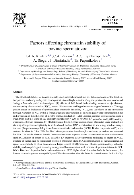
2.5.2.1. Acidic aniline blue (AB) staining. Sperm chromatin condensation is disturbed (NCI)
when lysine-rich somatic histones are not sufficiently substituted by arginine-rich protamines
during spermiogenesis. Histone-containing spermatozoa can be visualized using acidic AB staining solution, which reacts with lysine residues of persisting histones (Hingst et al., 1995). The
percentage of sperm heads stained partially or entirely dark blue (Fig. 1) was examined according
to the procedures described by Jeulin et al. (1986).
2.5.2.2. Acridine orange (AO) staining. The compact chromatin structure of mature bull spermatozoa renders their nuclear DNA resistant to mild acid hydrolysis (Barth and Oko, 1989). AO test
was carried out according to the method of Tejada et al. (1984) which measures the susceptibility
of sperm nuclear DNA to acid-induced denaturation in situ by quantifying the metachromatic shift
of AO fluorescence from green (native DNA) to red (denatured DNA). The percentage of sperm
heads fluorescencing a spectrum of colors ranging from yellowish green to partially or entirely
red (denatured DNA, Fig. 2) was calculated.
2.5.2.3. Sperm susceptibility to nuclear chromatin decondensation (NCD). The susceptibility of
bull sperm to NCD was tested according to the methods of Bedford et al. (1973) and Courtens et
al. (1989) with some modifications. For each semen sample, 100 sperm were assessed according
to the degree of NCD in smears stained with a modified ENS (Fig. 3) and the morphometric
dimensions of sperm heads were manually measured as described above. The ellipticity (E) of
sperm head was calculated (Beletti et al., 2005) from the following equation:
E=
length − width
length + width
The proportion (%) of sperm heads having ellipticity ≤0.20 was estimated and scored as cells
susceptible to NCD (Rodriguez-Martinez et al., 1985).
2.5.2.4. Quantitative transmission electron microscopy (TEM). The ultrastructural changes in
bull sperm nuclei after induction of NCD were evaluated according to the method described by
Bedford et al. (1973). Sperm cells were pre-fixed in 800 l of glutaraldehyde (2.50%) borax
buffer, post-fixed in 1% osmium tetroxide for 90 min, dehydrated in an ascending alcohol chain
(30, 50, 70, 96 and 100% ethanol), embedded in Epon 812, contrasted with uranyl acetate (1% in
absolute methanol) and lead citrate and examined in a TEM (JEOL TEM 2000 FXII) operating at
80 kV. Sperm heads were graded on a scale of 0–4 based upon the degree of NCD (Figs. 4 and 5).
�148
T.A.A. Khalifa et al. / Animal Reproduction Science 104 (2008) 143–163
Fig. 1. Bright-field microscopy micrographs (oil immersion, 1000× original magnification) showing the degree of nuclear
chromatin stability (DNA condensation) in bull spermatozoa after acidic aniline blue (AB) staining. Nuclear DNA of
mature bull sperm is highly condensed due to removal of lysine-rich histones and their replacement by arginine-rich
protamines. Sperm heads having stable chromatin structure appear grey to white (a) after AB staining. Because AB
selectively stains lysine residues of persisting histones, sperm heads having unstable chromatin structure appear partially
(b) or entirely (c) dark blue.
The mean of sperm head grades for each sample was estimated. In addition, the percentage of
sperm nuclei with single or multiple vacuoles was calculated.
2.6. Incubation of semen
All incubations of spermatozoa reported in experiments 1–7 were performed under restricted
oxygen availability at 23–25 ◦ C for 4 h in 1.50-ml sterilized airtight glass tubes filled to 90%
of their capacity with semen that was diluted at a concentration of 50 × 106 motile sperm/ml
(Bilodeau et al., 2000). SPM and SVS were assessed after dilution as well as after 4 h. Evaluation
of sperm viability, morphological abnormalities and head morphometry (experiment 1) as well
as NCI (experiments 1–7) was carried out at the end of incubation period.
2.7. Storage of semen samples
Samples used for measuring NCI were diluted (1:1) with cold (4 ◦ C) phosphate-buffered saline
(PBS), centrifuged (4 ◦ C) at 600 × g for 5 min and sperm pellet was resuspended in cold PBS to
�T.A.A. Khalifa et al. / Animal Reproduction Science 104 (2008) 143–163
149
Fig. 2. Fluorescence microscopy micrographs (oil immersion, 1000× original magnification) displaying assessment of
bull sperm susceptibility to nuclear chromatin instability after acridine orange (AO) staining. Sperm heads having stable
deoxyribonucleoprotein (DNP) or compact chromatin structure fluorescence green (views a) when AO binds to doublestranded (native) DNA by intercalation. Because of high-binding density of AO to single-stranded (denatured) DNA,
sperm heads having unstable DNP or decompacted chromatin fluorescence a spectrum of colors ranging from yellowish
green to partially or entirely red (views b–e).
obtain a final concentration of 25 × 106 sperm/ml. Afterwards, the samples were stored at −22 ◦ C
for 2 weeks and thawed at 35 ◦ C for 2 min (Love et al., 2002).
2.8. Experimental design
Experiment 1 aimed at studying the effects of successive ejaculations, SQC, individuality and
breed on the incidence of NCI in FE sperm. The first (n = 3) and second (n = 3) ejaculates were
collected from each bull, diluted with sodium citrate buffer (SCB) (Salisbury et al., 1978) and
incubated. Sub-samples were evaluated for SQC and NCI.
Experiment 2 was conducted to test the suitability of TEM for studying the effects of successive
ejaculations and individuality on the incidence of NCI in FE sperm. Two successive ejaculates
were collected from bulls 1 and 5. According to the results of AO staining in experiment 1, NCI
�150
T.A.A. Khalifa et al. / Animal Reproduction Science 104 (2008) 143–163
Fig. 3. Phase-contrast microscopy micrographs (oil immersion, 1000× original magnification) illustrating various degrees
of nuclear chromatin decondensation (NCD) after eosin-nigrosin staining. View A shows a non-decondensed sperm. Note
the tail is still attached to a normal size head which is elliptical and refractile. The midpiece of sperm axoneme is somewhat
thicker than the mainpiece and there is no change in outline or in density of the nucleus. Views B–C display early stages of
sperm NCD. Note elongation and coiling of sperm tail (B) as well as uniform thickening of sperm axoneme (C) having a
normal size head. Views D–J depict decondensed sperm cells. Note the moderate swelling of sperm heads and fibrillation
of tails (D–E) together with appearance of a narrow gap at the proximal end of midpiece (E, arrows) for initiation of
tail excision. On some occasions, darkening (F1–F2 and G4), granulation and loss of the refractile quality of the nucleus
(G2–G3) along with decrease of nuclear ellipticity (G1 and G4) are observed in slightly (F) and moderately (G) swollen
�T.A.A. Khalifa et al. / Animal Reproduction Science 104 (2008) 143–163
151
Fig. 4. Transmission electron micrographs showing susceptibility of bull spermatozoa to nuclear chromatin decondensation (NCD); longitudinal ultrathin sections through sperm heads (white arrows indicate nuclei). Views A (5000×) and, B
and C (12,000×) display nuclei having compacted aggregation of uniformly homogeneous dark dense (electron-opaque)
condensed chromatin. The longitudinal section (B, black arrow) through the base of condensed nucleus includes sperm
neck and midpiece. Note the appearance of a gap at the proximal end of midpiece, the disassembly of the connecting piece
of axoneme and initiation of tail excision. View D (25,000×) shows a mild degree of NCD. Note the slight cloudy appearance of nucleus and segregation of chromatin, particularly in the post-acrosomal (pa) region, into white (electron-lucent)
and black areas.
was significantly higher in sperm of bull 1 than in that of bull 5. Semen was diluted with SCB,
incubated and sub-samples were evaluated by quantitative TEM.
Experiment 3 was carried out to investigate the effect of dilution rates on NCI of FE semen. Two
successive ejaculates were collected from each bull, split, diluted at rates 1:1, 1:9 and 1:19 with
Tris buffer (TB) (Salisbury et al., 1978) and incubated. The mean (±SEM) sperm concentrations
sperm heads after detachment of tails. Grossly expanded sperm heads (H and J) refer to types of nuclei that have become
greatly enlarged with loss of their density and ellipticity (J2–J5). Grossly swollen sperm heads also lose their membranes
(J2–J4) and implantation fossae (H asterisk, J1–J5 and J7). In some cases, the tails are retained when the nuclei have
undergone moderate (D) or slight (I) degrees of decondensation.
�152
T.A.A. Khalifa et al. / Animal Reproduction Science 104 (2008) 143–163
Fig. 5. Transmission electron micrographs displaying susceptibility of bull spermatozoa to nuclear chromatin decondensation (NCD); longitudinal ultrathin sections through sperm heads (arrows indicate nuclei). Views I–L (25,000×) show
a high degree of NCD. Note the mosaic or coarse granular texture and aggregated pattern of chromatin, which is quite
uniform throughout the nucleus. On some occasions (J and L), the nuclei appear as if they initiated their decompaction at
the peripheral regions. Some nuclei (K and L) have chromatin organized into beaded “hub-like” structures of nucleosomal appearance representing areas of incomplete decondensation joined by a network of branching and anastomosing of
chromatin filaments.
(×106 /ml) in the diluted semen were 501.25 ± 86.09 (1:1), 99.92 ± 17.21 (1:9) and 49.67 ± 8.56
(1:19). Sub-samples were examined for NCI.
Experiment 4 was designed to study the effect of cooling of semen on NCI. The first (n = 5) and
second (n = 5) ejaculates were collected, combined for each bull (n = 6), treated as one sample and
divided into two parts. The first part (FE semen) was diluted with TB and incubated. The second
part was processed as CS semen and then incubated (10 straws/sample). Sub-samples from FE
and CS semen were assessed for the incidence of NCI.
�T.A.A. Khalifa et al. / Animal Reproduction Science 104 (2008) 143–163
153
Experiment 5 was performed to investigate the effect of freezing and thawing of semen on
NCI. The first (n = 5) and second (n = 5) ejaculates were collected, combined for each bull (n = 5),
treated as one sample and divided into two parts. The first part (FE semen) was diluted with TB and
incubated. The second part was processed as FT semen and then incubated (10 straws/sample).
Sub-samples from FE and FT semen were evaluated for the incidence of NCI.
Experiment 6 was undertaken to test the suitability of TEM for studying the effects of freezing
and thawing of semen and individuality on NCI. Two successive ejaculates were collected twice
from bulls 2 and 6, combined and treated as mentioned above in experiment 5. According to the
results of AO staining, spermatozoa of these bulls in each breed group had the highest rates of
increase in NCI after freezing and thawing of semen. Sub-samples from FE and FT semen were
evaluated by quantitative TEM.
Experiment 7 aimed at studying the relationship between sperm motion characteristics obtained
from CASA system and the incidence of NCI in FT semen. The first (n = 5) and second (n = 5)
ejaculates were collected, combined for each bull (n = 5), frozen, thawed and then incubated (10
straws/sample). At the beginning of incubation period, sub-samples were examined by CASA
system. After incubation, each sample was assessed for NCI.
Experiment 8 was conducted aiming at studying: (1) the effect of sperm selection through
a swim-up (SU) procedure on NCI, (2) whether there was a variation in NCI within a frozen
ejaculate of a single dairy bull and (3) the influence of within-ejaculate variation in NCI and
its interaction with variation of oocytes quality due to in vitro maturation time and/or season
on the developmental capacity of bovine embryos. All media used in IVF trials (n = 36) were
prepared according to Parrish et al. (1988), Vainas et al. (1994) and Schoenfelder and Einspanier
(2003). Non-cystic bovine ovaries were collected, irrespective of stage of the estrous cycle, at
a local abattoir and transported at 33 ◦ C in sterile PBS to the laboratory. Ovaries were washed
three times in PBS supplemented with d-(+) glucose anhydrous (1 mg/ml), sodium pyruvate
(0.036 mg/ml), penicillin G potassium (0.020 mg/ml) and streptomycin sulfate (0.040 mg/ml).
The cumulus-oocyte complexes (COCs) were recovered by aspiration of 2–8-mm antral follicles
using an 18-g needle connected to a suction pump (Medela® vario, vacuum machine, Switzerland) adjusted at 100 mmHg. Oocytes with a compact multilayered cumulus investment and evenly
granulated homogeneous cytoplasm were selected for in vitro maturation in a modified Parker
medium (MPM) supplemented with 10% estrous cow serum (ECS) and 22 g/ml ovine pituitary
FSH (OvagenTM , BW Alkmaar, Holland). COCs were matured for 18 or 24 h at 38.50 ◦ C in an
atmosphere of 5% CO2 in air with maximum humidity and then transferred to the fertilization
medium supplemented with bovine serum albumin (BSA, 6 mg/ml, fatty acid-free), sodium pyruvate (0.22 mg/ml) and heparin sodium salt (10 g/ml). Frozen semen was thawed (4–5 straws/IVF
trial) at 38 ◦ C for 30 s and only straws having ≥40% SPM and SVS ≥3 were pooled and spermatozoa were purified by a SU procedure according to the sperm selection method described by Parrish
et al. (1988). Aliquots (210 l) of FT semen were layered under 1 ml of the capacitation medium
(supplemented with 6 mg/ml BSA, 0.11 mg/ml sodium pyruvate and 0.05 mg/ml gentamycin) and
incubated for 1 h at 38.50 ◦ C in an atmosphere of 5% CO2 in air. The upper (800 l) and lower
(400 l) fractions were collected (Walters et al., 2004), centrifuged at 600 × g for 10 min and
sperm concentration was adjusted at 36 × 106 sperm/ml. Sub-samples from each fraction were
evaluated for the incidence of NCI. For IVF, 10 l of the upper fraction was co-incubated with
oocytes (18 × 103 sperm/oocyte) under mineral oil for 24 h at 38.50 ◦ C in an atmosphere of 5%
CO2 , 10% O2 and 85% N2 with maximum humidity. Presumptive zygotes were then cultured
for 48 h in a MPM supplemented with 10% ECS and the developmental capacity of oocytes was
evaluated under a stereomicroscope.
�154
T.A.A. Khalifa et al. / Animal Reproduction Science 104 (2008) 143–163
2.9. Data analysis
Data were analyzed according to the statistical procedures described by Rosenberger (1990)
and Snedecor and Cochran (1991) and with the use of SPSS package (SPSS® 11.01.1 Statistical
Software Inc., Chicago, 2001). In each experiment, homogeneity of variances and their normal
distribution were tested and coefficients of variation (CV) were calculated. Hypothesis testing
was performed by parametric tests, which included linear regression analysis (LRA), analysis of
variance (ANOVA), Student’s test (t-test) and chi-squared (χ2 ) analysis. Coefficients of correlation
(r) and of determination (r2 ) were recorded for each LRA model. If ANOVA revealed significant
differences among means (main effects), planned multiple comparisons of means were examined
by Duncan’s multiple range test. The intra-assay CV and repeatability were estimated for AB
and AO staining tests (Dobrinski et al., 1994; Franken et al., 1999). A probability (P) value of
≤0.05 was selected as a criterion for a statistically significant difference. All data were expressed
in percentages. Results are presented as means ± SEM.
3. Results
3.1. Experiment 1
The percentage of AB-positive sperm (Fig. 1) ranged from 0% in the second ejaculate of bull
6 to 19% in the first ejaculate of bull 3. The intra-assay CV of AB staining test ranged from 3.44
to 8.13%. The repeatability of AB test showed CV ranging from 6.99 to 11.90%. The percentages
of AB-positive sperm were not significantly influenced by individuality, breed or successive
ejaculations (Table 1). The incidence of AB-positive sperm was significantly influenced by sperm
viability (r = −0.40, P < 0.05), head abnormalities (r = 0.58, P < 0.0005) and percentage of sperm
with distal cytoplasmic droplets (r = 0.41, P < 0.014). The effects of the other SQC on AB-positive
sperm were not significant.
The percentage of sperm with denatured DNA (Fig. 2) varied from 0% in the second ejaculate
of bull 5 to 40% in the first ejaculate of bull 1. The intra-assay CV of AO test varied from 4.68 to
10.37%. The repeatability of AO test recorded CV ranging from 5.66 to 9.26%. The percentage of
sperm with denatured DNA in ejaculates of bull 1 (27.83 ± 4.70%) was significantly (P < 0.006)
higher than that in ejaculates of bulls 2 (10.00 ± 0.86%), 3 (14.50 ± 3.78%), 4 (15.75 ± 2.76%), 5
(10.50 ± 3.53) and 6 (11.33 ± 2.99%). Neither breed nor successive ejaculations had any significant effect on the proportions of sperm with denatured DNA (Table 1). The percentages of sperm
with denatured DNA were significantly affected by ejaculate volume (r = −0.33, P < 0.05), SPM
Table 1
Effect of ejaculation sequence on sperm nuclear chromatin instability (NCI) of Blonde d’Aquitaine (n = 3) and Limousine
(n = 3) bulls (experiment 1)
NCI measures (%)
Aniline blue staining
Acridine orange staining
Chromatin decondensation
Ejaculatesa
Overall
First (n = 18)
Second (n = 18)
12.03 ± 1.56
16.19 ± 2.32
34.61 ± 1.18
10.42 ± 1.14
13.78 ± 2.31
31.83 ± 1.43
Means ± SEM having dissimilar superscripts are significantly different at P < 0.0005.
a Non-significant differences between first and second ejaculates.
11.22 ± 0.96 a
14.99 ± 1.63 a
33.22 ± 0.94 b
�155
T.A.A. Khalifa et al. / Animal Reproduction Science 104 (2008) 143–163
(r = −0.36, P < 0.05), SVS (r = −0.32, P < 0.05), sperm viability (r = −0.43, P < 0.009) and mainpiece abnormalities (r = 0.34, P < 0.042). The effects of the other SQC on DNA denaturability
were not significant.
Sperm heads underwent a slight to gross swelling (NCD) in a regular, uniform manner (Fig. 3).
The rate of homogeneous nuclear swelling varied from 56% in the second ejaculate of bull 3 to 99%
in the first ejaculate of bull 6 with an overall mean of 86.44 ± 1.88%. The percentage of grossly
swollen sperm heads in ejaculates of LIM bulls (35.06 ± 0.75%) was significantly (P = 0.05) higher
than that in ejaculates of BDA bulls (31.39 ± 1.64%). The effects of successive ejaculations, breedby-successive ejaculations interaction and individuality on the proportions of grossly swollen
sperm heads were not significant (Table 1). The susceptibility of sperm to NCD was significantly
influenced by semen volume (r = −0.35, P < 0.05), sperm viability (r = −0.60, P < 0.01), head
abnormalities (r = 0.36, P < 0.05), decapitated sperm defect (r = 0.38, P < 0.024) and mainpiece
abnormalities (r = 0.60, P < 0.0005). The effects of the other SQC on sperm susceptibility to NCD
were not significant.
The response of spermatozoa to NCD was significantly (P < 0.0005) higher than that to AO or
AB tests (Table 1). No significant difference was found between sperm responses to AO and AB
tests. Significant relationships were detected between sperm responses to NCD and AO staining
(r = 0.64, P < 0.01), NCD and AB staining (r = 0.60, P < 0.01) and AO and AB tests (r = 0.49,
P < 0.05).
3.2. Experiment 2
TEM sections through sperm heads showed different grades of NCD with an overall mean
of 2.07 ± 0.11 (Figs. 4 and 5). Spermatozoa of bull 1 had a higher susceptibility (2.58 ± 0.10)
to NCD than those (1.58 ± 0.09) of bull 5 (P < 0.001). Spermatozoa of the first ejaculates were
more susceptible (2.35 ± 0.11) to NCD than those of the second ones (1.79 ± 0.11) (P < 0.001).
Ejaculates of bull 1 contained a higher percentage (7.50%) of nuclear vacuoles than those (2.50%)
of bull 5 (χ2 = 5.26, P < 0.05). The difference between percentages of nuclear vacuoles in the first
(7%) and second (3%) ejaculates was not significant.
3.3. Experiment 3
The overall effects of dilution rates (Table 2), breed, successive ejaculations and the interactions
between them on DNA denaturability were not significant. The percentage of sperm with denatured
DNA in ejaculates of bull 1 (25.83 ± 1.64%) was higher (P < 0.0005) than that in ejaculates
of the other bulls (8.17 ± 1.01% to 12.50 ± 0.62%). With the exception of bull 6, the second
Table 2
Effect of semen dilution rate on sperm nuclear chromatin instability (NCI) of Blonde d’Aquitaine (n = 3) and Limousine
(n = 3) bulls (experiment 3)
NCI measures (%)
Acridine orange staining
Chromatin decondensation
Dilution rates
Overall
1:1
1:9
1:19
12.25 ± 1.90 a
29.75 ± 0.98 a
13.42 ± 1.98 a
32.17 ± 0.28 b
13.42 ± 2.04 a
33.08 ± 0.16 c
13.03 ± 1.11 A
31.67 ± 0.41 B
Means ± SEM having dissimilar small letters within rows (P < 0.05) or capital letters within column “overall” (P < 0.0005)
are significantly different (n = 12).
�156
T.A.A. Khalifa et al. / Animal Reproduction Science 104 (2008) 143–163
Table 3
Influence of cooling and storage of semen at 4 ◦ C for 5 h on sperm nuclear chromatin instability (NCI) of Blonde
d’Aquitaine (n = 3) and Limousine (n = 3) bulls (experiment 4)
NCI measures (%)
Acridine orange staining
Chromatin decondensation
Semena
Overall
Fresh (n = 30)
Cooled (n = 30)
15.07 ± 2.66
34.94 ± 1.52
13.98 ± 3.46
37.88 ± 0.93
14.53 ± 2.16 a
36.41 ± 0.91 b
Means ± SEM having dissimilar superscripts are significantly different at P < 0.0005.
a Non-significant differences between fresh and cooled semen.
ejaculates contained lower (P < 0.05) percentages of sperm with denatured DNA than the first
ones. The effects of SPM and SVS on DNA denaturability were significant (r = −0.52 to −0.70,
P < 0.01). Increasing the rate of semen dilution was associated with increasing (P < 0.05) sperm
susceptibility to NCD (Table 2). The second ejaculates were more susceptible (P < 0.025) to NCD
than the first ones at dilution rates of 1:9 or 1:19. Individuality, breed or successive ejaculations
did not significantly influence NCD. Sperm response to NCD was higher (P < 0.0005) than that
to AO staining (Table 2). NCD was influenced (P < 0.01) by SVS after dilution of semen at a rate
of 1:19 (r = −0.54).
3.4. Experiment 4
Semen type did not significantly affect DNA denaturability (Table 3). Spermatozoa of BDA
bulls had a higher susceptibility (19.68 ± 3.91%) to DNA denaturation than those (9.37 ± 1.37%)
of LIM bulls (P < 0.027). Spermatozoa of bull 1 (32.10 ± 7.07%) had a higher vulnerability
(P < 0.0002) to DNA denaturation than those of bulls 2 (7.00 ± 1.10%), 4 (11.80 ± 3.24%), 5
(8.60 ± 1.83%) and 6 (7.70 ± 1.82%). The effects of SPM and SVS on DNA denaturability were
significant in CS semen (r = −0.38 to −0.59, P < 0.01). NCD was not significantly influenced by
semen type, breed or individuality (Table 3). Sperm response to NCD was significantly higher
than that to AO staining. NCD was influenced (P < 0.01) by SVS in CS semen (r = −0.40).
3.5. Experiment 5
Cryopreservation of semen increased (P < 0.05) sperm susceptibility to DNA denaturation
(Table 4). Neither individuality nor breed had any significant effect on DNA denaturability. The
effects of SPM and SVS on DNA denaturation were significant in FT semen (r = −0.52, P < 0.01).
Table 4
Impact of freezing and thawing of semen on sperm nuclear chromatin instability (NCI) of Blonde d’Aquitaine (n = 2) and
Limousine (n = 3) bulls (experiment 5)
NCI measures (%)
Acridine orange staining
Chromatin decondensation
Semen
Overall
Fresh (n = 25)
Frozen (n = 25)
11.64 ± 2.25 a
36.08 ± 1.67 a
18.40 ± 2.20 b
38.08 ± 1.28 a
15.02 ± 1.63 A
37.08 ± 1.05 B
Means ± SEM having unlike small letters within rows (P < 0.05) or capital letters within column “overall” (P < 0.0005)
are significantly different.
�T.A.A. Khalifa et al. / Animal Reproduction Science 104 (2008) 143–163
157
Sperm susceptibility to NCD did not vary significantly between semen types or breeds (Table 4).
In FT semen, bull 3 (31.75 ± 2.63%) was less susceptible (P < 0.05) to sperm NCD than bulls 2
(39.13 ± 1.86%), 4 (42.25 ± 2.44%) and 6 (41.38 ± 2.56%). Sperm response to NCD was significantly higher than that to AO staining (Table 4). NCD was influenced by SPM and SVS in FT
semen (r = −0.37, P < 0.05).
3.6. Experiment 6
Quantitative TEM (Figs. 4 and 5) revealed that the mean values of sperm head grades for
bulls 2 and 6 were 2.50 ± 0.11 and 2.32 ± 0.15, respectively (P > 0.05). FT semen had a higher
vulnerability (3.01 ± 0.09) to NCD than FE semen (1.69 ± 0.13) (P < 0.001). The percentages
of sperm nuclei with single or multiple vacuoles in fresh and FT semen of bulls 2 and 6 were
5.50, 3.50, 4.50 and 2.50%, respectively. The incidence of nuclear vacuoles was not significantly
influenced by semen type or individuality.
3.7. Experiment 7
The percentages of sperm with denatured DNA in FT semen were influenced by linearly
motile sperm (r = −0.36, P < 0.05), flagellar beat cross frequency (r = −0.29, P = 0.05), straight
line velocity (r = −0.42, P < 0.01), average path velocity (r = −0.44, P < 0.01) and curvilinear velocity (r = −0.38, P < 0.01). Progressively motile sperm, hyperactivated sperm, linearity,
straightness and amplitude of lateral head displacement had no significant effect on DNA
denaturability.
3.8. Experiment 8
The percentage of AB-positive sperm ranged from 1 to 11% with an overall mean of
4.13 ± 0.31%. After SU procedure, the upper fraction had a significantly (P < 0.025) lower proportion (2.24 ± 0.25%, CV = 55.80%) of AB-positive sperm than that (6.02 ± 0.66%, CV = 54.82%)
of the lower fraction. The percentage of sperm with denatured DNA varied from 0 to 24% with
an overall mean of 5.81 ± 0.72%. The difference between proportions of sperm with denatured
DNA in the upper (6.19 ± 1.04%, CV = 84.35%) and lower (5.42 ± 1.04%, CV = 92.55%) fractions was not significant. IVF rate varied from 52.63 to 83.87%. IVF rate in spring was significantly
(χ2 = 6.49) higher than that in summer (Table 5). Neither oocyte maturation time (OMT) (Table 6)
nor sperm susceptibility to DNA denaturation had any significant effect on IVF rates. At 18
but not 24 h of OMT, IVF rates were significantly (P < 0.01) influenced by AB-positive sperm
(r = −0.75). The developmental capacity of fertilized oocytes to four- or eight-cell embryos was
significantly (χ2 = 4.81–5.23) higher in spring than in winter (Table 5). Oocytes that matured
for 24 h were more likely to develop beyond the two-cell stage in spring and summer than in
winter (χ2 = 24.23, P < 0.001) (Table 6). AB-positive sperm had significant (P < 0.01) effects on
frequency of one-cell embryo (r = 0.45), four- and eight-cell embryos (r = −0.52) and 8- and 16cell embryos (r = −0.49). The interaction between OMT and AB-positive sperm was significant
(P < 0.01) at 18 h for frequency of one- and two-cell embryos (r = −0.75), four- and eight-cell
embryos (r = −0.88) and 4- to 16-cell embryos (r = −0.75). Sperm susceptibility to DNA denaturation had significant effects on the frequency of 8- and 16-cell embryos (r = −0.39, P < 0.05) and
4- to 16- cell embryos (r = −0.42, P < 0.02). The interaction between season and NCI measures
on IVEP traits was not statistically significant.
�158
T.A.A. Khalifa et al. / Animal Reproduction Science 104 (2008) 143–163
Table 5
Impact of season on in vitro embryo production characteristics (experiment 8)
Characteristics (%)
Fertilization ratea
1-Cell embryob
2-Cell embryoc
4-Cell embryoc
8-Cell embryoc
16-Cell embryoc
Seasons
Overall
Winter
Spring
Summer
67.01 (132/197) ab
13.64 (18/132) a
25.00 (33/132) a
28.79 (38/132) a
21.97 (29/132) a
10.61 (14/132) a
73.95 (264/357) a
3.03 (8/264) b
12.50 (33/264) b
40.53 (107/264) b
32.58 (86/264) b
11.74 (31/264) a
62.41 (88/141) b
3.41 (3/88) b
17.05 (15/88) ab
35.23 (31/88) ab
36.36 (32/88) b
7.96 (7/88) a
69.64 (484/695)
5.99 (29/484)
16.74 (81/484)
36.36 (176/484)
30.37 (147/484)
10.74 (52/484)
Different letters within rows denote significant differences (P < 0.05).
a Number of fertilized oocytes/number of inseminated oocytes.
b,c Stage of embryonic development/number of fertilized oocytes.
b Oocyte after fertilization showing extrusion of the second polar body.
Table 6
Influence of oocytes maturation time (OMT) on in vitro embryo production attributes in winter (W) vs. spring and summer
(SS) seasons (experiment 8)
Attributes (%)
Fertilization ratea
1- and 2-Cell embryosb
4- to 16-Cell embryosb
Seasons
OMT
Overall
18 h
24 h
W
65.79 (25/38) a
67.30 (107/159) a
67.01 (132/197) a
SS
Overall
67.73 (212/313) a
67.52 (237/351)
75.68 (140/185) a
71.80 (247/344)
70.68 (352/498) a
69.64 (484/695)
W
28.00 (7/25) a
41.12 (44/107) a
38.64 (51/132) a
SS
Overall
18.40 (39/212) a
19.41 (45/237)
14.29 (20/140) b
25.91 (64/247)
16.76 (59/352) b
22.73 (110/484)
W
SS
Overall
72.00 (18/25) a
81.60 (173/212) a
80.59 (191/237)
58.88 (63/107) a
86.43 (121/140) b
74.49 (184/247)
61.36 (81/132) a
83.52 (294/352) b
77.48 (375/484)
Non-significant differences between values of OMT within rows. Different letters within columns denote significant
differences (P < 0.001).
a Number of fertilized oocytes/number of inseminated oocytes.
b Stages of embryonic development/number of fertilized oocytes.
4. Discussion
During the past decade, evaluation of sperm genomic integrity brought us closer to a complete
and accurate assessment of male reproductive competence. In the present study, we used three
assays to evaluate stability of sperm DNP complex. The positive correlations between AB-reactive
cells and reduced resistance of sperm to DNA denaturation and NCD indicate that removal of
lysine-rich histones from sperm DNA is necessary for chromatin compaction (Balhorn, 1982).
Moreover, the low CVs obtained from intra-assay and repeatability data of AB and AO staining
suggest that both assays are suitable for evaluating the chromatin packaging quality of bull spermatozoa (Dobrinski et al., 1994; Franken et al., 1999). However, the percentage of sperm with
denatured DNA accounted for only 24.01% of the variation in AB-positive cells of bull semen,
indicating the low sensitivity of AO test in detecting histone-containing spermatozoa (Evenson,
1999). The high response of sperm to NCD compared with that to DNA denaturation points out
�T.A.A. Khalifa et al. / Animal Reproduction Science 104 (2008) 143–163
159
that bull sperm nucleoprotein is susceptible to solubilization by detergents rather than to hydrolysis by mild acids. This may explain the moderate correlations between NCD susceptibility and
the incidence of DNA denaturability and AB-positivity in spermatozoa.
The results of experiments 1–3 showed that the second ejaculates were superior to the first
ones with respect to chromatin stability. In the studies of Glogowski et al. (1994) on bulls and
Strzezek et al. (1995) on boars, increasing the frequency of semen collection was accompanied by increasing stability of sperm chromatin. It seems likely, therefore, that BDA and LIM
bulls require a high collection frequency or a short collection interval to obviate accumulation
and senescence of spermatozoa in the epididymis and/or ampullae (Barth and Oko, 1989). The
outcomes of sperm aging are peroxidative damage of membranes and destabilization of DNP
complex, leading to loss of sperm viability, motility and chromatin stability (Aitken, 2006). This
also explains the positive relationship observed in our study between sperm susceptibility to NCD
and decapitated sperm defect, which is considered as a marker of sperm senescence (Dobrinski
et al., 1994).
Dilution of semen to low cell numbers can reduce proteins and natural antioxidants along
with other components in seminal plasma necessary for preservation of sperm chromatin stability
(Bedford et al., 1973; Maxwell and Johnson, 1999). In agreement with these studies, we found that
dilution of bull semen to a concentration of 49.67 ± 8.56 × 10⁶ sperm/ml (1:19) brought about an
increase in sperm susceptibility to NCD. This may explain the negative influence of semen volume
on the vulnerability of sperm cells to NCD and DNA denaturation. Notwithstanding, we observed
that the second ejaculates were more susceptible to NCD than the first ones at dilution rates of 1:9
or 1:19. It is likely that successive ejaculations decrease the concentration of some constituents
in seminal plasma, such as zinc ions that contribute to the stability of sperm chromatin structure
(Rodriguez-Martinez et al., 1985).
The current study showed that cooling and storage of semen for a period identical to the
equilibration time prior to freezing did not cause any significant changes in chromatin stability. This is consistent with a previous finding on cooled bull semen (Paufler and Foote, 1967).
Cyropreservation of BDA and LIM semen induced a pronounced decrease in sperm chromatin
stability. These results are in accordance with the findings previously reported on FT semen of
bulls (Salisbury et al., 1964, 1978). In our opinion, the destabilizing effect of cryopreservation on
sperm chromatin may stem from the high ionic strength in frozen nuclei (Courtens et al., 1989)
and excessive intracellular influx of free calcium ions (Zhao and Buhr, 1995) leading to premature
acrosome reaction and activation of nucleoprotein-degrading enzymes such as acrosin (Zirkin et
al., 1980), endonucleases (Maione et al., 1997; Krzyzosiak et al., 2000) and phospholipases with
their toxic metabolites, lysolecithins (Upreti et al., 1999). Moreover, dilution and cryopreservation of bovine sperm particularly in egg yolk-based extenders decrease the levels of intra- and
extracellular antioxidant defenses resulting in exposure of spermatozoa to superphysiological concentrations of reactive oxygen species which contribute to loss of chromatin stability (Vishwanath
and Shannon, 1997; Bilodeau et al., 2000; Chatterjee and Gagnon, 2001). Chromatin instability
of FT sperm may arise also from the intracellular increase of seminal plasma deoxyribonuclease
activity (Quinn, 1968).
In the wake of experiments 2 and 6, we consider quantitative TEM as a suitable and accurate
method for evaluation of nuclear chromatin stability compared with the other methods. With a few
number of samples, TEM allowed us to assess directly the condensation status of sperm nuclei
and to estimate significant variations between bulls, successive ejaculates and semen type. TEM
also gave us additional information about the incidence of nuclear vacuolation that could not be
detected using the phase-contrast microscopy.
�160
T.A.A. Khalifa et al. / Animal Reproduction Science 104 (2008) 143–163
Throughout the present study, the decline in sperm resistance to NCI was generally concomitant with the decline in sperm motility, velocity and beat cross frequency. We also found a low
incidence of DNA denaturation in linearly motile sperm of FT semen. These results are in agreement with previous findings in FE and FT semen of bulls (Ballachey et al., 1988; Krzyzosiak
et al., 2000). Experiment 1 showed that high incidence of NCI was associated with high percentages of sperm with damaged plasma membrane and morphological abnormalities. These
results are supported by other reports in bulls (Ballachey et al., 1986; Karabinus et al., 1991;
Dobrinski et al., 1994). The negative correlations between the decrease of SQC and chromatin
stability suggest a possible testicular or epididymal origin of abnormal nuclear condensation
(Dobrinski et al., 1994). Moreover, we detected a significant improvement in chromatin stability of swim-up separated spermatozoa, which is consistent with a recent study on FT semen
of bulls (Hallap et al., 2005). Interestingly, we found high variations of NCI within a frozen
ejaculate of a single Holstein bull. It is likely that sperm cells differ in their resistance to the
destabilizing influence of freezing and/or thawing on chromatin structure (Duty et al., 2002). In
the epididymis of bulls, there is some variation in the rate of transport with mixing of spermatozoa of somewhat different ages. This may contribute significantly to within-sample or
sperm-to-sperm variability of chromatin resistance to cryopreservation (Bedford et al., 1973;
Mann and Lutwak-Mann, 1981). It must be recognized, however, that the level of NCI in this
ejaculate is not only much lower than that of BDA and LIM bulls but also very close to that
recorded in dairy bulls (Hallap et al., 2005). This may raise the question about the extent of
variation in chromatin stability between beef and dairy bulls. In the present study, the effect of
breed on chromatin stability was semen type dependent. While BDA had a higher resistance to
NCD than LIM in FE semen, the former showed a greater susceptibility to DNA denaturation
than the latter in CS semen. In FT semen, none of NCI measures significantly varied between
breeds.
Significant inverse relationships were observed between incidence of chromatin instability and the capacity of embryos to develop beyond the two-cell stage. This suggests that the
high variability of sperm chromatin quality found in this ejaculate after cryopreservation may
contribute to the efficiency of bovine IVEP. Corroborating our suggestion, Otoi et al. (1993)
concluded that the developmental capacity of embryos after insemination with FT sperm was
affected by factors associated with different straws from a single semen sample of the same
bull. It is likely that alteration of protamine packing around sperm DNA impedes expression
of the embryonic genome at the four- to eight-cell stage leading to apoptosis and developmental block, which is frequently arising in bovine embryos after the first cleavage (Fatehi et
al., 2006). Nonetheless, we found that sperm chromatin instability could reduce the fertilization rate and induce a developmental block in embryos at the first cell cycle after maturation
of oocytes for 18 h. This is supported by the study of Eid et al. (1994) who noticed that the
influence of paternal genome on bovine embryos could occur as early as the initiation of Sphase in the zygote. According to Hoshi et al. (1996), sperm bound to zona pellucida had
a level of chromatin instability lower than unbound sperm. Moreover, it has been shown that
oocytes and early embryos can repair sperm chromatin damage (Ashwood-Smith and Edwards,
1996). It is plausible, therefore, to speculate that maturation of oocytes for only 18 h may
decrease their ability to bind to sperm with stable chromatin structure and/or their capacity to
repair sperm with abnormal chromatin structure (Evenson et al., 2002). However, the effect of
season was independent of the effect of NCI on IVEP characteristics, indicating that the variation in oocytes quality due to season did not impair their capacity to repair sperm chromatin
abnormalities.
�T.A.A. Khalifa et al. / Animal Reproduction Science 104 (2008) 143–163
161
5. Conclusion
Individuality, bull breed, successive ejaculations and semen quality traits can be considered
as factors affecting stability of the transcriptionally silent sperm chromatin. Dilution of semen to
low cell numbers increases susceptibility of spermatozoa to chromatin decondensation. Cooling
and equilibration of semen in a Tris-egg yolk extender have no significant effect on stability
of sperm chromatin. Freezing and/or thawing of semen augment vulnerability of sperm cells to
DNA denaturation. Quantitative transmission electron microscopy can be considered as a suitable
method for evaluation of sperm resistance to nuclear chromatin decondensation. The variability in
sperm chromatin instability within a frozen semen ejaculate influences the developmental capacity
of bovine embryos.
Acknowledgements
This study was technically and financially supported by NAGREF, Veterinary Research Institute, Ionia, Thessaloniki, Greece. The authors wish to thank the members of AI center and
IVEP laboratory for their support. Appreciation is expressed to Dr. S. Belibasaki (NAGREF),
Dr. E. Vainas (NAGREF) and E.A. Rekka (Department of Pharmaceutical Chemistry, School of
Pharmacy, Aristotle University of Thessaloniki, Greece) for their valuable advice and technical
assistance. The authors would like to gratefully thank Christine Cooreman (translator—congress
interpreter, member of the Panhellenic Association of Translators and FIT) for her invaluable
assistance in the preparation and reviewing of the manuscript.
References
Ahmadi, A., Ng, S.C., 1999. Fertilizing ability of DNA-damaged spermatozoa. J. Exp. Zool. 284, 696–704.
Aitken, J.R., 2006. Sperm function tests and fertility. Int. J. Androl. 29, 69–75.
Anzar, M., He, L., Buhr, M.M., Kroetsch, T.G., Pauls, K.P., 2002. Sperm apoptosis in fresh and cryopreserved bull semen
detected by flow cytometry and its relationship with fertility. Biol. Reprod. 66, 354–360.
Ashwood-Smith, M.J., Edwards, R.G., 1996. DNA repair by oocytes. Mol. Hum. Reprod. 2, 46–51.
Balhorn, R., 1982. A model for the structure of chromatin in mammalian sperm. J. Cell Biol. 93, 298–305.
Ballachey, B.E., Miller, H.L., Jost, L.K., Evenson, D.P., 1986. Flow cytometry evaluation of testicular and sperm cells
obtained from bulls implanted with Zeranol. J. Anim. Sci. 63, 995–1004.
Ballachey, B.E., Evenson, D.P., Saacke, R.G., 1988. The sperm chromatin structure assay. Relationship with alternate
tests of semen quality and heterospermic performance of bulls. J. Androl. 9, 109–115.
Barth, A.D., Oko, R.J., 1989. Abnormal Morphology of Bovine Spermatozoa, 1st ed. Iowa State University Press, Ames,
USA.
Bedford, J.M., Bent, M.J., Calvin, H., 1973. Variations in the structural character and stability of the nuclear chromatin
in morphologically normal human spermatozoa. J. Reprod. Fertil. 33, 19–29.
Beletti, M.E., Costa, L.D.F., Viana, M.P., 2005. A comparison of morphometric characteristics of sperm from fertile Bos
taurus and Bos indicus bulls in Brazil. Anim. Reprod. Sci. 85, 105–116.
Bilodeau, J.F., Chatterjee, S., Sirard, M.A., Gagnon, C., 2000. Levels of antioxidant defenses are decreased in bovine
spermatozoa after a cycle of freezing and thawing. Mol. Reprod. Dev. 55, 282–288.
Bochenek, M., Smorag, Z., Pilch, J., 2001. Sperm chromatin structure assay of bulls qualified for artificial insemination.
Theriogenology 56, 557–567.
Chatterjee, S., Gagnon, C., 2001. Production of reactive oxygen species by spermatozoa undergoing cooling, freezing
and thawing. Mol. Reprod. Dev. 59, 451–458.
Courtens, J.L., Paquignon, M., Blaise, F., Ekwall, H., Ploen, L., 1989. Nucleus of the boar spermatozoon: structure and
modifications in frozen, frozen-thawed, or sodium dodecyl sulfate-treated cells. Mol. Reprod. Dev. 1, 264–277.
Dobrinski, I., Hughes, H.P., Barth, A.D., 1994. Flow cytometric and microscopic evaluation and effect on fertility of
abnormal chromatin condensation in bovine sperm nuclei. J. Reprod. Fertil. 101, 531–538.
�162
T.A.A. Khalifa et al. / Animal Reproduction Science 104 (2008) 143–163
Duty, S.M., Singh, N.P., Ryan, L., Chen, Z., Lewis, C., Huang, T., Hauser, R., 2002. Reliability of the comet assay in
cryopreserved human sperm. Hum. Reprod. 17, 1274–1280.
Eid, L.N., Parrish, J.J., 1995. Duration of G2 -phase and onset of M-phase during the first cell cycle of the bovine embryo
is dependent on bull in vivo fertility. Theriogenology 43, 205 (abstract).
Eid, L.N., Lorton, S.P., Parrish, J.J., 1994. Paternal influence on S-phase in the first cell cycle of the bovine embryo. Biol.
Reprod. 51, 1232–1237.
Evenson, D.P., 1999. Loss of livestock breeding efficiency due to uncompensable sperm nuclear defects. Reprod. Fertil.
Dev. 11, 1–16.
Evenson, D.P., Wixon, R., 2006. Clinical aspects of sperm DNA fragmentation detection and male infertility. Theriogenology 65, 979–991.
Evenson, D.P., Larson, K.L., Jost, L.K., 2002. Sperm chromatin structure assay: its clinical use for detecting sperm DNA
fragmentation in male infertility and comparisons with other techniques. J. Androl. 23, 25–43.
Fatehi, A.N., Bevers, M.M., Schoevers, E., Roelen, B.A.J., Colenbrander, B., Gadella, B.M., 2006. DNA damage in bovine
sperm does not block fertilization and early embryonic development but induces apoptosis after the first cleavages. J.
Androl. 27, 176–188.
Franken, D.R., Franken, C.J., de la Guerre, H., de Villiers, A., 1999. Normal sperm morphology and chromatin packaging:
comparison between aniline blue and chromomycin A3 staining. Andrologia 31, 361–366.
Gledhill, B.L., 1970. Enigma of spermatozoal deoxyribonucleic acid and male infertility: a review. Am. J. Vet. Res. 31,
539–545.
Glogowski, J., Strzezek, J., Jazdzewski, J., 1994. Intensity of 3 H-actinomycin D (3 H-AMD) binding to chromatin of bull
spermatozoa. Reprod. Domest. Anim. 29, 396–403.
Gordon, I., 1996. Reproduction in Cattle and Buffaloes. Controlled Reproduction in Farm Animals Series, vol. 1. CAB
International, UK.
Hallap, T., Nagy, S., Haard, M., Jaakma, U., Johannisson, A., Rodriguez-Martinez, H., 2005. Sperm chromatin stability
in frozen-thawed semen is maintained over age in AI bulls. Theriogenology 63, 1752–1763.
Hanada, A., Hiroe, K., Tomizuka, T., 1965. DNA content in bull spermatozoa during storage in yolk-citrate diluent at
4 ◦ C. Jpn. J. Anim. Reprod. 10, 109–113.
Harasymowycz, J., Ball, L., Seidel, G.E., 1976. Evaluation of bovine spermatozoal morphologic features after staining or
fixation. Am. J. Vet. Res. 37, 1053–1057.
Hingst, O., Blottner, S., Franz, C., 1995. Chromatin condensation in cat spermatozoa during epididymal transit as studied
by aniline blue and acridine orange staining. Andrologia 27, 275–279.
Hoshi, K., Katayose, H., Yanagida, K., Kimura, Y., Sato, A., 1996. The relationship between acridine orange fluorescence
of sperm nuclei and the fertilizing ability of human sperm. Fertil. Steril. 66, 634–639.
Januskauskas, A., Johannisson, A., Rodriguez-Martinez, H., 2003. Subtle membrane changes in cryopreserved bull semen
in relation with sperm viability, chromatin structure and field fertility. Theriogenology 60, 743–758.
Jeulin, C., Feneux, D., Serres, C., Jouannet, P., Guillet-Rosso, F., Belaisch-Allart, J., Frydman, R., Testart, J., 1986. Sperm
factors related to failure of human in vitro fertilization. J. Reprod. Fertil. 76, 735–744.
Karabinus, D.S., Evenson, D.P., Jost, L.K., Baer, R.K., Kaproth, M.T., 1990. Comparison of semen quality in young and
mature Holstein bulls measured by light microscopy and flow cytometry. J. Dairy Sci. 73, 2364–2371.
Karabinus, D.S., Evenson, D.P., Kaproth, M.T., 1991. Effects of egg yolk-citrate and milk extenders on chromatin structure
and viability of cryopreserved bull sperm. J. Dairy Sci. 74, 3836–3848.
Kjaestad, H., Ropstad, E., Berg, K.A., 1993. Evaluation of spermatological parameters used to predict the fertility of
frozen bull semen. Acta Vet. Scand. 34, 299–303.
Krzyzosiak, J., Evenson, D., Pitt, C., Jost, L., Molan, P., Vishwanath, R., 2000. Changes in susceptibility of bovine sperm
to in situ DNA denaturation during prolonged incubation at ambient temperature under conditions of exposure to
reactive oxygen species and nuclease inhibitor. Reprod. Fertil. Dev. 12, 251–261.
Livolant, F., 1984. Cholesteric organization of DNA in the stallion sperm head. Tissue Cell 16, 535–555.
Love, C.C., Thompson, J.A., Lowry, V.K., Varner, D.D., 2002. Effect of storage time and temperature on stallion sperm
DNA and fertility. Theriogenology 57, 1135–1142.
Madrid-Bury, N., Perez-Gutierrez, J.F., Perez-Garnelo, S., Moreira, P., Sanjuanbenito, B.P., Gutierrez-Adan, A., de la
Fuente Martinez, J., 2005. Relationship between non-return rate and chromatin condensation of deep frozen bull
spermatozoa. Theriogenology 64, 232–241.
Magistrini, M., Guitton, E., Levern, Y., Nicolle, J.Cl., Vidament, M., Kerboeuf, D., Palmer, E., 1997. New staining methods
for sperm evaluation estimated by microscopy and flow cytometry. Theriogenology 48, 1229–1235.
Maione, B., Pittoggi, C., Achene, L., Lorenzini, R., Spadafora, C., 1997. Activation of endogenous nucleases in mature
sperm cells upon interaction with exogenous DNA. DNA Cell Biol. 16, 1087–1097.
�T.A.A. Khalifa et al. / Animal Reproduction Science 104 (2008) 143–163
163
Mann, T., Lutwak-Mann, C., 1981. Male Reproductive Function and Semen, 2nd ed. Springer-Verlag, Berlin, Heidelberg,
New York.
Martin, G., Sabido, O., Durand, P., Levy, R., 2004. Cryopreservation induces an apoptosis-like mechanism in bull sperm.
Biol. Reprod. 71, 28–37.
Maxwell, W.M.C., Johnson, L.A., 1999. Physiology of spermatozoa at high dilution rates: the influence of seminal plasma.
Theriogenology 52, 1353–1362.
Meirelles, F.V., Caetano, A.R., Watanabe, Y.F., Ripamonte, P., Carambula, S.F., Merighe, G.K., Garcia, S.M., 2004.
Genome activation and developmental block in bovine embryos. Anim. Reprod. Sci. 82–83, 13–20.
Miller, D., 2000. Analysis and significance of messenger RNA in human ejaculated spermatozoa. Mol. Reprod. Dev. 56,
259–264.
Otoi, T., Tachikawa, S., Kondo, S., Suzuki, T., 1993. Effects of different lots of semen from the same bull on in vitro
development of bovine oocytes fertilized in vitro. Theriogenology 39, 713–718.
Parrish, J.J., Susko-Parrish, J.L., Winer, M.A., First, N.L., 1988. Capacitation of bovine sperm by heparin. Biol. Reprod.
38, 1171–1180.
Paufler, S.K., Foote, R.H., 1967. Influence of light on nuclear size and deoxyribonucleic acid content of stored bovine
spermatozoa. J. Dairy Sci. 50, 1475–1480.
Quinn, P.J., 1968. Deoxyribonuclease activity in semen. J. Reprod. Fertil. 17, 35–39.
Revell, S.G., Mrode, R.A., 1994. An osmotic resistance test for bovine semen. Anim. Reprod. Sci. 36, 77–86.
Rodriguez-Martinez, H., Ohanian, C., Bustos-Obregon, E., 1985. Nuclear chromatin decondensation of spermatozoa in
vitro: a method for evaluating the fertilizing ability of ovine semen. Int. J. Androl. 8, 147–158.
Rosenberger, J.L., 1990. Design of experiments for evaluating frozen sperm. J. Androl. 11, 89–96.
Salisbury, G.W., Birge, W., de la Torre, L., Lodge, J.R., 1961. Decrease in nuclear Feulgen-positive material (DNA) upon
aging in in vitro storage of bovine spermatozoa. J. Biophys. Biochem. Cytol. 10, 353–359.
Salisbury, G.W., Lodge, J.R., Baker, F.N., 1964. Effects of age of stain, hydrolysis time, and freezing of the cells on the
Feulgen-DNA content of bovine spermatozoa. J. Dairy Sci. 47, 165–170.
Salisbury, G.W., VanDemark, N.L., Lodge, J.R., 1978. Physiology of Reproduction and Artificial Insemination of Cattle,
2nd ed. W.H. Freeman and Company, San Francisco, USA.
Schoenfelder, M., Einspanier, R., 2003. Expression of hyaluronan synthases and corresponding hyaluronan receptors is
differentially regulated during oocyte maturation in cattle. Biol. Reprod. 69, 269–277.
Smorag, Z., Bochenek, M., Wojdan, Z., Sloniewski, K., Reklewski, Z., 2000. The effect of sperm chromatin structure on
quality of embryos derived from superovulated heifers. Theriogenology 53, 206 (abstract).
Snedecor, G.W., Cochran, W.G., 1991. Statistical Methods: Tests of Hypotheses, 8th ed. Iowa State University Press,
Ames, USA.
Strzezek, J., Kordan, W., Glogowski, J., Wysocki, P., Borkowski, K., 1995. Influence of semen-collection frequency on
sperm quality in boars, with special reference to biochemical markers. Reprod. Domest. Anim. 30, 85–94.
Sutovsky, P., McCauley, T.C., Sutovsky, M., Day, B.N., 2003. Early degradation of paternal mitochondria in domestic pig
(Sus scrofa) is prevented by selective proteasomal inhibitors lactacystin and MG132. Biol. Reprod. 68, 1793–1800.
Tejada, R.I., Mitchell, J.C., Norman, A., Marik, J.J., Friedman, S., 1984. A test for the practical evaluation of male fertility
by acridine orange (AO) fluorescence. Fertil. Steril. 42, 87–91.
Trounson, A., 1992. The production of ruminant embryos in vitro. Anim. Reprod. Sci. 28, 125–137.
Upreti, G.C., Hall, E.L., Koppens, D., Oliver, J.E., Vishwanath, R., 1999. Studies on the measurement of phospholipase
A2 (PLA2 ) and PLA2 inhibitor activities in ram semen. Anim. Reprod. Sci. 56, 107–121.
Vainas, E., Vafeiadis, D., Boscos, C., Tsakalof, P., 1994. In vitro maturation and fertilization of ova from the ovaries of
cows. Bull. Hellenic Vet. Med. Soc. 45, 31–36.
Vishwanath, R., Shannon, P., 1997. Do sperm cells age? A review of the physiological changes in sperm during storage
at ambient temperature. Reprod. Fertil. Dev. 9, 321–331.
Walters, A.H., Eyestone, W.E., Saacke, R.G., Pearson, R.E., Gwazdauskas, F.C., 2004. Sperm morphology and preparation
method affect bovine embryonic development. J. Androl. 25, 554–563.
Ward, W.S., Kimura, Y., Yanagimachi, R., 1999. An intact sperm nuclear matrix may be necessary for the mouse paternal
genome to participate in embryonic development. Biol. Reprod. 60, 702–706.
Zhao, Y., Buhr, M.M., 1995. Cryopreservation extenders affect calcium flux in bovine spermatozoa during a temperature
challenge. J. Androl. 16, 278–285.
Zirkin, B.R., Chang, T.S.K., Heaps, J., 1980. Involvement of an acrosinlike proteinase in the sulfhydryl-induced degradation of rabbit sperm nuclear protamine. J. Cell Biol. 85, 116–121.
�
 C. Rekkas