Efecto del virus de artritis encefalitis caprina en el aparato
reproductor de machos caprinos
Effect of the caprine arthritis encephalitis virus in the
reproductive system of male goats
Humberto Alejandro Martínez Rodríguez* Hugo Ramírez Álvarez*
Jorge Tórtora Pérez**
Álvaro Aguilar Setién*** Germán Isauro Garrido Fariña*
Juan Antonio Montaraz Crespo**
Abstract
The effect of the Caprine Arthritis Encephalitis Virus (CAEV) on the reproductive system of
the male goat was evaluated. Fourteen adult males distributed into 4 groups were studied:
I) negative control (uninfected animals) (n=3); II) animals experimentally infected with an
autochthonous CAEV (FES-C.UNAM isolate) (n=3); III) naturally infected animals (n=5); IV)
animals experimentally infected with CAEV reference strain from the American Type Culture
Collection (ATCC) (n=3). Blood and semen samples were collected every 30 days and animals were clinically evaluated (especially for possible external genital alterations) during 10
months. No differences in scrotal diameter, seminal motility, seminal pH, color and volume
were found among the four groups. Antibodies against CAEV were detected by ELISA in blood
serum and seminal fluid in animals infected with the autochthonous strain after 7 months, in
animals experimentally infected with the reference strain after 3 months and animals naturally
infected remained positive throughout the experiment. By Western Blot, antibodies against
proteins p14, p16, p19, p25, p27, p45, p71, p90 and gp135 thousand kDa were found in seminal
fluid and in blood serum against proteins p16, p19, p25, p27, p71, p90, and gp135 thousand
kDa. Animals were humanely sacrificed 10 months after inoculation. No pathological alterations were found in the genital organs of animals belonging to groups I and IV. In group II
only one animal was found with a granuloma in the head of the epididymis of the right testis
with a yellowish creamy substance. In group III one animal was found with hydrocele that
contained a serous liquid, another with edema in the head of the right testis and another
one with syncytia in seminal vesicle. Immunohistochemical studies showed CAEV antigens in
epithelial cells of the seminal vesicles, ampulla and bulbourethral glands in groups II, III and
IV. By PCR, CAEV was detected in seminal fluid at the time of sacrifice. No histological alterations were found except in one animal belonging to group III which presented syncytia in the
seminal vesicle. The presence of antigen in the tissues demonstrates that these may act as
disseminators of CAE.
Key words: CAPRINE ARTHRITIS ENCEPHALITIS, MALE REPRODUCTIVE SYSTEM,
SEMEN, SEMINAL INFECTION, IMMUNOHISTOCHEMISTRY.
Resumen
Se evaluó el efecto del virus de artritis encefalitis caprina (AEC) en el aparato reproductor
de machos caprinos. Catorce machos se dividieron en 4 grupos de estudio: I) testigos
no infectados (n=3), II) infectados experimentalmente con la cepa FES-C.UNAM (n=3),
III) naturalmente infectados (n=5) y IV) infectados experimentalmente con una cepa de
referencia de la colección americana de tipos de cultivo (ATCC) (n=3). Cada 30 días durante
todo el experimento (10 meses), se colectó sangre y semen para evaluar estado general
y posibles alteraciones en sus genitales externos. No se encontraron diferencias entre
Recibido el 26 de abril de 2004 y aceptado el 9 de noviembre de 2004.
*Unidad de Investigación Multidisciplinaria en Salud Animal, Facultad de Estudios Superiores-Cuautitlán, 54700, Cuautitlán,
Izcalli, Estado de México, México. Correo elecrónico: humberr@servidor.unam.mx
**Coordinación de Investigación y Estudios de Posgrado, Facultad de Estudios Superiores-Cuautitlán, 54700, Cuautitlán,
Izcalli, Estado de México, México.
***Unidad de Investigación en Inmunología, Instituto Mexicano del Seguro Social, Centro Médico Nacional Siglo XXI, Av.
Cuauhtémoc 330, Col. Doctores, 06720, México, D. F.
Vet. Méx., 36 (2) 2005
159
�los grupos en el diámetro escrotal, motilidad seminal, pH seminal, color y volumen del
semen. Fueron detectados anticuerpos contra AEC por ELISA en suero y líquido seminal, en
animales infectados con la cepa autóctona después de siete meses, los animales infectados
experimentales con la cepa de referencia tres meses después y los animales naturalmente
infectados permanecieron positivos durante todo el experimento. En líquido seminal se
detectaron por inmunotransferencia proteínas a p14, p16, p19, p25 p45, p71, p90 y gp135 mil
kDa y en suero anticuerpos contra las proteínas p16, p19, p25, p27, p71, p90 y gp 135 mil kDa.
Después de diez meses los animales fueron sacrificados. A la necropsia no se encontraron
alteraciones patológicas en los órganos genitales en los grupos I y IV. En el grupo II,
solo un animal se encontró con granuloma en cabeza de epidídimo del testículo derecho
con una sustancia cremosa amarillenta. En el grupo III, un animal fue encontrado con
hidrocele y líquido seroso, otro con edema en la cabeza del epidídimo del testículo derecho y
otro que presentó sincicios en vesícula seminal. Estudios inmunohistoquímicos evidenciaron
antígenos del virus de AEC viral, en células epiteliales de vesícula seminal, ámpula y glándulas
bulbouretrales en los grupos II, III y IV. Por PCR fue detectado el virus de AEC en líquido
seminal al final del experimento. Histológicamente no se encontraron alteraciones, excepto un
animal del grupo III que presentó sincicios en vesícula seminal. La presencia de antígeno en
los tejidos, demuestra que pueden actuar como diseminador de AEC.
Palabras clave: ARTRITIS ENCEFALITIS CAPRINA, REPRODUCTOR MACHOS, SEMEN,
INFECCIÓN SEMINAL, INMUNOHISTOQUÍMICA.
Introduction
Introducción
C
a artritis encefalitis caprina (AEC) es producida
por un virus de la familia retroviridae del
género lentivirus, produce infección persistente
caracterizada por un cuadro afebril que puede
producir poliartritis, mastitis e induración de la
glándula mamaria en adultos, problemas neumónicos
y en forma menos frecuente encefalomielitis en
cabritos.1 El diagnóstico serológico de la enfermedad
se realiza a partir de suero sanguíneo, leche o
calostro; la identificación viral se realiza a partir
del ADN de las células blanco, monocitos-macrófagos,
donde el agente se localiza en forma de provirus. 2
Se puede utilizar el líquido seminal caprino como
muestra para el diagnóstico, 3 pero poco se conoce
acerca del efecto del virus en el aparato reproductor
del macho.1,4 No se descarta la transmisión venérea, 5,6
como sucede con la inmunodeficiencia humana,7
la inmunodeficiencia bovina, 8 el Maedi-Visna 9 y la
leucemia felina.10 La enfermedad se introdujo a
México con animales importados y se correría el
riesgo de diseminarla aún más mediante el uso de
semen contaminado, importado o sementales con los
que se espera lograr mejorías genéticas 5,6,11 además
de otras formas más frecuentes de transmisión como
la del calostro de hembras enfermas.1,2 El control de
la enfermedad se fundamenta en reducir las posibles
fuentes de infección, ya que no existen inmunógenos
que puedan prevenirla.12-14
El presente trabajo se desarrolló con el fin de
detectar los posibles efectos del virus de AEC en el
aparato reproductor de machos caprinos.
aprine arthritis encephalitis (CAE) is produced by a virus of the retroviridae family in
the lentivirus genera. It produces a persistent
infection characterized by a feverless medical profile
that can produce polyarthritis, mastitis and induration of the mammary gland in adults, pneumonic
problems and, in lesser frequency, encephalomyelitis
in kids.1 The serological diagnosis of the disease is
done through blood serum, milk or colostrum; viral
identification is done through DNA of target cells,
monocytes-macrophages, where the agent is located
as a provirus. 2 Seminal fluid of goats can be used as
a diagnostic sample, 3 but little is known on the effect
of the virus on the male reproductive system.1,4 Venereal transmission is not ruled out, 5,6 similar to what
happens in human immunodeficiency,7 bovine immunodeficiency, 8 Maedi-Visna disease 9 and feline leukemia.10 The disease was introduced into Mexico trough
imported animals, and there would be a risk of further
dissemination through the use of contaminated or
imported semen, or through bucks with which genetic
improvement is sought 5,6,11 as well as through other
more frequent forms of transmission such as colostrums of sick females.1,2 The control of the disease is
based on the reduction of possible sources of infection because there are no antigens that can be used to
prevent it.12-14
The present study was developed in order to detect
the possible effects of the CAE virus in the male
reproductive system of bucks.
160
L
�Material and methods
Material y métodos
Animals used
Animales utilizados
A total of 14 bucks ranging from seven to 12 months
of age were used, which were assessed through immunodiffusion, ELISA and immunotransfer as well as
with the polymerase chain reaction (PCR) in order
to know their condition in relation to the CAE virus.
The animals were submitted to clinical assessment
of their general condition, mucosa and palpation
of the scrotal contents (testicles, epididymides, spermatic cord) before the experiment and every month
each time blood and semen samples were taken. Fecal
parasite studies were carried out and parasites were
removed with ivermectine. The study was completed
with a complete blood count of each of them.
With the results of the tests, the animals were
distributed into four groups: Group I, negative control, three uninfected animals of undefined breed;
Group II, three Nubian breed animals experimentally infected with a CAE strain isolated in the laboratory of FESC-UNAM, Mexico laboratory; Group
III, five naturally infected animals, three of Toggenburg breed and two of Alpine breed; and Group IV,
three undefined breed animals infected with a reference strain from the American Type Culture Collection (ATCC VR905).
The animals in groups II and IV were experimentally infected with a 10 6 syncytia forming units
(SFU)/ml dose via intravenous injection. All animals
in groups I, II and IV were negative to serological and
PCR tests that were carried out before viral infection.
The animals were kept under observation during the
10 months that the experiment lasted and they were
sacrificed at the eleventh month.
Se utilizaron 14 machos caprinos con edades de siete
a 12 meses, que fueron evaluados para conocer su
condición con respecto al virus de AEC, mediante
pruebas de inmunodifusión, ELISA e inmunotransferencia y con la reacción en cadena de la polimerasa
(PCR). A los animales se les realizó una evaluación
clínica de su condición general, mucosas y la palpación
del contenido escrotal (testículos, epidídimos, cordón
espermático) antes del experimento y mensualmente
cada vez que se tomaron muestras de sangre y semen.
Se les realizaron estudios coproparasitoscópicos y se
desparasitaron con ivermectina. El estudio se completó
con biometría hemática a cada uno de ellos.
Con los resultados de las pruebas, los animales
se distribuyeron en cuatro grupos: Grupo I, testigo
negativo, tres animales de raza indefinida, no
infectados. Grupo II, tres animales de raza Nubia,
infectados experimentalmente con una cepa de AEC
aislada en el laboratorio de la FESC-UNAM, México.
Grupo III, cinco animales naturalmente infectados,
tres de raza Toggerburg y dos Alpino. Grupo IV, tres
animales de raza indefinida, infectados con la cepa
de referencia de la American Type Culture Collection
(ATCC VR905).
Los animales de los grupos II y IV fueron infectados
experimentalmente con una dosis de 10 6 unidades
formadoras de sincitios (UFS) × mililitro, por vía
intravenosa. Todos los animales de los grupos I, II
y IV resultaron negativos en las pruebas serológicas
y de PCR, realizadas antes de la infección viral. Los
animales fueron mantenidos en observación los diez
meses que duró el experimento y al onceavo mes se
sacrificaron.
Serological assessment carried out
All animals were assessed through immunodiffusion* and through ELISA using commercially available tests** in accordance with the manufacturer’s
instructions.
Serum and semen collection
Of each group monthly blood samples were obtained
from the jugular vein with a “vacutainer” collection
system. The blood was later centrifuged in order to
obtain the serum and it was preserved at –20°C until
used. Semen was collected each month with a 1 amp
10-14 V electroejaculator, and scrotal diameter was
measured after the sample was taken. The percentage of viability was assessed in the semen (microscope
analysis repeated thrice, observing sperm movement);
Evaluaciones serológicas realizadas
Todos los animales fueron evaluados por inmunodifusión* y por ELISA, utilizando pruebas
comerciales** de acuerdo con el instructivo de los
fabricantes.
Obtención de suero y semen
De cada grupo se colectó mensualmente sangre de la
vena yugular, con la ayuda de sistema “vacutainer”,
la cual fue centrifugada para obtener el suero
y conservarlo a –20°C hasta su uso. Con
un
*Immunodiffusion CAEV/MMV Veterinary Diagnostic Technology. Wheat Ridge, CO, USA.
** Chekit CAEV/MMV. Bommeli, Behring Diagnostic, Bern, Switzerland.
Vet. Méx., 36 (2) 2005
161
�pH with reagent strips, volume and color15 were also
recorded. It was later centrifuged at 600 g for 20
minutes in order to separate the sperm cell interphase from the seminal fluid, and this was preserved
at –70°C for cell culture and immunotransfer assessment.7
Immunotransfer (Western Blot)
The serum and seminal fluid of the males in the test
were assessed from the first month until the tenth
month of the experiment with an immunotransfer
test, using as viral antigens those obtained from cells
of the synovial membrane of caprine fetus (CSMCF)
infected with the CAE virus isolated in Mexico and
the ATCC reference strain.12 The cells were kept in
Eagle DULBECCO medium with 10% bovine fetus
serum, 1% glucose, 25 mM of Earle salts, 100 IU per
ml of penicillin, 100 µg of streptomycin and 100 µg of
nystatin per ml, and incubated in a humid chamber
with 5% CO 2 at 37°C for ten days and 2-3 passes until
syncytia were observed. Later they were harvested
and clarified by centrifugation at 1500 g during 20
min, filtration with 0.45 µ diameter pore membranes,
purified in a 20% sucrose gradient and ultra-centrifuged at 51000 g for 2 hours.
Electrophoresis of the antigenic material was carried out in 12% polyacrilamide gels; 16 the proteins
were passively transferred to nitrocellulose paper and
the reaction was later blocked with 3% albumen. This
was incubated with a 100 µl volume of serum and
seminal fluid diluted 1/50 (first antibody) for 2 h at
37°C. Afterwards a goat anti-IgG-Fc* labelled with
peroxidase (1/1000) was used for 2 h (second antibody) and then developed with diaminobenzidin**
and hydrogen peroxide (0.25%), using in parallel the
ATCC virus as a positive control.
Polymerase chain reaction (PCR)
Using a PCR technique the presence of the genome
of the virus was identified from DNA extraction, as
well as viral demonstration in cells of the synovial
membrane infected with seminal fluid and macrophages (provirus). This test was carried out in the
samples from all males. The DNA extraction was carried out in accordance with the protocol of the commercially available diagnostic test*. The purified DNA
was analyzed with a spectrophotometer at 260/280
nm absorption, with readings of 1.7-2.1 and 50-100
ng/µl concentrations. Amplification of the provirus in
cellular DNA was carried out with a diagnostic
test** using 20 µl of purified DNA plus 5 µl
of each primer, the primers used were those for
a conserved region of the gag gene (forward
162
electroeyaculador con salida de 1 amper y 10-14
V, se tomó semen de cada uno de los animales
mensualmente, evaluando después a la toma de
muestra el diámetro escrotal. Se registró en el semen
el porcentaje de viabilidad (análisis microscópico del
semen por triplicado, observando el movimiento de
espermatozoides), pH medido con ayuda de tiras
reactivas, volumen y color,15 para luego centrifugar
a 600 g por 20 minutos y separar la interfase de
células espermáticas del líquido seminal, que se
conservó a –70°C para evaluaciones en cultivo celular
e inmunotransferencia.7
Inmunotransferencia (Western Blot)
El suero y el líquido seminal de los machos del
ensayo se evaluaron desde el primer mes al décimo
del experimento, mediante la prueba de inmunotransferencia, utilizando como antígenos virales los
obtenidos de células de membrana sinovial de
feto caprino (CMSFC) infectadas con el virus de
AEC aislado en México y la cepa de ATCC de
referencia.12 Las células se mantuvieron en medio
Eagle DULBECCO con 10% de suero fetal bovino,
1% de glucosa, sales de Earle 25 mM, penicilina 100
UI por ml, 100 µg de estreptomicina y 100 µg de
nistatina por ml e incubadas con 5% de CO 2 en
cámara húmeda a 37°C, durante diez días, con 2-3
pases hasta la observación de sincitios. Posteriormente
se cosechó y clarificó por centrifugación a 1 500 g
durante 20 min, filtrándolo en membranas con poros
de 0.45 µ de diámetro, purificadas en gradiente de
sacarosa al 20% y ultracentrifugada a 51 000 g por 2
h.
La electroforesis del material antigénico se realizó
en geles de poliacrilamida al 12%,16 las proteínas
fueron transferidas a papel de nitrocelulosa en forma
pasiva, para luego bloquearlas con albúmina al 3%.
Se incubó con un volumen de 100 µl de suero y
líquido seminal diluido 1/50 (primer anticuerpo)
por 2 h a 37°C. Posteriormente se usó una antiIgG-Fc* de cabra marcada con peroxidasa (1/1 000)
por 2 h (segundo anticuerpo), y se reveló finalmente
con diaminobencidina** y peróxido de hidrógeno
(0.25%), utilizando paralelamente el virus de ATCC
como testigo positivo.
Reacción en cadena de la polimerasa
(PCR)
Utilizando la técnica de PCR se identificó la presencia
del genoma del virus, a partir de la extracción del
* Calbiochem-Novabiochem Corporation, St Louis MO, USA.
**Sigma Immunochemical Company, St Louis MO, USA.
�oligo: 5´CCAGGGAATCCAATGCTAGTAAAGC 3´
from 1355-1379 bp, reverse oligo: 3´CCTGGCCTTAATGCTTGTGCTAACA5´ from 1518-1642 bp). These
amplified a 287 base pair fragment with under the following conditions: cycles, 1) 94ºC/3 min; 2) 94ºC/1
min; 3) 55ºC/1 min; 4) 72ºC/2 min; repeating step
tow 34 times; 6) 72ºC/5 min; 7) 40C,*** until electrophoresis was carried out by loading 10 µl of DNA in
a 3% agarose gel made with distilled water and 2 µl
of ethidium bromide added and run in 1X TAE† running buffer at 90-92 V for 45 min. 17
Reproductive tissue samples
All males were sacrificed at seven-day intervals for a
month, with prior desensitization by electrical shock,
in order to carefully collect the samples in each group.
The process was initiated with Group III, the animals
that were naturally infected, followed by Group II,
the animals that were infected with the strain isolated in Mexico, Group IV, the animals infected with
the ATCC strain, and finally Group I, the negative
control animals. Samples were taken from the testicles, epididymis (head and tail), seminal vesicles,
bulbourethral glands and seminal ampulla, which
were fixed in Bouin solution for the histopathology
and immunocytochemistry studies. The tissues were
then embedded in paraffin and 4 µ thick slices were
obtained, one part was used for immunohistochemistry tests and others were dyed with hematoxylin and
eosin. 18
Processing of samples for immunohistochemistry
The tissues that were embedded in paraffin and
cut where mounted on glass slides and the paraffin
removed in a hot water bath at 90°C for 15 min,
they were then washed with PBS. Endogenous peroxidase was removed by covering the slides with 3%
hydrogen peroxide for one hour in a humid chamber, washed once more with PBS, blocked with 2%
bovine serum albumin, incubated during one hour in
a humid chamber at ambient temperature and finally
washed once more with PBS. Each slide was covered
with 200 µl of 1/50 polyclonal serum positive to CAE
and incubated in a humid chamber for one hour at
ambient temperature. They were later washed with
PBS twice, incubated with a monoclonal anti-goat
antibody labeled with peroxidase* for 60 to 90 min in
a humid chamber and then they were washed again.
Five mg of diaminobenzidine chromogen were dissolved in 10 ml of 0.1 M Tris HCl pH 7.6 regulating
solution and 0.1 m of 3% H2 O 2 were added. This solution was placed on the glass slide and incubated for 7
ADN y la demostración viral en células de membrana
sinovial infectadas con líquido seminal y macrófagos
(provirus). Esta prueba se realizó con las muestras
de todos los machos. La extracción de ADN se llevó
a cabo de acuerdo con el protocolo de la prueba
de diagnóstico comercial*. El ADN purificado fue
analizado con la ayuda de un espectrómetro a 260/280
nm de absorción, con lecturas de 1.7-2.1 y con
concentraciones de 50-100 ng/µl. La amplificación
del provirus en el ADN celular se realizó con
la ayuda de una prueba diagnóstica,** utilizando
20 µl de ADN purificado más 5 µl de cada
iniciador, empleando para tal fin iniciadores de
una región conservada del gen gag oligo superior
5´CCAGGGAATCCAATGCTAGTAAAGC 3’ (de
1355-1379 pb) y oligo inferior 3´CCTGGCCTTAATG
CTTGTGCTAACA5´ (1 518 a 1 642 pb), amplificando
un fragmento de 287 pares de bases, con las siguientes
constantes: ciclos: 1) 94ºC/3 min; 2) 94ºC/1 min; 3)
55ºC/1 min; 4) 72ºC/2 min; 5) repetición del paso
2, 34 veces; 6) 72ºC/5 minutos; 7) 40C,*** hasta la
realización de la electrofóresis con 90-92 V por 45
min, en agarosa al 3% en agua destilada con 2 l de
bromuro de etidio, con10 µl de ADN y utilizando
como amortiguador de corrida TAE 1X.† 17
Muestras de tejido reproductor
Todos los machos fueron sacrificados previa
desensibilización con corriente eléctrica a intervalos
de siete días, durante un mes, para una toma de
muestras más cuidadosa en cada grupo. Se inició
con el grupo lll de animales infectados naturalmente,
seguidos del grupo ll infectados con la cepa aislada
en México, luego el grupo IV infectados con la
cepa ATCC y finalmente el grupo l de testigos
negativos. Se tomaron muestras de testículo, epidídimo
(cabeza y cola), vesículas seminales, glándulas
bulbouretrales y ámpula seminal, que se fijaron en
solución de Bouin para los estudios histopatológicos
e inmunocitoquímicos. Luego los tejidos se incluyeron
en parafina y se realizaron cortes de aproximadamente
4 µ de espesor, una parte se empleó en las pruebas
de inmunohistoquímica y otros fueron teñidos con la
técnica de hematoxilina y eosina.18
Procesamiento de las muestras para
la inmunohistoquímica
Los tejidos incluidos y cortados en parafina fueron
montados sobre portaobjetos y se desparafinaron en
*Qiagen, Quiamp DNA Blood, Valencia, CA, USA.
**PCR, Supermix, Life Technology, Alameda, CA, USA.
***Thermocycler PCT-100 MJ.research Inc, Watertown, MA, USA.
†Gibco. BRL, Paisley, Scotland.
Vet. Méx., 36 (2) 2005
163
�to 10 min; then washed with distilled water three to
five times. The formation of brown colored immune
complexes was observed, the slides were then immediately washed with distilled water for 5 min, and
counterdyed with Harris’ hematoxylin for 3 to 60 s.
Lastly they were washed with distilled water for 5 min
then dehydrated with 80%, 90%, 96% and 100% ethanol and finally cleared with xylol two times. As a
final step the slides were mounted with a synthetic
resin for their observation.18
Results
The serological studies by immunodiffusion, ELISA
and immunotransfer, as well as by PCR of groups I, II
and IV were negative at the beginning of the experiment. Group III remained positive to the serological
and PCR tests during the whole of the study.
The data found by the assessment of the testicles
and semen did not show significant variations during
the experiment (P > 0.05) between animals nor
between groups. The average of the general testicular diameter was 28 cm for all groups, the ejaculate
volume was 0.5 ml, the semen had a white creamy
color, motility varied between 80%-90% and the pH
was between 6.8 and 7.0. All of the results of the
complete blood count of the animals were within
normal parameters; the fecal parasite analysis after
treatment was negative to gastrointestinal nematodes
with counts less than 100 Eimeria spp oocysts.
Serology
Group I with seronegative males remained in this condition for the whole of the study until their sacrifice.
The animals in Group II, infected with the strain isolated in Mexico, seroconverted at seven months postinoculation by immunodiffusion and ELISA. Group
III, with males naturally infected, remained positive
throughout the study and, Group IV with animals
infected with the ATCC reference strain seroconverted at three months, which was checked by immunodiffusion, ELISA and immunotransfer tests.
Inmunotransfer
Serum from all groups, with the exception of the negative control uninfected group, recognized the main
proteins that are known to be expressed by the virus.19
The protein bands recognized by the serum of each
animal are summarized in Table 1. The serum of one
animal of Group I (uninfected) recognized two viral
proteins of p19 and p25 kDa in weight, the other two
did not recognize any band. The serum of animals in
Group II (inoculated with the FESC Mexican strain)
164
baño María a 90°C durante 15’, posteriormente se
lavaron con PBS. Se procedió a eliminar la peroxidasa
endógena de las muestras cubriendo los cortes con
peróxido de hidrógeno al 3% por una hora en
cámara húmeda, se lavaron nuevamente con PBS, se
bloquearon con albúmina sérica bovina al 2%, se
incubaron nuevamente una hora en cámara húmeda
a temperatura ambiente y se lavaron finalmente
con PBS. Cada corte fue cubierto con 200 µl de
suero policlonal positivo a AEC 1/50 y se incubó
en cámara húmeda durante una hora a temperatura
ambiente. Posteriormente se lavaron con PBS dos
veces, para incubar con un anticuerpo monoclonal
anti-anticuerpo de cabra peroxidado* de 60 a 90 min
en cámara húmeda y se volvieron a lavar. Se disolvieron
5 mg de diamino-bencidina como cromógeno en 10
ml de solución reguladora Tris HCl 0.1 M, pH 7.6
y se añadió 0.1 ml de H2 O 2 al 3%. Esta solución
se depositó sobre los portaobjetos y se incubó de 7
a 10 min; se lavó con agua destilada de tres a cinco
veces. Se observó la formación del complejo Ag-Ac
de coloración café, se lavaron inmediatamente los
cortes con agua destilada por 5 min y se realizó una
contratinción con hematoxilina de Harris durante 3
a 60 seg. Finalmente se lavaron con agua destilada
durante 5 min, se deshidrataron con alcohol etílico
de 80%, 90%, 96% y 100%, se aclararon con xilol dos
veces. Como paso final se montaron las laminillas con
resina sintética para su observación.18
Resultados
Los estudios serológicos por inmunodifusión, ELISA
e inmunotransferencia, así como de PCR, de los
grupos I, II y IV resultaron negativos al inicio
del experimento. El grupo III permaneció positivo
a las pruebas serológicas y de PCR durante todo el
estudio.
Los datos encontrados en las evaluaciones
testiculares y de semen no demostraron variaciones
significativas durante el experimento (P > 0.05) ni
entre los animales ni entre los grupos, el promedio
general del diámetro testicular fue de 28 cm para
todos los grupos, el volumen del eyaculado fue de 0.5
ml, el semen tenía color blanco cremoso, la motilidad
varió entre 80%-90% y el pH entre 6.8-7.0. Todos los
resultados de la biometría hemática en los animales
se ubicaron entre los rangos normales, el estudio
coproparasitoscópico posterior al tratamiento resultó
negativo a nematodos gastroentéricos, con cuentas
menores a 100 ooquistes de Eimeria spp.
*Anti-Goat IgG-Fc Calbiochem Novabiochem Corporation.
�Serología
recognized viral proteins of p25 and gp135 kDa in
molecular weight. The serum of the animals in Group
III (naturally infected) recognized proteins of p16,
p19, p25 and gp135 kDa. The serum of the animals of
Group IV (inoculated with the ATCC reference strain)
recognized viral proteins of p16, p25, p71, p90 and
gp135 kDa. A total of 52 protein bands were detected
with the serum of the animals assessed (Table 1).
The protein bands of the CAE virus recognized
by the antibodies of the seminal fluid of the infected
animals and controls are indicated in Table 2. The
seminal fluid of one animal in Group I (uninfected)
recognized a band of p19 kDa, the other two did not
recognize any. The seminal fluid of the animals in
Group II recognized five bands (p25, p27, p71, p90
and gp135 kDa), those of group III recognized seven
bands (p16, p19, p25, p27, p71, p90 and gp135) and
the animals of Group IV recognized six bands (p16,
El grupo I de machos seronegativos permaneció en
esta condición durante todo el tiempo de estudio hasta
su sacrificio. Los animales del grupo II, infectados con
la cepa aislada en México, seroconvirtieron a los siete
meses posinoculación por inmunodifusión y ELISA.
El grupo III, formado por machos naturalmente
infectados, permaneció positivo durante todo el
estudio y, finalmente, en el grupo IV, infectado
con la cepa de referencia de ATCC, los machos
seroconvirtieron a los tres meses, lo que se comprobó
por las pruebas de inmunodifusión, ELISA e
inmunotransferencia.
Inmunotransferencia
Los sueros de todos los grupos, a excepción del grupo
Cuadro 1
PROTEÍNAS DEL VIRUS DE LA AEC IDENTIFICADAS POR EL SUERO DE LOS ANIMALES
INFECTADOS Y TESTIGOS EN INMUNOTRANSFERENCIA
PROTEINS OF THE CAE VIRUS IDENTIFIED BY IMMUNOTRANSFER OF SERUM OF
INFECTED AND CONTROL ANIMALS
Total
Groups
Treatment
(number)
recognized
p16
p19
p25
p27
gag
Negative
FESC strain
Nat. Inf.
ATCC strain
p71
p90
pol
p135
proteins
env
I (1)
-
+
+
-
-
-
-
2
I (2)
-
-
+
-
-
-
-
1
I (3)
-
-
+
-
-
-
-
1
II (4)
+
+
+
-
+
-
+
5
II (5)
+
+
+
-
+
-
+
5
II (6)
+
+
-
-
-
-
+
1
III (7)
-
+
+
-
+
-
+
4
III (8)
+
-
+
-
-
-
-
2
III (9)
+
+
+
-
+
-
+
5
III (10)
+
+
+
-
+
-
+
5
III (11)
+
+
+
-
+
-
+
5
IV (12)
+
+
+
-
+
+
+
6
IV (13)
+
+
+
-
+
+
+
6
IV (14)
+
+
+
-
+
-
-
4
10
10
14
0
9
2
9
52
Total reactivity
Nat. Inf: naturally infected
Proteins coded by the gag gene: 16, 19, 25.
Proteins coded by the pol gene: 71.
Proteins coded by the env gene: 90, 135.
Vet. Méx., 36 (2) 2005
165
�p 19, p 27, p 71, p 90 and gp 135). Finally, as can
be observed in Table 2, the total number of proteins
detected by the antibodies of the seminal fluid of reactor animals was 63.
testigo no infectado, reconocieron las principales
proteínas que se han descrito expresa el virus.19 Las
bandas proteínicas reconocidas por el suero de cada
animal se resumen en el Cuadro 1. El suero de un
animal del grupo I (no infectado) reconoció dos
proteínas virales, con peso de p19 y p25 kDa, los
otros dos no reconocieron ninguna banda. El suero
de los animales del grupo II (inoculados con la
cepa mexicana FESC) reconoció proteínas virales con
un peso de p25 y gp135 kDa de peso molecular.
El suero de los animales del grupo III (infectados
naturalmente) reconoció las proteínas de p16, p19,
p25 y gp135 kDa. El suero de los animales del grupo
IV (inoculados con la cepa de referencia ATCC)
reconoció proteínas virales de p16, p25, p71, p90
y gp135 kDa. Detectándose un total de 52 bandas
Polymerase chain reaction
The results of the polymerase chain reaction confirmed the amplification of a 287 base pair genomic
fragment corresponding to the gag gene of the CAE
virus, which was identified when inoculating semen of
infected animals into cells of the synovial membrane
of caprine fetus.
Pathology
One of the animals in Group III, naturally infected,
Cuadro 2
PROTEÍNAS VIRALES IDENTIFICADAS POR ANTICUERPOS DEL LÍQUIDO
SEMINAL CONTRA EL VIRUS DE ARTRITIS ENCEFALITIS CAPRINA POR
INMUNOTRANSFERENCIA
VIRAL PROTEINS IDENTIFIED BY IMMUNOTRANSFER BY ANTIBODIES OF THE
SEMINAL FLUID AGAINST THE CAPRINE ARTHRITIS ENCEPHALITIS VIRUS
Total
recognized
Groups
Treatment
(number)
p16
p19
p25
p27
gag
Negative
FESC strain
Nat. Inf.
ATCC strain
p90
pol
p135
proteins
env
I (1)
-
+
-
-
-
-
-
1
I (2)
-
-
-
-
-
-
-
0
I (3)
-
-
-
-
-
-
-
0
II (4)
+
-
+
+
-
-
-
3
II (5)
+
+
+
+
-
+
+
6
II (6)
-
-
+
+
-
-
+
3
III (7)
+
+
+
+
-
+
+
6
III (8)
+
+
+
+
+
+
+
7
III (9)
+
+
+
+
+
+
+
7
III (10)
+
+
+
+
+
+
+
7
III (11)
+
+
+
+
+
+
+
7
IV (12)
+
+
-
+
+
+
+
6
IV (13)
+
+
-
+
+
+
-
5
IV (14)
+
+
-
+
+
+
-
5
10
10
8
11
7
9
8
63
Total reactivity
Nat. Inf: naturally infected
Proteins coded by the gag gene: 16, 19, 25.
Proteins coded by the pol gene: 71.
Proteins coded by the env gene: 90, 135.
166
p71
�was sacrificed due to locomotion problems because it
could not get up and stayed most of the time laying
down. Suppurating osteomyelitis in long bones was
found as well as in the femur-tibia-patella articulation
of both limbs; in the bacteriological analysis Staphylococcus epidermidis was identified.
In the necropsy there were no apparent pathological changes in the reproductive system of animals of
the uninfected control group and in those infected
with the ATCC strain of the virus. In the group
infected with the FESC-UNAM strain, one animal had
a lesion with an abscessing granulomatous appearance in the head of the right epididymis that had a
creamy yellow content from which Staphylococcus epidermidis was also isolated. One of the animals of the
naturally infected group had hydrocele with serous
content and another one had edema in the head of
the right epididymis.
proteínicas con el suero de los animales evaluados
(Cuadro1).
Las bandas de proteínas del virus de AEC
reconocidas por los anticuerpos del líquido seminal
de los animales infectados y testigos se indican en el
Cuadro 2. El líquido seminal de un animal de grupo
I (no infectados), reconoció una banda de p19 kDa,
los otros dos no reconocieron ninguna. El líquido
seminal de los animales del grupo II reconoció cinco
bandas (p25, p27, p71, p90 y 135kDa), los del grupo III
reconocieron siete bandas (p16, p19, p25, p27, p71, p90
y gp135) y los animales del grupo IV reconocieron seis
bandas (p16, p19, p27, p71, p90 y gp135). Finalmente,
como se observa en el Cuadro 2, el total de proteínas
detectadas por los anticuerpos del líquido seminal de
los animales reactores fue de 63.
Reacción en cadena de la polimerasa
Los resultados de la prueba de reacción en cadena
Cuadro 3
HISTOPATOLOGÍA DEL APARATO REPRODUCTOR DE MACHOS CAPRINOS
INFECTADOS CON AEC
HISTOPATHOLOGY OF THE REPRODUCTIVE SYSTEM OF MALE GOATS INFECTED WITH
CAE
Group
Tissue
(main damage)
Total
(l)
Negative
control
( ll )
FESC
strain
( lll )
Natural
infection
( lV )
ATCC
strain
a/b
a/b
a/b
a/b
2/3
0/3
1/5
0/3
0/3
1/3
1/5
0/3
3/3
3/3
3/5
3/3
Ampulla
7
(Sequestering of semen)
2/3
2/3
3/5
0/3
Bulbourethral glands
6
(Shedding of ephitelium)
Total
3/3
0/3
1/5
2/3
10
6
9
5
Testicle
3
(Alteration of the
germinal epithelium)
Epididymis
2
(Degeneration of
epithelium)
Seminal vesicle
12
(Shedding of epithelium)
a/: number of animals affected with mononuclear or macrophage infiltration
/b: total number of animals in the group
Vet. Méx., 36 (2) 2005
167
�de la polimerasa confirmaron la amplificación de
un fragmento genómico de 287 pares de bases,
correspondiente al gen gag del virus de AEC, que
fue identificado al inocular semen de los animales
infectados en células de membrana sinovial de feto
caprino.
Histopathology
The histopathological findings in the animals infected
by the CAE virus and in the uninfected controls were
not very consistent, few and, by their characteristics,
difficult to attribute to the virus. Their distribution by
Group is summarized in Table 3 and it can be noted
that even the animals in Group I had lesions.
The testicular alterations consisted in unapparent
peritubular mononuclear infiltration, changes in the
germination epithelium, aberrant mitosis and calcification. In one case (Group I) the epididymis showed
an abnormal density of sperm in the tubular lumen
(packaging) with presence of activated macrophages
phagocytizing sperm (Figure 1a), alteration that suggests the presence of an obstructive situation in the
organ. Furthermore in the epididymis mononuclear
infiltration was occasionally demonstrated and in one
case (Group III) a foreign-body type giant cell was
found associated to an efferent tubule in degeneration (Figure 2b).
In annex glands, lymphocyte infiltration was found,
with the occasional presence of macrophages and cells
in the glandular alveoli (Figure 3b). The observation
of activated macrophages, characterized by containing sperm nucleus in their cytoplasm, was constant in
association with the unexpected presence of seminal
material accumulated in the alveoli (Figures 1b and
2a). In two cases (Group III) the presence of foreignbody giant cells were observed, in one, in the seminal
Patología
Uno de los animales del grupo III, naturalmente
infectado, fue sacrificado debido a problemas
locomotores, ya que no se podía levantar y pasaba
la mayor parte del tiempo postrado; se encontró
osteomielitis supurativa en huesos largos y en la
articulación
femorotibiorrotuliana
de
ambos
miembros, en el análisis bacteriológico se identificóó
Staphylococcus epidermidis.
En la necropsia de los animales del grupo testigo, no
infectado, y en los infectados con virus de la cepa ATCC
no se presentaron cambios patológicos aparentes en
el tracto reproductor. En el grupo infectado con
la cepa FESC-UNAM, un animal presentó una
lesión de aspecto granulomatoso abscedativo en la
cabeza del epidídimo derecho, con un contenido
cremoso, amarillento, del que también se aisló
Staphylococcus epidermidis. Uno de los animales del
grupo naturalmente infectado, presentó hidrocele
con contenido de líquido seroso y otro edema en la
cabeza del epidídimo derecho.
Cuadro 4
INMUNOHISTOQUÍMICA DE ANIMALES POR GRUPO Y ÓRGANO QUE PRESENTARON
RESPUESTA POSITIVA AL VIRUS DE AEC
IMMUNOHISTOCHEMISTRY OF ANIMALS PER GROUP AND ORGAN THAT HAD A POSITIVE
REACTION TO CAE VIRUS
Negative
FESC
Natural
ATCC
control
strain
infection
strain
I
II
III
IV
a/b
a/b
a/b
a/b
Testicle
0/3
0/3
0/5
0/3
Epididymis
0/3
0/3
0/5
0/3
Seminal vesicle
0/3
3/3
3/5
3/3
Ampulla
0/3
2/3
3/5
0/3
Bulbourethral glands
0/3
3/3
5/5
3/3
Total
0
8
11
6
a/: number of animals positive to the immunocytochemistry reaction
/b: total number of animals in the group
I,II,III, and IV: animal groups
168
�Figura 1. Histopatología de animales infectados con cepa FES-C.UNAM con virus de AEC
1a) Epidídimo de macho negativo; empaquetamiento de espermatozoides, presencia de macrófagos en la luz, sugestivo de
situación obstructiva (140X).
1b) Ámpula deferente, animal inoculado con cepa FES.C, espermatozoides empaquetados en los acini con algunos macrófagos
(140X).
Histopathology of animals infected with the FESC-UNAM strain of CAE virus.
1a) Epididymis of negative male; sperm packaging, prescence of macrophages in the lumen suggesting an obstructive situation
(140X).
1b) Deferens ampulla, animal inoculated with FESC strain, sperm packaged in the acini with some macrophages (140X).
vesicle and in the other, in the ampulla of the vas deferens, associated to seminal material retained in the
glandular alveoli (Figure 3a). The glandular structure that most frequently had the changes described
above was the seminal vesicles, in particular mononuclear infiltrate in the alveoli lumen in 12 of 14
animals (Table 3). In the bulbourethral glands the
presence of mononuclear cells occurred in acini, but
they were also observed in the lumen of the secreting
conducts.
Immunohistochemistry
The monoclonal antibody against the CAE virus
showed the presence of viral antigen, mainly in the
seminal vesicle of infected males, while in the control uninfected group remained negative. The positive responses in the seminal vesicle were observed in
three animals (3/5) naturally infected, in the three
(3/3) males infected with the strain isolated in the
country (FESC), as well as in the three (3/3) infected
with the ATCC strain (Table 4). Furthermore a positive response in bulbourethral glands was observed
in three of the five males naturally infected (3/5)
and in three of the ones infected with the ATCC viral
strain (3/3) (Figures 4a, b, c, d). The antigen-antibody reaction was located mainly in the epithelium
of these accessory glands.
Histopatología
Los hallazgos histopatológicos en los animales
infectados por el virus de AEC y los testigos no
infectados fueron poco consistentes, escasos y por sus
características difíciles de atribuir al efecto del virus.
Su distribución por grupo se resume en el Cuadro 3
y se puede notar que aun los animales del grupo I
presentaron lesiones.
Las alteraciones testiculares consistieron en
infiltrados peritubulares poco aparentes de
mononucleares, cambios en el epitelio germinal,
mitosis aberrantes y calcificación. En un caso
(grupo I), el epidídimo presentó una densidad
anormal de espermatozoides en la luz tubular
(empaquetamiento), con presencia de macrófagos
activados fagocitando los espermatozoides (Figura
1a), alteración que sugiere la presencia de alguna
situación obstructiva en el órgano. También en
epidídimo se demostraron ocasionalmente infiltrados
de mononucleares y en un caso (grupo III), una
célula gigante de tipo cuerpo extraño asociada a un
túbulo eferente en degeneración (Figura 2b).
En glándulas anexas se presentaron infiltrados
linfocitarios, con presencia ocasional de macrófagos
y células descamadas en los alvéolos glandulares
(Figura 3b). La observación de macrófagos activados,
caracterizados
por
contener
núcleos
de
Vet. Méx., 36 (2) 2005
169
�Figura 2. Vesícula seminal de machos infectados con cepa FES-C.UNAM y epidídimo de machos naturalmente infectados con
virus de AEC.
2a) Vesícula seminal, de macho inoculado con cepa FES-C, con células mononucleares y material seminal en la luz del acini
glándular (140X HE).
2b) Epidídimo de un animal naturalmente infectado, Células gigantes de tipo cuerpo extraño, túbulo en degeneración (140X
HE).
Seminal vesicle of males infected with FESC-UNAM strain and epididymis of males infected naturally with CAE virus.
2a) Seminal vesicle of male inoculated with FESC-UNAM strain showing mononuclear cells and seminal material in the lumen
of the glandular acini (140X HE).
2b)Epididymis of a naturally infected animal, foreign body type giant, tubule in degeneration (140X HE).
Figura 3. Ámpula y vesícula seminal de macho naturalmente infectado e inoculado con cepa ATCC de AEC
3a) Ámpula deferente de macho naturalmente infectado, con macrófagos células multinucleadas que fagocitan espermatozoides en la luz del acini, (140X).
3b) Vesícula seminal de macho inoculado con cepa ATCC; mononucleares en la luz y células descamadas, con hemorragia.
Henle´s ampulla and seminal vesicle of a CAE naturally infected male and ATCC strain inoculated male.
3a) Henle´s ampulla of a naturally infected male with multinucleated marophages that phagocytize sperm in the lumen of the
acini (140X).
3b) Seminal vesicle of a male inoculated with ATCC strain; mononuclear cells in the lumen and shed cells with hemorrhage
Discussion
The presence of the viral antigen was demonstrated
in the reproductive system of male goats; nevertheless, no parameters such as testicle diameter, seminal
volume and sperm viability were affected,14 and the
animals did not show signs or alterations, in par-
170
espermatozoides en su citoplasma, fue una constante
en asociación a la inesperada presencia de material
seminal acumulado en los alveolos (Figuras 1b y 2a).
En dos casos (grupo III) se observaron en uno en
vesícula seminal y en otro en ámpula del deferente,
la presencia de células gigantes de cuerpo extraño
asociadas al material seminal retenido en los alveolos
�Figura 4. Inmunocitoquímica de animales
inoculados con virus de AEC.
a) Negativo, vesícula seminal (140X HE ).
b) Infectado naturalmente, vesícula seminal (100XHE) mononucleares en intersticio.
c) Cepa FES-C, Vesícula seminal (140X HE)
mononucleares en la luz del acini.
d) Cepa ATCC, vesícula seminal (140X HE)
respuesta positiva en epitelio del acini.
Inmunocytochementry of animals inoculated with CAE virus.
a) Negative, seminal vesicle (140X HE).
b) FESC strain, seminal vesicle (140x HE).
c) Naturally infected, seminal vesicle
(100XHE) mononuclear cells interstitium.
d) ATCC strain, seminal vesicle (140X HE)
positive response in acini epithelium.
ticular in the reproductive system, that would suggest a disease during the ten months of the study.
Sperm motility assessed on a slide at 37°C was always
above 80%. Nevertheless, sperm morphology was not
directly assessed with specific dyes therefore it was
not checked if the virus affected their morphology.
The absence of lesions or alterations in the reproductive system confirms the risk that infected males
may be used, that even though they are infected they
could maintain good reproductive parameters, not
assessed in this study. These animals could disseminate the virus to herds free of the disease either by
themselves or through the use of their semen in artificial insemination, or maintain the disease in the
herds as carriers because viral transmission has been
demonstrated through this way. 5,6
The animals in Group II, infected with the strain
isolated in Mexico, seroconverted in ELISA until
after seven months, compared to animals in group IV,
that were infected with the ATCC reference strain,
where the antibodies were detected at three months
post-infection. The native strain behaved biologically
different, although both were inoculated in the same
way and in the same dosage. The difference can be
attributed to the capacity of the virus to present frequent variations in its genomic expression that can
have an effect on its virulence and in the mechanisms of immune response induction. Changes in the
disease pattern have also been reported as a consequence of different susceptibilities associated to
breed; in this manner the Saanen breed, which was
not used in this study, is reported to be the most susceptible one.19,21 If this is the case, we would have to
glandulares (Figura 3a). Las vesículas seminales
fueron la estructura glandular que con mayor
frecuencia presentó los cambios descritos, en
particular el infiltrado de mononucleares en las
luces alveolares 12 de 14 (Cuadro 3). En glándulas
bulbouretrales la presencia de mononucleares ocurrió
en acinos, pero también se observaron en la luz de
los conductos de secreción.
Inmunohistoquímica
El anticuerpo monoclonal contra el virus de la
AEC evidenció la presencia de antígeno viral,
principalmente en la vesícula seminal de los machos
infectados, mientras el grupo testigo no infectado
permaneció negativo. Las respuestas positivas en
vesícula seminal se observaron en tres de los cinco
animales (3/5) infectados naturalmente, en los tres
(3/3) machos infectados con la cepa aislada en el
país (FESC), así como en los tres (3/3) infectados
con la cepa ATCC (Cuadro 4). Además, se observó
respuesta positiva en glándulas bulbouretrales en tres
de los cinco machos infectados naturalmente (3/5)
y en los tres infectados con la cepa ATCC (3/3).
(Figuras 4a, b, c, d). La reacción antígeno-anticuerpo
se localizó principalmente en los epitelios de estas
glándulas accesorias.
Discusión
Se demostró la presencia de antígeno viral en
el aparato reproductor de machos caprinos; sin
embargo, no se afectaron los parámetros: diámetro
Vet. Méx., 36 (2) 2005
171
�put forward the possibility of different susceptibility
between Nubian animals (Group II) and mixed breed
animals in Group IV. The number of animals used in
this experiment does not allow for conclusions to be
drawn in this sense. This virus can modify its effect on
the animal, immunologically modulating the expression of the cytokines (tumor necrosis factor alpha and
interleukin 6) and therefore cause unexpected resistance or susceptibility effects. 22,23
Serum antibodies recognized different proteins,
nevertheless, recognition of p 25 predominated in the
majority of the infected animals, and even uninfected
controls responded to p 19 and p 25, proteins that are
possibly expressed by caprine endogenous retrovirus
that could genetically modify the behavior of the challenging strain and generate variation in the expression of antigen proteins,24 but this was not assessed in
this study. The demonstration of the response against
at least three viral proteins that result from the expression of different genes (gag, pol and env) allows confirmation that the animals reactive in the immunotransfer test are indeed infected and thus avoid possible
false positives due to cross-reaction with endogenous
retroviral antigens. 24 The frequent response to p 19
and p 25 contrasts with other immunotransfer studies done on blood serum, 25 where the recognition of
proteins p 24 and p 14 predominated; both are the
expression of the gag gene.
The identification of antibodies in the serum or
seminal fluid that recognize three or more proteins
coded by different genomic fragments, and as such
correspond to different structural components of the
virus, is an indicator of the fact that the animals
have been infected and are potential disseminators. 3
The demonstration of antibodies in the seminal fluid
allows the use of the test to detect male goats that are
potentially capable of transmitting the disease, and
it is especially useful in the case of imported semen
where the males used cannot be assessed directly.
The demonstration of the genomic fragment of
the CAE virus through the polymerase chain reaction
confirmed the presence of the virus in the semen of
males infected naturally and experimentally as has
been described. 5-6 Viral elimination through seminal
fluid has been pointed out in other infections by retrovirus such as bovine viral leukaemia, bovine immunodeficiency and maedi-visna in sheep. 26 In this latter
case, it was associated to epididymitis and leukocytospermia, situation that increases the risk of dissemination and implies the reconsideration of the role of the
male in the epidemiology and control of the disease. 8
The fact that no clinical pathological changes were
found in the reproductive system and semen that are
attributable to the virus suggests that it does not affect
them significantly. The findings in the necropsy and
172
testicular, volumen seminal y viabilidad de
espermatozoides,14 los animales tampoco presentaron
ningún signo o alteración, en particular en el aparato
reproductor, que sugiriera alguna enfermedad
durante los diez meses que duró el estudio. La
motilidad espermática evaluada en portaobjetos a
37°C, siempre fue superior al 80%. Sin embargo, la
morfología de los espermatozoides no fue evaluada
directamente con tinciones específicas, por lo que
no se determinó si el virus afectaba su morfología.
La ausencia de lesiones o alteraciones del aparato
reproductor confirma el riesgo de que se puedan
utilizar machos infectados, que pese a la infección
pudieran mantener buenos parámetros reproductivos
no evaluados en este estudio. Estos animales podrían
diseminar el virus a rebaños libres por ellos mismos
o a través de su semen utilizado en inseminación
artificial o mantener la enfermedad en los rebaños
en carácter de portadores, ya que se ha demostrado
la transmisión viral por esta vía. 5,6
Los animales del grupo II, infectados con la cepa
aislada en México, seroconvirtieron en ELISA hasta
los siete meses, comparado con los animales del grupo
IV, que fueron infectados con la cepa de referencia
ATCC, en que los anticuerpos se detectaron a los tres
meses posinfección. La cepa autóctona se comportó
biológicamente en forma diferente, aunque ambas se
inocularon por la misma vía y en la misma dosis. La
diferencia puede atribuirse a la capacidad del virus
de presentar frecuentes variaciones en su expresión
genómica que pueden repercutir en su virulencia
y en los mecanismos de inducción de respuesta
inmune. También se han señalado cambios en los
patrones de enfermedad como consecuencia de
diferente susceptibilidad racial, se ha indicado como
más susceptible a la raza Saanen que no fue utilizada
en el presente estudio,19-21 de ser éste el caso habría
que postular diferente susceptibilidad entre animales
nubios (grupo II) y los “criollos” del grupo IV,
el número de animales empleados en este ensayo
no permite llegar a conclusiones en este sentido.
Este virus también puede modificar su efecto en el
animal, modulando inmunológicamente la expresión
de citocinas (factor de necrosis tumoral alfa e
interleucina 6) y provocar con ello efectos de
resistencia o susceptibilidad no esperados. 22,23
Los anticuerpos séricos reconocieron diferentes
proteínas; sin embargo, predominó el reconocimiento
de la p25 en la mayoría de los animales infectados,
e incluso testigos no infectados, respondieron a
p19 y p25, proteínas posiblemente expresadas por
retrovirus endógenos caprinos que pueden modificar
genéticamente el comportamiento de la cepa de
desafío y generar variación en la expresión de
proteínas antigénicas, 24 pero esto no se evaluó en
�histopathology in the different groups demonstrated
a greater number of defense cells in the animals than
in the controls, but it should be emphasized that
mononuclear cells and macrophages were also found
in animals not infected by CAE. The presence of macrophages in the reproductive system, in particular in
the acini lumen of the annex glands, ampulla, bulbourethral gland and seminal vesicle, allows the assumption that they would be the vehicle that explains
the viral presence in semen, because they are the
cells that transport the virus in a provirus form.1
The migration of infected macrophages towards the
reproductive tissue can be increased in inflammatory,
traumatic or other processes; nevertheless their presence in the lumen of glandular acini in infected and
control animals indicates their permanent migration
towards the seminal material. In individuals without
lesions in the reproductive system, the secretion of
the seminal vesicle represents 50% to 70% of the ejaculate volume followed by the secretion of the bulbourethral gland with 20% to 30% and the ampulla
with 10% approximately. 27
A study carried out in monkeys infected with the
simian immunodeficiency virus reports an increase of
multinucleated cells in the epididymis of an infected
animal and a moderate increase of mononuclear
cells in the seminal vesicle, with degeneration and
hyperplasia of the epithelium in another.18 On the
other hand, studies carried out with murine retrovirus report that they can induce a marked epididymitis. 28 In the present study, infiltration with mononuclear cells, macrophages and giant cells was present, mainly in the seminal vesicle of animals naturally infected. This group had been infected for a
longer period of time since they were selected and
included in the work because they were positive to
CAE; the time they had been infected by the disease
was unknown.
The immunohistochemical findings in the naturally and experimentally infected animals indicated
the presence of the viral antigen, mainly associated
to the epithelium of the bulbourethral gland (11 of
11) and of the seminal vesicle (9 of 11). It has been
pointed out that the CAE virus can replicate in epithelial cells of the mammary gland, 29 endothelium 30
and, recently, in cells of the granular layer, therefore
the epithelium of the annex glands could be used
by the virus in their replication. 31 The replication of
the leukemia virus in human T cells has been demonstrated in epithelium of the reproductive system. 32
Furthermore, azoospermia has been described with
lymphocyte and macrophage infiltration in interstitial areas of the testicle in patients infected with the
human immunodeficiency virus, although not in all
of the patients assessed. 33 These observations sug-
el presente estudio. La demostración de respuesta
contra al menos tres proteínas virales que resultan
de la expresión de diferentes genes (gag, pol y
env), permite asegurar que los animales reactores
en la inmunotransferencia están infectados y evita
posibles falsos positivos por reacciones cruzadas con
antígenos de retrovirus endógenos. 24 La frecuente
respuesta a p19 y p25 contrasta con otros estudios
de inmunotransferencia en suero sanguíneo, 25 en los
que predomina el reconocimiento a las proteínas p24
y p14, ambas expresión del gen gag.
El identificar anticuerpos en suero o líquido
seminal, que reconocen a tres o más proteínas
codificadas por diferentes fragmentos genómicos y en
consecuencia corresponden a distintos componentes
estructurales del virus, es indicador de que los
animales han sido infectados y son potenciales
diseminadores. 3 La demostración de anticuerpos en
el líquido seminal permite emplear la prueba para
detectar machos caprinos, potencialmente capaces
de transmitir la enfermedad y resulta en especial útil
para el caso de semen importado en que los machos
empleados no pueden ser evaluados de manera
directa.
La demostración del fragmento genómico del
virus de AEC por medio de la prueba de reacción
en cadena de la polimerasa confirmó la presencia
viral en el semen de los machos infectados en forma
natural y experimental como se ha notificado. 5-6
La eliminación seminal ha sido señalada en otras
infecciones por retrovirus, como leucemia viral
bovina, inmunodeficiencia bovina y maedi-visna
en ovinos. 26 En este último caso se asoció con
epididimitis y leucocitoespermia, situación que
incrementa el riesgo de diseminación e implica
reconsiderar al macho en la epidemiología y control
de la enfermedad. 8
El hecho de no haber encontrado cambios
clínico-patológicos en el aparato reproductor y el
semen atribuibles al virus, sugiere que éste no
lo afecta significativamente. Los hallazgos en la
necropsia e histopatológicos en los diferentes grupos,
demostraron un mayor número de células de defensa
en los animales infectados que en los testigos, pero
se debe destacar que mononucleares y macrófagos se
encontraron también en los animales no infectados
por AEC. La presencia de macrófagos en el tracto
reproductor, en particular en la luz acinar de
las glándulas anexas, ámpula, vesícula seminal y
bulbouretral, permite asumir que serían el vehículo
que explica la presencia viral en el semen, por ser
las células que transportan al virus en forma de
provirus.1 La migración de macrófagos infectados al
tejido reproductor se puede incrementar en procesos
inflamatorios, traumáticos o de otro origen; sin
Vet. Méx., 36 (2) 2005
173
�gest that the epithelium of the reproductive system
in infected male goats, especially those of the annex
glands, can serve as multipliers of the CAE virus and
favor dissemination through semen therefore being
able to detect positive reactors in serum or seminal
plasma is a necessary control measure that could
reduce the dissemination of the disease.
The changes observed in the histology of the
reproductive system of goats infected and free of CAE
did not allow the definition of lesions attributable to
the virus and, in consequence, their possible effect
on the reproductive system as suggested by other
authors.1,34
Changes in the parameters assessed of testicular
volume, seminal color, volume and pH, and sperm
motility were not demonstrated between infected and
uninfected animals. The lack of specific signs or
lesions in the reproductive system and the normal
reproductive behavior of the bucks infected with
CAE increase the risk of introduction into herds and
act as disseminators and reservoirs of the disease.
The results show the presence of viral antigen and
the virus itself in the reproductive system of males
infected by CAE, as well as lesions that, even though
they cannot be attributed specifically to the virus,
facilitate the release of infecting material into the
semen. 5,6
The use of seropositive male goats implies the risk
of disseminating the disease through the semen. Artificial insemination programs, as well as the use of
imported semen obtained from infected bucks, can
play an important role in the epizootiology of the disease. In Mexico a direct mount system predominates;
the exchange, loan or sale of bucks could therefore
disseminate the disease from one herd to the other.
Because the male acts as a reservoir of the disease,
and the fact that the buck is usually the most longlived animal, allows an increase by several factors of
the risk of dissemination of the disease in free herds.
Acknowledgments
We thank the National Council for Science and
Technology in Mexico (Conacyt) for the doctoral
scholarship number 125047; the project was partially
financed by PAPIIT-DGAPA number IN201798, Conacyt number 34964-B and the chair “Study of infectious
diseases produced by retrovirus” code 5.10 UNAMFESC.
Referencias
1. Rowe J D, East N E. Risk factors for transmission and
methods for control of caprine arthritis-encephalitis
virus infection. Vet Clin North Am Food Anim
Retrovirus Pract 1997; 13 (I): 25-53.
174
embargo, su presencia en la luz de los acinos
glandulares, de animales infectados y testigos, indica
su permanente migración al material seminal. En
individuos sin lesiones en el aparato reproductor,
la secreción de vesícula seminal representa de 50%
a 70% del volumen del eyaculado, seguido de la
secreción de la glándula bulbouretral con 20%-30%
y el ámpula con 10%, aproximadamente. 27
Un estudio realizado en monos infectados con el
virus de la inmunodeficiencia del simio, señala el
incremento de células multinucleadas en el epidídimo
de un animal infectado y moderado incremento de
mononucleares en vesícula seminal, con degeneración
e hiperplasia del epitelio en otro.18 Por otra parte,
estudios realizados con retrovirus murinos informan
que pueden inducir una marcada epididimitis. 28
En el presente estudio se presentaron infiltrados
de mononucleares, macrófagos y células gigantes,
principalmente en la vesícula seminal de los animales
naturalmente enfermos. Este grupo tenía más tiempo
de infectado, ya que fueron seleccionados e incluidos
en el trabajo por ser positivos a AEC, desconociéndose
desde cuándo estaban afectados por la enfermedad.
Los hallazgos inmunohistoquímicos en el grupo
de animales natural y experimentalmente infectados,
indicaron presencia de antígeno viral, principalmente
asociado al epitelio de la glándula bulbouretral (11
de 11) y al de la vesícula seminal (9 de 11). Se ha
señalado que el virus de la AEC puede replicarse en
células epiteliales de glándula mamaria, 29 endotelios 30
y recientemente en células de la granulosa, por lo
que estos epitelios de las glándulas anexas podrían
ser utilizados por el virus en su multiplicación. 31
También se ha demostrado la replicación del virus
de la leucemia de células T humanas, en epitelios
del aparato reproductor. 32 Asimismo, se ha descrito
azoospermia con infiltrado de linfocitos y macrófagos
en intersticios del testículo, en individuos infectados
con el virus de la inmunodeficiencia humana, aunque
no en todos los individuos evaluados. 33 Lo observado
sugiere que los epitelios del tracto reproductor de
machos caprinos infectados, en especial los de las
glándulas anexas, pueden servir como multiplicadores
del virus de la AEC y propiciar su diseminación por el
semen, por lo que detectar reactores positivos séricos
o en el plasma seminal es una medida de control
necesaria que podría reducir la diseminación de la
enfermedad.
Los cambios observados en la histología del
aparato reproductor de los caprinos infectados y libres
de AEC no permitieron definir lesiones atribuibles al
virus y, en consecuencia, su posible efecto sobre el
aparato reproductor, como sugieren otros autores.1,34
No se demostraron cambios en los parámetros
evaluados de volumen testicular, color, volumen y
�2. East N E, Rowe JD, Dahlberg JE, Theilen GH,
Pedersen NC. Modes of transmission of caprine
arthritis encephalitis virus infection.Small Rumin Res
1993;10: 251-261.
3. Martinez HA, Ramirez AH, Tortora PJL, Montaraz
CJA. Detection of antibodies in seminal liquid of
animals infected with goats arthritis encephalitis
by western blotting. International Conference and
Workshop on animals Retroviruses 2000 September
3-5. Queens´ College Cambridge UK: 62.
4. Novelo BP, Martinez RHA, Tortora PJ, Ramirez AH.
Serologic diagnosis of caprine arthritis encephalitis
using an immunodiffusion test on bucks. 7th
International Conference on goats 2000 May 15-21
Tours. France. 819 pp. Round. Table 3. Detection and
prevention of CAE. International Goats Association
2000. Proceedings. Tome II. Nouzilly. France.37380.
5. Travassos C, Benoit C, Valas S, Da Silva A, Perrin
G. Détection du virus de l´arthritis encéfalite caprine
dans le sperma de boucs infectés expérimentalement.
Vet Res 1998; 29:578-579.
6. Travassos CE, Benoit C., Valas S, Da Silva AG, Perrin
G. Caprine arthritis-encephalitis virus in semen of
naturally infected bucks. Small Rumin Res 1999;
32:101-106.
7. Van Voorhis BJ, Martinez A, Mayer K, Anderson
DJ. Detection of Human immunodeficiency virus
type 1 in semen from seropositive men using culture
and polimerasa chain reaction deoxyribonucleic
acid amplification techniques. Fertil Steril 1991;
55:588-594.
8. Nash JW, Hanson LA, Coats KSC. Bovine
immunodeficiency virus in stud bull semen. Am J Vet
Res 1995; 56:760-763.
9. De la Concha-Bermejillo A, Magnus-Corral S, Brodie
SJ, Demartin JC. Venereal shedding of ovine lentivirus
in infected rams. Am J Vet Res 1996; 57: 684-688.
10. Jordan HL, Liang Y, Hudson LC, Tompkins WA.
Shedding of feline immunodeficiency virus in semen
of domestic cats during acute infection. Am J Vet Res
1999; 60:211-215.
11. Nazara SJ, Trigo FJ, Suberbie E, Madrigal V. Estudio
serológico de la artritis encefalitis caprina en
México.Téc Pec Méx 1985; 48:99-101.
12. Vitu C, Russo P, Vignoni M, Arthrite-Encephalite
caprine: Essai D´une preparation vaccinale adjuvee-II.
Etude de la reponse anticorps. Com Immun Microbiol
Infect Dis 1993;2:137-144.
13. Russo P, Vitu C, Fontaine JJ, Vignoni M. Arthriteencéfalite caprine : Essai d´une préparation vaccinale
adjuvée.I Étude clinique et virologique. Com Immun
Microbiol Infect Dis 1993;16:131-136.
14. Beyer JC, Chebloune Y, Lakhal-Mselli L, Hötzel I,
Mcwhirter-Kumpula N, Cheevers WP. Immunization
with plasmad DNA expressing the caprine arthritisencephalitis virus envelope gene. Quantitative and
qualitative aspects of antibodies response to viral
surface glycoprotein. Vaccine 2001; 19:1643-1651.
15. Foote RH. Value of testicular and sperm profiles
pH seminal y motilidad espermática, entre animales
infectados y no infectados. La falta de signos
o lesiones específicas en el aparato reproductor
y el comportamiento reproductivo normal de los
sementales infectados por AEC, incrementa el riesgo
de que sean introducidos a los rebaños y actúen como
diseminadores y reservorios de la enfermedad. Los
resultados demuestran presencia de antígeno viral
y del propio virus en el aparato reproductor de
los machos infectados por AEC y de lesiones, que
si bien no pueden atribuirse específicamente al
virus, facilitan la salida de material infectante en el
semen. 5,6
El hecho de utilizar machos caprinos seropositivos
como reproductores implica el riesgo de diseminación
de la enfermedad por el semen. Los programas de
inseminación artificial, así como el uso de semen
importado obtenido de sementales infectados, puede
jugar un papel relevante en la epizootiología de la
enfermedad. En México predomina el sistema de
monta directa, intercambio, préstamos o venta de
sementales, podrían así diseminar la enfermedad de
un rebaño a otro. Al actuar el macho como reservorio
de la enfermedad y siendo el semental generalmente
el animal más longevo, permite que varias veces
aumente el riesgo de diseminación de la enfermedad
a hatos libres.
Agradecimientos
Se agradece al Consejo Nacional de Ciencia y
Tecnología, de México, la beca de doctorado número
125047, proyecto parcialmente financiado por el
PAPIIT-DGAPA número IN201798, Conacyt número
34964-B y la cátedra Estudio de las enfermedades
infecciosas producidas por retrovirus, clave 5.10
UNAM-FESC.
in optimizing reproductive success: lessons learned
from selective breeding programs of domestic and
laboratory animals. Sperm measures and reproductive
success. Institute for Healt Policy Analysis. Forum on
Science health and environmental risk assessment.
In:Burger Jr EJ, Tardiff RG, Scialli AR, Zenick H,
editors. Progress in Clinical and Biological Research
32. New York, Ithaca. Alan R. Liss, Inc. 1989:107-126.
16. Laemmli VK. Cleavage of structural proteins during
the assembly of the head of bacteriophage T4. Nature
1970; 227: 680-685.
17. Clavijo A, Thorsen J. Chemilumininescent detection
of caprine arthitis encephalitis virus with a PCRgenerated single stranded non radiolabelled probe.
Vet Microbiol 1995; 43:295-305.
18. Miller CJ, Vogel P, Alexander NJ, Dandekar AS,
Hendrickx AG, MarxP A. Pathology and localization
Vet. Méx., 36 (2) 2005
175
�of simian immunodeficiency virus in the reproductive
tract of chronically infected male Rhesus macaques. Lab
Invest 1994; 70:255-262.
19. Ellis TM, Wilcox GE, Robinson WF. Antigenic variation
of caprine arthritis encephalitis virus during persistent
infection of goats. J Gen Virol 1987; 68:3145-3152.
20. Adams DS, Klevjer-Anderson P, Carlson JL, McGuire
TC, Gorham JR. Transmission and control of caprine
artritis-encephalitis virus. Am J Vet Res 1983;
44:837-840.
21. Cheveers WP, Knowles DP, McGuire TC, Cunningham
DR, Adams DS, Gorham JR. Chronic disease in goats
orally infected with two isolates of the caprine arthritis
encephalitis lentivirus. Lab Invest 1988; 58: 510-517.
22. LechnerF, Machado J, Bertoni G, Seow HF, Dobbelaere
DA, Peterhans E. Caprine arthritis encephalitis virus
dysregulates the expression of cytokines in
macrophages. J Virol 1997; 71:7488-7497.
23. Adeyemo O, Gao RJ, Lan HC. Cytoquine production
in vitro by macrophages of goats with caprine arthritis
encephalitis. Cell Mol Biol 1997; 43:1031-1037.
24. York DF, Vigne R, Verwoerd DW, Querat G. Nucleotide
sequence of jaagsiekte retrovirus, an exogenous and
endogenous type D and B retrovirus of sheep and goats.
J Virol 1992; 66:4930-4939.
25. Belov L, Whalley MJ. Virus-specific polipeptides of
caprine arthritis encephalitis virus recognized by
monoclonal antibodies to virion protein p24 and p14. J
Gen Virol 1988;69:1097-1103.
26. Meas S, Usui T, Ohashi K, Sugimoto Ch, Onuma M.
Vertical transmission of bovine leukemia virus and
bovine immunodeficiency virus in dairy cattle herds.
Vet Microbiol 2002; 84:275-282.
176
27. Kashuba ADM, Dyer JR, Kramer LM, Raasch RH, Eron
JJ, Cohens MS. Antiretroviral-Drugs concentration in
semen: implications for sexual transmission of human
immunodeficiency virus type 1. Antimicrob Agents
Chemother 1999; 43:1817-1826.
28. Kiessling AA, Crowell RC, Connell RS. Spermassociated retrovirus in the mouse epididymis. Proc
Natl Acad Sci USA 1987; 84:8667-8671.
29. Mselli-Lakhal L, Guiguen F, Fornazero C, Du J, Favier
C, Durand J, et al. Goat milk epithelial cells are
permissive to CAE infection in vitro. Virology 1999;
259:67-73.
30. Jan CL, Greenland T, Gounel F, Balleydier S, Mornex
JF. Activation of small ruminant aortic endothelial
cells after in vitro infection by caprine arthritis
encephalitis virus. Res Vet Sci 2000; 69:225-231.
31. Lamara A, Fieni F, Mselli-Lakhal L, Taintuier D,
Chebloune Y. Efficient replication of caprine arthritisencephfalitis virus in goats granulosa cells. Virus Res
2001; 79:165-172.
32. Zacharopoulus VR, Phillips DM. Cell-mediated
HTLV-I infection of a cervix-derived epithelial cellline. Microb Pathog 1997; 23:225-233.
33. Pudney J, Anderson D. Orchitis and human
immunodeficiency virus type 1 infected cells in
reproductive tissues from men with the acquired
immune deficiency syndrome. Am J Pathol 1991;
139:149-161.
34. Ali O A. Caprine arthritis-encephalitis related changes
in the uterus of a goat. Vet Rec 1987;121:131-132.
�
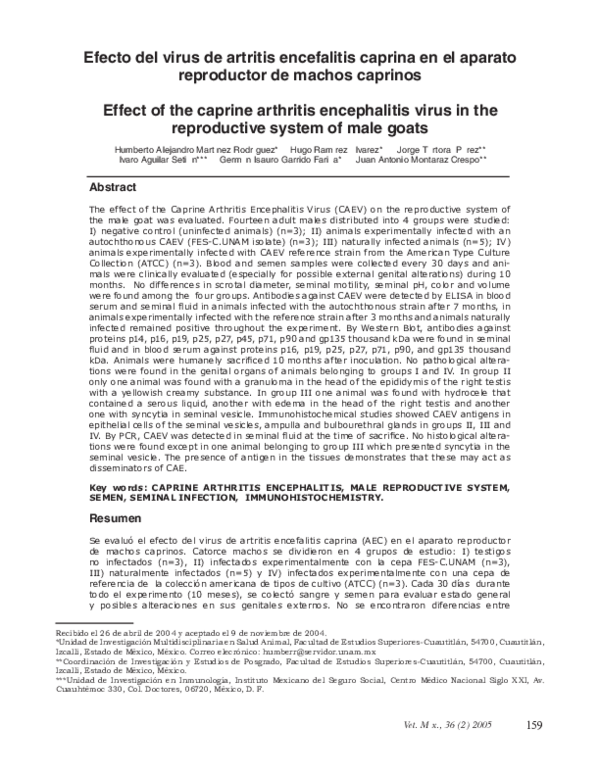
 Jorge Tortora