Comparative Biochemistry and Physiology, Part C 142 (2006) 356 – 364
www.elsevier.com/locate/cbpc
Vitellogenin induction in the endangered goodeid fish Girardinichthys
viviparus: Vitellogenin characterization and estrogenic effects of
polychlorinated biphenyls☆
Armando Vega-López a,⁎, Laura Martínez-Tabche a , Maria Lilia Domínguez-López b ,
Ethel García-Latorre b , Eva Ramón-Gallegos c , Alejandra García-Gasca d
a
c
Laboratorio de Toxicología Acuática, Escuela Nacional de Ciencias Biológicas, IPN. Prol. Carpio y Plan de Ayala s/n,
Col. Plutarco Elías Calles “Casco de Santo Tomás”, D.F. CP 11340, México
b
Laboratorio de Inmunoquímica, Escuela Nacional de Ciencias Biológicas, IPN. Prol. Carpio y Plan de Ayala s/n,
Col. Plutarco Elías Calles “Casco de Santo Tomás”, D.F. CP 11340, México
Laboratorio de Citopatología Ambiental. Escuela Nacional de Ciencias Biológicas, IPN. Prol. Carpio y Plan de Ayala s/n,
Col. Plutarco Elías Calles “Casco de Santo Tomás”, D.F. CP 11340, México
d
Laboratorio de Biología Molecular, CIAD Mazatlán. Mazatlán Sinaloa, México
Received 23 June 2005; received in revised form 24 October 2005; accepted 1 November 2005
Available online 27 December 2005
Abstract
Vitellogenin (VTG) is a widely used biomarker in studies of endocrine disruption induced by xenobiotics such as polychlorinated biphenyls
(PCBs). This study evaluates the estrogenic effects of these compounds on the black-fin goodeid Girardinichthys viviparus, an endangered fish
species in Mexico with a reduced range of distribution due to pollution of its natural environment. Adult fish born in the laboratory were exposed
to half the LC0 of Inerteen® commercial PCB mixture. VTG was determined through an inhibition enzyme-linked immunosorbent assay (ELISA)
using a homologous–heterologous system. Male and female fish were killed after 1, 2, 4, 8 and 16 days of exposure. The distal third of each
specimen was used for analysis. VTG was obtained from cultured hepatocytes and blood serum of males previously exposed to 17β-estradiol.
VTG molecular mass was 348 kDa. PCBs were found to elicit greater estrogenic effects on VTG induction in males than in females (p b 0.05) and
sex differences were noted. Time-dependent VTG induction kinetics in males and a stationary phase in females were also observed.
© 2005 Elsevier Inc. All rights reserved.
Keywords: Endocrine disruption; Girardinichthys viviparus; Goodeid fish; Vitellogenin; PCBs; Waterborne exposure; Sex differences
1. Introduction
Vitellogenin (VTG) is a characteristic protein in females,
associated with egg production (Tata and Smith, 1979; Wiley et
al., 1979; Nagler and Idler, 1990) in both oviparous and
viviparous species, and it is used as a biomarker for monitoring
xenobiotics that mimic estrogen (Fukada et al., 2003). Synthesis
☆
This paper is part of a special issue of CBP dedicated to “The Face of Latin
American Comparative Biochemistry and Physiology” organized by Marcelo
Hermes-Lima (Brazil) and co-edited by Carlos Navas (Brazil), Tania ZentenoSavín (Mexico) and the editors of CBP. This issue is in honour of Cicero Lima
and the late Peter W. Hochachka, teacher, friend and devoted supporter of Latin
American science.
⁎ Corresponding author. Tel.: +52 55 57 29 63 00x62343.
E-mail address: avegadv@terra.com (A. Vega-López).
1532-0456/$ - see front matter © 2005 Elsevier Inc. All rights reserved.
doi:10.1016/j.cbpc.2005.11.009
of this biomolecule is regulated by the endocrine control axis,
where external stimuli condition internal stimuli and there is a
feedback from the latter to the axis (Nagler and Idler, 1990; Singh
and Singh, 1991; Arukwe and Goksøyr, 2003). Despite endocrine
control by the organism, certain man-made and natural substances
are capable of altering these processes (García et al., 1997;
Bowman et al., 2000; Nicolas, 2001; Spengler et al., 2001; Corsi
et al., 2003; MacLatchy et al., 2003). Such compounds are known
as endocrine disruptors (Nicolas, 1999; Arukwe and Goksøyr,
2003). In vitro and in vivo studies have shown that polychlorinated biphenyls (PCBs) in commercial mixtures elicit estrogenic
and anti-estrogenic effects, and toxic response is related to the
number of chlorine atoms in the PCB molecule (Gierthy et al.,
1997; Vakharia and Gierthy, 2000; Bonefeld-Jørgensen et al.,
2001). Toxicity and biotransformation are inversely proportional
�A. Vega-López et al. / Comparative Biochemistry and Physiology, Part C 142 (2006) 356–364
to the number of chlorine atoms. There is evidence that octa-,
nona- and decachlorobiphenyls are not biotransformed (Groten et
al., 1999) and endocrine disruption potential is species-dependent. When VTG is synthesized due to false stimuli, a number of
important alterations may follow. For instance, the energy
investment involved in reproducing outside normal periods may
be enormous (Stancel et al., 1995). Males bear the corresponding
genes for VTG synthesis and a series of complications such as
reduced fertility and feminization arise due to presence of this
phospholipoprotein (Tata and Smith, 1979; Maitre et al., 1984,
1986; Le Guellec et al., 1988; Mori et al., 1998; Okoumassoun et
al., 2002). In extreme cases, this large-sized protein may lead to
death from kidney failure (Pajor et al., 1990; Sultan et al., 1995;
Schultz et al., 2003). Although endocrine disruption studies have
been conducted on several indicator species, few of them involve
species of intrinsic ecological importance such as the black-fin
goodeid Girardinichthys viviparus (Bustamante). The latter is a
live-bearing fish endemic to Mexico as well as an endangered
species with a reduced range of distribution (NOM-059-ECOL,
1994). G. viviparus lives near the largest urban population center
in Mexico and must endure the impact of domestic inputs and
industrial wastewater in its natural environment, along with the
general disappearance of the latter (Díaz-Pardo and OrtízJiménez, 1985). An electric power plant that makes routine use
of PCBs is located near its habitat. It is thus essential to carry out
several environmental risk-assessment studies to protect this
species. In the case of the present study, our goals have been to
assess the toxic effects of PCBs at sublethal concentrations, by
waterborne exposure to PCBs, on vitellogenin induction in the
black-fin goodeid G. viviparus as well as to find any sex-linked
differences in these responses and characterize the protein in this
particular species.
2. Methodology
2.1. Fish
Since the present study involves a protected species,
collection of the parent group of fish was conducted subsequent
to evaluation and authorization by Mexican authorities (Oficio
No./SGPA/DGVD/02750). Fish were collected from reservoirs
near Lake Texcoco in the State of Mexico. Pollutant impact in
these reservoirs is relatively small as they are filled by deep-well
waters. Fish were kept in the laboratory at 25 °C under natural
daylight with fresh food (Daphnia pulex and Artemia salina) or
pellets available ad libitum, in 200-L glass aquariums at a
density of 0.5 g fish/L water until reproduction occurred. All
specimens in the study were 8-month-old adults born in the
laboratory. Four months prior to beginning tests, fish were
separated by sex, a task made easier by their sexual dimorphism,
to ensure a state of rest in female gonad development through
elimination of the visual stimuli associated with courtship.
2.2. Lethal toxicity tests
Tests followed the criteria established by USEPA (OPPTS
850.1075): five concentrations were performed in triplicate in a
357
glass aquarium with seven fish in static medium at a density of
0.8 g fish/L. Dimethyl sulphoxide (DMSO, Sigma) was used as
a vehicle for the PCBs. Pure DMSO dissolved in semi-hard
synthetic water was used for the control group. The LC50 was
estimated after 96 h by the Probit method. LC0 was calculated
using a semi-logarithmic method since the latter is capable of
determining extreme concentrations.
2.3. Sublethal toxicity tests
Nominal PCBs concentrations was dosed at half the LC0 in
DMSO as well as the solvent control were prepared in semihard synthetic water in semi-static medium that was completely
renewed every 4 days. Three males and three females were
selected at random after 1, 2, 4, 8 and 16 days of exposure. Fish
were anesthetized with xylocaine (30 mg/L) for several seconds
and killed via fast freezing to − 70 °C followed by cervical
dislocation. Because of the small size of this species (males:
25.54 ± 2.8 mm LP and 0.39 ± 0.02 g; females: 39.55 ± 4.6 mm
LP and 1.20 ± 0.018 g), the distal third of each specimen
(without fins) was homogenized in phosphate buffered saline
(PBS, pH 7.5) with protease inhibitor (Aprotinin, 3 mg/mL
Sigma), centrifuged at 1500×g (5 min) and stored at − 70 °C
until VTG analysis. The Inerteen® chromatographic profile was
obtained using a CG Varian 3400 coupled to a Saturn II mass
detector with programmed temperatures, in a 0.25-mmdiameter, 30-m-long Restec capillary column preheated to
40 °C, with the injector set at 200 °C and detector at 280 °C.
2.4. VTG quantification
VTG purification: VTG was obtained from blood serum of
male fish exposed to 17β-estradiol (E2) in aqueous medium
(1.0 μg/L) that was completely renewed every 2 days (Sherry
et al., 1999; Lattier et al., 2003; Van den Belt et al., 2003). After
2 weeks, fish were anesthetized and completely bled through the
caudal vein. VTG was purified via triple differential precipitation (Sherry et al., 1999) and filtration in a diethyl-amino-ethyl–
cellulose (DEAE–cellulose) column in a KCl gradient (0.01–
0.5 M) as suggested by Wiley et al. (1979). VTG fractions were
obtained at KCl concentrations of 0.2–0.25 M and concentrated
by dialysis in 12,000 Da bags. Protein concentrations were
determined according to Bradford (1976) using bovine serum
albumin (Merck) as a standard.
VTG was also obtained in vitro from cultured hepatocytes.
Anesthetized adult males were killed by cervical dislocation.
Perfusion, separation, and cell culture were performed following the techniques proposed by several authors (Maitre et al.,
1986; Kordes et al., 2002; Rouhani et al., 2002) with certain
modifications. Livers were rinsed in sterile PBS containing a
2% antibiotic mix (10,000 U/mL penicillin, 10,000 U/mL
streptomycin, 25 μg/mL amphotericin) and dissected. Liver
fractions were shaken for 25 min in 0.05% type IV collagenase
(Sigma) and filtered through sterile cloth. Then the collagenase
was neutralized with the same amount of culture medium and
hepatocytes were centrifuged and re-suspended in culture
medium. A total of 3 × 10⁵ hepatocytes were placed per well
�358
A. Vega-López et al. / Comparative Biochemistry and Physiology, Part C 142 (2006) 356–364
in 24-well flat-bottom plates. Cell cultures were kept at 25 °C in
phenol red-free DMEM (Dulbecco's modification of Eagle's
medium) with 2% bovine fetal serum. After 16 h, the culture
medium was changed and after 48 h, a medium with E2
dissolved in ethanol at a concentration of 1 × 10− 10 M was
added. This culture medium was changed after 96, 144, 192,
240 and 288 h, and VTG obtained each time from the harvested
medium. VTG was purified via triple differential precipitation
and filtration in a DEAE–cellulose column as previously
described.
VTG obtained from the cultured hepatocytes and blood sera
was denatured by heating with 2-mercaptoethanol (0.0143 M)
and characterized by sodium dodecyl sulfate–polyacrylamide
gel electrophoresis (SDS–PAGE) under reducing conditions
(Korsgaard and Ladegaard, 1998; Kordes et al., 2002; Van den
Belt et al., 2003). Electrophoretic separation was performed
using a spacing gel and a 20% running gel with molecular mass
markers of 14,200 to 66,000 Da (Sigma).
As it is difficult to obtain purified VTG from G. viviparus in
sufficient amounts for immunization and since cross-reactivity
has been previously observed with other species (Huggeu et al.,
2003; Mylchreest et al., 2003; Nilsen et al., 2004), the rainbow
trout Oncorhynchus mykiss was selected as a substitute.
Rainbow trout weighing 300–400 g were used to obtain VTG
for immunization and ELISA protocol. VTG was induced via
two intraperitoneal injections of E2 in saline solution (5.0 mg/kg
mass) administered on days 0 and 5. On day 10, fish were
anesthetized and partially bled through the caudal vein. Blood
samples were taken and VTG purified as previously described.
Fish were revived by filling the mouth and gills with cold
running water until they recovered from the anesthetic.
2.5. ELISA protocol for VTG
2.5.1. Polyclonal anti-VTG serum
A young male New Zealand rabbit was immunized with
rainbow trout vitellogenin (omVTG) according to the following
procedure.
First dose: 500 μg omVTG in 500 μL complete Freund's
adjuvant, given subcutaneously at various sites. Second dose:
an equal amount but in incomplete Freund's adjuvant,
administered 15 days later. Third, fourth, fifth and sixth doses
(days 30, 31, 32 and 39): 250 μg omVTG in saline solution via
intramuscular injection. On day 46, an anesthetic (20 mg/kg
ketamin + 5 mg/kg xilacin) was applied and blood drawn via
intracardiac puncture. The polyclonal anti-omVTG serum was
obtained by centrifugation and stored at − 70 °C.
2.5.2. Inhibition of hybrid ELISA
A calibration curve was done as follows. 96-well flat-bottom
plates were used. The wells were coated with 7.0 ng of omVTG
(homologous) in 100 μL of carbonate buffer pH 9.0–9.5 (1.5 g
Na2CO3 + 2.93 g NaHCO3/1000 L) and incubated overnight at 4
°C. Wells were washed two times with PBS–0.05% Tween 20
and blocked for 1 h at 37 °C with a 0.25% gelatin (Sigma) in
PBS. Different amounts of the heterologous G. viviparus VTG
(gvVTG) (1, 2, 4, 8 15, and 30 ng) were mixed with 100 μL of
the polyclonal serum anti-omVTG diluted 1:100 in PBS–
gelatin solution in a 1.5-mL microcentrifuge tube, agitated in a
vortex, added to the wells and incubated for 1 h at 37 °C. The
wells were washed three times (5 min) with PBS–0.05% Tween
20 and 100 μL of the conjugate (peroxidase-conjugated goat
anti-rabbit gamma globulin, Sigma) at a dilution of 1:4000 was
added. Incubation and washing process were done as above.
100 μL of the substrate (10 mg of o-phenylendiamine + 10 μL of
H2O2 in 25 mL citrate solution pH 5.0) was added. The reaction
was stopped with 30 μL of 7 M H2SO4 after 25 min. The color
intensity was inversely proportional to the amount of gvVTG at
A490. For the gvVTG analysis in the different samples, 5 μL of
homogenized fish sample was mixed with 100 μL of the
polyclonal serum anti-omVTG diluted 1:100. The procedure
was done as described for the calibration curve.
To validate the detection limit (MDL) of the inhibition of
hybrid ELISA method the test was run with 2.0 ng of gvVTG,
for 3 consecutive days, 24 wells each time. All calibration
curves and samples were run six times during 3 consecutive
days. Accuracy of the method, was determined by spiked
samples (adding three known concentrations of gvVTG to the
samples) and proceeding according to the described
methodology.
2.6. Statistics
MDL was estimated using the formula: MDL = 2 * αn−1
absorbance * CAC / Average absorbance; where 2 = 95.45%
reliability, αn−1 = the standard deviation of the samples, and
CAC = the mean sample concentration derived from the
calibration curve. The method quantification limit (MQL) was
obtained by means of the formula: MQL = MDL * tn−1; where
t = the “t” score of tables with 95% confidence at n − 1. Method
precision was estimated through the coefficient of variation: %
CV = (αn−1replicates / mean) * 100. Accuracy was determined by
the recovery average: %R = [(ECA) − (C) / (A)] * 100; where
ECA = the experimental concentration of the spiked sample;
C = sample concentration; and A = the concentration of the
gvVTG used to spike samples (CENAM, 1993).
Results were subjected to a two-way ANOVA and the means
compared using a two-way Student's t-test. Minimum probability criterion for significant differences was set at p ≤ 0.05.
3. Results
More than 46 different compounds were detected in the
Inerteen® analysis (4 mono-, 5 di-, 7 tri- and 31 tetrachlorobiphenyls). Tri-and tetrachlorobiphenyls with different chlorination patterns were predominant (data not shown). Penta-,
hexa- and heptachlorobiphenyls were also observed.
An LC50 equivalent to 17.04 mg PCBs/L (y = − 4.3644 +
7.6082 * X; χ2 = 0.3563, p b 0.05) was observed. The LC0 in
the black-fin goodeid was 1.84 mg PCBs/L.
Output of gvVTG was less than 2.0 mg in both cell cultures
and blood sera, but larger amounts were obtained in vivo. The
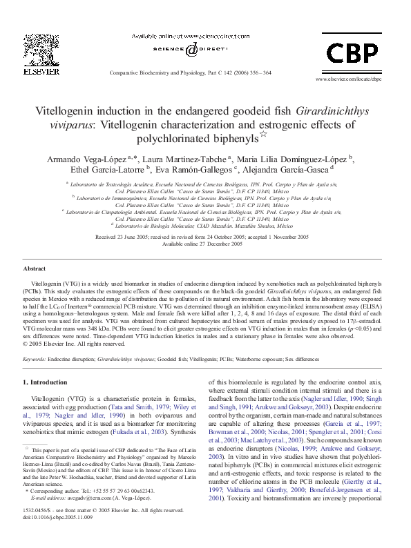
gvVTG elusion pattern in the DEAE–cellulose column (Fig. 1)
shows two well-defined peaks within the KCl gradient (0.2–
�A. Vega-López et al. / Comparative Biochemistry and Physiology, Part C 142 (2006) 356–364
359
600
500
gvVTG
µg protein
400
300
200
100
0
0.2
-100
0.2
0.2
0.225 0.225 0.225 0.225 0.225 0.25
0.25
0.25
0.25
0.25 0.275
KCl concentration (M)
Fig. 1. DEAE–cellulose column chromatographic patterns of VTG obtained from Girardinichthys viviparus exposed to 17β-estradiol. Protein concentration obtained
by the UV method (280 nm) via linear regression with a bovine albumin curve. [Note: Negative values are due to the effect of statistical ones (slope and origin) in the
calibration curve].
0.25 M) reported as typical of VTG (Wiley et al., 1979; De
Vlaming et al., 1980; Parks et al., 1999). SDS–PAGE
electrophoresis showed seven polypeptide bands of 64,000,
62,000, 60,000, 59,000, 46,000, 43,000 and 14,000 Da adding
up to 348,000 Da (Fig. 2).
Inhibition of polyclonal anti-omVTG serum by heterologous
gvVTG was observed from 27.07% for 1 ng of gvVTG to
71.52% for 30 ng of gvVTG in the calibration curve of the
hybrid ELISA. The following values were determined:
CV = 28.87% (25.60–32.15%), MDL = 0.049 ng gvVTG
(p b 0.05) and MQL = 0.09 ng gvVTG. Accuracy was within
the range %R = 86 to 107. This method is suitable based on
statistical parameters due to the correlation coefficient obtained
and the level of significance of the measurements in the
calibration curve (r2 = 0.983, p b 0.001).
In the control group, females had average base values of
0.009 pg gvVTG/mg protein/g tissue/g fish. The protein was not
detected in control males. PCBs elicited estrogenic effects in
males and females at significant levels from day 2 of exposure.
Average gvVTG was up to 10 times greater (p b 0.05) in males
Fig. 2. SDS–PAGE (20% running gel) of G. viviparus VTG under reducing
conditions, stained with Coomassie Brilliant Blue R250. Lane 1–4: G. viviparus
VTG at different concentrations (1.5, 1.0, 0.5 and 0.3 μg, respectively). Lane m:
molecular mass markers (molecular masses on left side are those of the
commercial markers). Arrowheads indicate the three main polypeptides of VTG
(64,000, 43,000 and 14,000 Da).
than females (0.96 vs. 0.09 pg VTG/mg protein/g tissue/g fish,
respectively). Figs. 3 and 4 show the induction kinetics of this
biomolecule. PCBs-treated females showed significant differences from controls from day 2 to day 16 of exposure relative to
control. PCBs-treated males exhibited differences starting on
day 1 relative to control. Estrogenic effects in males were
clearly time-dependent after day 2, and showed significant
differences between all days of exposure (Fig. 4). In contrast,
induction was more or less constant in females after day 2 (Fig.
3). CV was 24.5% (21.2–27.8%, p b 0.05) in females and 19.4%
(14.2–24.7%, p b 0.05) in males. Accuracy of spiked samples
was within the range %R = 71 to 114.
4. Discussion
The two peaks in the elusion profile derived from the
DEAE–cellulose column indicate that the two VTG polypeptide chains (α and β) are present. Based on their position, we
deduced that the α chain is lighter in this species, as has been
reported by several authors for different species such as the
fathead minnow Pimephales promelas, sheepshead minnow
Cyprinodon variegatus, zebrafish Danio rerio, white perch
Morone america, and rainbow trout O. mykiss (Parks et al.,
1999; Bowman et al., 2000; Fenske et al., 2001; Hiramatsu et
al., 2002; Van den Belt et al., 2003). Results found by the former
authors about the VTG mass agree with our observations
regarding the black-fin goodeid VTG (348 kDa). Though
similarities in the VTG of G. viviparus and other species are
undoubtedly interesting, it is more noteworthy that the presence
of this biomolecule in the Goodeidae has been established for
the first time in this study as well as confirmed by us after partial
sequencing of the VTG mRNA and registration in GenBank
Accession No. AY845859 (data reported elsewhere).
Other authors have found higher VTG molecular mass (De
Vlaming et al., 1980; Maitre et al., 1984; Korsgaard and
Ladegaard, 1998; Fukada et al., 2003). In view of the
�360
A. Vega-López et al. / Comparative Biochemistry and Physiology, Part C 142 (2006) 356–364
pg VTG/mg proteins/g tissue/g fish
0.16
0.14
Control
D*
A*
PCBs
0.12
C*
B*
0.1
0.08
0.06
0.04
0.02
0
1
2
4
8
16
Exposure time (days)
Fig. 3. Vitellogenin induction kinetics in female G. viviparus exposed to PCBs [half the LC0 (0.92 mg/L)] during a 16-day period and DMSO control in semi-hard
synthetic water. Significant differences were not found after day 2 of exposure: (A) day 2 vs. day 1, p b 0.05; (B) day 4 vs. day 2, ns; (C) day 8 vs. day 4, ns; (D) day 16
vs. day 8, ns. *Significant differences relative to control. ns = not significant. Results were subjected to a two-way ANOVA and the set of triplicates compared using a
two-ways Student's t-test. Minimum probability criterion for significant differences was set at p ≤ 0.05.
differences in the values reported by these two groups of
authors, the fact that this protein shows significant variations in
molecular weight must be further examined. For instance, it is
estimated that VTG can weigh anywhere between 250 to
600 kDa (Norberg and Haux, 1985; Arukwe and Goksøyr,
2003). Such disparities are a clear indication of little
evolutionary conservatism in this biomolecule, even among
members of the same family, as observed by us in VTG of the
black-fin goodeid and the butterfly split-fin goodeid Ameca
splendens (379 kDa). MacLatchy et al. (2003) discuss VTG
variations in different species as well as loss of the primary
sequence of this protein as noted by us.
In the present study, gvVTG was quantified by means of a
hybrid ELISA in an inhibition format showing suitable
detection and quantification. To be able to compare our data
with previously reported results, we used similar units to
estimate limits (MDL = 0.5 ng/mL and MQL = 0.9 ng/mL
gvVTG). Different detection limits have been reported in
2
D***
Control
pg VTG/mg proteins/g tissue/g fish
1.8
PCBs
1.6
C**
1.4
1.2
1
B**
0.8
**
A*
0.6
0.4
0.2
0
1
2
4
8
16
Exposure time (days)
Fig. 4. Vitellogenin induction kinetics in male G. viviparus exposed to PCBs [half the LC0 (0.92 mg/L)] during a 16-day period and DMSO control in semi-hard
synthetic water. [Note: VTG not detected in the control]. Significant differences were found between all days of exposure: (A) day 2 vs. day 1, p b 0.05; (B) day 4 vs.
day 2, p b 0.01; (C) day 8 vs. day 4, p b 0.01; (D) day 16 vs. day 8, p b 0.05. Significant differences relative to control: * p b 0.05, **p b 0.01, ***p b 0.001. Results were
subjected to a two-way ANOVA and the set of triplicates compared using a two-ways Student's t-test. Minimum probability criterion for significant differences was set
at p ≤ 0.05.
�A. Vega-López et al. / Comparative Biochemistry and Physiology, Part C 142 (2006) 356–364
validation of various ELISAs using a sandwich format based on
monoclonal and polyclonal antibodies. Nilsen et al. (2004)
report MDLs of 0.1 ng/mL and 0.6 ng/mL in the fathead
minnow, P. promelas, and the Japanese medaka, Oryzias
latipes, respectively. Brion et al. (2002) recorded 0.4 ng/mL
MDL in D. rerio, while Holbech et al. (2001) found 0.2 ng/mL
in this same species. Using polyclonal antibodies, Fenske et al.
(2001) reported 2–3 ng/mL, again in D. rerio. In contrast, it has
been observed that VTG quantification with different ELISA
formats is suitable as long as the MDL is within the range of
concentration of the samples (Nilsen et al., 2004). In regard to
the working range of the calibration curve (1–30 ng gvVTG), it
is similar to the ranges reported by previously mentioned
authors. The intra- and inter-assay coefficient of variation was
slightly higher than those reported by Brion et al. (2002),
Holbech et al. (2001) and Fukada et al. (2003). However, this
deviation may be due to reduced sensitivity during the Ag–Ab
hybrid reaction (Mylchreest et al., 2003). We consider
inhibition of polyclonal antibodies with heterologous VTG in
solution to be a more precise method since we know the
amounts of protein used, and the uncertainty related to wellcoating is eliminated. A further aspect of method validation is
accuracy. Our results show suitable recovery averages, since
during this type of biological reactions an equal bimolecular
reactivity cannot be expected due to several sources of variation
associated with matrix interference. Certain quality control
aspects observed during our study lead us to conclude that a
hybrid ELISA in an inhibition format is sufficiently sensitive,
accurate and precise for VTG analysis in small-sized species,
especially when not enough purified VTG is available. It is
nonetheless necessary to test for cross-reactivity between the
homologous and heterologous systems.
A weak endocrine potential from commercial PCB mixtures
has been considered (Gierthy et al., 1997; Arcaro et al., 1998;
Bonefeld-Jørgensen et al., 2001; Carlson and Williams, 2001).
Our results differ from those of previous reports in that the
estrogenic effects elicited by the commercial mixture used in
this study were significantly different from the control. For
example, the amount of PCB-induced VTG on day 16 was up to
15 times more in females, than in the females control group. In
the PCBs-treated males, this protein was detected in the range of
2.0 pg VTG/mg protein/g tissue/g fish whereas in the male
control fish, VTG was not detected. These results are
comparable to the amount elicited by the most potent VTG
inducers in fish: 17α-ethynylestradiol (EE2) and E2 (Holbech et
al., 2001; Brion et al., 2002; Panter et al., 2002; Andersen et al.,
2003; Mylchreest et al., 2003; Versonnen and Janssen, 2004).
These findings establish the endocrine disruption potential of
Inerteen® in this goodeid fish under experimental conditions.
Unfortunately, detailed action mechanisms involved in egg yolk
protein induction have not been fully studied (Nicolas, 1999;
Arukwe and Goksøyr, 2003). However, it is necessary to
consider that VTG is not the only oogenic protein; for instance,
Fujita et al. (2005) comment that the choriogenin levels (H and
L types) in serum of masu salmon Oncorhynchus masou were
higher than VTG in pre-vitellogenic growth phase; but on the
vitellogenic phase, the VTG were the most abundant oogenic
361
protein coincident with the higher E2 levels. Also, the former
authors comment that the initiation of choriogenin and VTG
synthesis and their inducibility appear to be species-dependent.
Sex differences in VTG production are associated with
steroid metabolism and cytochrome P450 activity. Some PCBs
and dioxins have both estrogenic and anti-estrogenic potential
(Spink et al., 1990) mediated by an aryl hydrocarbon receptor
(AhR). Such interactions include a down-regulation of estrogen
receptors (ER), ER ligand–E2 interference with response genes
in DNA, and/or cytochrome P450 induction, specifically of the
enzymes CYP1A1 and CYP1A2 involved in E2 metabolism
(Bonefeld-Jørgensen et al., 2001). For instance, in females,
increased estrogen levels have been found to lead to a decrease
in mixed oxidase function (MOF) enzymes, as in the case of the
brook trout Salvelinus fontinalis (Stegeman et al., 1982), the
winter flounder Pleuronectes americanus and the scup Stenotomus chrysops (Gray et al., 1991). However, sex differences
may also be explained by genetic regulation, since MOF activity
is higher in males and testosterone 6β-hydroxylase activity is
higher in females, as observed in the winter flounder (Gray et
al., 1991). These observations show that the comparatively
higher activity of CYP1A in male G. viviparus (unpublished
data) leads to formation of PCBs metabolites with greater
efficiency and possibly estrogenic potential (Figs. 3 and 4). In
addition, suppression of P450 catalytic activity elicited by E2 is
suggested to take place at pre-transcriptional levels (mRNA for
P4501A) while endogenous regulation may overcome the
exogenous regulation exerted by high concentrations of
inductors of this enzyme superfamily (Elskus et al., 1992).
Another aspect contributing to increased blood levels of
VTG induced by PCB mixtures may be the lack of target organs
in males. Relative to VTG induction kinetics by PCBs exposure,
Petit et al. (1997) found in a primary culture of rainbow trout
hepatocytes, an induction of gene expression of VTG after
2 days exposure to Aroclor 1221 and Aroclor 1248. Flouriot et
al. (1995) inform an induction of gene expression of VTG in
rainbow trout hepatocytes after over 30 days of xenobiotics
exposure. In juvenile zebrafish, D. rerio, a VTG half-life of
2.4 days has been observed when induced by a single exposure
to EE2 (Andersen et al., 2003). These reports are in agreement
with our observations, since VTG blood levels in female G.
viviparus remained relatively constant throughout the exposure
to xenobiotics. This supports the assumption of a fast uptake of
induced amounts of this protein by oocytes as observed by
Fujita et al. (2005). In contrast, VTG induction in male P.
promelas has been reported to have two different-length stages
(Schmid et al., 2002), the first stage lasting approximately
2 days, and the second one, 21 days. These findings support our
observations, since this protein increased with time in male G.
viviparus reaching maximum levels on day 16 of observation.
VTG induction kinetics elicited by PCBs in male G. viviparus
also appeared to be two-staged, although the first stage took
place during the first 24 h while the second one continued for at
least 16 days.
One of the most unsettling characteristics of PCBs endocrine
disruption is related to the structure and biotransformation of
these xenobiotics and to ER–AhR interactions. Groten et al.
�362
A. Vega-López et al. / Comparative Biochemistry and Physiology, Part C 142 (2006) 356–364
(1999) find that phase I (oxidation by MOF) takes place in liver
microsomes by way of an arene to form an intermediate
metabolite (arene oxide or epoxide), and the consequence of this
biotransformation is a biphenylol. This oxidation is enhanced in
meta and para positions (Bonefeld-Jørgensen et al., 2001).
Gierthy et al. (1997) found the estrogenic effects elicited by
PCB metabolites in human adenocarcinoma MCF-7 cells are
enhanced by para-OH and ortho substitution, and these events
are related to the affinity of these biphenylols for ER. The
authors explained this process as an effect of the structural
similarity between biphenylols and E2, or the conformational
restriction derived from ortho substitution (Bonefeld-Jørgensen
et al., 2001). In MCF-7 cells, ortho-OH and meta-OH
metabolites do not compete with ER, while para-OH
metabolites do so depending on their concentration (Vakharia
and Gierthy, 2000). The Inerteen® chromatographic profile
shows all these related compounds are present in the
commercial mixture used in our study, di-ortho and ortho
substituted compounds being predominant. These compounds
are capable of interacting with ER. Metabolite formation due to
MOF activity may also be conjectured. Metabolites have been
shown to form in the presence of liver microsomes and
NADPH, although chromatographic confirmation is still
lacking (Vakharia and Gierthy, 2000). Such events lead us to
assume that the black-fin goodeid has the ability to biotransform
these xenobiotics into metabolites of enhanced estrogenic
capability. Bonefeld-Jørgensen et al. (2001) mention that
evaluation of commercial PCB mixtures is important since it
allows prediction of toxic, particularly estrogenic, effects. These
authors mention that di-ortho substitutions also have an affinity
for ER. However, when mixing different di-ortho biphenyls
(PCB # 138, 2,2′,3,4,4′,5-hexachlorobiphenyl; PCB # 153,
2,2′,4,4′,5,5′-hexachlorobiphenyl; and PCB # 180,
2,2′,3,4,4′,5,5′-heptachlorobiphenyl) at 3μM concentrations,
anti-estrogenic effects at ER level were observed in MCF-7
cells. In addition to the concentration used, these effects may be
further explained by the affinity of PCB # 138 to link to
androgen receptors. Gierthy et al. (1997) report anti-estrogenic
effects elicited by 3,4′,5-trichlorobiphenyl, while none were
observed for its metabolite (3,4′,5-trichloro-4-biphenylol). It is
thus evident that the in vivo interactions of commercial PCB
mixtures are quite complex in G. viviparus; nonetheless, we
were able to establish presence of estrogenic effects in this
species.
Although the toxic effects of the commercial PCB mixture
were estrogenic in their manifestation in both G. viviparus
sexes, the possibility of anti-estrogenic and anti-androgenic
interactions has not been ruled out, since all these interactions
are considered endocrine disruptions. Nevertheless, the ecological significance of these disorders must be evaluated since
endocrine disruption is closely tied to physiological and
hormonal cycles and shows strong sex-linked seasonal
variation. Detoxification schemes to get rid of these xenobiotics
must also be considered, as such schemes may be speciesdependent. Toxic response modifications are vitally important
to assess risk of exposure to endocrine disruptors in the natural
environment.
References
Andersen, L., Holbech, H., Gessbo, A., Norrgren, L., Petersen, G.I., 2003.
Effects of exposure to 17alpha-ethinylestradiol during early development on
sexual differentiation and induction of vitellogenin in zebrafish (Danio
rerio). Comp. Biochem. Physiol. C 134, 365–374.
Arcaro, K.F., Vakharia, D.D., Yang, Y., Gierthy, J.F., 1998. Lack of
synergy by mixtures of weakly estrogenic hydroxylated polychlorinated
biphenyls and pesticides. Environ. Health Perspect. 106 (Suppl 4),
1041–1046.
Arukwe, A., Goksøyr, A., 2003. Eggshell and egg yolk proteins in fish: hepatic
proteins for the next generation: oogenetic, population, and evolutionary
implications of endocrine disruption “Review”. Comp. Hepat. http://www.
comparative-hepatology.com/content/2/I/4.
Bonefeld-Jørgensen, E.C., Andersen, H.R., Rasmussen, T.H., Vinggaard, A.M.,
2001. Effect of highly bioaccumulated polychlorinated biphenyl congeners
on estrogen and androgen receptor activity. Toxicology 158, 141–153.
Bowman, C.J., Kroll, K.J., Hemmer, M.J., 2000. Estrogen-induced vitellogenin
mRNA and protein in sheepshead minnow (Cyprinodon variegatus). Gen.
Comp. Endocrinol. 120, 300–313.
Bradford, M., 1976. A rapid and sensitive method for the quantitation of
microgram quantities of protein utilizing the principle of protein–dye
binding. Anal. Biochem. 72, 248–254.
Brion, F., Nilsen, B.M., Eidem, J.K., Goksoyr, A., Porcher, J., 2002.
Development and validation of an enzyme-linked immunosorbent assay to
measure vitellogenin in the zebrafish (Danio rerio). Environ. Toxicol.
Chem. 21, 1699–1708.
Carlson, D.B., Williams, D.E., 2001. 4-Hydroxy-2′,4′,6′-trichlorophenyl and 4hydroxy-2′,3′,4′,5′-tetrachlorobiphenyl are estrogenic in rainbow trout.
Environ. Toxicol. Chem. 20, 351–358.
CENAM, 1993. Acreditamiento para laboratorios de química, guía en la
interpretación de la serie de normas EN45000 y la guía ISO/IEC25,
Eurachem guidance document no. 1/WELAC guidance document no.
WGD2. Secretaría de Economía, Centro Nacional de Metrología, Área de
Metrología de Materiales. Querétaro, México. pp. 1–48.
Corsi, I., Mariottinni, M., Sensini, C., Lancini, L., Focardi, S., 2003. Fish as
bioindicators of brackish ecosystem health: integrating biomarker responses
and target pollutant concentrations. Oceanol. Acta 26, 129–138.
De Vlaming, V.L., Wiley, H.S., Delahunty, G., Wallace, R.A., 1980. Goldfish
(Carassius auratus) vitellogenin: induction, isolation, properties and
relationship to yolk proteins. Comp. Biochem. Physiol. B 67, 613–623.
Díaz-Pardo, E., Ortíz-Jiménez, D., 1985. Reproduccción y ontogenia de
Girardinichthys viviparus (Pisces: Goodeidae). An. Esc. Nac. Cienc. Biol.
30, 45–60.
Elskus, A.A., Pruell, R., Stegemann, J.J., 1992. Endogenously-mediated,
pretranslational suppression of cytochrome P4501A in PCB-contaminated
flounder. Mar. Environ. Res. 34, 97–101.
Fenske, M., van Aerle, R., Brack, S., Tyler, C.R., Segner, H., 2001.
Development and validation of a homologous zebrafish (Danio rerio
Hamilton–Buchanan) vitellogenin enzyme-linked immunosorbent assay
(ELISA) and its application for studies on estrogenic chemicals. Comp.
Biochem. Physiol. C 129, 217–232.
Flouriot, G., Pakdel, F., Ducouret, B., Valotaire, Y., 1995. Influence of
xenobiotics on rainbow trout liver estrogen receptor and vitellogenin gene
expression. J. Mol. Endocrinol. 15, 143–151.
Fujita, T., Fukada, H., Shimizu, M., Hiramatsu, N., Hara, A., 2005. Annual
changes in serum levels of two choriogenins and vitellogenin in masu
salmon, Oncorhynchus masou. Comp. Biochem. Physiol. B 141, 211–217.
Fukada, H., Fujiwara, Y., Takahashi, T., Hiramatsu, N., Sullivan, C.V., Hara, A.,
2003. Carp (Cyprinus carpio) vitellogenin: purification and development of
a simultaneous chemiluminescent immunoassay. Comp. Biochem. Physiol.
A 134, 615–623.
García, E.F., McPherson, R.J., Martin, T.H., Poth, R.A., Greeley Jr., M.S., 1997.
Live cell estrogen receptor binding in prespawning female largemouth bass
Micropterus salmoides, environmentally exposed to polychlorinated
biphenyls. Arch. Environ. Contam. Toxicol. 32, 309–315.
Gierthy, J.F., Arcaro, K.F., Floyd, M., 1997. Assessment of PBC estrogenicity in
a human breast cancer line. Chemosophere 34, 1495–1505.
�A. Vega-López et al. / Comparative Biochemistry and Physiology, Part C 142 (2006) 356–364
Gray, E.S., Woodin, B.R., Stegeman, J.J., 1991. Sex differences in hepatic
monooxygenases in winter flounder (Pseudopleuronectes americanus) and
scup (Stenotomus chrysops) and regulation of P450 forms by estradiol. J.
Exp. Zool. 259, 330–342.
Groten, J.P., Cassee, F.R., van Bladeren, P.J., de Rosa, C.T., Ferrón, V.J., 1999.
Mixtures. In: Marquardt, H., Shäfer, S.G., McClellan, R.O., Welsch, F.
(Eds.), Toxicology. Academic Press, San Diego, USA, pp. 711–728.
Hiramatsu, N., Hara, A., Hiramatsu, K., Fukada, H., Weber, G.M., Denslow, D.,
Sullivan, C., 2002. Vitellogenin-derived yolk proteins of white perch
Morone americana: purification, characterization and vitellogenin-receptor
binding. Biol. Reprod. 67, 655–667.
Holbech, H., Andersen, L., Petersen, G.I., Korsgaard, B., Pedersen, K.L.,
Bjerregaard, P., 2001. Development of an ELISA for the vitellogenin in
whole body homogenate of zebrafish (Danio rerio). Comp. Biochem.
Physiol. C 130, 119–131.
Huggeu, D.B., Foran, C.M., Brooks, B.W., Weston, J., Peterson, B., Marsh, K.
E., La Point, T.W., Schlenk, D., 2003. Comparison on in vitro and in vivo
bioassays for estrogenicity in effluent from North American municipal
wastewater facilities. Toxicol. Sci. 72, 77–83.
Kordes, S., Rieber, E.P., Guitzeit, H.O., 2002. An in vitro vitellogenin bioassay
for the oestrogenic substances in the medaka (Oryzias latipes). Aquat.
Toxicol. 58 (3–4), 151–164.
Korsgaard, B., Ladegaard, K., 1998. Vitellogenin in Zoarces viviparus:
purification, quantification by ELISA and induction by estradiol-17β and
4-nonylphenol. Comp. Biochem. Physiol. C 120, 159–166.
Lattier, D.L., Reddy, T.V., Gordon, D.A., Lazorchak, J.M., Smith, M., Willimas,
D.E., Wiechman, B., Flick, R.W., Miracle, A.L., Toth, G.P., 2003. 17α
Ethynylestradiol-induced vitellogenin gene transcription quantified in livers
of adult males, larvae, and gills of fathead minnows (Pimephales promelas).
Environ. Toxicol. Chem. 21, 2385–2393.
Le Guellec, K., Lawless, K., Valotaire, Y., Kress, M., Tenniswood, M., 1988.
Vitellogenin gene expression in male rainbow trout (Salmo gairdneri). Gen.
Comp. Endocrinol. 71, 359–371.
MacLatchy, D.L., Courtenay, S.C., Rice, C.D., Van Der Kraak, J., 2003.
Development of a short-term reproductive endocrine bioassay using steroid
hormone and vitellogenin end points in the estuarine mummichog (Fundulus
heteroclitus). Environ. Toxicol. Chem. 22, 996–1008.
Maitre, J.-L., Le Guellec, C., Derrien, S., Tenniswood, M., Valotaire, Y., 1984.
Measurement of vitellogenin from rainbow trout by rocket immunoelectrophoresis: application to the kinetic analysis of estrogen stimulation in the
male. Can. J. Biochem. Cell Biol. 63, 982–987.
Maitre, J.-L., Valotaire, Y., Guguen-Guillouzo, C., 1986. Estradiol-17β
stimulation of vitellogenin synthesis in primary culture of male rainbow
trout hepatocytes. In Vitro Cell. Dev. Biol. 22, 337–343.
Mori, T., Matsumoto, H., Yokota, H., 1998. Androgen-induced vitellogenin
gene expression in primary cultures of trout hepatocytes. J. Steriod Biochem.
67, 133–141.
Mylchreest, E., Snajdr, S., Korte, J.J., Ankley, G.T., 2003. Comparison of
ELISAs for the detecting vitellogenin in the fathead minnow (Pimephelas
promelas). Comp. Biochem. Physiol. C 134, 251–257.
Nagler, J.J., Idler, D.R., 1990. Ovarian uptake of vitellogenin and other very
high density lipoproteins in winter flounder (Pseudopleuronectes americanus) and their relationship with yolk proteins. Biochem. Cell. Biol. 68,
330–335.
Nicolas, J.-M., 1999. Vitellogenesis in fish and the effects of polycyclic aromatic
hydrocarbon contaminants. Aquat. Toxicol. 45, 77–90.
Nicolas, J.-M., 2001. Environmental xenoestrogens, antiandrogen and disorders
of male sexual differentiation. Mol. Cell. Endocrinol. 178, 99–105.
Nilsen, B.M., Berg, K., Eidem, J.K., Kristiansen, S.I., Brion, F., Porcher, J.M.,
Goksøyr, A., 2004. Development of quantitative vitellogenin–ELISAs for
fish tests species used in endocrine disruptor screening. Anal. Bioanal.
Chem. 378, 621–633.
NOM-059-ECOL, 1994. Norma oficial mexicana que determina las especies y
subespecies de flora y fauna silvestres terrestres y acuáticas en peligro de
extinción, amenazadas, raras y las sujetas a protección especial y que
establece especificaciones para su protección. PODER EJECUTIVO,
SECRETARÍA DE DESARROLLO SOCIAL, Diario Oficial (primera
sección) Lunes 16 de mayo de 1994. México.
363
Norberg, B., Haux, C., 1985. Induction, isolation and a characterization of the
lipid content of plasma vitellogenin from two Salmo species: rainbow trout
(Salmo gairdneri) and sea trout (Salmo trutta). Comp. Biochem. Physiol. B
81, 869–876.
Okoumassoun, L.-E., Averrill-Bates, D., Gagné, F., Marion, M., Denizeau,
F., 2002. Assessing the estrogenic potential of organochloride pesticides
in primary cultures of male rainbow trout (Oncorhynchus mykiss)
hepatocytes using vitellogenin as a biomarker. Toxicology 178,
193–207.
Pajor, A.M., Stegeman, J.J., Thomas, P., Woodin, B.R., 1990. Feminization of hepatic microsomal cytochrome P-450 system in brook trout
by estradiol, testosterone, and pituitary factors. J. Exp. Zool. 253,
51–60.
Panter, G.H., Hutchinson, T.H., Lange, R., Lye, C.M., Sumpter, J.P., Zerulla, M.,
Tyler, C.R., 2002. Utility of a juvenile fathead minnow screening assay for
the detecting (anti-)estrogenic substances. Environ. Toxicol. Chem. 21,
319–326.
Parks, L., Cheek, A.O., Denslow, N.D., Heppell, S.A., McLachlan, J.A.,
LeBlanc, G.A., Sullivan, C.V., 1999. Fathead minnow (Pimephales
promelas) vitellogenin: purification, characterization and quantitative
immunoassay for the detection of estrogenic compounds. Comp. Biochem.
Physiol. C 123, 113–125.
Petit, F., Le Goof, P., Cravédi, J.-P., Valotaire, Y., Pakdel, F., 1997. Two
complementary bioassays for screening the estrogenic potency of xenobiotics: recombinant yeast for trout estrogen receptor and trout hepatocytes
cultures. J. Mol. Endocrinol. 19, 321–335.
Rouhani, T., van Holsteijn, I., Letcher, R., Giesy, J.P., van den Berg, M., 2002.
Effects of primary exposure to environmental and natural estrogens on
vitellogenin production in carp (Cyprinus carpio) hepatocytes. Toxicol. Sci.
67, 75–80.
Sherry, J., Gamble, A., Hudson, P., Solomon, K., Hock, B., Marx, A., Hanse, P.,
1999. Vitellogenin induction in fish as an indicator of exposure to
environmental estrogens. In: Rao, S.S. (Ed.), Impact Assessment of
Hazardous Aquatic Contaminants. Concepts and Approaches. Lewis
Publishers, Boca Raton, USA, pp. 123–162.
Schmid, T., Gonzalez-Valero, J., Rufli, H., Dietrich, DR., 2002. Determination
of vitellogenin kinetics in male fathead minnows (Pimephales promelas).
Toxicol. Lett. 131, 65–74.
Schultz, I.R., Skillman, A., Nicolas, J.-M., Cyr, D.G., Nagler, J.J., 2003. Shortterm exposure to 17α-ethynylestradiol decreases the fertility of sexually
maturing male rainbow trout (Oncorhynchus mykiss). Environ. Toxicol.
Chem. 22, 1272–1280.
Singh, P.B., Singh, T.P., 1991. Impact of γ-BHC on sex steroid levels and their
modulation by ovine luteinizing hormone-releasing hormone and Mystus
gonadotropin in the fresh-water catfish Heteropneustes fossilis. Aquat.
Toxicol. 21, 93–102.
Spengler, P., Körner, W., Metzger, J.W., 2001. Substances with estrogenic
activity in effluents of sewage treatment plants in Southwestern Germany: 1.
Chemical analysis. Environ. Toxicol. Chem. 20, 2133–2141.
Spink, D.C., Lincoln, D.W., Dickerman, H.W., Gierthy, J.F., 1990. 2,3,7,8Tetrachloro-dibenzo-p-dioxin causes an extensive alteration of 17β-estradiol
metabolism in MCF-7 breast tumor cells. Proc. Natl. Acad. Sci. U. S. A. 87,
6917–6921.
Stancel, G.M., Boettger-Tong, H.L., Chiappetta, C.C., Hyder, S.M., Kirkland, J.
L., Murthy, L., Loose-Mitchell, D.S., 1995. Toxicity of endogenous and
environmental estrogens: what is the role of element interactions? Environ.
Health Perspect. 103 (Suppl. 3), 29–33.
Stegeman, J.J., Pajor, A.M., Thomas, P., 1982. Influence of estradiol and
testosterone on cytochrome P-450 and monooxygenase activity in
immature brook trout, Salvelinus fontinalis. Biochem. Pharmacol. 31,
3979–3989.
Sultan, C., Balaguer, P., Terouanne, B., Georget, V., Paris, F., Jeandel, C.,
Lumbroso, S., Sumpter, J.P., 1995. Feminized responses in fish to
environmental estrogens. Toxicol. Lett. 82/83, 737–742.
Tata, J.R., Smith, D.F., 1979. Vitellogenesis: a versatile model for the hormonal
regulation of gene expression. Recent Prog. Horm. Res. 35, 47–95.
USEPA. OPPTS 850.1075. Ecological effects test guidelines: Fish acute toxicity
tests, freshwater and marine.
�364
A. Vega-López et al. / Comparative Biochemistry and Physiology, Part C 142 (2006) 356–364
Vakharia, D.D., Gierthy, J.F., 2000. Use of combined human liver microsome–
estrogen receptor binding assay to assess potential estrogen modulating
activity of PCB metabolites. Toxicol Lett. 114, 55–65.
Van den Belt, K., Verheyen, R., Witters, H., 2003. Comparison of vitellogenin
responses in zebrafish and rainbow trout following exposure to environmental estrogens. Ecotoxicol. Environ. Saf. 56, 271–281.
Versonnen, B.J., Janssen, C.R., 2004. Xenoestrogenic effects of ethinylestradiol
in zebrafish (Danio rerio). Environ. Toxicol. 19 (3), 198–206.
Wiley, H.S., Opresko, L., Wallace, R., 1979. New methods for the purification
vertebrate vitellogenin. Anal. Biochem. 97, 145–152.
�
 Alejandra García-Gasca