Mol Diagn Ther 2009; 13 (3): 137-151
1177-1062/09/0003-0137/$49.95/0
REVIEW ARTICLE
ª 2009 Adis Data Information BV. All rights reserved.
Molecular Diagnostics in Tuberculosis
Basis and Implications for Therapy
Seetha V. Balasingham, Tonje Davidsen, Irena Szpinda, Stephan A. Frye and Tone Tønjum
Centre for Molecular Biology and Neuroscience, Institute of Microbiology, University of Oslo, Norway, and Centre for
Molecular Biology and Neuroscience, Institute of Microbiology, Oslo University Hospital (Rikshospitalet), Oslo, Norway
Contents
Abstract . . . . . . . . . . . . . . . . . . . . . . . . . . . . . . . . . . . . . . . . . . . . . . . . . . . . . . . . . . . . . . . . . . . . . . . . . . . . . . . . . . . . . . . . . . . . . . . . . . . . . . . . . . 137
1. Detection of Mycobacteria in Clinical Specimens . . . . . . . . . . . . . . . . . . . . . . . . . . . . . . . . . . . . . . . . . . . . . . . . . . . . . . . . . . . . . . . . . . . . 138
1.1 Recent Advances in the Cultivation of Mycobacteria . . . . . . . . . . . . . . . . . . . . . . . . . . . . . . . . . . . . . . . . . . . . . . . . . . . . . . . . . . . . 138
1.2 Direct Detection of Mycobacterium tuberculosis Nucleic Acids in Clinical Specimens . . . . . . . . . . . . . . . . . . . . . . . . . . . . . . . . . 139
1.3 Integration of Molecular Diagnostics with Clinical Practice . . . . . . . . . . . . . . . . . . . . . . . . . . . . . . . . . . . . . . . . . . . . . . . . . . . . . . . . 140
1.4 Tuberculosis Diagnostics in Children . . . . . . . . . . . . . . . . . . . . . . . . . . . . . . . . . . . . . . . . . . . . . . . . . . . . . . . . . . . . . . . . . . . . . . . . . . . . 141
1.5 Emergence of Non-Tuberculous Mycobacteria (NTM): New Species are Discovered . . . . . . . . . . . . . . . . . . . . . . . . . . . . . . . . . . 142
1.5.1 Identification of NTM Species . . . . . . . . . . . . . . . . . . . . . . . . . . . . . . . . . . . . . . . . . . . . . . . . . . . . . . . . . . . . . . . . . . . . . . . . . . . . 143
1.5.2 The Burden of Unidentified Mycobacteria in Clinical Laboratories . . . . . . . . . . . . . . . . . . . . . . . . . . . . . . . . . . . . . . . . . . . . . 143
1.5.3 Follow-Up of Positive NTM Cultures: When and How? . . . . . . . . . . . . . . . . . . . . . . . . . . . . . . . . . . . . . . . . . . . . . . . . . . . . . . . . 144
2. M. tuberculosis Drug Susceptibility Testing . . . . . . . . . . . . . . . . . . . . . . . . . . . . . . . . . . . . . . . . . . . . . . . . . . . . . . . . . . . . . . . . . . . . . . . . . . . 144
2.1 Drug Susceptibility Testing by Culture . . . . . . . . . . . . . . . . . . . . . . . . . . . . . . . . . . . . . . . . . . . . . . . . . . . . . . . . . . . . . . . . . . . . . . . . . . . 144
2.2 Direct Detection of Drug Resistance Markers for M. tuberculosis . . . . . . . . . . . . . . . . . . . . . . . . . . . . . . . . . . . . . . . . . . . . . . . . . . . . 144
3. Molecular Epidemiological Markers: M. tuberculosis Strain Characterization. . . . . . . . . . . . . . . . . . . . . . . . . . . . . . . . . . . . . . . . . . . . . . 145
4. Genome Instability in M. tuberculosis and its Human Host. . . . . . . . . . . . . . . . . . . . . . . . . . . . . . . . . . . . . . . . . . . . . . . . . . . . . . . . . . . . . . 146
4.1 Genome Instability is the Basis for Strain Variability and Drug Resistance Development in M. tuberculosis . . . . . . . . . . . . . . . . . 146
4.2 Molecular Diagnostics for Assessing Host Susceptibility . . . . . . . . . . . . . . . . . . . . . . . . . . . . . . . . . . . . . . . . . . . . . . . . . . . . . . . . . . . . 146
5. Implications of Genome Instability and DNA Repair for Modern Mycobacterial Diagnostics and Treatment. . . . . . . . . . . . . . . . . . . 147
5.1 Perspectives on Diagnostics. . . . . . . . . . . . . . . . . . . . . . . . . . . . . . . . . . . . . . . . . . . . . . . . . . . . . . . . . . . . . . . . . . . . . . . . . . . . . . . . . . . 147
5.2 Perspectives on Multidrug-Resistant and Extensively Drug-Resistant M. tuberculosis . . . . . . . . . . . . . . . . . . . . . . . . . . . . . . . . . . . . 147
6. Conclusions . . . . . . . . . . . . . . . . . . . . . . . . . . . . . . . . . . . . . . . . . . . . . . . . . . . . . . . . . . . . . . . . . . . . . . . . . . . . . . . . . . . . . . . . . . . . . . . . . . . . 148
Abstract
The processing of clinical specimens in the mycobacterial diagnostic laboratory has undergone remarkable improvements during the last decade. While microscopy and culture are still the major backbone
for laboratory diagnosis of tuberculosis on a worldwide basis, new methods including molecular diagnostic
tests have evolved over the last two decades. The majority of molecular tests have been focused on
(i) detection of nucleic acids, both DNA and RNA, that are specific to Mycobacterium tuberculosis, by
amplification techniques such as polymerase chain reaction (PCR); and (ii) detection of mutations in
the genes that are associated with resistance to antituberculosis drugs by sequencing or nucleic acid
hybridization. Recent developments in direct and rapid detection of mycobacteria, with emphasis on
M. tuberculosis species identification by 16S rRNA gene sequence analysis or oligohybridization and
strain typing, as well as detection of drug susceptibility patterns, all contribute to these advances. Generally,
the balance between genome instability and genome maintenance as the basis for evolutionary development, strain diversification and resistance development is important, because it cradles the resulting
M. tuberculosis phenotype.
Balasingham et al.
138
At the same time, semi-automated culture systems have contributed greatly to the increased sensitivity
and reduced turnaround time in the mycobacterial analysis of clinical specimens. Collectively, these
advances are particularly important for establishing the diagnosis of tuberculosis in children.
More basic and operational research to appraise the impact and cost effectiveness of new diagnostic
technologies must, however, be carried out. Furthermore, the design and quality of clinical trials evaluating
new diagnostics must be improved to allow clinical and laboratory services that would provide rapid
response to test results. Thus, important work remains before the new diagnostic tools can be meaningfully
integrated into national tuberculosis control programs of high-burden countries.
Rapid and accurate diagnosis of symptomatic tuberculosis is
critical for the control of this serious disease. The resurgence of
tuberculosis worldwide has been accompanied by an increase in
the incidence of multidrug-resistant (MDR) tuberculosis on all
continents.[1,2] At the same time, a number of other nontuberculous mycobacterial (NTM) species are emerging as
causes of disease.
Remarkable progress has been made in improving the speed
and quality of mycobacteriology diagnostic services. Still, there
is clearly a demand for more rapid and reliable laboratory
methods for the diagnosis of Mycobacterium tuberculosis infections for public health and therapeutic reasons worldwide.[3]
In well-established tuberculosis control programs where diagnostic access has been ensured, efforts to interrupt disease
transmission have until now been hampered by the lack of
sensitivity and late detection of smear microscopy and culture.
Fortunately, recent technical progress in diagnostics is resulting
in a number of improved tools, including some appropriate for
low-income settings.
Molecular methods present many advantages compared
with conventional diagnostics. Results are quick, reliable and
reproducible, and even mixed cultures can be analyzed. DNA
probes are extensively used by clinical laboratories for identification of the most commonly encountered mycobacterial
species.[4,5] Since automated DNA sequencing and subsequent
analysis has become so accessible and affordable, PCR-based
DNA-sequence analysis for the identification of mycobacteria
has been taken into use by many clinical laboratories for species
identification. Significant advances have been made by these
molecular tools for mycobacterial diagnostics. For future use,
microarray-based techniques hold great potential because they
are easy to perform, can readily be automated, and many
expression activities or species can be investigated in a single
reaction.
This review describes some of the recent advances in tuberculosis diagnostic technologies and puts them into the perspective of therapeutic implications. In this context, recent
developments in direct and rapid detection of mycobacteria
ª 2009 Adis Data Information BV. All rights reserved.
with emphasis on M. tuberculosis, species identification by 16S
rRNA gene sequence analysis or oligohybridization and strain
typing, as well as detection of drug susceptibility patterns, are
assessed. The important balance between M. tuberculosis
genetic instability and genome maintenance as the basis for
evolutionary development, strain diversification and resistance
development is also addressed. Thereby, the possibilities and
limitations of the repertoire of molecular methods we can use
to characterize the resulting M. tuberculosis phenotype are
elucidated.
1. Detection of Mycobacteria in Clinical Specimens
1.1 Recent Advances in the Cultivation of Mycobacteria
Rapid detection of mycobacterial growth and susceptibility
testing in liquid media have greatly reduced the time by which
the overall processing of culture-positive samples is completed.
This procedure has also reduced efforts invested in culturenegative samples. An important aspect of mycobacterial rapid
diagnosis is the implementation of automated or semiautomated liquid culture systems. Early detection of growth is
achieved by monitoring the increased CO2 or decreased O2
tension in the culture medium. The introduction of the radiometric BACTEC system (Becton Dickinson [BD Diagnostics,
Sparks, MD, USA]), and subsequently the mycobacterial
growth indicator tube (MGIT) system, (Becton Dickinson [BD
Diagnostics]) represent major improvements in the cultivation
of mycobacteria by providing rapid detection and a high recovery rate of mycobacteria, usually within 10–20 days.[6-8]
Species-specific nucleic acid probes have significantly improved
the opportunity for rapid confirmation of culture results for
several mycobacterial species.[9] The use of DNA amplification
techniques such as PCR for species identification of mycobacteria,
particularly M. tuberculosis, from early BACTEC cultures
has also been favorably explored.[9] Still, days to weeks may be
required for sufficient growth for identification and subsequent
susceptibility testing, particularly in paucibacillary samples.
Mol Diagn Ther 2009; 13 (3)
Rapid Detection of Mycobacteria
139
The detection of minimal growth at an early stage combined with the use of nucleic acid-based tests for mycobacterial
species identification and their drug susceptibility markers
provides a unique opportunity for more rapid diagnostics.
However, the long mycobacterial generation time and time
required for growth, and less than optimal media, are still
considered as limiting factors for mycobacterial culture systems. Potentially, novel media with key supplements hold
promise for significant improvements in culture-based mycobacterial diagnostics.
1.2 Direct Detection of Mycobacterium tuberculosis
Nucleic Acids in Clinical Specimens
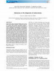
For direct detection of M. tuberculosis in clinical samples,
representative and adequate amounts of the clinical specimen
must be sent to the laboratory quickly in order to allow
optimal sensitivity in the detection of mycobacteria (figure 1).
Clinical specimens from any organ where tuberculosis is suspected should be submitted for analysis. For M. tuberculosis,
a number of nucleic acid amplification techniques are available
as commercial or in-house prepared kits. In order to shorten
diagnostic delay, nucleic acid amplification of mycobacteriumspecific genes has been used to detect M. tuberculosis directly
in clinical samples, and has demonstrated reliability and high
sensitivity. The availability of new kits, and accumulated
experience with nucleic acid amplification techniques for
M. tuberculosis detection in most laboratories, have yielded
improved sensitivity and specificity of these tests.
Clinical specimen
Culture
BACTEC™ MGIT™ 960
Direct detection
• Microscopy
• PCR/LCR for identification
• MTB rifampin resistance
Species identification and typing
• 16S rRNA hybridization (MTB and MAC),
16S rRNA gene PCR sequencing (NTM)
• RFLP- or spoligo typing
Susceptibility testing
• Rifampin resistance (PCR oligohybridization/sequencing)
• Rapid culture
Fig. 1. The logistics of handling clinical specimens with regard to mycobacterial diagnostics. The clinician should be alerted at every step to
promote rapid diagnosis and optimal treatment. LCR = ligase chain reaction;
MAC = Mycobacterium avium; MGIT = mycobacterial growth indicator
tube; MTB = Mycobacterium tuberculosis; NTM = non-tuberculous mycobacteria; PCR = polymerase chain reaction; RFLP = restriction fragment
length polymorphism.
ª 2009 Adis Data Information BV. All rights reserved.
Several groups have previously validated PCR assays for the
identification of M. tuberculosis directly in clinical specimens
(table I).[4,5,10-16] Although 16S rRNA and particularly the 16S
rRNA gene is the most commonly used target, multiple nucleic
acid targets other than the 16S rRNA gene have rendered a high
sensitivity and representative species-specific differentiation,
such as insertion sequence (IS) elements[25,26] and the genes
encoding the 32 kD and 65 kD proteins.[27,28] Nucleic acid
amplification techniques other than PCR – such as transcription-mediated amplification of RNA[29] and, more recently,
strand displacement[22] and Qb-replicase probe amplification
assays[21] – have also been used.
For example, the ligase chain reaction (LCR) test, the LCx
M. tuberculosis assay (Abbott, Chicago, IL, USA), uses the
gene encoding the protein Antigen b (PAB) as the target template for detection of M. tuberculosis directly in clinical specimens.[30] In this assay, detection of M. tuberculosis is performed
by specific probe amplification employing the LCR.[18,31] In
comparison with culture, the sensitivity of LCR was 96.7% for
smear-positive samples and 72.0% for smear-negative samples,
respectively. The specificity of the LCR test was challenged by
including samples containing M. marinum and M. ulcerans,
which are the species that are most closely related to
M. tuberculosis outside the M. tuberculosis complex.[32]
The two commercially available amplification tests approved by the US FDA – the Amplified MTD (M. tuberculosis
Direct) test (Gen-Probe, San Diego, CA, USA) and the Cobas
Amplicor MTB assay (Roche Diagnostics, Mannheim,
Germany) – had excellent performance when used for testing
smear-positive specimens (sensitivity >95%, specificity 100%).
The sensitivity was lower (83–85%) when the test was used for
testing smear-negative specimens, though the specificity stayed
high (99%).[4] On the basis of these data, the FDA recommended the use of nucleic acid amplification tests only for
smear-positive respiratory specimens from patients who had
not received antituberculosis drugs for ‡7 days or within the last
12 months.[5] Following the initial FDA approval, Gen-Probe
enhanced the performance of the MTD test. A large-scale
study further revealed the overall sensitivity, specificity, positive predictive value (PPV), and negative predictive value
(NPV) of the enhanced MTD test to be 94.7, 100, 100, and
98.4%, respectively, in respiratory specimens.[10] The corresponding values were 89.4, 100, 100, and 98.3%, respectively, in
smear-negative respiratory specimens. This enhanced version
of the MTD test was eventually approved by the FDA for
testing respiratory specimens, regardless of the smear status.
The tests have been validated for the performance in respiratory
specimens, while the currently increasing number of specimens
Mol Diagn Ther 2009; 13 (3)
Balasingham et al.
140
Table I. Some examples of performance of commercial kits in direct detection of Mycobacterium tuberculosis (MTB) by nucleic acid amplification
Amplification method
Nucleic acid
target
Test name
Manufacturer
Sensitivity (%)
PCR
16S rRNA gene
Cobas Amplicor
MTB test
Roche
91.7–95.2
LCR
Antigen b gene
LCx
Abbott
92.1–96.7
72
8,9
TMA
16S rRNA
Amplified MTD
Gen-Probe
6.5–95.2/92.6
Extrapulmonary
10,20
Qb replicase amplification
NASBA
23S rRNA
NucliSens QT
Organon Teknika
Co.
91.1–95.8
SDA
IS6110
BD ProbeTec ET
Becton Dickinson
92.1–98.5
Roche
71.0 (93.0)
Real-time PCR
16S rRNA gene
smear pos
Cobas TaqMan MTB
References
smear neg
12,16,17
21
40.3–53.1
22,23
15,24
LCR = ligase chain reaction; NASBA = nucleic acid sequence-based amplification; neg = negative; PCR = polymerase chain reaction; pos = positive;
SDA = strand displacement amplification; TMA = transcription mediated amplification.
from extrapulmonary sites is an additional challenge that often
yields lower sensitivity and specificity (tables I and II).
The Cobas Amplicor MTB assay has maintained a reasonable sensitivity and specificity in smear-positive respiratory
specimens since its initial approval by the FDA.[11,13,14] However, it is limited by a slow block cycler amplification process
and time-consuming colorimetric detection of the amplification
products, which may affect the turnaround time. Recently, the
Cobas Amplicor MTB assay integrated real-time techniques
using the LightCycler 2.0 instrument in the Cobas TaqMan
MTB test (Roche Applied Science, Penzberg, Germany). The
procedure for template DNA extraction remains the same as
that used in the Cobas Amplicor assay. With the use of the
LightCycler instrument to detect the amplification products,
the turnaround time can be shortened. In 146 clinical specimens
evaluated, good agreement (100% sensitivity, 98.6% specificity)
between the LightCycler and Cobas Amplicor assays was
reported.[15] However, further validation of this test is currently
warranted.
1.3 Integration of Molecular Diagnostics
with Clinical Practice
The early studies on nucleic acid amplification tests were
largely laboratory based, emphasizing culture results as a major
endpoint, and neglected the integration of available clinical
information into the decision-making process.[5] In reality, it
is mandatory to consider the degree of clinical suspicion of
tuberculosis in determining the clinical utility of nucleic acid
amplification tests.
A number of subsequent studies have addressed the use of
nucleic acid amplification in the clinical setting. Prospective
ª 2009 Adis Data Information BV. All rights reserved.
studies have evaluated the usefulness of PCR to rule out pulmonary tuberculosis in hospitalized patients.[41,59] The sensitivity in smear-negative patients was 73% and 53% in airway and
extrapulmonary specimens, respectively. Thus, PCR was found
to be a useful tool to evaluate patients for tuberculosis within
the first hospital day. In a multicenter prospective trial, a total
of 338 patients with symptoms and signs consistent with active
pulmonary tuberculosis and complete clinical diagnosis were
stratified by the clinical investigators to be at low (£25%),
intermediate (26–75%), or high (>75%) relative risk of having
tuberculosis.[60] Based on low, intermediate, and high clinical
suspicion of tuberculosis following a comprehensive clinical
diagnosis, the sensitivity of the enhanced MTD test was 83%,
75%, and 87%, respectively, and the corresponding specificity
was 97%, 100%, and 100%. Thus, for complex diagnostic problems such as tuberculosis, assessments of clinical risk can
provide important information about the predictive values
more likely to be experienced in clinical practice.
A number of in-house nucleic acid amplification tests have
been developed over the years. On the whole, the commercial
tests have sensitivity and specificity similar to or better than
those of in-house tests for respiratory specimens.[33,61] While
originally intended to facilitate early diagnosis of pulmonary
tuberculosis, these tests have also been extensively studied and
used in patients with extrapulmonary tuberculosis (table II).
For tuberculous meningitis, for example, there have been quite
a number of reports on direct detection by PCR, although test
performance has been variable.[42-44,62] In one study, the initial
low sensitivity of 33% in cerebrospinal fluid (CSF) could be
elevated to 83% by decreasing the cut-off values for positive
results of the MTD test.[42] In a large-scale study from Sweden
that analyzed 154 CSF samples using the Cobas Amplicor
Mol Diagn Ther 2009; 13 (3)
Rapid Detection of Mycobacteria
141
test, the sensitivity, specificity, PPV, and NPV were 55.6%,
97.2%, 55.6%, and 97.2%, respectively.[43] In a systematic review
and meta-analysis on the diagnostic accuracy of nucleic acid
amplification tests for tuberculous meningitis,[42] the overall
estimates of sensitivity and specificity in 14 studies utilizing
commercial tests were 0.56 and 0.98, respectively. In summary,
based on current evidence, commercial nucleic acid amplification tests show a potential role in confirming the diagnosis of
tuberculous meningitis, although their overall low sensitivity
possibly precludes exclusion of the disease with certainty.[42]
The results also emphasize that multiple samples from each
patient should be tested in order to allow sufficient accuracy
in detecting M. tuberculosis in the specimens by nucleic acid
detection. A certain prioritization of samples subjected to
the nucleic acid detection assay should be based on clinical
indications and risks with regard to infection transmission
and patient isolation policy.
The routine use of nucleic acid amplification techniques to
detect M. tuberculosis directly in clinical specimens has been
hampered for a variety of reasons, such as lack of sensitivity
and/or specificity, contamination problems, high cost, and relatively labor-intensive and complex procedures required for
sample preparation, amplification, and detection.[26,27,63] The
principal factor lowering the sensitivity of amplification techniques in general is the presence of interfering substances in
clinical specimens.[16,26,27] Insufficient amounts of the microbial DNA present in the sample is a real problem, especially as
tuberculosis infections are often paucibacillary. Mycobacteria
or DNA are often unevenly distributed, particularly in the
mucous material in sputum, and may cause an arbitrary sampling effect. This also applies to cultured samples. The elimination of factors inhibitory for amplification of nucleic acids in
clinical specimens still remains a challenge in the use and acceptance of such assays in the diagnostic setting. The specificity
of direct detection assays should be challenged by including
strains and clinical samples containing the very closely related
species M. marinum and M. ulcerans.[32]
The particular challenge for the nucleic acid amplification
tests currently is to provide an early diagnosis of tuberculosis in
smear-negative patients. The potential for influencing patient
outcome is much greater when the acid-fast bacterial smear is
negative. In addition, more automation and lower assay expenses are generally required. Still, using amplification techniques in the identification of M. tuberculosis directly in clinical
samples offers unique improvements in this diagnostic field.
1.4 Tuberculosis Diagnostics in Children
Tuberculosis in children is a major health problem, especially in developing countries. Children exposed to adults with
smear-positive pulmonary tuberculosis have a high risk for
infection, and this increases with the degree of contact.[64,65] In
countries with a high incidence of tuberculosis, risk for infection among children in contact with adults with tuberculosis is
Table II. Overview of the sensitivity and specificity of the polymerase chain reaction, using either in-house or commercial methods in the direct detection
of Mycobacterium tuberculosis in clinical specimens
Anatomical site
Sensitivity (%)
Specificity (%)
References
Respiratory specimens
77.1–100
99.3–100
19,33
Gastric aspirates
44–60
93.7–98
20,23
Lymph node (fresh tissue)
71.6–87.5
NA
24,34
Pleural fluid
27.3–81
90–100
35-39
Pleural biopsy
90–92
100
40
Cerebrospinal fluid
56–58
97–98
41
Ascites fluid
31.4–56
98
42-45
Liver biopsy tissue (paraffin-embedded)
58–88
96–100
46,47
Urine
55.8–95.6
98.1–98.9
48,49
Pulmonary
Extrapulmonary
Skin
60–80
100
50,51
Peripheral blood
30.4–100
NA
52-54
Blood marrow
42–73.2
NA
55,56
Paraffin-embedded tissues
60–68
NA
57,58
NA = not available.
ª 2009 Adis Data Information BV. All rights reserved.
Mol Diagn Ther 2009; 13 (3)
142
30–40%.[66,67] Most children who develop tuberculosis disease
experience pulmonary manifestation, but about 25–35% of
children have an extrapulmonary presentation.[68] Two forms
of extrapulmonary tuberculosis are most common in children.
1. Lympho-hematogenous disease: tubercle bacilli are disseminated to distant sites in all cases of tuberculosis infection.[69] This
dissemination is clinically silent in most cases, but can be the
origin of miliary or extrapulmonary tuberculosis in the
immediate or distant future. The most common clinically
significant disseminated tuberculosis is miliary disease, which
occurs when massive numbers of tubercle bacilli are released into
the bloodstream, causing disease in two or more organs.[70,71]
2. Tuberculosis of the CNS: especially tuberculosis meningitis
(TBM), which is the most serious complication of tuberculosis
in children and occurs in about 4% of children with tuberculosis.[72] The overall mortality has been reported to be 13%,
with approximately half of the survivors developing permanent
neurological sequelae.[73] Tuberculomas are less common
manifestations of CNS infection, usually characterized by
solitary brain lesions.
The diagnosis of tuberculosis in children is traditionally
determined with chest radiography, tuberculin skin test, and
mycobacterial staining/culture, although these diagnostic
methods may not always be positive in children with tuberculosis. The best culture specimen for pulmonary tuberculosis in
children is early morning gastric aspirates. Gastric lavage has
an even higher yield for M. tuberculosis than bronchoalveolar
lavage in children with pulmonary tuberculosis.[74,75] The
standard approach for collecting gastric aspirates is to hospitalize the child and collect three aspirates on consecutive
mornings before gastric emptying is stimulated either by being
ambulatory or by eating. In blood count and cell differential
tests, most typically there are several hundreds to several
thousand white blood cells/mm3, with an early predominance
of polymorphonuclear cells followed by a high proportion
of lymphocytes.[76]
Advances in diagnostic methods, such as the nucleic acid
amplification tests described above and immune-based methods, are increasingly being used for detecting tuberculosis in
children. The immune-based assays detect interferon (IFN)-g
secreted by T cells in response to antigens that are specific to
M. tuberculosis and different from other mycobacteria. The
early secretary antigen target (ESAT-6) is present in all
M. tuberculosis complexes, but absent from all strains of
M. bovis bacillus Calmette-Guérin (BCG) and most environmental bacteria.[77] The development of a new ex vivo enzymelinked immunospot (ELISPOT) assay of ESAT-6-specific
ª 2009 Adis Data Information BV. All rights reserved.
Balasingham et al.
IFNg-secreting antigens by Lalvani and colleagues has enabled
the differentiation of M. tuberculosis infection from BCG
immunization in both adults and children.[78]
The immune system of young children is less developed than
that of adults, and the risk of developing active tuberculosis
disease is therefore higher in young children. The chance of
developing tuberculosis disease is greatest shortly after infection. Childhood tuberculosis cases indicate recent community
transmission, and thus reflect the effectiveness of tuberculosis
control efforts, particularly the contact investigation. As
transmission normally occurs from adult to child, each child
case provides a window by which to observe the effectiveness of
the contact investigation surrounding an index or source case.
As such, evaluation of childhood tuberculosis rates provides
the opportunity to assess a core component of any tuberculosis
control program.[79] Therefore, a good tuberculosis control
program that will ensure early diagnosis and treatment of
adults with infectious forms of tuberculosis is the best way
also to prevent tuberculosis in children.
Tuberculosis in infants and children <4 years of age is much
more likely to spread throughout the body via the bloodstream.
Because of this, children are at much greater risk of developing
tuberculous meningitis than are adults, and the risk is particularly high in HIV-infected children. Tuberculous meningitis
can be devastating in children, with deafness, blindness, paralysis, and mental retardation as some of the consequences.[80]
Mortality resulting from tuberculosis meningitis also continues
to be very high. For these reasons, prompt diagnosis and immediate treatment of tuberculosis are critical in pediatric care.
1.5 Emergence of Non-Tuberculous Mycobacteria (NTM):
New Species are Discovered
NTM are mycobacteria that belong to species other than the
M. tuberculosis complex or M. leprae[81] (table III). The impact
of the presence of NTM in airway samples and other clinical
specimens is often difficult to evaluate;[82] most often the findings are rejected as being only colonization or environmental
contamination.[83] NTM often colonize the airways without
causing disease; however, NTM can sometimes cause pulmonary infection in immunocompetent individuals.[84,85] Because of their ubiquitous nature, random detection or
contamination by NTM can also occur.[81] Only a clinically
validated effect of antimycobacterial treatment can conclusively determine if there really is a causal connection between
the finding of NTM in a clinical specimen and an infection.
At the same time, the prevalence of NTM among clinical
mycobacterial isolates in industrialized countries is increasing,
Mol Diagn Ther 2009; 13 (3)
Rapid Detection of Mycobacteria
Table III. Important non-tuberculous mycobacteria that can cause airway
infections
Frequently involved in airway infections
Slow growers
M. avium complex including M. intracellulare
M. genavense
M. kansasii
M. malmoense
M. simiae
M. szulgai
M. xenopi
Rapid growers
M. fortuitum complex (M. fortuitum, M. peregrinum)
M. abscessus
M. chelonae
M. mucogenicum
Seldomly occurring or causing of disease
M. asiaticum
M. bohemicum
M. branderi
143
defined evidence from biopsy analyses and prospective studies
supports the causative relationship between the isolation of
strains belonging to the M. avium complex and the presence
of small peripheral nodules with or without focal bronchiectasies.[86] Other mycobacterial species that can cause a
tuberculosis-like clinical picture are M. kansasii, M. abscessus,
M. fortuitum, M. chelonae, M. haemophilum, M. malmoense and
M. intracellulare, and new mycobacterial entities are emerging
in this context.[86] Because of the very slow progression of
non-cavitary lung disease, the question still remains whether
one should be proactive and treat these patients with multiple
broad-spectrum drugs, or whether one should be apprehensive
and monitor the patients with frequent microbiology testing,
blood infection tests, and radiological examinations.
Since 1990, an increasing number of reports on the finding of
NTM in the lower airways in patients with cystic fibrosis have
been presented.[87] Prospective studies with screening of cystic
fibrosis patients indicate a prevalence of NTM of approximately 13%.[87] The differentiation of airway colonization from
infection that can contribute to the progress of the underlying
disease can be particularly difficult in cystic fibrosis.
M. conspicuum
M. celatum
M. gastri
M. gordonae
M. interjectum
M. intermedium
M. lentiflavum
M. scrofulaceum
M. shimoidei
M. terrae complex
1.5.1 Identification of NTM Species
Due to sensitive cultivation conditions used in combination
with molecular classification based on 16S rRNA gene PCRsequencing, new mycobacterial species are continuously being
discovered (table III; figure 1).[88] At the same time, many
clinical isolates of NTM are excluded as results of contamination (table III).[88] The diagnosis of an infection with NTM is
not easy, since it must be differentiated from bacteria that represent colonization or contamination.
M. triplex
M. tusciae
M. haemophilum – cause of RES and skin infections
Mycobacteria most often found as contamination
M. gordonae
M. phlei
M. smegmatis
M = Mycobacterium; RES = reticuloendothelial system.
including in children.[83,86] Reports on human lung disease
associated with NTM, particularly those that belong to the
M. avium complex, have also involved hosts that do not exhibit
the traditional risk factor such as known pulmonary disease or
conditions that can alter the local or systemic immune system.[86] The steady increase in incidence of lateral neck cysts in
children infected with NTM supports this notion.[86] More
ª 2009 Adis Data Information BV. All rights reserved.
1.5.2 The Burden of Unidentified Mycobacteria
in Clinical Laboratories
Modern gene technology has to a large extent improved the
classification and basis for taxonomy of mycobacteria.[88] In
addition to the use of phylogenetic nucleic acid sequences for
mycobacterial identification, the patterns of unique mycolic
acids in these bacteria has allowed the application of lipid
profiling as a means of species identification. Routine use of
high-performance liquid chromatography (HPLC) of mycolic
acids and 16S rDNA sequence analysis contributes to a large
extent to the identification of most mycobacterial isolates.[32,88]
However, isolates of new mycobacterial entities constantly
arise that cannot be identified as belonging to a recognized
mycobacterial species.[88] Many NTM isolates from humans
come from the airways and can potentially be regarded as
Mol Diagn Ther 2009; 13 (3)
Balasingham et al.
144
nonsignificant. Even though the occurrence of mycobacteria in
clinical specimens is increasing, several studies show that the
presence of NTM in sputum or secretions from bronchoalveolar lavage are correlates with infection. On the other hand,
the detection of mycobacteria in clinical specimens must not
cause diagnostic delay or inappropriate treatment with regard
to other diseases. It is important that the finding of NTM is
carefully evaluated during the hunt for M. tuberculosis – but
very critically, so that differential diagnostics such as cancer
screening are not in any way delayed.
Some NTM isolates, however, come from normally sterile
areas of the body (blood, pleural biopsy, CSF, intravenous
catheters, or pus) and can therefore not so easily be dismissed
as non-important. The taxonomy of genus Mycobacterium
therefore seems to be far from elucidated, and new species
of this genus are emerging all the time. The reporting of
uncommon mycobacterial isolates is therefore valuable in
this context.
There is a fine line between the benefits of sensitive mycobaterial diagnostics and the problem of over-diagnosis. Although it is known that NTM should be considered in all cases
when acid-fast bacteria are detected by microscopy, such a
finding can represent a trap in clinical medicine and cause both
maltreatment and delayed detection of pulmonary diseases
of other etiology, e.g. cancer. Reliable DNA-based methods
combined with close contact between the clinical microbiologist/microbiology laboratory and the clinician is important for optimal handling of suspected mycobacterial
infections.
1.5.3 Follow-Up of Positive NTM Cultures: When and How?
Follow-up specimens for culture are taken once the infection
is clinically established, as it is culture, and not microscopy or
PCR, that is the test decisive in defining whether treatment
is successful. In this context, it can be difficult to get representative sputum from children, and a gastric aspirate is a
good alternative (as described in section 1.4). Bronchoalveolar
lavage (BAL) is used a lot in hospitals and outpatient clinics.
However, it is extremely important to perform appropriate
cleaning and disinfection of the bronchoscopy equipment in
order to avoid cross-contamination of mycobacteria.
The macrolide-azide antibiotics with demonstrable effect
towards the M. avium complex strains represent a considerable
improvement in the outcome of the different drug combinations employed in treatment. Even though the indication for
treatment against NTM must be clear and critically evaluated,
the potential for prevention of significant bronchiectatic lung
ª 2009 Adis Data Information BV. All rights reserved.
disease warrants a proactive diagnostic approach for the
identification of NTM in their various clinical presentations.
Recognition of the various forms of lung diseases associated
with NTM can be a prerequisite for heightened clinical
awareness and suspicion of NTM as a potential agent in lower
airway infections.
2. M. tuberculosis Drug Susceptibility Testing
2.1 Drug Susceptibility Testing by Culture
Rapid detection of mycobacteria and their markers for drug
susceptibility is an important field that is in active development.[89] MDR M. tuberculosis strains can be detected by
a number of different assays.[90-99] Assay methods are often
difficult to standardize, and the World Health Organization/International Union Against Tuberculosis and Lung
Disease Global Project on Anti-Tuberculosis Drug Resistance
Surveillance (WHO/IUATLD) is attempting to produce
standardized drug resistance data worldwide.[100] Broth-based
methods are faster than solid media systems, and commercial
systems, the BACTEC 460 or MGIT 960, are arguably the
fastest methods that exist today and permit testing to be completed within 7 days. However, these methods are expensive and
some older versions require disposal of radioactive material.
Novel phenotypic methods that utilize mycobacteriophages
have shown promise.
Drug susceptibility testing of NTM is warranted only after
conference between the clinician and clinical microbiologist,
and agreement on the clinical indication. The NTM species
most often in question are the M. avium complex, M. kansasii
and M. malmoense (table III). Drug susceptibility testing of
NTM can be performed on liquid and/or solid media. Drugs
included in the analysis are clarithromycin, amikacin, rifabutin,
ciprofloxacin, ethambutol, ethambutol/ciprofloxacin, and
ethambutol/clarithromycin. Rapid-growing mycobacteria can
be subjected to drug susceptibility and minimum inhibitory
concentration (MIC) testing by using E-tests (AB Biodisk,
Sweden), but this is only done on evident clinical suspicion.
2.2 Direct Detection of Drug Resistance Markers
for M. tuberculosis
Globally, the emergence of MDR and extensively drugresistant (XDR; also referred to as extreme drug-resistant)
strains of M. tuberculosis is an increasing problem that adversely affects patient care and public health. MDR strains of
M. tuberculosis are resistant to at least the two main first-line
Mol Diagn Ther 2009; 13 (3)
Rapid Detection of Mycobacteria
145
tuberculosis drugs (isoniazid and rifampin [rifampicin]), while
XDR strains are MDR strains that also are resistant to three or
more of the six classes of second-line drugs. Direct detection of
M. tuberculosis drug resistance markers is particularly important for the monitoring of patients carrying MDR strains.
In contrast to other bacteria, resistance of M. tuberculosis is
exclusively associated with chromosomal mutations. Recently
developed molecular biological techniques have significantly
helped in understanding the basis of drug action and resistance
mechanisms in this organism. The molecular detection systems require knowledge of the genes encoding the drug target
(rpoB for rifampin; the inhA/mabA, katG, oxyR and ahpC genes
for isoniazid) and the mutations producing resistance. In this
regard, the gene targets most frequently exhibiting point
mutations associated with drug resistance have been identified
(table IV).
Genotypic methods allow earlier detection of drug resistance,
while conventional approaches are cumbersome or lack sensitivity or specificity. New real-time PCR methods to detect
rifampin- and isoniazid-resistant M. tuberculosis strains in a
single reaction tube have been designed[101] and employed to
characterize resistant isolates in Spain by real-time PCR and
DNA sequencing. Full concordance of the results of the PCR
with the sequencing data was obtained. In addition, a blind test
was performed with a panel of 15 different susceptible and resistant strains from throughout Spain, and these results were also
in 100% agreement with the sequencing data.[101] This was the
first assay based on rapid-cycle PCR able to simultaneously detect in a single reaction tube a large variety of mutations associated with rifampin resistance (12 different mutations affecting
eight independent codons, including the most prevalent mutations at positions 526 and 531) and the most frequent isoniazid
resistance mutations. This design could be a model for new, rapid
genotypic methods able to simultaneously detect a wide variety of
antibiotic resistance mutations. Apart from DNA sequencing,
these genotypic methods are limited in that not all resistance
mechanisms are known. The alleles conferring mycobacterial
antibiotic resistance are ever-evolving, and new point mutations
that may or may not confer drug resistance occur, requiring DNA
sequence analysis for some loci to give the complete overview.
However, the availability of relatively rapid (4–5 days) susceptibility testing using MGIT 960 liquid cultures somewhat
alleviates the need for direct detection of other antibiotic
resistance markers than rifampin and isoniazid.
Table IV. Genetic targets relevant for direct detection of antimycobacterial
drug resistance development
Antimycobacterial agents
Genetic marker(s)
Primary agents
Rifampin
rpoB (RNA polymerase B subunit)
Isoniazid
katG (catalase-peroxidase)
inhA (enoyl-acyl carrier protein reductase)
ahpC (alkyl-hydroperoxide reductase)
kasA (keto-acyl synthase)
ndh (NADH dehydrogenase)
Pyrazinamide
pncA (pyrazinamidase)
Streptomycin
rpsL (S12), rrs (16S rDNA)
Ethambutol
embB (arabinosyl transferase)
Secondary agents
Capreomycin
Kanamycin
Cycloserine
Ethionamide
inhA (enoyl-acyl carrier protein reductase)
Alternative agents
Rifapentine
rpoB
Rifabutin
rpoB
Amikacin
Quinolones, ciprofloxacin
gyrA, gyrB (DNA gyrase)
NADH = nicotinamide adenine dinucleotide (reduced).
ª 2009 Adis Data Information BV. All rights reserved.
3. Molecular Epidemiological Markers:
M. tuberculosis Strain Characterization
Mycobacterial strain typing is important, both for the analysis of the spread of tuberculosis and for monitoring the development of antibiotic resistance. Molecular fingerprinting of
M. tuberculosis is particularly challenging due to its clonal
nature. By using the transposon IS6110, an internationally
accepted restriction fragment length polymorphism (RFLP)
procedure for M. tuberculosis strains has been used for epidemiological studies.[102] However, IS6110-RFLP requires
culturing, DNA extraction, and Southern hybridization, and
may take as long as 4–5 weeks. Moreover, IS6110 fingerprinting may be of limited use, since some M. tuberculosis strains do
not harbor a copy of IS6110, whereas others may contain only
one to five copies, which do not provide a sufficient resolution
pattern. Development of rapid typing methods remains important, and alternative PCR-based techniques are particularly
promising, as they may facilitate both rapid diagnosis and
molecular typing of tuberculosis. Repeat amplification by using
the conventional IS6110-RFLP typing has therefore been
supplemented by PCR-based methods such as spoligotyping
and double-repetitive-element (DR)-typing.[102] However,
the PCR-based methods available may not be sufficiently
Mol Diagn Ther 2009; 13 (3)
Balasingham et al.
146
discriminatory when used alone. For this reason, a combination of methods with spoligotyping as a first-line test followed
by DR-PCR as a rapid alternative strategy for M. tuberculosis
typing has recently been suggested.[102]
Multilocus sequence typing (MLST) has resolved the strain
diversification in many bacterial species. However, mycobacterial genomes have relatively low spontaneous mutation
and recombination rates, and have demonstrated (to date) a
lack of horizontal gene transfer, rendering them relatively
conserved and static genomes (see section 4). The resolution of
MLST in the M. tuberculosis complex is consequently insufficient for strain discrimination. Epidemiology therefore
needs to use comparatively low-resolution tools for adequate
strain discrimination or advanced typing tools (see section 5.1).
4. Genome Instability in M. tuberculosis
and its Human Host
4.1 Genome Instability is the Basis for Strain Variability
and Drug Resistance Development in M. tuberculosis
Intracellularly, the genome of M. tuberculosis may sustain
considerable damage as a result of exposure to oxidative and
nitrosative stress, both during replicative periods and during
the dormant phase. The expression patterns of genes involved
in genome maintenance and DNA repair are therefore likely to
be essential for the intracellular survival of M. tuberculosis in
the hostile environment of the macrophage phagolysozome.
Recent experiments have strengthened this hypothesis, as both
M. tuberculosis replicative DNA polymerase (DnaE2) and
nucleotide excision repair mutants show reduced virulence in
mice.[103] Nevertheless, the characterization of M. tuberculosis
DNA repair components is still in its infancy and is based
mostly on sequence homology searches.[104] Importantly, mutations in genes involved in the repair of DNA damage contribute to increased genome instability and mutation rates in
several pathogens, including those causing disease in children.[105,106] A hypermutator phenotype may be beneficial under specific selective pressures.[107] In a clinical setting, this is
highly relevant to antibiotic resistance. In M. tuberculosis, the
DNA polymerase DnaE2 has been shown to be a major mediator of induced mutagenesis in mice and play a role in the
emergence of drug resistance.[108] Also, polymorphisms in the
mutT and ogt genes have been identified in the W-Beijing
phylogenetic lineage.[109] However, in this latter case, the
mutator phenotype does not appear to increase the prevalence
of drug resistance in clinical isolates.[110] A general role for
ª 2009 Adis Data Information BV. All rights reserved.
hypermutators in the emergence of clinically relevant antibiotic
resistance awaits further studies.
Mycobacterial antibiotic resistance per se occurs by chromosomal point mutations (table IV). Resistance development
is thus a result of the balance between DNA damage, recombination, replication fidelity, and mutation rate on the one
hand, and genome maintenance on the other (tables V and VI).
Knowledge of this genetic basis elucidates the defined repertoire of molecular methods that can be used to characterize
the resulting M. tuberculosis phenotype, i.e. plasmid characterization is irrelevant, but single nucleotide polymorphisms
(SNPs) are most relevant.
4.2 Molecular Diagnostics for Assessing Host Susceptibility
The future of the molecular diagnostic tests for tuberculosis
is no longer limited to the mycobacterial genomes. The elucidation of the human genome has led to the discovery of more
susceptibility or resistance genes associated with tuberculosis.[112,113] These genes are related to effective killing of intracellular mycobacteria or granuloma formation. The effector
mechanisms include (i) the iron-scavenging function of the
macrophage transport proteins, which compete with the siderophores of mycobacteria; and (ii) the activation of macrophage function by vitamin D, by antigen presentation, and even
by cytokines, cytokine receptors, and intracellular signaling
molecules, which are all part of the immunological pathway of
activation for a T-cell helper-1 response.[114] Important examples are the natural resistance-associated macrophage protein (NRAMP1, gene name SLC11A1), the vitamin D receptor
(VDR), and the human leukocyte antigen (HLA)-DR2 and
HLA-DQB1 loci, located on chromosomes 15 and X.[115-117]
The importance of the mutations involving the IFNg receptors
1 and 2 (IFNGR1 and IFNGR2), STAT1, and interleukin
Table V. Main characteristics of the Mycobacterium tuberculosis genome
sequences[111]
Factor/trait
Characteristics
Genome size
4.4 Mb, 4000 genes
G + C content
66% (high G + C content)
Phylogenetic age
Long generation time makes this a
phylogenetically young bacterial species
Genome instability
High frequency of spontaneous mutations
Sparse homologous recombination
insertion sequence elements
Genome dynamics
Relatively static genome
G + C = guanosine + cytosine.
Mol Diagn Ther 2009; 13 (3)
Rapid Detection of Mycobacteria
147
Table VI. Comparison of the DNA repair profile in Escherichia coli and Mycobacterium tuberculosis. Genome maintenance and gene instability are essential in
the modulation of mycobacterial mutation and recombination rates, and thus influence the development of drug resistance and epidemiological variation
DNA repair pathways
E. coli
M. tuberculosis
Base excision repair (BER)
phrB, ada, ogt, nei, mutT, tag1, alkA, ung, mutY, nth, fpg
ada::alkAa, ogt, mutT b, mpg, ung, mutY, nth, fpg b, nei b
Nucleotide excision repair (NER)
uvrA, uvrB, uvrC, uvrD, mfd
uvrA, uvrB, uvrC, uvrD, mfd
Mismatch repair (MMR)
mutL, H, S
Not identified c
Recombinational repair
recB, recC, recD, recF, recO, recR, recQ d
recB; recC, recD, recF, recO, recR, ercc3 e
SOS repair
umuC, umuD d, dinP, polB d
umuC, dinP
a The alkA and ada genes are linked, encoding a fused gene product.
b Four fpg/nei and four mutT orthologs are found in M. tuberculosis.
c Some components that are lacking, such as an MMR system, make M. tuberculosis a natural mutator.
d recQ, umuD and polB genes are lacking in M. tuberculosis.
e ercc3 is present in M. tuberculosis, but absent in E. coli and most other bacterial species.
(IL)12R b1 (IL12RB1), associated with IFNg-mediated
immunity, is uncertain in tuberculosis, although they have been
found to be linked to disseminated diseases caused by atypical
mycobacteriosis and other intracellular pathogens.[118,119] The
use of microarrays for a host genome survey of tuberculosis
susceptibility is not too far from reality.
5. Implications of Genome Instability and DNA Repair
for Modern Mycobacterial Diagnostics and Treatment
5.1 Perspectives on Diagnostics
M. tuberculosis complex species display relatively static
genomes and 99.9% nucleotide sequence identity. The study of
the evolutionary history of such monomorphic bacteria is a
difficult and challenging task. SNP analysis of DNA repair,
recombination, and replication (3R) genes in a comprehensive
selection of M. tuberculosis complex strains representing the
global scenario yielded surprisingly high levels of polymorphisms in these genes compared with house-keeping genes, making it possible to distinguish between 80% of clinical isolates.
The relaxed fidelity of 3R genes thus reflected may allow the
occurrence of adaptive variants, among which some will survive. In the context of molecular diagnosis, this work is important, since 3R-based phylogenetic trees represent a new tool
for distinction between M. tuberculosis complex strains.[120]
This situation, and the consequent lack of fidelity in genome
maintenance, may serve as a starting point for deciphering
the evolution of antibiotic resistance, fitness for survival, and
pathogenicity, possibly conferring a selective advantage in
certain stressful situations.[121] Furthermore, detection of
M. tuberculosis strains directly in clinical samples through
ª 2009 Adis Data Information BV. All rights reserved.
PCR amplification of mycobacterium-specific genes has also
demonstrated the specificity of recA and pps1 inteins for
this complex and thus the feasibility of using intein-coding
sequences as a new target for PCR diagnosis has also been
demonstrated.[122] Indeed, the recA and pps1 genes of
M. tuberculosis were found to be interrupted by an intein sequence at the RecA-a and Pps1-b sites, respectively, while NTM
species fail to demonstrate these insertions. Besides, the MtuPps1
has been shown to possess an endonuclease activity. In addition
to the PCR amplification of recA and pps1 intein genes as a tool
for diagnosis, the specific endonuclease activity could represent a
new molecular approach to identify M. tuberculosis.
5.2 Perspectives on Multidrug-Resistant and Extensively
Drug-Resistant M. tuberculosis
A combination of insufficient treatment, noncompliance,
and the molecular basis for drug resistance comprise the basis
for the development of mycobacterial drug resistance currently
observed. It is questionable whether or when new antimycobacterial agents will be developed, as the pharmaceutical
industry does not see this as a fortuitous business. For the industry to be interested in investing in adequate rounds of drug
target screenings, a reasonable market size is a prerequisite. The
development of new drugs against MDR M. tuberculosis, although warranted, would require large investments to treat a
relatively small group of patients who generally are more
noncompliant than other patient groups. Such investment
cannot be expected, and academia can hardly provide the
funding it takes to undertake the large-scale screening required
for providing the drug candidates warranted. Directly observed
therapy (DOT) is therefore as important as ever.
Mol Diagn Ther 2009; 13 (3)
Balasingham et al.
148
6. Conclusions
Prompt and accurate diagnosis of symptomatic patients is a
cornerstone of global tuberculosis control strategies. Remarkable progress has recently been made, upgrading the speed
and quality of mycobacteriology diagnostic services in industrialized countries, but for most of the world where tuberculosis is a large public health burden, those gains are still
unrealized. Deficiencies in current case-finding tools in diseaseendemic countries have made it difficult to ensure access to
good diagnostics at all health service levels, leaving too many
patients undiagnosed. While significant advances have been
made in the rapid and accurate diagnosis of M. tuberculosis, the
molecular biology methods used in the research laboratory to
elucidate the mechanisms of drug resistance cannot be transferred to all the medical centers delivering patient care. These
methods require skilled operators, cumbersome protocols and
relatively high expenses. A number of companies that already
have a large investment in M. tuberculosis diagnostics are
adapting their high-throughput technology to drug susceptibility testing. These methodologies are not applicable to the
developing world, not only because of the costs involved, but
through a lack of the infrastructure that is required to operate
these machines and deliver specimens to the point of testing.
Alternative technologies for diagnostics and drug susceptibility
testing that do not rely on an investment in expensive hardware,
and that have the potential for use in the field, are thus
warranted.
We are now in an exciting era, when new molecular information is generated by many variants of nucleic acid-based
techniques, providing new developments in rapid diagnosis
and susceptibility testing for microbial agents, including
M. tuberculosis. Technical progress in mycobacterial diagnostics is resulting in a number of improved tools, including
some appropriate for low-income settings.[17] Often, the use of
these tests can reduce the need for invasive diagnostic procedures, which are both costly and pose an added risk to the
patient. Particularly in smear-negative patients, the nucleic acid
amplification tests could provide more rapid diagnosis of tuberculosis and subsequent initiation of therapy, and allow
earlier discharge of hospitalized patients. Despite the progress
in mycobacterial diagnostics and control, new diagnostic
approaches and therapies should be sought. These should be
implemented along with well-functioning and integrated tuberculosis control programs. Fortunately, technical progress in
diagnostics is resulting in a number of improved tools, including some appropriate for low-income settings. Important work
remains, however, before new diagnostic tools can be meanª 2009 Adis Data Information BV. All rights reserved.
ingfully integrated into national tuberculosis control programs
in high-burden countries and before tuberculosis control strategies can take them fully into account. The design and quality
of clinical trials evaluating new diagnostics must be improved,
clinical and laboratory services that would allow rapid response
to test results need to be established, and basic and operational
research to appraise the impact and cost effectiveness of new
technologies for mycobacterial diagnostics and therapy must
be carried out.
Acknowledgments
Financial support from the Research Council of Norway, the European
Union FW6 project TBadapt (contract no. 037919) and the Laurine
Maarschalks Fund is greatly acknowledged. The authors have no conflicts
of interest that are directly relevant to the content of this review.
References
1. Bifani PJ, Mathema B, Kurepina NE, et al. Global dissemination of the
Mycobacterium tuberculosis W-Beijing family strains. Trends Microbiol
2002 Jan; 10 (1): 45-52
2. Glynn JR, Whiteley J, Bifani PJ, et al. Worldwide occurrence of Beijing/W
strains of Mycobacterium tuberculosis: a systematic review. Emerg Infect Dis
2002 Aug; 8 (8): 843-9
3. Frieden TR, Sterling TR, Munsiff SS, et al. Tuberculosis. Lancet 2003 Sep 13;
362 (9387): 887-99
4. Woods GL. Molecular techniques in mycobacterial detection. Arch Pathol
Lab Med 2001 Jan; 125 (1): 122-6
5. American Thoracic Society Workshop. Rapid diagnostic tests for tuberculosis: what is the appropriate use? Am J Respir Crit Care Med 1997 May;
155 (5): 1804-14
6. Hanscheid T, Monteiro C, Cristino JM, et al. Growth of Mycobacterium
tuberculosis in conventional BacT/ALERT FA blood culture bottles allows
reliable diagnosis of Mycobacteremia. J Clin Microbiol 2005 Feb; 43 (2):
890-1
7. Diraa O, Fdany K, Boudouma M, et al. Assessment of the Mycobacteria
Growth Indicator Tube for the bacteriological diagnosis of tuberculosis.
Int J Tuberc Lung Dis 2003 Oct; 7 (10): 1010-2
8. Tortoli E, Benedetti M, Fontanelli A, et al. Evaluation of automated BACTEC MGIT 960 system for testing susceptibility of Mycobacterium
tuberculosis to four major antituberculous drugs: comparison with the
radiometric BACTEC 460TB method and the agar plate method of proportion. J Clin Microbiol 2002 Feb; 40 (2): 607-10
9. Cormican MG, Glennon M, Riain UN, et al. Evaluation of a PCR assay
for detection of Mycobacterium tuberculosis in clinical specimens. Diagn
Microbiol Infect Dis 1995 Aug; 22 (4): 357-60
10. Gamboa F, Fernandez G, Padilla E, et al. Comparative evaluation of initial
and new versions of the Gen-Probe Amplified Mycobacterium Tuberculosis
Direct Test for direct detection of Mycobacterium tuberculosis in respiratory
and nonrespiratory specimens. J Clin Microbiol 1998 Mar; 36 (3): 684-9
11. Reischl U, Lehn N, Wolf H, et al. Clinical evaluation of the automated
COBAS AMPLICOR MTB assay for testing respiratory and nonrespiratory
specimens. J Clin Microbiol 1998 Oct; 36 (10): 2853-60
12. Bogard M, Vincelette J, Antinozzi R, et al. Multicenter study of a commercial,
automated polymerase chain reaction system for the rapid detection of
Mycobacterium tuberculosis in respiratory specimens in routine clinical
practice. Eur J Clin Microbiol Infect Dis 2001 Oct; 20 (10): 724-31
Mol Diagn Ther 2009; 13 (3)
Rapid Detection of Mycobacteria
149
13. Levidiotou S, Vrioni G, Galanakis E, et al. Four-year experience of use of the
Cobas Amplicor system for rapid detection of Mycobacterium tuberculosis
complex in respiratory and nonrespiratory specimens in Greece. Eur J Clin
Microbiol Infect Dis 2003 Jun; 22 (6): 349-56
31. Ausina V, Gamboa F, Gazapo E, et al. Evaluation of the semiautomated
Abbott LCx Mycobacterium tuberculosis assay for direct detection of
Mycobacterium tuberculosis in respiratory specimens. J Clin Microbiol 1997
Aug; 35 (8): 1996-2002
14. Fegou E, Jelastopulu E, Sevdali M, et al. Sensitivity of the Cobas
Amplicor system for detection of Mycobacterium tuberculosis in respiratory and extrapulmonary specimens. Clin Microbiol Infect 2005 Jul; 11 (7):
593-6
32. Tønjum T, Welty DB, Jantzen E, et al. Differentiation of Mycobacterium
ulcerans, M. marinum, and M. haemophilum: mapping of their relationships
to M. tuberculosis by fatty acid profile analysis, DNA-DNA hybridization,
and 16S rRNA gene sequence analysis. J Clin Microbiol 1998 Apr; 36 (4):
918-25
15. Burggraf S, Reischl U, Malik N, et al. Comparison of an internally controlled,
large-volume LightCycler assay for detection of Mycobacterium tuberculosis
in clinical samples with the COBAS AMPLICOR assay. J Clin Microbiol
2005 Apr; 43 (4): 1564-9
16. Tønjum T, Klintz L, Bergan T, et al. Direct detection of Mycobacterium
tuberculosis in respiratory samples from patients in Scandinavia by polymerase chain reaction. Clin Microbiol Infect 1996; 2 (2): 127-31
17. Boehme CC, Nabeta P, Henostroza G, et al. Operational feasibility of using
loop-mediated isothermal amplification for diagnosis of pulmonary tuberculosis in microscopy centers of developing countries. J Clin Microbiol
2007 Jun; 45 (6): 1936-40
18. Lindbråthen A, Gaustad P, Hovig B, et al. Direct detection of Mycobacterium
tuberculosis complex in clinical samples from patients in Norway by ligase
chain reaction. J Clin Microbiol 1997 Dec; 35 (12): 3248-53
19. Wang SX, Tay L. Evaluation of three nucleic acid amplification methods
for direct detection of Mycobacterium tuberculosis complex in respiratory
specimens. J Clin Microbiol 1999 Jun; 37 (6): 1932-4
20. Gomez-Pastrana D, Torronteras R, Caro P, et al. Comparison of amplicor,
in-house polymerase chain reaction, and conventional culture for
the diagnosis of tuberculosis in children. Clin Infect Dis 2001 Jan; 32 (1):
17-22
21. An Q, Buxton D, Hendricks A, et al. Comparison of amplified Q beta replicase and PCR assays for detection of Mycobacterium tuberculosis. J Clin
Microbiol 1995 Apr; 33 (4): 860-7
22. Walker GT, Nadeau JG, Spears PA, et al. Multiplex strand displacement
amplification (SDA) and detection of DNA sequences from Mycobacterium
tuberculosis and other mycobacteria. Nucleic Acids Res 1994 Jul 11; 22 (13):
2670-7
23. Kang EY, Choi JA, Seo BK, et al. Utility of polymerase chain reaction for
detecting Mycobacterium tuberculosis in specimens from percutaneous
transthoracic needle aspiration. Radiology 2002 Oct; 225 (1): 205-9
33. Yuen KY, Yam WC, Wong LP, et al. Comparison of two automated DNA
amplification systems with a manual one-tube nested PCR assay for diagnosis of pulmonary tuberculosis. J Clin Microbiol 1997 Jun; 35 (6): 1385-9
34. Bruijnesteijn Van Coppenraet ES, Lindeboom JA, Prins JM, et al. Real-time
PCR assay using fine-needle aspirates and tissue biopsy specimens for rapid
diagnosis of mycobacterial lymphadenitis in children. J Clin Microbiol 2004
Jun; 42 (6): 2644-50
35. Querol JM, Minguez J, Garcia-Sanchez E, et al. Rapid diagnosis of pleural
tuberculosis by polymerase chain reaction. Am J Respir Crit Care Med 1995
Dec; 152 (6 Pt 1): 1977-81
36. Mitarai S, Shishido H, Kurashima A, et al. Comparative study of amplicor
Mycobacterium PCR and conventional methods for the diagnosis of
pleuritis caused by mycobacterial infection. Int J Tuberc Lung Dis 2000 Sep;
4 (9): 871-6
37. Villegas MV, Labrada LA, Saravia NG. Evaluation of polymerase chain
reaction, adenosine deaminase, and interferon-gamma in pleural fluid for
the differential diagnosis of pleural tuberculosis. Chest 2000 Nov; 118 (5):
1355-64
38. Nagesh BS, Sehgal S, Jindal SK, et al. Evaluation of polymerase chain reaction for detection of Mycobacterium tuberculosis in pleural fluid. Chest 2001
Jun; 119 (6): 1737-41
39. Pai M, Flores LL, Hubbard A, et al. Nucleic acid amplification tests in the
diagnosis of tuberculous pleuritis: a systematic review and meta-analysis.
BMC Infect Dis 2004 Feb 23; 4 (6): 6
40. Hasaneen NA, Zaki ME, Shalaby HM, et al. Polymerase chain reaction of
pleural biopsy is a rapid and sensitive method for the diagnosis of tuberculous pleural effusion. Chest 2003 Dec; 124 (6): 2105-11
41. Campos M, Quartin A, Mendes E, et al. Feasibility of shortening respiratory
isolation with a single sputum nucleic acid amplification test. Am J Respir
Crit Care Med 2008 Aug 1; 178 (3): 300-5
24. Kidane D, Olobo JO, Habte A, et al. Identification of the causative organism
of tuberculous lymphadenitis in Ethiopia by PCR. J Clin Microbiol 2002
Nov; 40 (11): 4230-4
42. Lang AM, Feris-Iglesias J, Pena C, et al. Clinical evaluation of the GenProbe Amplified Direct Test for detection of Mycobacterium tuberculosis
complex organisms in cerebrospinal fluid. J Clin Microbiol 1998 Aug; 36 (8):
2191-4
25. Eisenach KD, Cave MD, Bates JH, et al. Polymerase chain reaction amplification of a repetitive DNA sequence specific for Mycobacterium tuberculosis. J Infect Dis 1990 May; 161 (5): 977-81
43. Jonsson B, Ridell M. The Cobas Amplicor MTB test for detection of Mycobacterium tuberculosis complex from respiratory and non-respiratory clinical
specimens. Scand J Infect Dis 2003; 35 (6-7): 372-7
26. Sjøbring U, Mecklenburg M, Andersen AB, et al. Polymerase chain reaction
for detection of Mycobacterium tuberculosis. J Clin Microbiol 1990 Oct;
28 (10): 2200-4
44. Pai M, Flores LL, Pai N, et al. Diagnostic accuracy of nucleic acid amplification tests for tuberculous meningitis: a systematic review and metaanalysis. Lancet Infect Dis 2003 Oct; 3 (10): 633-43
27. Böddinghaus B, Rogall T, Flohr T, et al. Detection and identification of
mycobacteria by amplification of rRNA. J Clin Microbiol 1990 Aug; 28 (8):
1751-9
45. Desai MM, Pal RB. Polymerase chain reaction for the rapid diagnosis of
tuberculous meningitis. Indian J Med Sci 2002 Nov; 56 (11): 546-52
28. Soini H, Skurnik M, Liippo K, et al. Detection and identification of mycobacteria by amplification of a segment of the gene coding for the 32kilodalton protein. J Clin Microbiol 1992 Aug; 30 (8): 2025-8
46. Diaz ML, Herrera T, Lopez-Vidal Y, et al. Polymerase chain reaction for
the detection of Mycobacterium tuberculosis DNA in tissue and assessment
of its utility in the diagnosis of hepatic granulomas. J Lab Clin Med 1996
Apr; 127 (4): 359-63
29. Jonas V, Alden MJ, Curry JI, et al. Detection and identification of Mycobacterium tuberculosis directly from sputum sediments by amplification
of rRNA. J Clin Microbiol 1993 Sep; 31 (9): 2410-6
47. Alcantara-Payawal DE, Matsumura M, Shiratori Y, et al. Direct detection of
Mycobacterium tuberculosis using polymerase chain reaction assay among
patients with hepatic granuloma. J Hepatol 1997 Oct; 27 (4): 620-7
30. Andersen AB, Hansen EB. Structure and mapping of antigenic domains of
protein antigen b, a 38,000-molecular-weight protein of Mycobacterium
tuberculosis. Infect Immun 1989 Aug; 57 (8): 2481-8
48. Moussa OM, Eraky I, El-Far MA, et al. Rapid diagnosis of genitourinary
tuberculosis by polymerase chain reaction and non-radioactive DNA
hybridization. J Urol 2000 Aug; 164 (2): 584-8
ª 2009 Adis Data Information BV. All rights reserved.
Mol Diagn Ther 2009; 13 (3)
150
Balasingham et al.
49. Kafwabulula M, Ahmed K, Nagatake T, et al. Evaluation of PCR-based
methods for the diagnosis of tuberculosis by identification of mycobacterial
DNA in urine samples. Int J Tuberc Lung Dis 2002 Aug; 6 (8): 732-7
68. Ussery XT, Valway SE, McKenna M, et al. Epidemiology of tuberculosis
among children in the United States: 1985 to 1994. Pediatr Infect Dis J 1996
Aug; 15 (8): 697-704
50. Arora SK, Kumar B, Sehgal S. Development of a polymerase chain reaction
dot-blotting system for detecting cutaneous tuberculosis. Br J Dermatol 2000
Jan; 142 (1): 72-6
69. Van Zwanenberg D. The influence of the number of bacilli on the development of tuberculous disease in children. Am Rev Respir Dis 1960 Jul; 82:
31-44
51. Quiros E, Bettinardi A, Quiros A, et al. Detection of mycobacterial DNA in
papulonecrotic tuberculid lesions by polymerase chain reaction. J Clin Lab
Anal 2000; 14 (4): 133-5
70. Kim JH, Langston AA, Gallis HA. Miliary tuberculosis: epidemiology,
clinical manifestations, diagnosis, and outcome. Rev Infect Dis 1990 JulAug; 12 (4): 583-90
52. Schluger NW, Condos R, Lewis S, et al. Amplification of DNA of Mycobacterium tuberculosis from peripheral blood of patients with pulmonary
tuberculosis. Lancet 1994 Jul 23; 344 (8917): 232-3
71. Hussey G, Chisholm T, Kibel M. Miliary tuberculosis in children: a review
of 94 cases. Pediatr Infect Dis J 1991 Nov; 10 (11): 832-6
53. Folgueira L, Delgado R, Palenque E, et al. Rapid diagnosis of Mycobacterium
tuberculosis bacteremia by PCR. J Clin Microbiol 1996 Mar; 34 (3): 512-5
54. Honore S, Vincensini JP, Hocqueloux L, et al. Diagnostic value of a nested
polymerase chain reaction assay on peripheral blood mononuclear cells from
patients with pulmonary and extra-pulmonary tuberculosis. Int J Tuberc
Lung Dis 2001 Aug; 5 (8): 754-62
55. Lombard EH, Victor T, Jordaan A, et al. The detection of Mycobacterium
tuberculosis in bone marrow aspirate using the polymerase chain reaction
[published erratum appears in Tuber Lung Dis 1995 Oct; 76 (5): 471]. Tuber
Lung Dis 1994 Feb; 75 (1): 65-9
56. Akcan Y, Tuncer S, Hayran M, et al. PCR on disseminated tuberculosis in
bone marrow and liver biopsy specimens: correlation to histopathological
and clinical diagnosis. Scand J Infect Dis 1997; 29 (3): 271-4
57. Sumi MG, Mathai A, Sheela R, et al. Diagnostic utility of polymerase chain
reaction and immunohistochemical techniques for the laboratory diagnosis
of intracranial tuberculoma. Clin Neuropathol 2001 Jul-Aug; 20 (4): 176-80
58. Park DY, Kim JY, Choi KU, et al. Comparison of polymerase chain reaction
with histopathologic features for diagnosis of tuberculosis in formalin-fixed,
paraffin-embedded histologic specimens. Arch Pathol Lab Med 2003 Mar;
127 (3): 326-30
59. Cohen RA, Muzaffar S, Schwartz D, et al. Diagnosis of pulmonary tuberculosis using PCR assays on sputum collected within 24 hours of hospital
admission. Am J Respir Crit Care Med 1998 Jan; 157 (1): 156-61
60. Catanzaro A, Perry S, Clarridge JE, et al. The role of clinical suspicion in
evaluating a new diagnostic test for active tuberculosis: results of a multicenter prospective trial. JAMA 2000 Feb 2; 283 (5): 639-45
61. Huang TS, Liu YC, Lin HH, et al. Comparison of the Roche Amplicor
Mycobacterium assay and Digene SHARP Signal System with in-house
PCR and culture for detection of Mycobacterium tuberculosis in respiratory
specimens. J Clin Microbiol 1996 Dec; 34 (12): 3092-6
62. Pfyffer GE, Kissling P, Jahn EM, et al. Diagnostic performance of amplified
Mycobacterium tuberculosis direct test with cerebrospinal fluid, other nonrespiratory, and respiratory specimens. J Clin Microbiol 1996 Apr; 34 (4):
834-41
63. Forbes BA, Hicks KE. Direct detection of Mycobacterium tuberculosis in
respiratory specimens in a clinical laboratory by polymerase chain reaction.
J Clin Microbiol 1993 Jul; 31 (7): 1688-94
64. Grzybowski S, Barnett GD, Styblo K. Contacts of cases of active pulmonary
tuberculosis. Bull Int Union Tuberc 1975; 50 (1): 90-106
65. Loudon RG, Williamson J, Johnson JM. An analysis of 3,485 tuberculosis
contacts in the city of Edinburgh during 1954-1955. Am Rev Tuberc 1958
Apr; 77 (4): 623-43
66. Almeida LM, Barbieri MA, Da Paixao AC, et al. Use of purified protein
derivative to assess the risk of infection in children in close contact with
adults with tuberculosis in a population with high Calmette-Guerin bacillus
coverage. Pediatr Infect Dis J 2001 Nov; 20 (11): 1061-5
67. Lienhardt C, Fielding K, Sillah J, et al. Risk factors for tuberculosis infection
in sub-Saharan Africa: a contact study in The Gambia. Am J Respir Crit
Care Med 2003 Aug 15; 168 (4): 448-55
ª 2009 Adis Data Information BV. All rights reserved.
72. Kumar D, Watson JM, Charlett A, et al. Tuberculosis in England and Wales
in 1993: results of a national survey. Public Health Laboratory Service/
British Thoracic Society/Department of Health Collaborative Group.
Thorax 1997 Dec; 52 (12): 1060-7
73. Farinha NJ, Razali KA, Holzel H, et al. Tuberculosis of the central nervous
system in children: a 20-year survey. J Infect 2000 Jul; 41 (1): 61-8
74. Abadco DL, Steiner P. Gastric lavage is better than bronchoalveolar lavage
for isolation of Mycobacterium tuberculosis in childhood pulmonary tuberculosis. Pediatr Infect Dis J 1992 Sep; 11 (9): 735-8
75. Somu N, Swaminathan S, Paramasivan CN, et al. Value of bronchoalveolar
lavage and gastric lavage in the diagnosis of pulmonary tuberculosis in
children. Tuber Lung Dis 1995 Aug; 76 (4): 295-9
76. Starke JR. Tuberculosis in children. Semin Respir Crit Care Med 2004 Jun;
25 (3): 353-64
77. Behr MA, Wilson MA, Gill WP, et al. Comparative genomics of BCG vaccines by whole-genome DNA microarray. Science 1999 May 28; 284 (5419):
1520-3
78. Lalvani A, Nagvenkar P, Udwadia Z, et al. Enumeration of T cells specific for
RD1-encoded antigens suggests a high prevalence of latent Mycobacterium
tuberculosis infection in healthy urban Indians. J Infect Dis 2001 Feb 1;
183 (3): 469-77
79. Kimerling ME, Barker JT, Bruce F, et al. Preventable childhood tuberculosis
in Alabama: implications and opportunity. Pediatrics 2000 Apr; 105 (4): E53
80. van der Weert EM, Hartgers NM, Schaaf HS, et al. Comparison of diagnostic
criteria of tuberculous meningitis in human immunodeficiency virus-infected
and uninfected children. Pediatr Infect Dis J 2006 Jan; 25 (1): 65-9
81. Pfyffer GE, Brown-Elliott BA, Wallace Jr RJ. Mycobacterium: general
characteristics, isolation, and staining procedures. In: Murray PR, Baron EJ,
Jorgensen JH, et al., editors. Manual of clinical microbiology. 8th ed.
Washington, DC: ASM Press, 2003: 532-59
82. Griffith DE, Aksamit T, Brown-Elliott BA, et al. An official ATS/IDSA
statement: diagnosis, treatment, and prevention of nontuberculous mycobacterial diseases. Am J Respir Crit Care Med 2007 Feb 15; 175 (4): 367-416
83. Glassroth J. Pulmonary disease due to nontuberculous mycobacteria. Chest
2008 Jan; 133 (1): 243-51
84. Koh WJ, Kwon OJ, Lee KS. Nontuberculous mycobacterial pulmonary
diseases in immunocompetent patients. Korean J Radiol 2002 Jul-Sep; 3 (3):
145-57
85. Stout JE. Evaluation and management of patients with pulmonary nontuberculous mycobacterial infections. Expert Rev Anti Infect Ther 2006 Dec;
4 (6): 981-93
86. Nolt D, Michaels MG, Wald ER. Intrathoracic disease from nontuberculous
mycobacteria in children: two cases and a review of the literature. Pediatrics
2003 Nov; 112 (5): e434
87. Esther Jr CR, Henry MM, Molina PL, et al. Nontuberculous mycobacterial
infection in young children with cystic fibrosis. Pediatr Pulmonol 2005 Jul;
40 (1): 39-44
88. Cheng VC, Yew WW, Yuen KY. Molecular diagnostics in tuberculosis. Eur J
Clin Microbiol Infect Dis 2005 Nov; 24 (11): 711-20
Mol Diagn Ther 2009; 13 (3)
Rapid Detection of Mycobacteria
89. Wade MM, Zhang Y. Mechanisms of drug resistance in Mycobacterium
tuberculosis. Front Biosci 2004 Jan 1; 9: 975-94
90. Kim BJ, Lee KH, Park BN, et al. Detection of rifampin-resistant Mycobacterium tuberculosis in sputa by nested PCR-linked single-strand conformation polymorphism and DNA sequencing. J Clin Microbiol 2001 Jul;
39 (7): 2610-7
151
107. Giraud A, Matic I, Tenaillon O, et al. Costs and benefits of high mutation
rates: adaptive evolution of bacteria in the mouse gut. Science 2001 Mar 30;
291 (5513): 2606-8
108. Boshoff HI, Reed MB, Barry III CE, et al. DnaE2 polymerase contributes to
in vivo survival and the emergence of drug resistance in Mycobacterium
tuberculosis. Cell 2003; 113 (2): 183-93
91. Nash KA, Gaytan A, Inderlied CB. Detection of rifampin resistance in Mycobacterium tuberculosis by use of a rapid, simple, and specific RNA/RNA
mismatch assay. J Infect Dis 1997 Aug; 176 (2): 533-6
109. Rad ME, Bifani P, Martin C, et al. Mutations in putative mutator genes of
Mycobacterium tuberculosis strains of the W-Beijing family. Emerg Infect
Dis 2003; 9 (7): 838-45
92. Piana A, Orru M, Masia MD, et al. Detection of isoniazid and rifampin
resistance in Mycobacterium tuberculosis strains by single-strand conformation polymorphism analysis and restriction fragment length polymorphism. New Microbiol 2003 Oct; 26 (4): 375-81
110. Lari N, Rindi L, Bonanni D, et al. Mutations in mutT genes of Mycobacterium
tuberculosis isolates of Beijing genotype. J Med Microbiol 2006 May;
55 (Pt 5): 599-603
93. Yue J, Shi W, Xie J, et al. Detection of rifampin-resistant Mycobacterium
tuberculosis strains by using a specialized oligonucleotide microarray. Diagn
Microbiol Infect Dis 2004 Jan; 48 (1): 47-54
94. Watterson SA, Wilson SM, Yates MD, et al. Comparison of three molecular
assays for rapid detection of rifampin resistance in Mycobacterium tuberculosis. J Clin Microbiol 1998 Jul; 36 (7): 1969-73
95. Ruiz M, Torres MJ, Llanos AC, et al. Direct detection of rifampin- and
isoniazid-resistant Mycobacterium tuberculosis in auramine-rhodaminepositive sputum specimens by real-time PCR. J Clin Microbiol 2004 Apr;
42 (4): 1585-9
96. Mikhailovich V, Lapa S, Gryadunov D, et al. Identification of rifampinresistant Mycobacterium tuberculosis strains by hybridization, PCR, and
ligase detection reaction on oligonucleotide microchips. J Clin Microbiol
2001 Jul; 39 (7): 2531-40
97. Leone G, van Schijndel H, van Gemen B, et al. Molecular beacon probes
combined with amplification by NASBA enable homogeneous, real-time
detection of RNA. Nucleic Acids Res 1998 May 1; 26 (9): 2150-5
111. Cole ST, Brosch R, Parkhill J, et al. Deciphering the biology of Mycobacterium tuberculosis from the complete genome sequence. Nature 1998;
393 (6685): 537-44
112. Mira MT, Alcais A, Nguyen VT, et al. Susceptibility to leprosy is associated
with PARK2 and PACRG. Nature 2004 Feb 12; 427 (6975): 636-40
113. Hawn TR, Dunstan SJ, Thwaites GE, et al. A polymorphism in Tollinterleukin 1 receptor domain containing adaptor protein is associated with
susceptibility to meningeal tuberculosis. J Infect Dis 2006 Oct 15; 194 (8):
1127-34
114. Ragno S, Romano M, Howell S, et al. Changes in gene expression in macrophages infected with Mycobacterium tuberculosis: a combined transcriptomic and proteomic approach. Immunology 2001 Sep; 104 (1): 99-108
115. Selvaraj P, Chandra G, Jawahar MS, et al. Regulatory role of vitamin D
receptor gene variants of Bsm I, Apa I, Taq I, and Fok I polymorphisms on
macrophage phagocytosis and lymphoproliferative response to Mycobacterium tuberculosis antigen in pulmonary tuberculosis. J Clin Immunol
2004 Sep; 24 (5): 523-32
98. El-Hajj HH, Marras SA, Tyagi S, et al. Detection of rifampin resistance in
Mycobacterium tuberculosis in a single tube with molecular beacons. J Clin
Microbiol 2001 Nov; 39 (11): 4131-7
116. Uma H, Selvaraj P, Reetha AM, et al. Influence of HLA-DR antigens on
lymphocyte response to Mycobacterium tuberculosis culture filtrate antigens
and mitogens in pulmonary tuberculosis. Tuber Lung Dis 1999; 79 (4):
199-206
99. Bockstahler LE, Li Z, Nguyen NY, et al. Peptide nucleic acid probe detection
of mutations in Mycobacterium tuberculosis genes associated with drug
resistance. Biotechniques 2002 Mar; 32 (3): 508-10, 12, 14
117. Shams H, Klucar P, Weis SE, et al. Characterization of a Mycobacterium
tuberculosis peptide that is recognized by human CD4+ and CD8+ T cells in
the context of multiple HLA alleles. J Immunol 2004 Aug 1; 173 (3): 1966-77
100. Aziz MA, Wright A, Laszlo A, et al. Epidemiology of antituberculosis
drug resistance (the Global Project on Anti-Tuberculosis Drug Resistance
Surveillance): an updated analysis. Lancet 2006 Dec 16; 368 (9553):
2142-54
118. Bellamy R, Ruwende C, Corrah T, et al. Variations in the NRAMP1 gene and
susceptibility to tuberculosis in West Africans. N Engl J Med 1998 Mar 5;
338 (10): 640-4
101. Marin M, Garcia de Viedma D, Ruiz-Serrano MJ, et al. Rapid direct detection of multiple rifampin and isoniazid resistance mutations in Mycobacterium tuberculosis in respiratory samples by real-time PCR. Antimicrob
Agents Chemother 2004 Nov; 48 (11): 4293-300
102. Crawford JT. Genotyping in contact investigations: a CDC perspective. Int J
Tuberc Lung Dis 2003; 7 (12 Suppl. 3): S453-7
103. Darwin KH, Nathan CF. Role for nucleotide excision repair in virulence
of Mycobacterium tuberculosis. Infect Immun 2005 Aug; 73 (8): 4581-7
104. Dos Vultos T, Mestre O, Tønjum T, et al. DNA repair in Mycobacterium
tuberculosis revisited. FEMS Microbiol Rev 2009 May; 33 (3): 471-87
105. Oliver A, Canton R, Campo P, et al. High frequency of hypermutable Pseudomonas aeruginosa in cystic fibrosis lung infection. Science 2000 May 19;
288 (5469): 1251-4
106. Richardson AR, Yu Z, Popovic T, et al. Mutator clones of Neisseria
meningitidis in epidemic serogroup A disease. Proc Natl Acad Sci U S A
2002 Apr 30; 99 (9): 6103-7
ª 2009 Adis Data Information BV. All rights reserved.
View publication stats
119. Lopez-Maderuelo D, Arnalich F, Serantes R, et al. Interferon-gamma and
interleukin-10 gene polymorphisms in pulmonary tuberculosis. Am J Respir
Crit Care Med 2003 Apr 1; 167 (7): 970-5
120. Dos Vultos T, Mestre O, Rauzier J, et al. Evolution and diversity of clonal
bacteria: the paradigm of Mycobacterium tuberculosis. PLoS ONE 2008
Feb 6; 3 (2): e1538
121. Tønjum T, Seeberg E. Microbial fitness and genome dynamics. Trends
Microbiol 2001; 9 (8): 356-8
122. Saves I, Lewis LA, Westrelin F, et al. Specificities and functions of the recA
and pps1 intein genes of Mycobacterium tuberculosis and application for
diagnosis of tuberculosis. J Clin Microbiol 2002 Mar; 40 (3): 943-50
Correspondence: Prof. Tone Tønjum, Centre for Molecular Biology and
Neuroscience, Institute of Microbiology, Oslo University Hospital
(Rikshospitalet), Sognsvannsvn. 20, NO-0027 Oslo, Norway.
E-mail: tone.tonjum@rr-research.no
Mol Diagn Ther 2009; 13 (3)
Molecular diagnostics in tuberculosis: basis and implications for therapy
Molecular diagnosis & therapy, 2009
The processing of clinical specimens in the mycobacterial diagnostic laboratory has undergone remarkable improvements during the last decade. While microscopy and culture are still the major backbone for laboratory diagnosis of tuberculosis on a worldwide basis, new methods including molecular diagnostic tests have evolved over the last two decades. The majority of molecular tests have been focused on (i) detection of nucleic acids, both DNA and RNA, that are specific to Mycobacterium tuberculosis, by amplification techniques such as polymerase chain reaction (PCR); and (ii) detection of mutations in the genes that are associated with resistance to antituberculosis drugs by sequencing or nucleic acid hybridization. Recent developments in direct and rapid detection of mycobacteria, with emphasis on M. tuberculosis species identification by 16S rRNA gene sequence analysis or oligohybridization and strain typing, as well as detection of drug susceptibility patterns, all contribute to t......Read more