SHORT COMMUNICATIONS
DOI: 10.7589/2015-02-044
Journal of Wildlife Diseases, 51(3), 2015, pp. 000–000
# Wildlife Disease Association 2015
A Recently Discovered Pathogenic Paramyxovirus, Sosuga Virus, is
Present in Rousettus aegyptiacus Fruit Bats at Multiple Locations
in Uganda
Brian R. Amman, 1 Cesar G. Albariño,1 Brian H. Bird, 1 Luke Nyakarahuka,2 Tara K. Sealy, 1
Stephen Balinandi,3 Amy J. Schuh,1 Shelly M. Campbell,1 Ute Ströher,1 Megan E. B. Jones,1,4,7
Megan E. Vodzack,5,8 DeeAnn M. Reeder,5 Winyi Kaboyo,6 Stuart T. Nichol,1 and Jonathan S.
Towner1,9 1Centers for Disease Control and Prevention, Viral Special Pathogens Branch, 1600 Clifton Rd.
NE, Atlanta, Georgia 30333, USA; 2Uganda Virus Research Institute, PO Box 49, Wilson Rd. and Nakiwago Rd.,
Entebbe, Uganda; 3Centers for Disease Control and Prevention, Viral Special Pathogens Branch, PO Box 49,
Wilson Rd. and Nakiwago Rd., Entebbe, Uganda; 4College of Veterinary Medicine, University of Georgia, 501
D.W. Brooks Drive, Athens, Georgia 30602, USA; 5Department of Biology, Bucknell University, 1 Dent Drive,
Lewisburg, Pennsylvania 17837, USA; 6Uganda Ministry of Health, PO Box 7272, Plot 6 Lourdel Rd., Nakasero,
Kampala, Uganda; 7Current address: San Diego Zoo Institute for Conservation Research, 15600 San Pasqual
Valley Road, Escondido, California 92027, USA; 8Current address: School of Public Health, Johns Hopkins
University, 3400 North Charles Street, Baltimore, Maryland 21218, USA; 9Corresponding author
(email: jit8@cdc.gov)
ABSTRACT:
In August 2012, a wildlife biologist
became ill immediately following a 6-wk field
trip to collect bats and rodents in South Sudan
and Uganda. After returning to the US, the
biologist was admitted to the hospital with
multiple symptoms including fever, malaise,
headache, generalized myalgia and arthralgia,
stiffness in the neck, and sore throat.
Soon after admission, the patient developed
a maculopapular rash and oropharynx
ulcerations. The patient remained hospitalized
for 14 d. Several suspect pathogens, including
viral hemorrhagic fever viruses such as Ebola
viruses and Marburg viruses, were ruled out
through standard diagnostic testing. However,
deep sequencing and metagenomic analyses
identified a novel paramyxovirus, later named
Sosuga virus, in the patient’s blood. To
determine the potential source, bat tissues
collected during the 3-wk period just prior to
the onset of symptoms were tested for Sosuga
virus, and several Egyptian rousette bats
(Rousettus aegyptiacus) were found to be
positive. Further analysis of archived Egyptian
rousette tissues collected at other localities in
Uganda found additional Sosuga virus–positive
bats, suggesting this species could be a potential
natural reservoir for this novel paramyxovirus.
Key words: Bats, paramyxovirus, Rousettus aegyptiacus, Sosuga virus, spillover, wildlife biologist.
with a novel paramyxovirus, provisionally
named Sosuga virus (Albariño et al. 2014).
Initially, the biologist worked for 3 wk in
remote areas of South Sudan collecting
bats and rodents, but later, the individual
traveled to Kibaale, Uganda, for a second
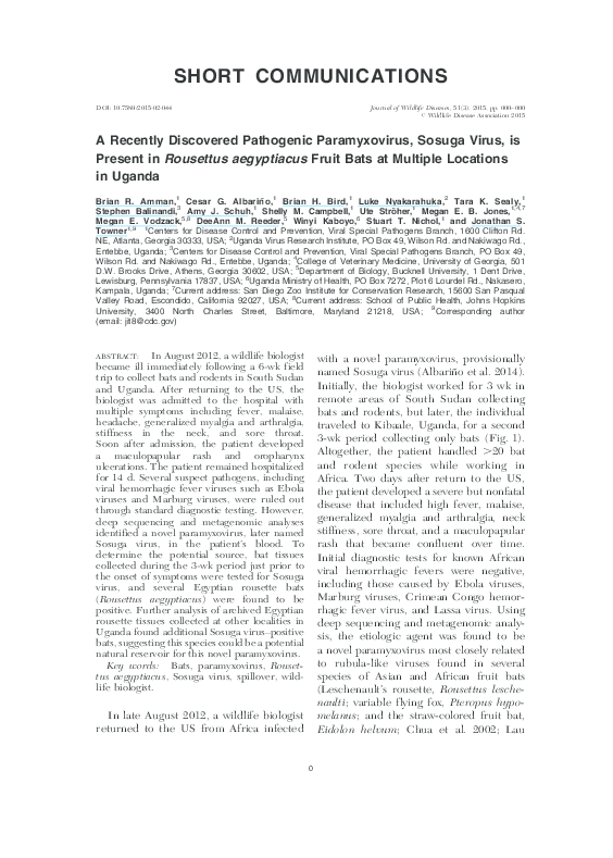
3-wk period collecting only bats (Fig. 1).
Altogether, the patient handled .20 bat
and rodent species while working in
Africa. Two days after return to the US,
the patient developed a severe but nonfatal
disease that included high fever, malaise,
generalized myalgia and arthralgia, neck
stiffness, sore throat, and a maculopapular
rash that became confluent over time.
Initial diagnostic tests for known African
viral hemorrhagic fevers were negative,
including those caused by Ebola viruses,
Marburg viruses, Crimean Congo hemorrhagic fever virus, and Lassa virus. Using
deep sequencing and metagenomic analysis, the etiologic agent was found to be
a novel paramyxovirus most closely related
to rubula-like viruses found in several
species of Asian and African fruit bats
(Leschenault’s rousette, Rousettus leschenaulti; variable flying fox, Pteropus hypomelanus; and the straw-colored fruit bat,
Eidolon helvum; Chua et al. 2002; Lau
In late August 2012, a wildlife biologist
returned to the US from Africa infected
0
�0
JOURNAL OF WILDLIFE DISEASES, VOL. 51, NO. 3, JULY 2015
FIGURE 1.
Map of Uganda showing capture locations of bats tested for Sosuga virus.
et al. 2010; Drexler et al. 2012; Baker et al.
2013; Albariño et al. 2014).
It is unclear how the biologist became
infected with Sosuga virus. Interviews with
the patient revealed that appropriate levels
of personal protective equipment (PPE)
were used during animal capture and
processing in Kibaale, Uganda, including
the use of disposable Tyvek suits coupled
with powered air-purifying respirators
(PAPRs; 3M, St. Paul, Minnesota, USA).
However, inconsistent adherence to PPE
practices did occur during the earlier
South Sudan work. Incubation periods
with other human paramyxovirus infections
vary greatly and are generally in the range
of 1–3 wk (Sartwell 1950; Goh et al. 2000;
Playford et al. 2010). This fact made the
field work in Kibaale, the 3-wk period just
prior to symptom onset, the most plausible
time for exposure to the virus. Taking these
variables into account, combined with the
close genetic relationship between Sosuga
virus and the other fruit bat–borne rubulalike viruses, efforts to identify the virus
source were focused on bats caught and
necropsied at the Kibaale field site in
Uganda (Table 1). There, bats were captured using a harp trap (Bat Conservation
and Management, Inc., Carlisle, Pennsyl-
�SHORT COMMUNICATIONS
TABLE 1. Bats captured in southwestern and central
Uganda and tested for Sosuga virus by quantitative
reverse transcriptase PCR (qRT-PCR) on pooled
liver/spleen samples. Shown are total number and
percent of positive bats by species. Results for
Egyptian rousettes (Rousettus aegyptiacus) are
further subdivided by sex and age. QENP
represents bats collected at Python Cave, Queen
Elizabeth National Park. Egyptian rousettes caught
in Kibaale were captured at Butogota Cave. All
animal work was performed in accordance with
a Centers for Disease Control and Prevention
Animal Care and Use Committee approved protocol.
Locality and species
Kibaale Aug 2012
Epomophorus labiatus
Lissonycteris angolensis
Hipposideros spp.
Rousettus aegyptiacus
Female
Male
Adult
Juvenile
QENP Aug 2009
Rousettus aegyptiacus
Female
Male
Adult
Juvenile
QENP Nov 2009
Rousettus aegyptiacus
Female
Male
Adult
Juvenile
Kitaka Nov 2012
Rousettus aegyptiacus
Female
Male
Adult
Juvenile
n
No. qRT-PCR
positive (%)
262
18
1
122
68
54
77
45
0 (0)
0 (0)
0 (0)
3 (2.5)
3 (4.4)
0 (0.0)
2 (2.6)
1 (2.2)
401
196
205
237
161
3
2
1
0
3
(0.7)
(1.0)
(0.5)
(0.0)
(1.9)
408
187
221
165
243
15 (3.6)
4 (2.1)
11 (5.0)
9 (5.5)
6 (2.5)
400
203
197
233
167
41
19
22
22
19
(10.2)
(9.4)
(11.2)
(9.4)
(11.4)
vania, USA) placed at the entrance of
a cave roost (Butogota Cave; 0u47951.300N,
31u2927.420E). The specific use of PPE is
detailed by Towner et al. (2011). Briefly,
PPE, consisting of a caving helmet, full
face respirator with P100 filters, Tyvek
coveralls, rubber gum boots, and biteresistant leather gloves over double-layered
latex gloves, was worn at all times during
bat captures. Necropsies were performed
at a central processing station away from
public access. Liver, spleen, heart, lung,
and kidney tissue aliquots were taken and
0
placed directly in chaotropic lysis buffer
known to have virucidal properties, and
samples were also frozen in liquid nitrogen. During necropsies, PPE included
double latex gloves, disposable gowns,
and PAPRs.
Testing for Sosuga virus in bat specimens was carried out using a highly
sensitive quantitative reverse transcriptase
PCR (qRT-PCR) assay targeting the NP
gene, which had been initially developed
for detection and quantitation of Sosuga
virus in patient blood (Albariño et al.
2014). Briefly, total nucleic acid from
pooled bat liver/spleen tissue was extracted as described by Amman et al.
(2012). All tissues were flash-frozen in
liquid nitrogen in the field during necropsy and stored continuously frozen until
processing. Of all the Egyptian rousettes,
also known as Egyptian fruit bats (Rousettus aegyptiacus), caught at Butogota
Cave (Fig. 1), 2.5% (3/122) were PCR
positive for Sosuga virus, whereas the 262
Ethiopian epauletted fruit bats (Epomophorus labiatus), 18 Angolan rousettes
(Lissonycteris angolensis), and one roundleaf bat (Hipposideros spp.) caught in
the same general vicinity were negative
(Table 1). To determine if testing of other
bat tissues was more sensitive, liver,
spleen, heart, kidney, lung, and blood
from each of the three virus-positive bats
(bats 841, 867, and 926) were tested
separately by qRT-PCR, and only spleen
was positive for Sosuga virus RNA.
To determine if Sosuga virus infection
of Egyptian rousettes was common across
Uganda, pooled liver/spleen tissue samples from approximately 1,200 archived
Egyptian rousette bats from other locations were tested by qRT-PCR. Egyptian
rousettes are a reservoir for Marburg
viruses, and extensive bat samples were
still available from previous studies at
Python Cave in Queen Elizabeth National
Park (0u16937.920S, 30u397.200E in August
2009 and November 2009; Amman et al.
2012), and more recently from Kitaka
Mine (0u7950.340S, 30u18932.180E) in
�0
JOURNAL OF WILDLIFE DISEASES, VOL. 51, NO. 3, JULY 2015
�SHORT COMMUNICATIONS
October 2012 (Amman et al. 2014).
Evidence of Sosuga virus was found in
bats from all three Egyptian rousette
collections tested, dating back to August
2009. The highest number of positive bats,
41 total (10% overall), was found in the
Kitaka Mine in October 2012 (Table 1).
Both Kitaka Mine and Python Cave are
approximately 130 km from Butogota
Cave in Kibaale and well within reported
Egyptian rousette dispersal ranges of up to
500 km (Jacobsen and Du Plessis 1976;
Amman et al. 2012), thus making intermixing between the populations likely.
All samples with Sosuga virus qRT-PCR
cycle threshold (Ct) values ,35 were
additionally subjected to reverse transcriptase PCR (RT-PCR) using primers specific for a 331-nucleotide region in the HN
gene as well as heminested RT-PCR using
primers specific for a 127-nucleotide region in the NP gene. Because of the low
levels of RNA found in tissues, the
sequence was determined from only 11
bats: one bat caught at the Kibaale field
site and 10 from the Kitaka Mine in 2012.
These sequences were subjected to
a BLAST (NCBI 2014) search to confirm
identity and analyzed with other known
rubula-like paramyxoviruses, including
true rubula viruses (mumps), to generate
0
phylogenies showing the inclusion of
Sosuga virus within the rubula-like virus
clade in the family Paramyxoviridae
(Fig. 2). A more detailed phylogenetic
placement of Sosuga virus within the
Paramyxoviridae was described by Albariño et al. (2012). The bat from the Kibaale
field site (bat 926) was positive in the NP
assay only, and the sequence was identical
to the patient isolate. For the 10 bats from
the Kitaka Mine that were positive in the
NP assay (Fig. 2A), seven had sequences
identical to the patient isolate, while three
bats differed by one nucleotide. Four
Kitaka bats were additionally positive in
the HN assay and differed by 6/331 (2%)
nucleotides or less from each other and
the sequence of the virus found in the
infected biologist (Fig. 2B). Parallel attempts at virus isolation in Vero E6 cells
and suckling mice were performed on
those specimens with Ct values less than
35 using methods described in Albariño et
al. (2012). Unfortunately, isolation attempts were negative (data not shown),
presumably due to the low viral loads in
the tissues.
The sequence data presented herein are
limited in information regarding the exact
placement of Sosuga virus within the
Paramyxoviridae. They exhibit very little
r
FIGURE 2. Phylogenetic analysis of Sosuga virus sequences determined from reverse transcriptase PCR
(RT-PCR) amplification of NP and HN genes from bats. Nucleotide sequences corresponding to (A) 127nucleotide and (B) 331-nucleotide fragments of the NP and HN genes, respectively, and those from eight
representative rubula-like viruses, including comparable sequence fragments from the patient (Sosuga), were
aligned using the MUSCLE algorithm (CLC Genomics Workbench version 6.0.1; CLC Bio, Cambridge,
Massachusetts, USA). Mumps virus sequence was used as an out-group. Phylogenetic analysis was conducted
with a Bayesian algorithm (Mr. Bayes, Geneious version 6.1.5, www.geneious.com/). Bat sample localities are
represented by color: Blue, Kiballe (Butagota Cave); Red, Python Cave; Green, Kitaka Mine. HN sequences
were extracted from the complete genomic sequences in GenBank: KF774436 (Sosuga virus [SosV]),
GU128082 (Tuhoko virus 3), U128081 (Tuhoko virus 2), GU128080 (Tuhoko virus 1), AF298895 (Tioman
virus), NC_007620 (Menangle virus), JX051319 (Achimota virus 1), JX051320 (Achimota virus 2), NC_002200
(mumps virus). Posterior probability values are shown at each node. Scale is in substitutions/site. A more
detailed phylogenetic placement of Sosuga virus within the virus family Paramyxoviridae was described in
Albariño et al. (2012). GenBank accession numbers are as follows: Sosuga virus KF774436, Mumps
NC_002200, Achimota virus 1 JX051319, Achimota virus 2 JX051320, Menangle virus NC_007620, Tioman
virus AF298895, Tuhoko virus 1 GU128080, Tuhoko virus 2 GU128081, Tuhoko virus 3 GU128082, Sosuga
virus from bats HN gene partial sequence, Bat-1605 KP150637, Bat-1319 KP150638, Bat-1271 KP150639,
Bat-1624 KP150640, Sosuga virus from bats NP gene partial sequence, Bat-926 KP150641, Bat-1302
KP150642, Bat-1319 KP150643, Bat-1516 KP150644, Bat-1541 KP150645, Bat-1571 KP150646, Bat1605 KP150647.
�0
JOURNAL OF WILDLIFE DISEASES, VOL. 51, NO. 3, JULY 2015
variation (#2% for the HN and ,1% for
the NP sequences) but clearly identify
Sosuga as a rubula-like virus. For comparison, Hendra virus exhibited #1% variation during multiple separate introductions
over a 2-yr period (Marsh et al. 2010).
Moreover, we show that Egyptian rousette
populations in multiple locations across
Uganda were actively infected with Sosuga
virus over a 3-yr period. This finding is
consistent with Tuhoko3 virus, the nearest
known relative of Sosuga virus, being found
in Leschenault’s rousette in Asia (Lau et al.
2010).
Given the wildlife biologist’s exposure
to bats in Uganda during the 3 wk prior to
onset of illness, these Egyptian rousettes
were the probable source of the infection.
The wide distribution and detection of the
virus at multiple time points suggest the
Egyptian rousette could be a reservoir
species, although that was not formally
demonstrated here. If so, the extensive
range of these bats across Sub-Saharan
Africa would predict a wide distribution of
the Sosuga virus. It is difficult to predict if
a paramyxovirus closely related to Sosuga
virus, such as Tuhoko virus, is capable of
productively infecting humans. However,
Drexler et al. (2012) report that bats
appear to be the ancestral source of
paramyxoviruses and that viruses in this
family are known for their promiscuity,
having spilled over into multiple orders of
mammalian fauna.
We thank the Uganda Virus Research
Institute, the Uganda Ministry of Health,
and the Uganda Wildlife Authority for
their assistance during past collection
efforts. We also thank E. Ervine for
assistance with creating the map of
Uganda. Funding for this study was provided by the US Department of Health
and Human Services. The findings and
conclusions in this report are those of the
authors and do not necessarily represent
the views of the Centers for Disease
Control and Prevention or Health and
Human Services.
LITERATURE CITED
Albariño CG, Foltzer M, Towner JS, Rowe LA,
Campbell S, Jaramillo CM, Bird BH, Reeder
DM, Vodzak ME, Rota P. 2014. Novel paramyxovirus associated with severe acute febrile
disease, South Sudan and Uganda, 2012. Emerg
Infect Dis 20:211–216.
Amman BR, Carroll SA, Reed ZD, Sealy TK,
Balinandi S, Swanepoel R, Kemp A, Erickson
BR, Comer JA, Campbell S, et al. 2012. Seasonal
pulses of Marburg virus circulation in juvenile
Rousettus aegyptiacus bats coincide with periods
of increased risk of human infection. PLoS
Pathog 8:e1002877.
Amman BR, Nyakarahuka L, McElroy AK, Dodd
KA, Sealy TK, Schuh AJ, Shoemaker TR,
Balinandi S, Atimnedi P, Kaboyo W, et al.
2014. Marburgvirus resurgence in Kitaka mine
bat population after extermination attempts,
Uganda. Emerg Infect Dis 20:1761–1762.
Baker KS, Todd S, Marsh GA, Crameri G, Barr J,
Kamins AO, Peel AJ, Yu M, Hayman DT, Nadjm
B. 2013. Novel, potentially zoonotic paramyxoviruses from the African straw-colored fruit bat
Eidolon helvum. J Virol 87:1348–1358.
Chua KB, Wang LF, Lam SK, Eaton BT. 2002. Full
length genome sequence of Tioman virus, a novel
paramyxovirus in the genus Rubulavirus isolated
from fruit bats in Malaysia. Arch Virol 147:1323–
1348.
Drexler JF, Corman VM, Muller MA, Maganga GD,
Vallo P, Binger T, Gloza-Rausch F, Rasche A,
Yordanov S, Seebens A, et al. 2012. Bats host major
mammalian paramyxoviruses. Nat Commun 3:1–12.
Goh KJ, Tan CT, Chew NK, Tan PSK, Kamarulzaman A, Sarji SA, Wong KT, Abdullah BJJ, Chua
KB, Lam SK. 2000. Clinical features of Nipah
virus encephalitis among pig farmers in Malaysia. N Engl J Med 342:1229–1235.
Jacobsen NHG, Du Plessis E. 1976. Observations on
the ecology and biology of the Cape fruit bat
Rousettus aegyptiacus leachi in the eastern
Transvaal. S Afr J Sci 72:270–273.
Lau S, Woo P, Wong B, Wong A, Tsoi H, Wang M,
Lee P, Xu H, Poon R, Guo R. 2010. Identification and complete genome analysis of three
novel paramyxoviruses, Tuhoko virus 1, 2 and 3,
in fruit bats from China. Virology 404:106–116.
Marsh GA, Todd S, Foord A, Hansson E, Davies K,
Wright L, Morrissy C, Halpin K, Middleton D,
Field HE, et al. 2010. Genome sequence
conservation of Hendra virus isolates during
spillover to horses, Australia. Emerg Infect Dis
16:1767–1769.
National Center for Biotechnology Information
(NCBI). 2014. BLAST. http://blast.ncbi.nlm.
nih.gov/Blast.cgi?PROGRAM5blastn&PAGE_
�SHORT COMMUNICATIONS
TYPE5BlastSearch&LINK_LOC5blasthome.
Accessed December 2014.
Playford EG, McCall B, Smith G, Slinko V, Allen G,
Smith I, Moore F, Taylor C, Kung YH, Field H.
2010. Human Hendra virus encephalitis associated with equine outbreak, Australia, 2008.
Emerg Infect Dis 16:219–223.
Sartwell PE. 1950. The distribution of incubation
periods of infectious disease. Am J Epidemiol
51:310–318.
View publication stats
0
Towner JS, Amman BR, Nichol ST. 2011. Significant
zoonotic diseases identified in bats: Filoviruses.
In: Investigating the role of bats in emerging
zoonoses, Newman SH, Field H, Epstein J, de
Jong C, editors. Food and Agriculture Organisation of the United Nations, Rome, Italy,
pp. 123–135.
Submitted for publication 13 February 2015.
Accepted 12 March 2015.
�
 Luke Nyakarahuka