Atlas of Clinical and
Surgical Orbital Anatomy
Commissioning Editor: Russell Gabbedy
Development Editor: Nani Clansey
Editorial Assistant: Kirsten Lowson
Project Manager: Glenys Norquay/Nancy Arnott
Designer: Charles Gray
Illustrator: Thomas G. Waldrup, MSMI
Marketing Manager(s) (UK/USA): Gaynor Jones/Helena Mutak
Atlas of Clinical and
Surgical Orbital Anatomy
Second Edition
Jonathan J. Dutton MD, PhD, FACS
Professor and Vice Chair of Ophthalmology
The University of North Carolina
Chapel Hill,
North Carolina
USA
Illustrations by:
Thomas G. Waldrop, MSMI
© 2011, Elsevier Inc. All rights reserved.
First edition 1994
Second edition 2011
No part of this publication may be reproduced or transmitted in any form or by any means, electronic or
mechanical, including photocopying, recording, or any information storage and retrieval system, without
permission in writing from the publisher. Details on how to seek permission, further information about
the Publisher’s permissions policies and our arrangements with organizations such as the Copyright
Clearance Center and the Copyright Licensing Agency, can be found at our website: www.elsevier.com/
permissions.
This book and the individual contributions contained in it are protected under copyright by the Publisher
(other than as may be noted herein).
Notices
Knowledge and best practice in this field are constantly changing. As new research and experience
broaden our understanding, changes in research methods, professional practices, or medical
treatment may become necessary.
Practitioners and researchers must always rely on their own experience and knowledge in
evaluating and using any information, methods, compounds, or experiments described herein.
In using such information or methods they should be mindful of their own safety and the safety
of others, including parties for whom they have a professional responsibility.
With respect to any drug or pharmaceutical products identified, readers are advised to check the
most current information provided (i) on procedures featured or (ii) by the manufacturer of each
product to be administered, to verify the recommended dose or formula, the method and duration
of administration, and contraindications. It is the responsibility of practitioners, relying on their own
experience and knowledge of their patients, to make diagnoses, to determine dosages and the best
treatment for each individual patient, and to take all appropriate safety precautions.
To the fullest extent of the law, neither the Publisher nor the authors, contributors, or editors,
assume any liability for any injury and/or damage to persons or property as a matter of products
liability, negligence or otherwise, or from any use or operation of any methods, products, instructions,
or ideas contained in the material herein.
Saunders
British Library Cataloguing in Publication Data
Dutton, Jonathan J.
Atlas of clinical and surgical orbital anatomy. – 2nd ed.
1. Eye-sockets–Anatomy–Atlases. 2. Eye-sockets–
Surgery–Atlases.
I. Title
611.8’4-dc22
ISBN-13: 978-1-4377-2272-7
Library of Congress Cataloging in Publication Data
A catalog record for this book is available from the Library of Congress.
Printed in China
Last digit is the print number:
9
8
7
6
5
4
3 2
1
“The learning and knowledge that we have is, at the most, but little compared
with that of which we are ignorant.”
Plato, 428-348 BC
“The known is finite, the unknown infinite, intellectually we stand on an islet in the midst of an
illimitable ocean of inexplicability. Our business in every generation is to reclaim
a little more land.”
T.H. Huxley, 1887
With the second edition of this book, we continue to explore further into the realm of orbital anatomy.
We hope thereby that we are able to contribute, however slightly,
to Huxley’s precious intellectual land.
This page intentionally left blank
About the Authors
JONATHAN J. DUTTON, M.D., Ph.D. is currently Professor
and Vice Chair of Ophthalmology at The University of North
Carolina at Chapel Hill. He completed his masters and
doctorate degrees in zoology, evolutionary biology, and vertebrate paleontology at Harvard University in 1970, and joined
the faculty of Princeton University as Sinclair Professor of
Vertebrate Paleontology from 1970 to 1973. Between 1965
and 1973 he conducted ten research expeditions to East Africa
and published widely on vertebrate morphology and mammalian evolution. After returning to school and receiving
his M.D. degree in 1978, and going on to residency training
at Washington University Medical School, he completed a
research fellowship in glaucoma at Washington University,
and another fellowship in oculoplastic and orbital surgery at
the University of Iowa. From 1983 to 1999 he was Professor
of Ophthalmology and head of the Oculoplastic and Orbital
Service at Duke University Medical Center. He served as CEO
and Medical Director of the Atlantic Eye and Face Center
in Cary, NC from 2000-2003 and then joined the full-time
faculty at the University of North Carolina at Chapel Hill,
where he is currently Professor and Vice Chair. Dr Dutton
is senior preceptor of an ASOPRS-approved fellowship
program that has trained 15 fellows. He specializes in oculoplastic reconstructive and orbital surgery, thyroid eye disease,
and periorbital and intraocular ophthalmic oncology.
THOMAS G. WALDROP, M.S.M.I. received his Master of
Science degree in medical illustration from the Medical College
of Georgia in 1978. He directed the ophthalmic photography
and ultrasound section of the Retina Institute in St Louis before
establishing his medical illustration service in Hillsborough,
North Carolina in 1980. Since then, he has worked closely with
the Duke University Eye Center producing ophthalmic illustrations for publication, and he has collaborated with Dr. Dutton
on several major atlases of ophthalmic surgery.
vii
This page intentionally left blank
Preface to the First Edition
Few areas in ophthalmology have proven to be as elusive
or difficult to teach as orbital anatomy. The grasp of clinical
diagnostic techniques, and the development of sophisticated
surgical skills seem far removed from the mundane and
often boring tasks of plowing through pages of descriptive
anatomic detail. Idealized artistic drawings have often failed
to accurately portray true anatomic relationships with other
structures. Photographs of clinical dissections are usually so
cluttered with extraneous structures as to make interpretation of individual anatomic systems impossible. The result
has been a poor understanding of orbital anatomy, not only
among ophthalmologists, but also among neurosurgeons
and otolaryngologists who frequently pursue lesions into
the orbit.
During the past decade there has been a renewed interest
in clinical eyelid and orbital anatomy. Detailed dissections
and reinterpretations have markedly altered our concepts of
functional morphology of such structures as Whitnall’s ligament, the medial canthal tendon, orbital fascial septa, the
lower eyelid retractors, and the levator aponeurosis. This
has resulted in the development of new surgical procedures
based on such concepts, and the resurrection and successful modification of older, long abandoned operations. With
the growing appreciation of anatomical and functional relationships, older, non-physiologic procedures are slowly
giving way to those directed at the site of pathology, and
aimed at the restoration of normal anatomic structure and
physiology. Without an intimate knowledge of the anatomy
of these regions, the modern surgeon dealing with orbital
and eyelid disorders can no longer function adequately. Nor
can progress occur in the evolution of newer and even more
physiologically appropriate therapeutic techniques.
Of all the subjects in medicine, the study of anatomy is
perhaps the most visual. Few of us can easily commit to
memory the numerous and frequently antiquated names
given to anatomic structures. Even more confusing are the
spatial relationships of different anatomic systems and their
common variants. Often we rely on simple images, mental
drawings that depict key landmarks in familiar juxtapositions
that can be recalled during clinical evaluations or surgical
operations. Most of us have divined various tricks to visually
reconstruct complex anatomic detail from two-dimensional
artistic renderings, or from confusing cadaver dissections.
It is this very process of conjuring up prepackaged eidetic
images that led to the concept of the present book.
The illustrations presented in the following pages combine
the best features of several different techniques. Anatomic
details and relationships are based on several human orbits
cut into 300 histologic sections at 150 microns thickness. For
each anatomic system (e.g. bones, arteries, nerves, etc.) each
section was projected to 3X magnification and traced onto
a transparent mylar sheet. Accurate registration was assured
through the use of precut feduciary markings within the
blocks, and adjustments for differential shrinkage and warpage were made visually. The mylar sheets were then stacked
in layered fashion and the resulting three-dimensional reconstructed images were used to prepare the final illustrations.
Translation into various orientations was performed visually
from these base views, and from measurements calculated
from the original histologic series. These techniques allowed
us to image each anatomic system in isolation, or in combination with other structures by overlay of the appropriate
Mylar transparencies. We have attempted to choose some
views and angles not typical in some other atlases of orbital
anatomy, but which we feel will enhance the visual concepts.
Where possible, instead of cutting and reflecting structures
to show deeper layers, we have kept structures intact, making them transparent to more accurately demonstrate relationships of features behind them. The result is a series of
illustrations that create in the reader’s mind a series of visual
patterns that can more easily be recalled.
Each chapter focuses on a different anatomic system,
such as extraocular muscles, arteries, or orbital nerves. In a
series of reconstructions we sequentially add and silhouette
adjacent structures to illustrate them in their proper threedimensional perspective. Each chapter begins with a coronal
view of the orbit as seen when facing the human head. The
anatomic system of interest is pictured first in isolation to
show its essential features. Additional systems are then
added, beginning with the extraocular muscles, to demonstrate anatomic relationships. Finally the orbital bones are
added. This series of images are then repeated in the lateral
and superior aspects. Such transformations help translate
morphological relationships into more familar surgical
views. Other images at unique orientations and magnifications are used where necessary to illustrate specific anatomic
detail.
This book is intended as a visual atlas. The text presents
introductory material, embryology, discussions of variability, explanations of concepts, and descriptions of structures and functions that are difficult to display in pictures
alone. The text also describes anatomic details in a logical
sequence that follows regional, functional, or morphologic
criteria that will help the reader create meaningful mental
images. Since our goal is clinical anatomy, wherever possible, clinically relevant correlations are included to relate
normal anatomic structure to pathologic states or to surgical
procedures.
For each chapter we include a collection of full-color
illustrations with appropriate labels. Because of the exquisite
ix
Preface to the First Edition
detail in the original histologic sections, we include as a
separate chapter a series of photomicrographs illustrating
the histologic cross-sectional anatomy of the orbit.
Following a series of coronal sections through the orbit, we
illustrate of each anatomic system or structure at appropriate
magnification. In the final chapter we include a series of
computerized tomographic scans and magnetic resonance
images. These are figured in both the coronal and axial
orientations, along with corresponding reconstructions for
anatomic correlation.
For those students of orbital anatomy interested in details
of structure, functional morphology, and clinical correlations,
we suggest a careful reading of the text in conjunction with
a systematic sequential review of the illustrations. For those
more familiar with orbital anatomy who may wish only to
x
review certain anatomic systems or structures for teaching
or in preparation for surgery, the illustrations may be used
independent of the text. While we do not intend reference
citations to be encyclopedic, we do include sources for new
findings or controversial interpretations.
It is sincerely hoped that this volume will enhance the
teaching of orbital anatomy for the clinician, and serve as a
stimulus for further investigation of anatomic and functional
relationships which are so essential for progress. This
volume should prove valuable for the resident and practicing
physician in ophthalmology, otolaryngology, plastic surgery,
neurosurgery, dermatology, neuroradiology and all others
who diagnose and treat diseases of the eyelids and orbit.
Jonathan J. Dutton and Thomas G. Waldrop
Preface to the Second Edition
In 1994, we published the first edition of this book.
Gratifyingly, this book was well received, and won awards
for the best medical illustrations for 1994, as well as
recognition as one of the 100 most important books
published in ophthalmology in the 20th century (Thompson
HS, Blanchard DL. Arch Ophthalmol 2001; 119:761-763).
Our goal at that time was to produce a visual atlas of orbital
and eyelid anatomy, describing anatomic details in a logical
sequence following regional, functional, or morphologic criteria. These mental or eidetic images would help the reader
create meaningful mental pictures that can be recalled from
memory, like reading the pages of an open book. Since our
goal was clinical anatomy, we included some clinically relevant correlations related to normal anatomic structures, and
to some pathologic conditions.
Anatomy of relatively well-known regions of the body
tends to be rather stable, with few significant changes in
knowledge, at least with respect to major structures. However,
during the 16 years since publication of the first edition, a
great deal of new information has been added to the medical
literature, especially as regards eyelid anatomy, the orbital
fascial connective tissue structures, and extraocular muscle pulley systems. Some refinements also have been made
to our understanding of other anatomic systems, including the vascular, neural, and muscular systems. All of these
findings have been updated in the current edition. We have
added a section on facial anatomy to the Eyelid Anatomy
chapter that is relevant to facial and SOOF lift procedures.
Also, we added a new chapter on the cavernous sinus, since
many orbital structures and pathologic conditions involving
the orbital apex also involve the cavernous sinus and middle cranial fossa, so that knowledge of anatomic continuity
between these structures is important. References have been
updated throughout, and a number of new or modified illustrations have been added to several chapters based on recent
anatomic findings. We also added new subheadings to most
chapters, in order to more clearly delineate specific areas of
information. We expanded sections on clinical correlations
in all chapters, to better relate disease processes with anatomic structures.
As we stated in the first edition, for those students of
orbital anatomy interested in details of structure, functional
morphology, and clinical correlations, we suggest a careful
reading of the text in conjunction with a systematic sequential review of the illustrations. For those more familiar
with orbital anatomy who may wish only to review certain
anatomic systems or structures, the illustrations can be used
independent of the text.
Jonathan J. Dutton and Thomas G. Waldrop
xi
This page intentionally left blank
Contents
1.
Cavernous Sinus . . . . . . . . . . . . . . . . . . 1
2.
Osteology of the Orbit . . . . . . . . . . . . 15
3.
Extraocular Muscles . . . . . . . . . . . . . . 29
4.
Orbital Nerves . . . . . . . . . . . . . . . . . . . 51
5.
Arterial Supply to the Orbit . . . . . . . 83
6.
Venous and Lymphatic Systems . . . . 99
7.
Orbital Fat and Connective
Tissue Systems . . . . . . . . . . . . . . . . . . 111
8.
The Eyelids and Anterior Orbit . . . 129
9.
The Lacrimal Systems . . . . . . . . . . . . 165
10.
Histologic Anatomy of the Orbit . . . 175
11.
Radiographic Correlations . . . . . . . 227
Index . . . . . . . . . . . . . . . . . . . . . 257
xiii
This page intentionally left blank
CHAPTER
1
Cavernous Sinus
The cavernous sinus (CS) is a very important intracranial,
extradural anatomic region that contains many structures
vital for visual function. Numerous disease processes along
the skull base and in the cavernous sinus can have a major
impact on vision or on ocular motility. Yet, this anatomic
structure remains quite unfamiliar to most ophthalmologists and orbital surgeons. It serves as a critical venous drainage route for both the orbit and the cranial base.16 It also
transmits arterial and neural structures from the intracranial
compartment into the orbital apex.
The term cavernous sinus has been in use for 275 years,
ever since Jacobus Winslow proposed it in 1734, reflecting
his concept of a single trabeculated venous cavern similar to
the corpus cavernosus of the penis.42 His concept was incorrect, yet the term has persisted in the medical literature. It
is clear from modern studies that the CS is neither cavernous nor is it an intradual sinus, but rather it is a plexus or
network of extremely thin-walled veins associated with adipose tissue. Parkinson27 emphasized the inappropriateness
of this term on anatomical grounds. Hashimoto12 recommended following Parkinson’s lead in using the term “lateral sellar compartment” (LSC)26 for this structure in its
broader sense, and restricting the term “cavernous sinus” to
the more limited venous pathways within the LSC. In 2003,
Tobenas-Dujardin et al.38 proposed the term “inter-periostodural space” which they believed would better reflect the real
anatomic pattern. However, this has not gained widespread
usage. While the term lateral sellar compartment might
be anatomically more accurate, the term cavernous sinus
remains in widespread use, especially outside the specialty
of neurosurgery. Furthermore, the International Federation
of Associations of Anatomists (IFAA) did not adopt an alternative terminology for the cavernous sinus in its most recent
edition of Terminologia Anatomica 1998.37 Therefore, for
the present chapter we will use the classic terminology, using
the term cavernous sinus for both the neural and venous
components.
Embryology
The early development of the cavernous sinus is complex.
Our current understanding is based on the seminal studies
of Padget23 as well as more recent works.9,18 By the 3 mm
(28-day) embryonic stage two longitudinal venous channels,
the anterior cardinal veins, are laid down and extend along
the ventrolateral surface of the developing brain, on the
medial side of the cranial nerve roots. Three pairs of venous
channels develop from these to form the superior cerebral,
middle cerebral, and inferior cerebral veins. Most of each cardinal vein atrophies, except for a segment of each vein in the
region of the trigeminal ganglion which becomes the forerunner of the cavernous sinus, and another segment more
posteriorly which becomes the internal jugular vein.
By the 8 mm (36-day) embryonic stage the primitive
supraorbital vein arises in the superficial tissues dorsal to
the developing eye. It initially drains backward between
the trigeminal and trochlear nerves into an anterior dural
plexus, which will become the superior sagittal and transverse sinuses. A new anastomosis appears from the supraorbital vein that diverts blood over the incipient annulus of
Zinn into the venous plexus of the future cavernous sinus.
By the 11 mm (40-day) stage the initial formation of the
chondrocranium is seen around the anterior notochord, surrounded by primitive mesenchyme. At the 14.5 mm (44-day)
stage chondrification begins in the future greater and lesser
wings of the sphenoid bone and in the dorsum sellae.38 At the
same time the trigeminal (gasserian) ganglion forms, along
with its three major peripheral divisions. In the 23–25 mm
(50-day) embryo the hypophysis and diaphragma sellae
become differentiated in the region of the developing cavernous sinus. The lateral wall of the cavernous sinus is partially
developed as a meningeal layer enclosing several cranial
nerves, but the medial wall is not yet formed. By the 31 mm
(56-day) embryo a well developed cavernous sinus with a
definitive cavernous carotid artery and sympathetic plexus is
present, containing two venous compartments, one on each
side of the midline. Cranial nerves III, IV, VI, and the three
branches of the trigeminal nerve are all differentiated and
located in their approximate adult relationships.
In the 70–90 mm (13–15-week) fetal stage small ossification centers are seen in the body, greater wings, and lesser
wings of the sphenoid bone. At the same time ossification
is beginning in the cartilaginous petrous portion of the temporal bone.12 The primordium of the dura mater and subarachnoid membrane are already seen lining the area of the
cavernous sinus on either side of the body of the sphenoid.
The pituitary gland is lined by an inner capsule and an outer
meningeal layer, forming the definitive medial wall of the cavernous sinus. Many small irregularly shaped lumens develop
within the mesenchyme of the cavernous sinus region, and
these venous channels gradually enlarge with further fetal
development. These channels meander and intertwine, and
are lined only by an endothelial layer with no smooth muscle. These venous channels communicate with other venous
channels. Posteriorly they drain to the basilar venous sinus
1
1
Cavernous Sinus
and then to the jugular bulb; posteroinferiorly with the inferior petrosal sinus and then into the pterygoid venous plexus
through the foramen lacerum; and posterosuperiorly with
the superior petrosal sinus and then into the sigmoid sinus.
The cavernous sinuses on each side communicate with each
other through one or more intercavernous sinuses situated
between the dural layers, below the pituitary gland.
The gasserian ganglion is situated posterior to the developing cavernous sinus on either side, over the tip of the petrous
bone and lateral to the dorsum sellae. The three branches
of the trigeminal nerve run forward from the gasserian ganglion. The ophthalmic branch (V1) and the maxillary branch
(V2) run anteriorly in the lateral wall of the cavernous sinus,
within the loose inner connective tissue endosteal layer.
The oculomotor (III) and trochlear (IV) nerves enter the
cavernous sinus near the posterior clinoid process and also
run anteriorly within the lateral wall to the superior orbital
fissure. The abducens nerve (VI) runs through the basilar
venous plexus and then enters the cavernous sinus; it courses
forward within the venous channels of the sinus just lateral
to the internal carotid artery, and passes into the superior
orbital fissure. Third order sympathetic nerve fibers enter the
cranium through the foramen lacerum and become associated with these cranial nerves and vascular elements. The
internal carotid artery (ICA) enters the skull base through the
future carotid canal. It then penetrates the floor of the cavernous sinus inferolateral to the cartilaginous sphenoid bone.
As the sella turcica develops, the ICA gradually assumes the
S-shaped configuration seen in the adult.
During the 128–183 mm (18–23-week) stage of fetal
development further ossification occurs in the sphenoid bone
as it expands in the anterolateral directions. By the 230 mm
(28-week) fetal stage a thick periosteum is seen over the
surface of sphenoid bone. Dura is distinguishable along the
lateral wall of the cavernous sinus as a definite meningeal
layer separate from the overlying arachnoid membrane and
the inner endosteal layer that is continuous with the periosteum of the sphenoid bone. Superiorly the meningeal layer
folds to contribute to the diaphragma sellae over the pituitary gland. Within the mesenchyme of the cavernous sinus
large well-defined venous lumens are now present. The mesenchymal tissue between lumens gradually thins to become
membranes separating the individual vascular channels. Small arteries and autonomic nerve fascicles are now
apparent within these membranous walls.
In the 150–200 mm (21–25-week) fetal stage, blood flow
through the cavernous sinus rapidly increases, probably due
to alterations in neighboring venous pathways. Nerve fascicles become surrounded by collagen fibers forming sheaths.
Simultaneous with formation of the cavernous sinus is
development of the pituitary gland, which forms an important
element adjacent to and above the bilateral cavernous sinuses.
During the 2–3 mm (21-day) embryonic stage the gland
originates from two distinct ectodermal tissues. A finger-like
protrusion, called Rathke’s pouch, grows upward as a dorsal
evagination from the stomodeum, or mouth, just anterior
to the bucco-pharyngeal membrane. It differentiates into
glandular epithelium characteristic of endocrine glands. The
infundibulum is a ventral evagination from the floor of the
third ventricle of the diencephalon just caudal to the developing optic chiasm from the same tissue.1 It differentiates into
the exocrine component of the pituitary gland. During the
2
second month of embryonic development, Rathke’s pouch
wraps around the infundibulum, and differentiates into
the anterior lobe, or adenohypophysis, of the pituitary gland.
The infundibulum differentiates into the pituitary stalk
and the posterior lobe, or neurohypophysis, of the gland.
Ultimately, the two portions grow together to form the definitive pituitary gland. As the cavernous sinus continues to
develop, the enclosing dural and endosteal sheaths conform
to the body of the pituitary gland to form the medial walls of
the sinus, as well as the roof and the diaphragma sellae that
separates the gland from the optic chiasm.
Anatomy of the adult cavernous sinus
The cavernous sinus is a paired structure located near the center of the head on either side of the sella turcica and pituitary
gland, and posterior to the sphenoid sinus. It is defined as
the space between the superior orbital fissure anteriorly, the
posterior petroclinoid fold and clivus dura mater posteriorly,
and the inner surface of the middle cranial fossa inferolaterally, where the meningeal and periosteal layers of the dura
meet and fuse.12 It measures 8 to 10 mm in antero-posterior
length, and 5 to 7 mm in height.17 The lateral wall of the
sinus is more complex, composed of a superficial (outer)
meningeal layer of dura, and a deeper (inner) layer containing several cranial nerves. The cavernous sinus is therefore
surrounded by this dural envelope, and contains a venous
plexus, a short segment of the internal carotid artery, and the
abducens nerve (VI). The venous plexus is fed by veins draining from the face, orbit, nasopharynx, cerebrum, cerebellum,
and brainstem. It empties into the basilar venous system as
well as into the petrosal venous sinuses. Within the lateral
wall of the cavernous sinus run the oculomotor (III) and trochlear (IV) nerves, and the first two divisions (V1 and V2)
of the trigemimal nerve. These latter structures, therefore,
are not technically within the cavernous sinus, but are only
associated with its lateral wall.
The bony boundaries of the cavernous sinus
The cavernous sinus lies within the middle cranial base. The
latter is bounded anteriorly and laterally by the greater wing
of the sphenoid bone, and posteriorly by the clivus and the
anterior aspect of the petrous temporal bone. The body of
the sphenoid bone makes up the floor of the middle cranial fossa and contains the sella turcica, situated between
the anterior and posterior clinoid processes. The sella turcica consists of the tuberculum sellae anteriorly between the
cranial openings of the optic canal. Behind it is the pituitary
fossa, and the posterior extent of the sella is bounded by the
dorsum sellae.
The cavernous sinus lies lateral to the body of the sphenoid bone, and over the top of the petrous apex of the temporal bone. The posterior portion of the sinus rests against
the lateral edge of the dorsum sellae, and its anterior portion
extends to the superior orbital fissure beneath the anterior
clinoid process and the lesser wing of the sphenoid. Laterally
the sinus extends to the junction of the sphenoid body and
the greater wing, but does not include the foramen rotundum, foramen ovale, and the foramen spinosum. The latter
three foramina are located just lateral to the lateral wall of
the cavernous sinus. Inferiorly, the sinus extends to the lower
Anatomy of the Adult Cavernous Sinus
border of the carotid sulcus, a groove along the lateral aspect
of the sphenoid body in which lies the intracavernous portion of the internal carotid artery.
Lateral to the anterior clinoid process and extending superolaterally beneath the lesser sphenoid wing is the superior
orbital fissure (SOF) which marks the anterior most extent
of the cavernous sinus. It opens into the orbital apex, and
transmits cranial nerves III, IV, VI, and branches of the ophthalmic division of the trigeminal nerve (V1). Just posterior
and slightly inferior to the SOF, in the floor of the middle
cranial fossa, is the foramen rotundum, lateral to the sphenoid sinus. It lies lateral to the cavernous sinus and transmits the maxillary division (V2) of the trigeminal nerve
into the pterygopalatine fossa. The foramen ovale lies about
1 cm posterior and lateral to the foramen rotundum and carries the mandibular branch (V3) of the trigeminal nerve into
the infratemporal fossa. The foramen lacerum is an irregular
opening posteromedial to the f. ovale and transmits the internal jugular vein as it exits the cranium. In the petrous apex,
near its junction with the sphenoid and occipital bones, lies
the carotid canal which continues anteromedially to open
into the f. lacerum.
Anteriorly, the anterior clinoid process is a rounded projection extending from the lesser wing of the sphenoid bone. It
extends above the anterior roof of the cavernous sinus, and
forms the lateral wall of the optic canal. Inferomedially, the
lesser sphenoid wing and clinoid process are joined by the
optic strut to the body of the sphenoid bone. The strut separates the optic canal from the superior orbital fissure. It also
forms the floor of the optic canal and the anterior roof of the
cavernous sinus. The posterior face of the optic strut has a
depression to accommodate the anterior bend of the intracavernous carotid artery beneath the anterior clinoid process.
The dural folds
The cavernous sinus has four walls that mark its boundaries and delimit its anatomic extent. Dural folds help define
boundaries of the cavernous sinus and provide important
landmarks for surgery in this anatomic location. Anteriorly,
dural structures extend from the upper and lower portions
of the anterior clinoid process and surround the internal
carotid artery, forming upper and lower rings in the region
where the artery forms a sharp anterior bend. The segment
of the carotid artery that lies between the upper and lower
dural rings is the clinoid portion and lies within the anteriormost portion of the cavernous sinus. The floor of the sinus is
composed of endosteum (periosteum) which also covers the
body of the sphenoid bone, and is continuous with periosteum of the middle cranial fossa.
The medial wall of the sinus is divided into a lower sphenoidal portion and an upper sellar portion. The lower
sphenoidal part of the medial wall overlies the body of
the sphenoid bone and a horizontal groove for the carotid
artery, the carotid sulcus. It is covered by endosteum continuous with periosteum of the floor of the middle cranial
fossa. The bone separating the sphenoid sinus from the cavernous sinus is very thin in this region, less than 0.5 mm in
most individuals,17 and may even have spontaneous dehiscences so that the sphenoid sinus may be separated from
the cavernous sinus only by layers of endosteum and sinus
mucosa. The upper sellar portion of the medial wall is lined
by a meningeal layer continuous with the diaphragma sellae
above. Controversy exists as to the existence of the endosteal
layer in this region. Songtao et al.34 recently reported a distinct inner layer (lamina propria), between the dural layer
and the pituitary gland, that also contributed to the medial
wall in two-thirds of specimens studied.
The roof of the cavernous sinus is formed by dural folds
extending from the petrous apex to the anterior clinoid
process (anterior petroclinoid ligament), from the petrous
apex to the posterior clinoid process (posterior petroclinoid
ligament), and between the anterior and posterior clinoid
processes (interclinoid ligament). The diaphragma sellae
completes the roof. The latter is composed of two layers, an
outer superficial meningeal layer, and a deep layer of endosteum.4 These layers form the dura, and are continuous anteriorly with dura that covers the planum sphenoidale over the
body of the sphenoid bone, and posteriorly with the dura
that covers the dorsum sellae and clivus. The meningeal layer
is also continuous with the outer lateral wall of the cavernous sinus, the upper dural ring of the carotid artery, and the
optic sheath. 6,15,35,39,41 The endosteal layer is continuous with
the inner lateral wall of the cavernous sinus, the periosteum
of the middle cranial fossa, the lower dural ring of the carotid
artery, and periorbita of the orbital cavity. The junction of the
superior and medial walls of the cavernous forms the medial
edge of the diaphragma over the pituitary gland. In the center of the diaphragma sellae is an opening through which
the pituitary stalk passes. The size of this opening varies from
<4 mm to >8 mm, and Campero et al.4 proposed the resulting differences in resistance could play a role in determining
the direction of growth of pituitary adenomas.
The lateral wall of the cavernous sinus is the most complex. Posteriorly it forms the medial edge of Meckel’s cave
along the petrous apex, and extends anteriorly to the lateral edge of the superior orbital fissure. The vertical extent
of the lateral wall is from the petroclinoid dural fold superiorly to the carotid sulcus inferiorly along the body of the
sphenoid bone.5 The lateral wall is bounded by a multilayered membrane consisting of several inner endosteal layers
that are continuous with the endosteum of the sinus floor
where it adheres to the sphenoid bone, and an outer meningeal layer that also covers the medial side of the temporal
lobe of the brain.43,44 From superior to inferior, cranial nerves
III, IV, V1 and V2 lie within the inner endosteal layers of the
lateral wall. These nerves, therefore, are anatomically separated from the venous channels that form the vascular component of the cavernous sinus. Marinkovic et al.19 reported
the inner layers of the lateral wall to consist of three layers of
endosteum in the human fetus; an outer layer of dense connective tissue containing the trochlear nerve (IV), and a middle layer containing loose connective tissue in which runs
the oculomotor nerve (III), as well as the ophthalmic (V1)
and maxillary (V2) divisions of the trigeminal nerve. They
reported an inner layer of endosteum running in the venous
channels containing the abducens nerve (VI). Umansky et
al.40,41 found that in the adult the oculomotor, trochlear, and
trigeminal nerves were included within a single irregular
deep lateral wall layer. This possibly represents the fused second and third layers of Marinkovic et al.19
The broad posterior dural wall of the cavernous sinus
extends from the posterior clinoid process and upper clivus
medially, to the petrous apex laterally along the upper edge
3
1
Cavernous Sinus
of the petroclival fissure. The upper edge of the posterior wall
extends to the posterior petroclinoid dural fold, which passes
from the petrous apex to the posterior clinoid process. The
lateral edge of the posterior wall is situated just medial to
the opening of Meckel’s cave, which contains the trigeminal
nerve and ganglion. Just lateral to the dorsum sellae, the posterior cavernous sinus opens into the basilar sinus, and communicates with the superior and inferior petrosal sinuses.
The intercavernous sinuses that connect the cavernous
sinuses on each side pass between the dural and endosteal
layers along the floor of the sella turcica, between the pituitary gland and the body of the sphenoid bone.
Nerves of the cavernous sinus
Five cranial nerves or branches pass through the cavernous sinus or travel in its walls en route from their origin in
the brain stem to their orbital and extraorbital targets. The
oculomotor, trochlear, and the first two divisions of the
trigeminal nerve lie in the lateral wall of the sinus between
the superficial dural and deep reticular endosteal layers. The
abducens nerve runs within the sinus in a reticular layer that
may be separate or part of that investing the ICA. In addition, a plexus of sympathetic nerve fibers accompanies the
carotid artery and several nerve branches along their course
through the sinus.13
The oculomotor nerve
The oculomotor nerve (III) exits the brain and runs in the
interpeduncular fossa between the superior cerebellar and
posterior cerebral arteries. It pierces the roof of the cavernous
sinus posteriorly through the center of the oculomotor trigone, lateral to the posterior clinoid process. As it penetrates
the lateral portion of the posterior petroclinoid ligament it
acquires its own dural sheath. The nerve continues anteriorly within the deep endosteal layer of the lateral sinus wall.
The oculomotor nerve continues forward, passes beneath the
base of the anterior clinoid process, and branches into its
superior and inferior divisions just before passing through
the superior orbital fissure into the orbital apex. As it runs
through the SOF, the oculomotor nerve is covered by a
perineurium and a thin connective tissue sheath that blends
with the superolateral margin of the annulus of Zinn. The
nerve carries motor fibers to the superior rectus and levator
palpebrae superioris muscles (superior division), and to the
medial and inferior rectus muscles, and the inferior oblique
muscles (inferior division). It also carries preganglionic parasympathetic visceral efferent fibers to the ciliary ganglion
(see Chapter 4).
The trochlear nerve
The trochlear nerve (IV) exits the dorsal surface of the
midbrain just below the inferior colliculus in the cerebellomesencephalic fissure. It curves anteriorly in the ambient
cistern around the lateral aspect of the tectum and tegmentum, and proceeds in an anterolateral and slightly inferior
direction to penetrate the tentorium. The nerve runs forward
following the edge of the anterior petroclinoid ligament and
pierces the lower part of the posterior wall of the cavernous sinus posterolateral to the oculomotor nerve. The trochlear nerve courses just inferior to the third nerve within the
endosteal layer of the lateral sinus wall. As it passes beneath
4
the anterior clinoid process, the trochlear nerve moves
upward along the lateral surface of the oculomotor nerve
and crosses over it to enter the orbit through the superior
orbital fissure above the annulus of Zinn. It continues medially in the superior orbit to provide motor innervation to the
superior oblique muscle.
The abducens nerve
The abducens nerve (VI) leaves the pontomedullary sulcus
and courses anterosuperiorly in the prepontine cistern. It
pierces dura overlying the basilar venous plexus on the clivus
and enters a dural channel called Dorello’s canal. The nerve
continues superiorly and medially over the clivus and passes
beneath the posterior petroclinoid ligament where it enters
the posterior cavernous sinus. It then passes around the lateral
side of the intracavernous carotid artery, within the endosteal
layer that surrounds it. As the abducens nerve passes forward
it is joined by sympathetic fibers from the carotid autonomic
plexus.29 It then continues forward between and medial to
the oculomotor and ophthalmic nerves (V1). Anteriorly, the
abducens nerve gradually assumes a more inferior position
relative to the ophthalmic nerve, so that as it enters the superior orbital fissure it lies medial and inferior to V1. Near the
SOF the abducens nerve divides into as many as five separate
rootlets.11 These pass through the annulus of Zinn to provide
motor innervation to the lateral rectus muscle.
The trigeminal nerve
The trigeminal nerve (V) is the largest cranial nerve, and
arises from the lateral pons. It is a mixed nerve providing
sensory innervation, proprioceptive, and nociceptive information from the head and face, as well as motor function to
the muscles of mastication. A small motor and larger sensory
root run anterolaterally, superior to the petrous apex. These
roots enter a subarachnoid and dural outpouching known
as Meckel’s cave located in a small depression on the apex of
the petrous portion of the temporal bone, just at the posterior edge of the cavernous sinus. The sensory nerve fascicles
are joined by preganglionic parasympathetic fibers from the
greater superficial petrosal nerve, and gradually coalesce to
form the gasserian ganglion. The motor root passes beneath
the ganglion and exits the cranium through the foramen
ovale where it immediately joins the mandibular branch of
the trigeminal nerve (V3) en route to muscles of mastication.
The gasserian ganglion also receives sympathetic filaments
from the carotid plexus, and gives off sensory fibers to the
tentorium and dura of the middle cranial fossa.
Three nerve trunks emerge anteriorly from the gasserian
ganglion; the ophthalmic, maxillary, and mandibular nerves,
each exiting the cranium via a separate foramen or fissure.
The ophthalmic nerve (V1, or first division of the trigeminal
nerve) is the smallest of the three trunks and contains only
sensory fibers. It carries sensory innervation from the cornea, ciliary body and iris, the lacrimal gland, the conjunctiva,
and from the skin of the upper eyelid, forehead, scalp and
nose. Tracing this branch forward, it arises from the upper
part of the gasserian ganglion as a short flattened band. It
enters the cavernous sinus posteriorly where it passes forward within the deep endosteal layer of the lateral cavernous
sinus wall, below the oculomotor and abducens nerves. Near
the anterior end of the cavernous sinus the ophthalmic nerve
gives off a small recurrent branch which passes between the
Anatomy of the Adult Cavernous Sinus
layers of the tentorium. The main trunk then divides into
three branches, the frontal, lacrimal, and nasociliary nerves
that pass into the orbit through the superior orbital fissure.
The nasociliary nerve enters the orbit through the oculomotor foramen of the annulus of Zinn, into the intraconal compartment between the superior and inferior branches of the
oculomotor nerve (see Chapter 4). The frontal and lacrimal
nerves enter the orbit above the annulus into the superior
extraconal orbital space. Occasionally the lacrimal nerve
is absent, and sensory fibers reach the lacrimal gland and
superolateral eyelid via the zygomaticotemporal branch of
the maxillary nerve (V2). Sympathetic fibers from the cavernous plexus accompany the ophthalmic nerve into the
orbital apex.
The maxillary nerve (V2) carries sensory information from
the lower eyelid and cheek, the upper lip, the gums above the
incisor and canine teeth, the nasal mucosa, palate and roof of
the pharynx, and from the maxillary, ethmoid, and sphenoid
sinuses. Tracing it forward, it arises from the central portion
of the gasserian ganglion and enters the cavernous sinus
where it runs for a short distance within the lateral wall. It
exits the inferior sinus and penetrates the floor of the middle
cranial fossa through the foramen rotundum, which is situated on a line between the superior orbital fissure and the
foramen ovale. The nerve then crosses the pterygopalatine
fossa, passes over the back of the maxillary bone, and enters
the orbit though the inferior orbital fissure to become the
infraorbital nerve. The maxillary nerve gives off a number of
branches. The middle meningeal nerve is given off immediately after the maxillary nerve leaves the gasserian ganglion;
it accompanies the middle meningeal artery and supplies the
dura mater of the middle cranial fossa. Within the pterygopalatine fossa the maxillary nerve gives off two sphenopalatine branches that course to the sphenopalatine ganglion.
The latter is a sympathetic ganglion receiving sensory, motor
and sympathetic fibers distributed to the region of the pharynx, palate, and mouth. The alveolar branches emerge just
before the maxillary nerve enters the inferior orbital fissure.
They supply the upper gums and adjacent portions of the
oral mucosa, nasal mucosa, and the maxillary sinus, and
communicate with the alveolar nerves to supply the upper
teeth.
The mandibular nerve (V3) does not pass through the
cavernous sinus but exits the cranium lateral to the sinus
through the foramen ovale. It carries sensory information
from the lower lip, the lower gums and teeth, the chin and
jaw, and parts of the external ear. The motor branches of the
trigeminal nerve are distributed in the mandibular nerve and
innervate the masseter, temporalis, medial and lateral pterygoid muscles, as well as the tensor veli palatini, mylohyoid,
anterior belly of the digastric, and tensor tympani muscles.
Numerous small sympathetic nerve fibers surrounding the
ICA coalesce within the cavernous sinus into discreet fiber
bundles. These leave the ICA and join the abducens nerve
for a few millimeters before crossing over to the ophthalmic
nerve. They accompany the ophthalmic nerve into the orbit
(see Chapter 4).
Internal carotid artery and its branches
The internal carotid artery (ICA) is the only artery in the
body that travels completely through a venous structure. It
runs a complex course from the bifurcation of the common
carotid artery in the neck, into the cranium, and then takes
a serpinginous path through the cranial base and cavernous
sinus before terminating at the anterior and middle cerebral arteries. In 1938, Fischer7 published a seminal paper in
which he described five segments of the carotid artery based
on its angiographic course and its displacement by various
intracranial anomalies. While this nomenclature became
widely used, it did not relate the segments of the ICA to specific anatomic compartments and it numbered the segments
in the opposite direction of blood flow. In recent decades,
many attempts have been made to correct these inaccuracies,
but they often introduced unnecessary complexity. In 1996,
Bouthillier et al.3 proposed a classification that described segments of the ICA with a numerical scale following the direction of blood flow, and identified segments according to
surrounding anatomy and the compartments through which
the artery travels. These segments were as follows: cervical,
petrous, lacerum-cavernous, clinoid, ophthalmic, and communicating segments. More recently, Ziyal et al.46 proposed
a more simplified classification by omitting the lacerum segment and combining the ophthalmic and communicating
segments. While a final classification system is still a matter
of debate, for the present chapter we have chosen to use a
more simplified modified anatomic description.
The cervical segment (C1) of the ICA begins at the common carotid artery bifurcation in the neck. It runs superiorly
within the carotid sheath, in company with the internal jugular vein, the vagus nerve, a venous plexus, and sympathetic
nerves. Where the ICA enters the carotid canal, this sheath
divides into an inner layer that becomes periosteum of the
bony canal, and an outer layer that becomes periosteum of
the external cranial surface.
The petrous segment (C2) of the ICA begins at the
entrance of the exocranial osteum of the carotid canal on
the ventral surface of the petrous portion of the temporal
bone. It ascends vertically within the periosteum of the canal
for a distance of about 10 mm and then turns anteromedially as a horizontal segment for about 20 mm anterior to the
cochlea. Inside the carotid canal the ICA is surrounded by
a venous plexus extension from the cavernous sinus, and a
network of sympathetic fibers from the cervical sympathetic
trunk. The ICA may give off one or two small inconsistent
branches from these initial segments. The caroticotympanic
branch arises from the vertical segment and enters the tympanic cavity through a small foramen in the canal. The vidian branch (artery of the pterygoid canal) may sometimes
arise from the horizontal segment and provides an anastomotic connection with the external carotid system through
the pterygopalatine fossa. The petrous segment of the ICA
ends at the distal (intracranial) osteum of the carotid canal
as it opens into the canalicular portion of the foramen lacerum (see Chapter 2).
The lacerum segment (C3) is not recognized in all classification schemes of the ICA. When recognized, the lacerum
segment begins at the cranial end of the carotid canal on
the posterior side of the cannalicular portion of the foramen
lacerum. The artery passes across (over) the foramen lacerum and then turns vertically along the body of the sphenoid
bone just lateral to the dorsum sellae. At this point the ICA
lays inferomedial to the posterior surface of the gasserian
ganglion within Meckel’s cave. As it ascends onto the
5
1
Cavernous Sinus
sphenoid bone, the vessel passes beneath a connective tissue band, the petrolingual ligament. This is an extension of
periosteum bridging between the petrous apex posteriorly and
the lingual process of the sphenoid bone at the anterior edge
of the foramen lacerum. The transition between the lacerum
and cavernous segments occurs at the upper end of this ligament. As with other segments of the ICA, the artery is accompanied by a venous plexus and sympathetic nerve fibers.
The cavernous segment (C4) of the ICA begins at the superior margin of the petroligual ligament. As it ascends onto
the sphenoid body, the vessel penetrates dura to enter the
posterior cavernous sinus just lateral to the posterior clonoid
process. The artery makes an anterior-ward bend (the posterior bend of the ICA) and runs horizontally forward in a
horizontal groove, the carotid sulcus, along the sphenoid
bone. The ICA continues forward to the anterior clinoid
process where it bends sharply upward as the anterior loop
(anterior bend of the ICA), medial to the anterior clinoid
process. Anteriorly, the two layers of the lateral cavernous
sinus wall separate as they rotate into a horizontal position to
envelop the anterior clinoid process and part of the anterior
ICA loop. The deep fibrous layer of the lateral wall forms an
incomplete dural ring around the carotid artery forming the
proximal or lower ring. This marks the actual anterior roof
of the cavernous sinus and the end of the cavernous segment
of the ICA.
The vertical upward loop of the clinoid segment (C5) of
the ICA begins at the proximal dural ring and ends a short
distance above this at the distal or upper dural ring. The latter is a complete ring of dura extending from the superficial
layer of the lateral wall of the cavernous sinus as it passes
over the anterior clinoid process and surrounds the ICA. This
upper ring is fused with the adventitia of the ICA laterally. It
is continuous with the falciform ligament superiorly, with
the roof of the cavernous sinus and the anterior clinoid process laterally, and with the diaphragma sellae medially.32 The
clinoid segment of the ICA between the two dural rings is
not intracavernous, but a venous plexus, continuous with the
anterior sinus channels, often extends through the incomplete lower dural ring and surrounds the ICA to the level of
the upper ring.
Above the upper ring, the ICA becomes intradural as it
enters the subarachnoid space and is situated between the
anterior clinoid process laterally and the carotid sulcus of
the basisphenoid bone medially, just posterior to the optic
canal. The ophthalmic segment (C6) of the ICA begins at
the upper dural ring and ends just before the origin of the
posterior communicating artery. Two arterial branches arise
from this segment, the superior hypophyseal artery and the
ophthalmic artery (OA). The former supplies portions of the
pituitary gland. The OA emerges from the anterior surface of
the ophthalmic segment of the ICA immediately beneath the
optic nerve. It runs anteriorly and slightly laterally below the
optic nerve and on the upper surface of the optic strut, and
then forward into the optic canal inferolateral to the nerve. As
it passes through the optic canal along with the optic nerve,
the ophthalmic artery pierces dura so that when it emerges
at the orbital apex the artery is extradural in location, inferolateral to the optic nerve and sheath. In 10% of individuals,
the ophthalmic artery may arise from the clinoid or even the
cavernous segments,31 or more rarely from the inferolateral
trunk from the cavernous segment of the ICA.46 In such cases,
6
the OA may enter the orbit through the superior orbital fissure instead of the optic canal.
The communicating segment (C7) of the ICA begins just
before the origin of the posterior communicating artery and
ends at the bifurcation into the anterior and middle cerebral
arteries. In some classification schemes the ophthalmic and
communicating segments are combined into a single supraclinoid segment.
Within the cavernous sinus the ICA gives origin to several
arterial branches.13 The most proximal branch is the meningohypophyseal trunk, arising lateral to the dorsum sellae
close to the first bend in the ICA and just above the foramen
lacerum. Although there is some variability in branching pattern,14 this trunk usually gives rise to three further branches,
the tentorial (Bernasconi Cassinari artery), inferior hyposphyseal, and dorsal meningeal (or clival) arteries. In about
30% of individuals, one or another of these branches can
arise directly from the ICA. These branches supply portions
of the oculomotor, trochlear, and abducens nerves.15 These
vessels also supply blood to the roof of the cavernous sinus,
the tentorium, the dura of the clivus, the capsule of the pituitary gland, and the floor of the sella turcica.
The inferolateral trunk (ILT) arises from the horizontal segment of the intracavernous ICA and gives rise to four
branches. The tentorial branch supplies blood to the oculomotor and trochlear nerves, whereas small twigs from the
ILT supply the abducens nerve. The orbital branch provides
blood to the ophthalmic division of the trigeminal nerve,
and to the orbital portions of cranial nerves III, IV, and VI.
The maxillary branch nourishes the maxillary division of the
trigeminal nerve, and the mandibular branch perfuses
the mandibular division and portions of the gasserian
ganglion.19
McConnell’s capsular artery is the third, variably present
branch from the ICA and supplies the capsule of the pituitary
gland and walls of the sella turcica.20 Arteriovenous fistulae
may occur from rupture of the ICA or any of these intracaverous arterial branches.
Venous relationships
The cavernous sinus contains four major venous spaces,31
with a variable amount of fatty connective tissue distributed
between the channels. These serve as major venous drainage routes for the orbit and skull base. The orbital ophthalmic veins drain into the anteroinferior venous space, situated
just behind the superior orbital fissure in a concavity within
front of the anterior loop of the carotid artery.11 This space
extends anteriorly to the confluence of the superior and inferior ophthalmic veins just within the cavernous sinus. The
posterosuperior venous space is located between the posterior half of the sinus roof and the posterior ascending part of
the intracavernous carotid artery. It drains posteriorly into a
confluence composed of the basilar sinus, the inferior petrosal sinus, and the superior petrosal sinus. The larger inferior
petrosal sinus is the most important of these, draining blood
from the cavernous sinus to the jugular bulb or to the lower
sigmoid sinus. The medial venous space is situated between
the carotid artery and the pituitary gland, and the very narrow lateral venous space lies between the carotid artery and
the lateral wall of the cavernous sinus. The latter is often so
narrow as to only accommodate the abducens (VI) nerve
Clinical Correlations: Orbital Apex/Cavernous Sinus Syndromes
that runs through it. Small tributaries interconnect the lateral
venous spaces with the pterygoid venous plexus via variable
emissary veins that pass through foramina in the skull base
(e.g. the foramen Vasalius). A venous plexus surrounds the
maxillary nerve within the foramen ovale as it exits Meckel’s
cave and drains through the lateral space to the pterygoid
plexus. The superficial middle cerebral veins also drain into
the lateral venous space. A very small fifth venous space,
called the clinoid space, extends upward from the anteroinferior space along the carotid artery between the lower and
upper dural rings.
The cavernous sinus venous channels collect blood from
the orbit via the superior and inferior ophthalmic veins. It
also receives venous blood from the cerebral hemispheres
via the middle and inferior cerebral veins, and from dura
through tributaries of the middle meningeal veins. The cavernous sinus drains posteriorly into the basilar sinus which
extends posterior to the dorsum sellae and interconnects the
left and right cavernous sinuses. It also drains backward into
the jugular bulb by way of the superior petrosal sinus, and
into the transverse sinus via the inferior petrosal sinus. Under
some circumstances, the cavernous sinus can also drain forward through the ophthalmic veins into the facial veins. In
about one-third of individuals a tiny foramen Vesalius is present in the posterior part of the greater sphenoid wing, medial
to the foramen ovale.10 This opening transmits an emissary
vessel, the vein of Vesalius, from the cavernous sinus to the
pterygoid venous plexus. This vessel can transmit infection
from the pterygoid plexus into the cavernous sinus in cases
of facial cellulitis.
The cavernous sinuses on each side are commonly connected by one or more intercavernous sinuses. These connections lie within the sella turcica, anterior, posterior or
beneath the pituitary gland. They are lined inferiorly by
endosteum covering the sphenoid bone, and superiorly by
meninges covering the pituitary gland. In some cases these
channels are absent, and in others the anterior and posterior
intercavernous sinuses, together with the cavernous sinuses
proper, form a circular sinus around the pituitary gland.32
There remains some controversy as to whether the cavernous sinus is in reality a cavity of unbroken trabeculated
venous caverns, or a plexus of veins that merge and divide as
they pass through the cavernous sinus space.2,24,36 However,
both concepts are, in part, correct.31 Some veins, such as the
superior ophthalmic vein, maintain their integrity through
part of the sinus, whereas in other areas large venous dural
sinuses predominate. Here, the venous spaces are lined by
a basal membrane surrounded by fibrous connective tissue,
but without smooth muscle.16
has not achieved widespread usage, and here we will use the
classic terms orbital apex, superior orbital fissure, and anterior cavernous sinus, since these are well entrenched in the
medical literature.
The superior orbital fissure (SOF) is a bony opening between the orbital apex and the middle cranial fossa.
The fissure is an apostrophe-shaped opening with a wider
rounded portion inferomedially, and a narrow elongated
portion superolaterally. It lies in the sphenoid bone between
the body and lesser wing medially, and between the lesser
and greater wings laterally. The bony fissure is divided into
three anatomic regions by the annulus of Zinn.33 The upper
and lateral-most narrow portion of the fissure lies above the
annulus and is lined by dura of the middle cranial fossa.
This dural layer continues on the orbital side of the fissure
where it blends into periorbita and fibers of the annulus of
Zinn. This portion of the superior orbital fissure transmits
the orbitomeningeal artery and dural veins that communicate between the middle cranial fossa and the orbital venous
network. It also transmits the superior ophthalmic vein in
its lower portion.30 Neural elements passing through this segment of the SOF include the trochlear nerve, and the frontal
and lacrimal branches of the ophthalmic nerve.8 The trochlear
and frontal nerves ascend as they pass through the SOF, and
move medially so as to enter the orbit into the superior extraconal space. The lacrimal nerve runs just above the superior
ophthalmic vein and passes above the superolateral portion
of the annulus.
The inferior portion of the SOF lies beneath the annulus
and is continuous inferiorly with the inferior orbital fissure
(IOF), which separates the orbital apex from the pterygopalatine fossa. The inferior orbital fissure is bridged by the inferior smooth orbital muscle of Müller, and its lateral wall is
covered by dura of the middle cranial fossa. This compartment transmits the inferior ophthalmic vein into the lower
portion of the cavernous sinus.
The larger central portion of the SOF is situated just lateral
to the sphenoid body, below the optic strut, and above the
posterior maxillary strut. It is surrounded on the orbital side
by the central opening of the annulus of Zinn (also known
as the common annular tendon). All structures passing
through this segment will enter the intraconal orbital space,
and therefore mostly serve extraocular muscle or ocular functions. These structures include the superior and inferior divisions of the oculomotor nerve, the nasociliary branch of the
ophthalmic nerve, and the abducens nerve. Each of these
neural elements is covered by a perineurium and is wrapped
in a layer of connective tissue. These fuse to the superolateral
margin of the central annulus as they pass through it.
The cavernous sinus to orbit transition
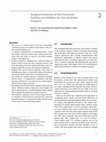
Clinical correlations: orbital apex/cavernous
sinus syndromes
While we usually consider the orbital apex and cavernous sinus as separate anatomic entities, the anatomy of the
superior orbital fissure area is important as a continuous
transition zone between the two regions. Parkinson25,27,28
considered the orbital apex, superior orbital fissure, and the
cavernous sinus to be connected via a continuous venous
link bridging these structures. Since that time a number of
anatomic studies have reaffirmed Parkinson’s concept.21,22,35
Froelich et al.8 proposed the term lateral sellar orbital junction (LSOJ) to define this transitional zone. However, this
Lesions occurring at the cavernous sinus—orbital apex transition zone frequently result in ocular or orbital dysfunction.
Symptoms are useful in defining the precise anatomic localization of such lesions, and this can be valuable for diagnosis and therapeutic planning. Several syndromes have been
used to characterize the symptom complex associated with
lesions in this area.45 The term superior orbital fissure syndrome
is often associated with lesions located just anterior to the
7
1
Cavernous Sinus
orbital apex, and involves structures passing through the
central annulus of Zinn, as well as those above the annulus.
Symptoms involve multiple cranial nerve palsies involving
the oculomotor, trochlear, and abducens nerves, as well as
the ophthalmic division of the trigeminal nerve, but not the
optic nerve. Orbital apex syndrome is associated with lesions
at the apex involving both the superior orbital fissure and
the optic canal. It involves dysfunctions of cranial nerves as
seen in the SOF syndrome, as well as the optic nerve. More
posterior lesions can produce a cavernous sinus syndrome,
and may include features of the orbital apex syndrome, as
well as Horner’s syndrome, and possible involvement of the
maxillary division of the trigeminal nerve. While these various syndromes differ in their exact anatomic locations, the
pathologies causing them are similar. Therefore, we will follow Yeh and Foroozan45 in applying the term orbital apex
syndrome to all of these syndromes for convenience of
discussion.
Orbital apex syndrome can result from diseases involving the cavernous sinus and/or the orbital apex. Typical signs
and symptoms depend upon the specific anatomic structures
involved, but frequently include ophthalmoplegia, trigeminal sensory loss, Horner’s syndrome, proptosis, chemosis,
and facial pain. Etiologies are numerous and may be infectious and non-infectious inflammatory conditions, vascular
anomalies, neoplastic lesions, and trauma.
Inflammatory syndromes include Herpes zoster, Tolosa
Hunt syndrome, sarcoidosis, Churg-Strauss syndrome,
Wegener’s granulomatosis, giant cell arteritis, and thyroid orbitopathy. Orbital pseudotumor is a non-specific idiopathic
inflammatory process that may involve any orbital structure including those of the orbital apex, cavernous sinus, and
optic nerve. With inflammatory lesions, the onset of symptoms is frequently more abrupt than with other causes, and
often includes pain. Infectious etiologies include fungal infections such as Mucormycosis and Aspergillosis, bacterial infections, and tuberculosis. Cavernous sinus thrombophlebitis is a
potentially lethal condition caused by bacterial or fungal invasion complicating sinusitis in immunocompromized patients.
Neoplastic tumors are a frequent cause of cavernous sinus
and orbital apex syndromes, and may arise as primary lesions
8
in the surrounding tissues or secondary to distant malignancies. Primary tumors include meningiomas, neurofibromas,
gliomas, pituitary gland tumors, and tumors extending from
parasellar regions such as nasopharyngeal malignancies, or
from the orbit as with lacrimal gland tumors. Metastatic
tumors to the cavernous sinus are most often from the breast,
prostate, or lung, and lymphomas can involve the orbit or
the cavernous sinus and adjacent sinuses.
Vascular lesions that can cause a cavernous sinus syndrome include aneurysms of the internal carotid artery or
its intracavernous branches. Rupture of such an aneurysm
or a vascular tear following trauma can result in a carotidcavernous fistula. Such fistulas can be direct, where there is
a direct communication between the carotid artery and the
cavernous venous channels, or indirect where the communication is with small branches of the carotid artery. The former type has a higher blood flow, and presents with abrupt
onset of proptosis, chemosis, ophthalmoplegia, and possibly loss of vision. The latter type tends to have slower blood
flow, progresses more slowly, is associated with less severe
symptoms, and may resolve spontaneously.
Localization of lesions affecting the cavernous sinus
is important in the differential diagnosis of cavernous
sinus syndrome. From the above anatomic discussions, it
should be apparent that intracavernous neural structures
can be affected differently in various parts of the sinus.
Sensory deficits are frequently seen with cavernous sinus
lesions. The maxillary nerve (V2) exits the sinus posteriorly, whereas the ophthalmic nerve (V1) courses through
the sinus to the superior orbital fissure. A lesion in the
anterior or middle sinus would be expected to affect V1
but not necessarily V2. Within the lateral sinus wall run
from top to bottom the oculomotor nerve (III), the trochlear nerve (IV), and V1, and in the posterior cavernous sinus, V2. With expanding lesions from above, the
motor nerves will be affected before any sensory deficit.
The abducens nerve (VI) does not run in the lateral wall
but within the sinus immediately lateral to the cavernous ICA. Being relatively unprotected, isolated sixth nerve
palsies are seen earlier with ICA aneurysms or with other
intracavernous lesions.
Clinical Correlations: Orbital Apex/Cavernous Sinus Syndromes
Anterior clinoid
process
Chiasmatic groove
Optic canal
Tuberculum sellae
Sella turcica
Superior orbital
fissure
Carotid groove
Posterior clinoid
process
Foramen rotundum
Clivus
Foramen lacerum
Foramen Vasalius
Petrous bone
Foramen
ovale
Internal acoustic
meatus
Figure 1-1 Bony sella turcica and clinoid processes limiting the cavernous sinus.
Carotid artery,
intradural segment
Diaphragma sellae
Optic chiasm
CN III
CN IV
Pituitary gland
Caverous sinus
CN V1
Carotid artery, horizontal
intracavernous segment
CN VI
CN V2
Sphenoid sinus
CN V3
Figure 1-2 Cross section through the mid cavernous sinus.
9
1
Cavernous Sinus
Optic nerve
Carotid artery
Diaphragma
sellae
Pituitary gland
Posterior clinoid
process
CN III
CN IV
CN VI
CN V2
Meckel’s cave
CN V3
Figure 1-3 Dura mater of the cranial base and nerve roots entering the cavernous sinus.
Optic nerve
Carotid artery
Interclinoid
ligament
Superior orbital
fissure
Anterior petroclinoid
ligament
Cavernous
sinus
CN V1
CN V2
Figure 1-4 Outer layer of the lateral wall of the cavernous sinus.
10
Pituitary stalk
Posterior
petroclinoid
ligament
Gasserian ganglion
Clinical Correlations: Orbital Apex/Cavernous Sinus Syndromes
Roof of the
cavernous sinus
Carotid artery
Pituitary gland
CN III
CN V1
CN IV
CN VI
CN V2
Trigeminal nerve
CN V3
Figure 1-5 Inner layer of the lateral wall of the cavernous sinus showing cranial nerves 3, 4, and 5.
CN III, superior
division
Intracavernous
carotid artery
CN III, inferior
division
CN VI
Inferolateral trunk
Cavernous sinus
Figure 1-6 Cavernous sinus with the lateral wall removed; cranial nerves 3, 4, and 5 are cut; cranial nerve 6 and the carotid artery are shown within the
sinus cavity.
11
1
Cavernous Sinus
Ophthalmic artery
Interclinoid ligament
Upper dural ring
Lower dural
CN VI
Posterior
petroclinoid
ligament
Figure 1-7 Cavernous sinus, medial wall, and dural ligaments.
Superior rectus
muscle
CN V1,
nasociliary nerve
Optic nerve
Ophthalmic artery
CN III,
superior division
CN V1,
lacrimal nerve
CN V1,
frontal nerve
Superior
ophthalmic vein
Trochlear nerve
CN III,
inferior division
CN VI
Medial rectus
muscle
Annulus of Zinn
Inferior rectus
muscle
Inferior
ophthalmic vein
Figure 1-8 Annulus of Zinn with major neural and vascular elements passing through to the orbital apex.
12
Lateral rectus
muscle
References
References
1. Amar AP, Weiss MH: Pituitary anatomy and physiology.
Neurosurg Clin N Am 13:11, 2003.
2. Bedford MA: The “cavernous” sinus. Br J Ophthalmol 50:41,
1966.
3. Bouthillier A, van Loveren HR, Keller JT: Segments of the
internal carotid artery: A new classification. Neurosurgery
38:425, 1996.
4. Campero A, Martins C, Yasuda, AL: Microsurgical anatomy
of the diaphragma sellae and its role in directing the pattern of growth of pituitary adenomas. Neurosurgery 62:717,
2008.
21. Morard M, Tcherekayev V, de Tribolet N: The superior orbital
fissure: A microanatomical study. Neurosurgery 35:1087,
1994.
22. Natori Y, Rhoton AL Jr: Microsurgical anatomy of the superior orbital fissure. Neurosurgery 36:762, 1995.
23. Padget DH: The development of the cranial venous system
in man from the viewpoint of comparative anatomy. Contrib
Embryol 247:79, 1956.
24. Parkinson D: Carotid cavernous fistula: Direct repair
with preservation of the carotid artery. Technical note.
J Neurosurg 38:99, 1973.
25. Parkinson D: Surgical anatomy of the lateral sellar compartment (cavernous sinus). Clin Neurosurg 36:219, 1990.
5. Conti M, Prevedello DM, Madhok R, et al: The antero-medial
triangle: The risk of cranial nerves ischemia at the cavernous sinus lateral wall. Anatomy cadaveric study. Clin Neurol
Neurosurg 110:682, 2008.
26. Parkinson D: Lateral sellar compartment. History and anatomy. J Craniofac Surg 5:55, 1995.
6. Dolenc VV: Anatomy and Surgery of the Cavernous Sinus. Wien,
Springer-Verlag, 1989, pp 3–137.
28. Parkinson D: Extradural
J Neurosurg 92:585, 2000.
7. Fischer E: Die Lageabweichungen der vorderen hirnarterie im
gefässbild. Zentralbl Neurochir 3:399, 1938.
29. Parkinson D, Johnston J, Chaudhuri A: Sympathetic connections to the fifth and sixth cranial nerves. Anat Rec 191:221,
1978.
8. Froelich S, Abdel KM, Aziz A, et al: The transition between
the cavernous sinus and orbit. In: Dolenc VV, Rogers L
(eds): Cavernous Sinus. New York, Springer Wien, 2009,
p 27.
9. Gilmore SA: Developmental anatomy of the intracranial
venous system: A review of dural venous sinus development.
In: Hakuba A (ed): Surgery of the Intracranial Venous System.
Tokyo, Springer, 1996, pp 3-13.
10. Gupta N, Ray B, Ghosh S: Anatomic characteristics of foramen vesalius. Katmandu Univ Med J 3:155, 2005.
11. Harris FS, Rhoton AL Jr: Anatomy of the cavernous sinus.
A microscopical study. J Neurosurg 45:169, 1976.
12. Hashimoto M, Yokota A, Yamada H, Okudera T:
Development of the cavernous sinus in the fetal period:
A morphological study. Neurol Med Chir 40:140, 2000.
13. Inoue T, Rhoton AL Jr, Theele D, Barry ME: Surgical
approaches to the cavernous sinus: A microsurgical study.
Neurosurgery 26:903, 1990.
14. Isolan G, de Oliveira E, Mattos JP: Microsurgical anatomy
of the arterial compartment of the cavernous sinus. Arq
Neuropsiquiatr 63:259, 2005.
15. Kawase T, van Loveren H, Keller JT, Tew JM: Meningeal architecture of the cavernous sinus. Clinical and Surgical implications. Neurosurgery 39:527, 1996.
27. Parkinson D: Lateral sellar compartment O.T. (cavernous sinus):
History, anatomy, terminology. Anat Rec 251:486, 1998.
neural
axis
compartment.
30. Reymond J, Kwiatkowski J, Wysocki J: Clinical anatomy of
the superior orbital fissure and the orbital apex. J CranioMaxilofac Surg 36:346, 2008.
31. Rhoton AL, Jr: The middle cranial base and cavernous sinus.
In: Dolenc VV, Rogers L (eds): Cavernous Sinus. New York,
Springer Wien, 2009, pp 3–25.
32. Seoane E, Rhoton AL Jr, de Oliveira EP: Microsurgical anatomy of the dural collar (carotid collar) and rings around the
clinoid segment of the internal carotid artery. Neurosurgery
42:869, 1998.
33. Shi X, Han H, Zhao J, Zhou C: Microsurgical anatomy of the
superior orbital fissure. Clin Anat 20:362, 2007.
34. Songtao Q, Yuntao L, Jun P, et al: Neurosurgery 64:1, 2009.
35. Spektor S, Piontek E, Umansky F: Orbital venous drainage
into the anterior cavernous sinus space: Microanatomic relationships. Neurosurgery 40:532, 1997.
36. Taptas JN: The so-called cavernous sinus: A review of the controversy and its implications for neurosurgeons. Neurosurgery
11:712, 1982.
37. Terminologia Anatomica. Federative Committee on Anatomical
Terminology. Stuttgart, Thieme, 1998, p 292.
16. Keller JT, Leach JL, van Loveren HR, et al: Venous anatomy of
the lateral sellar compartment. In: Dolenc VV, Rogers L (eds):
Cavernous sinus. New York, Springer Wien, 2009, pp 35–52.
38. Tobenas-Dujardin AC, Dupare F, Laquerriere A, et al:
Embryology of the walls of the lateral sellar compartment:
Apropos of a continuous series of 39 embryos and fetuses
representing the first six months of intrauterine life. Surg
Radiol Anat 25:252, 2003.
17. Knappe UJ, Konerding MA: Medial wall of the cavernous
sinus: Microanatomcal diaphanoscopic and episcopic investigation. Acta Neurochir 151:961, 2009.
39. Umansky F, Nathan H: The lateral wall of the cavernous
sinus. With special reference to the nerves to it. J Neurosurg
56:228, 1982.
18. Lasjaunias P, Bernstein A, Raybaud C. Intracranial venous
system. In: Lasjaunias P, Bernstein A (eds): Functional Vascular
Anatomy of the Brain, Spinal Cord and Spine. Berlin, Springer,
1987, pp 223–266.
40. Umansky F, Valarezo A, Elidan J: The superior wall of the cavernous sinus. A microanatomical study. J Neurosurg 81:914,
1994.
19. Marinkovic S, Gibo H, Vucevic R, Petrovic P: Anatomy of the
cavernous sinus region. J Clin Neurosci 8(Suppl.):78, 2001.
20. McConnell EM: The arterial blood supply of the human
hypophysis cerebri. Anat Rec 115:175, 1953.
41. Umansky F, Valarezo A, Piontek E, Spektor S: Surgical anatomy of the cavernous sinus and dural folds of the parasellar
region. In: Kobayashi S, Goel A, Hongo K (eds): Neurosurgery
of Complex Tumors and Vascular Lesions. Edinburgh, Churchill
Livingstone, 1997, p 156.
13
1
Cavernous Sinus
42. Winslow JB: Exposition Anatomique de la Structure du Corps
Humain. Vol. II. London, N. Prevast, 1734, p 29.
45. Yeh S, Foroozan R: Orbital apex syndrome. Curr Opin
Ophthalmol 15:490, 2004.
43. Yasuda A, Campero A, Martins C, et al: Microsurgical anatomy and approaches to the cavernous sinus. Operat Neurosurg
56:4, 2005.
46. Ziyal IM, Özgen T, Skhar LN, et al: Proposed classification
of segments of the internal carotid artery: Anatomical study
with angiographical interpretation. Neurol Med Chir (Tokyo)
45:184, 2005.
44. Yasuda A, Campero A, Martins C, et al: The medial wall of
the cavernous sinus: Microsurgical anatomy. Neurosurgery
55:179, 2004.
14
CHAPTER
2
Osteology of the Orbit
Embryology
The bony orbit develops from mesenchyme that encircles
the optic vesicle in early embryonic development. Individual
bones develop from a complex series of ossifications of two
types. Endochondral bones ossify secondarily after they
are preformed in cartilage. Membranous, or dermal bones,
ossify directly from connective tissue without a cartilaginous
precursor. The first cranial bone to appear embryologically is
the maxillary bone, first recognizable at the 16 mm (6-week)
embryonic stage. It is not preformed in cartilage, but arises
from dermal elements as an intramembranous ossification
in the region of the canine tooth. This is followed shortly
by secondary ossification centers in the orbitonasal area
and premaxilla.8 The primordial maxillary sinus does not
appear until the 320 mm (32-week) fetal stage. At the 30 mm
(7-week) stage additional intramembranous ossifications
mark the first appearance of the frontal, zygomatic, and palatine bones. As these centers enlarge, they make contact with
adjacent ossifications, forming suture lines. The zygomatic
and maxillary bones establish contact during the 70 mm
(13-week) stage, and the zygomaticofrontal fissure is established
at the 145 mm (20-week) stage. The zygomaticosphenoid fissure
closes at about the time of birth.
The sphenoid bone arises from both endochondral and
intramembranous ossifications. The lesser wing of the sphenoid and the optic canal begin as cartilaginous structures at
the 25 mm (7-week) stage. Ossification begins at the region
of the future optic strut in the 75 mm (13-week) fetus, and
along the superior rim of the optic canal at the 118 mm
(16-week) stage. The greater wing of the sphenoid bone is
preformed in cartilage during the 52 mm (12-week) stage,
and begins to ossify by the 67 mm (13-week) stage. All the
elements of the sphenoid bone, both endochondral and
intramembranous, finally join to form a single element in
the 125 mm (18-week) fetus. The sphenoid bone enlarges
and makes contact with the frontal bone, closing the lateral
and superior orbital walls by the 220 mm (26-week) stage.8
The ethmoid bone begins as part of the cartilaginous chondrocranium in the 25 mm (7-week) embryo. Ossification
begins in the 220 mm (26-week) stage on the lateral portion,
at what will become the lamina papyracea. By the 320 mm
(32-week) stage ossification is nearly complete, except for the
nasal septum, which remains cartilaginous. The ethmoid air
cells develop between the 220 and 320 mm (26–32-week)
stages. The lacrimal bone develops as a thin intramembranous ossification beginning in the 75 mm (13-week) fetus.
The orbital bones form around the developing optic
cup and stalk. Initially, the optic vesicles are positioned
170–180° apart, on opposite sides of the forebrain, reflecting
their earlier phylogenetic vertebrate configuration. During
the 4- to 8-week embryonic stages the optic cups begin to
rotate anteriorly as the primordial orbital bones are laid
down around them. By 3 months of fetal development, the
orbital axes form an angle of about 105° between them and
at birth, this angle is reduced to 45°. Only relatively slight
additional remolding occurs during childhood. Failure of
complete rotation results in the clinical condition of hypertelorism, whereas over rotation causes hypotelorism.22
Malpositions in ossification of orbital bones may result in
reduced orbital volume and proptosis, as seen in Crouzon
disease.
The adult bony orbit
In the adult, the bony orbit is roughly pyramidal in shape.
Its volume in the average individual is approximately 25
cm3, but published measurements of volume vary considerably using either direct filling or CT imaging techniques
from a mean of 17.05 cm3 to 29.30 cm3.1,9,19,34,42,50 Within the
orbit the eye contributes about 7.2 cm3 based on the average diameter of about 24 mm. However, a myopic eye will
be larger and a hyperopic eye will be smaller. Each change of
0.5 mm in diameter will result in a volumetric change of
about 0.45 cm3. Thaller56 measured the volume of enucleated
eyes by a volume displacement technique and found the average
volume to be 8.15 cm3.
The anterior entrance of the orbit forms a rough rectangle measuring approximately 43 mm (36–47 mm) wide
by 34 mm (26–42 mm) high.42 The orbit attains its widest
dimensions at about 15 mm behind the bony rim. As in all
other higher primates, the human orbit is completely closed
behind by the sphenoid bone, except for the superior and
inferior orbital fissures. The orbits are directed more forward
than in other mammals, and their anterior-posterior central
axes form a 45° angle between them. The two lateral orbital
walls subtend a 90° angle between them. The four walls of
each orbit converge posteriorly toward the orbital apex where
the optic canal and superior orbital fissure pass into the middle cranial fossa.
The overall dimensions of the orbit are quite variable,
especially its depth. Thus, the surgeon cannot rely on precise
measurements as a guide to the exact location of the optic
canal or superior orbital fissure. Nor can the position of the
15
2
Osteology of the Orbit
ethmoidal foramina, the bridging over of the infraorbital
canal, or of soft tissue structures within the orbit be accurately determined preoperatively. Therefore, extreme caution must be exercised in posterior orbital dissections in any
orbital surgery. During exploration of the orbital floor for
entrapment of the inferior rectus muscle following trauma or
in orbital decompression, the inferior orbital fissure may be
encountered inferolaterally as little as 10–15 mm behind the
rim. Dissection along the floor should not extend more than
40 mm posterior to the orbital rim, since the floor ends at
the posterior wall of the maxillary sinus, and therefore does
not extend to the apex.
The orbital rim
The orbital rim is rounded and thickened, and serves to protect the eye from facial impacts. The superior rim is the most
prominent due to expansion of the underlying frontal sinus.
It is more protuberant in adult males. Its significance has been
a matter of debate for over 100 years,48 but the most often
cited explanation for it is that it developed to counter biomechanical stress associated with mastication.15 Experimental
data have demonstrated mastication-related strain in the
interorbital and supraorbital regions. However, the degree is
very small compared to other parts of the facial skeleton, and
therefore does not support masticatory stress as a major evolutionary force in development of the supraorbital ridge.25
The medial third of the superior orbital rim is interrupted
by a notch or foramen for passage of the supraorbital neurovascular bundle. One or both sides will have an open notch
in 75% of all orbits. In 50% of individuals at least one side
may be closed to form a foramen.39,59 The notch is situated
about 25–30 mm from the facial midline.5,7,59 The location
of this notch is an important guide in avoiding injury to the
supraorbital nerve during brow and forehead surgery.
The orbital rim is flatter and less prominent between the
supraorbital notch and the medial canthal ligament. A number of important neurovascular structures emerge here, including the supratrochlear and infratrochlear nerves, and the dorsal
nasal artery. Just inside the rim at the superomedial corner of
the orbit is the cartilaginous trochlea of the superior oblique
tendon. Surgical access to the medial wall through a frontoethmoidal incision may interrupt these neural structures with
resultant glabellar and forehead anesthesia. If necessary for
orbital access, the trochlea can be disinserted by elevating the
periosteum.
Medially, the orbital rim extends downward to the posterior lacrimal crest and ends at the inferior entrance to the
nasolacrimal canal. The anterior lacrimal crest begins just
above the medial canthal ligament, and passes downward
into the inferior orbital rim. The medial rim is, therefore,
discontinuous at the lacrimal sac fossa. Between the anterior
and posterior lacrimal crests is the lacrimal sac fossa formed
at the junction of the maxillary and lacrimal bones. The fossa
measures about 16 mm in vertical length, 4–9 mm in width,
and 2 mm in depth.4 Just in front of and parallel to the anterior lacrimal crest is a vertical groove in the frontal process of
the maxillary bone for a nutrient branch of the infraorbital
artery. During dacryocystorhinostomy surgery this groove
may be mistaken for the medial edge of anterior lacrimal
crest. Brisk bleeding may occur from rupture of this vessel,
but it is easily controlled.
16
The inferior orbital rim is formed by the maxillary bone
medially and the zygomatic bone laterally. The infraorbital
foramen, conducting the infraorbital artery and nerve, is
located 4–10 mm below the central portion of the inferior
rim. During surgery on the orbital floor, care must be taken
not to elevate periosteum anterior to the central rim for more
than about 4 mm, since this may injure these neurovascular
structures.
The orbital rim is thickest laterally. Here it is formed by
the frontal process of the zygomatic bone and the zygomatic
process of the frontal bone. These two elements meet at the
frontozygomatic suture line near the superotemporal corner of the orbit. This suture line is an important landmark
for removing the lateral rim during orbital surgery, because
the anterior cranial fossa lies 5–15 mm above this horizontal level. This is a weak suture and is frequently the site of
separation following facial trauma. About 10 mm below the
frontozygomatic suture line, about 4–5 mm inside the rim
is a small mound, the lateral orbital tubercle of Whitnall. It
serves for insertion of the posterior crus of the lateral canthal ligament, Lockwood’s inferior suspensory ligament, the
lateral horn of the levator aponeurosis, the lateral check ligament and pulley system of the lateral rectus muscle, and
the deep layer of the orbital septum. Proper realignment of
these structures after lateral orbital surgery or repair of rim
fractures is essential for normal cosmetic and functional
reconstruction.
The entire orbital rim is buttressed by adjacent bones and
is frequently involved in complex facial fractures. The surgeon must be alert to the normal anatomic and functional
relationships between the orbital bones and the nasal cavity,
paranasal sinuses, cranial vault, and the temporomandibular joint.
The medial orbital wall
The medial walls of the orbits are approximately parallel to each other and to the mid-sagittal plane. The separation between the two orbits is approximately 24 mm from
the medial wall of one to the medial wall of the other. The
medial wall measures an average of 42 mm (range 32–53 mm)
in horizontal length from the anterior lacrimal crest to the
optic canal.38 The medial wall of each orbit is formed by
four osseous elements, the maxillary, lacrimal, ethmoid, and
sphenoid bones. Anteriorly, the thick frontal process of the
maxillary bone lies at the inferior medial rim. It contains
the anterior lacrimal crest and forms the anterior portion of
the lacrimal sac fossa. The lacrimal bone is a small, thin and
fragile plate situated just posterior to the maxillary process. It
forms the posterior portion of the lacrimal sac fossa. Running
vertically along its midpoint is the posterior lacrimal crest. The
suture between the maxillary and lacrimal bones generally
lies along the mid-vertical line within the lacrimal sac fossa.
However, in 8% of individuals this suture lies more posteriorly, occasionally nearly to the posterior lacrimal crest.6
In such cases the thicker maxillary bone underlies most of
the lacrimal sac fossa. As a result, creation of a bony osteum
during dacryocystorhinostomy surgery can be more difficult than usual, and will frequently require a burr to remove
excess bone.
Behind the posterior lacrimal crest is the lamina papyracea, which forms most of the lateral wall of the ethmoid
The Adult Bony Orbit
labyrinth. It contributes 4–6 cm2 to the orbital wall surface.
This is exceptionally fragile, measuring only 0.2–0.4 mm in
thickness. However, it is made more rigid by the honeycombed bony laminae surrounding the ethmoid air cells,
which usually number 3–8. Resistance of the medial wall to
static loading is greater when the lamina papyracea is smaller
in area, when the number of air cells is greater, or when their
individual sizes are smaller.47 Song et al.53 showed that medial
wall fractures are more frequent, compared to floor fractures,
when there are fewer ethmoid air cells, or when a larger
area of lamina papyracea is supported by each sinus septum. The fragility of this bone is also associated with its easy
displacement into the orbit with expanding lesions in the
ethmoid sinus.26 Following trauma, a 3 mm “blow-out” medial
displacement of the lamina papyracea may result in a 5%
increase in orbital volume, and 1.0–1.5 mm of enophthalmos.43 The lamina papyracea offers only a minimal barrier
to the spread of infection from the ethmoid sinus into the
orbit,58 sometimes resulting in the orbital edema, cellulitis, and abscess formation that is sometimes associated with
ethmoid sinusitis. Surgery along the medial wall, or probing
instrumentation during enucleation surgery may easily penetrate the lamina papyracea, with the possible complication
of orbital emphysema or infection.13
Superiorly the ethmoid bone joins the orbital roof at the
fronto-ethmoid suture line. This level approximately marks
the roof of the ethmoid sinus labyrinth and the floor of the
anterior cranial fossa. Just medial to the labyrinth, on either
side of the intracranial crista galli, is the cribriform plate. This
may extend 5–10 mm below the level of the fronto-ethmoid
suture line in some individuals. The root of the middle nasal
turbinate separates the cribriform plate on each side from the
superior ethmoid air cells. This relationship must be born in
mind during surgery along the medial wall, and the frontoethmoid suture line is a useful landmark indicating the safe
upper limit for bony dissection.
At the level of the lacrimal sac fossa the anterior cranial
fossa may be as little as 1 mm, or as much as 30 mm above
the upper border of the medial canthal ligament. The mean
value is 8.3 mm.33 This distance tends to correlate with the
size of the frontal sinus, being larger when the sinus is more
extensive. At the level of the posterior lacrimal crest this distance shortens to 0–19 mm (mean of 6.5 mm), as the floor of
the anterior cranial fossa slopes downward and backward. In
as many as 20% of normal individuals this distance may be
3 mm or less,33 and this may explain the occasional occurrence
of a CSF leak during creation of a bony osteum in dacryocystorhinostomy surgery. This complication is more likely when
the medial canthal ligament landmark is removed.33,41 It is,
therefore, safest to leave the ligament attached, and to use
this structure as a guide to placement of the upper border of
the bony osteum.
The anterior and posterior ethmoidal foramina usually
lie within the fronto-ethmoid suture line. These openings
transmit branches from the ophthalmic artery and nasociliary nerve passing out of the orbit. There is great variability in
the position of these foramina and in 10–20% of cases one
or both of these canals may lie outside (usually above) the
fronto-ethmoid suture line as a variant or racial difference.61
The posterior ethmoid foramen may sometimes be absent,
and both foramina may be multiple. The anterior ethmoidal
foramen is located about 22 mm (range 14–30 mm) behind
the anterior lacrimal crest. However, it is located within the
more narrow range of 20–25 mm behind the crest in twothirds of individuals.11,32 The posterior ethmoidal foramen lies
33 mm (range 25–41 mm) from the anterior lacrimal crest,36,42
approximately 4–15 mm anterior to the optic canal. The anterior and posterior ethmoid foramina transmit the ethmoidal
nerves and arteries into the anterior cranial fossa and to the
nasal and sinus mucosa. The positions of these foramina are
clinically important since they relate to important cranial
structures such as the cribriform plate, and to the optic foramen. They are key landmarks during surgery along the medial
orbital wall. Injury to the ethmoidal arteries can cause excessive orbital bleeding during surgery. Subperiosteal hematoma
following trauma frequently results from rupture of one of
these arteries, and management requires access to the medial
wall with ligation or cautery of the bleeding vessel.
Posterior to the ethmoid bone is the body of the sphenoid
bone that forms the short posterior portion of the medial
wall. The sphenoid body lies between the two orbital apices
and contains the sphenoid sinus. The optic canal is situated
in the superomedial portion of the orbital apex, enclosed by
the body of the sphenoid medially, the lesser wing of the
sphenoid superiorly, and the optic strut inferolaterally.
The lacrimal sac fossa is a depression in the anterior inferomedial orbit.27 It is bounded by the anterior and posterior
lacrimal crests and measures about 4–9 mm in width and
16 mm in height. The fossa is formed by the frontal process
of the maxillary bone anteriorly and by the lacrimal bone
posteriorly. The nasolacrimal canal is a bony tube extending
from the lacrimal sac fossa to the inferior nasal meatus, and
it contains the membranous nasolacrimal duct. The canal
measures about 5 mm in diameter and is bordered by three
bones, the maxilla, the lacrimal, and the inferior turbinate
bones. The canal runs inferolateral and slightly posterior
in the medial wall of the maxillary bone. It measures about
12–15 mm in length.
The orbital floor
The orbital floor is a very thin plate composed of three
bones (maxillary, zygomatic, and palatine). Its surface
forms a triangular segment extending from the maxillaryethmoid buttress on the medial side, horizontally to the
inferior orbital fissure on the lateral side, and from the inferior orbital rim back to the posterior wall of the maxillary
sinus. The floor contributes 3–5 cm2 to the overall orbital
wall surface. It is strengthened by the infraorbital canal
which runs anteroposteriorly through it near its midline or
sometimes closer to its lateral border. One or more trabeculae in the roof of the maxillary sinus are sometimes present
and they serve also to buttress the floor. Nevertheless, the
orbital floor shows the greatest degree of deformation with
static loading of any of the orbital walls.47 This explains the
high rate of floor fractures associated with blunt trauma.
A 3 mm downward displacement of the entire floor results
in an increase of about 1.5 cm3 (5%) to the orbital volume,
and about 1.0–1.5 mm of enophthalmos.
The major contribution to the floor is from the orbital
plate of the maxillary bone, which also forms the roof of the
maxillary sinus. Anterolaterally, the zygomatic bone contributes to the orbital rim and a small portion of the floor just in
front of the anterior border of the inferior orbital fissure. The
17
2
Osteology of the Orbit
palatine bone lies at the extreme posterior end of the floor,
near the orbital apex. In adults, it is usually fused with the
maxillary bone. The floor is bounded medially by the maxilloethmoid suture line, and anterolaterally by the zygomaticomaxillary suture. From the inferior orbital rim, the floor dips
downward, where it reaches its lowest point. This is about
1.5–2.0 mm below the rim in children and young adults, but
reaches 3.0 mm in older adults.40 From here the floor slopes
upward to the orbital apex at an angle of about 18–22° to the
horizontal Frankfort plane (inferior orbital rim [orbitale] to
the upper border of the bony ear canal [porion]).
In the mid and posterior orbit, the floor ends at the inferior orbital fissure, and the posterior extent of the maxillary
sinus. It is important to keep in mind that the orbital floor
does not extend all the way to the apex, but rather ends at
the pterygopalatine fossa. The floor is, therefore, the shortest
of the orbital walls, extending only about 35–40 mm from
the inferior rim to the posterior wall of the maxillary sinus.
However, the distance from the rim at the infraorbital canal to
the optic canal is greater, measuring 48 mm (range 41–57 mm).
During surgical exploration of orbital fractures or during floor
decompressions in thyroid orbital disease, dissection need
not be carried further than the posterior sinus wall. However,
in cases of compressive optic neuropathy in Graves’ disease,
it is essential to obtain an adequate decompression closer
to the orbital apex.2,30 This can be achieved on the medial
wall by opening the orbit into the posterior ethmoid sinus or
even into the sphenoid sinus. On the lateral wall the thicker
portion of the lateral sphenoid wing can be burred down to
the inner plate or even to the dura.
The infraorbital sulcus lies within the posterior portion
of the orbital floor. This fissure runs approximately in the
center of the floor from posterior to anterior, and carries
the maxillary division of the trigeminal nerve and the associated infraorbital branch of the maxillary artery from the
pterygopalatine fossa. At about the mid portion of the floor
the sulcus usually becomes bridged-over by a thin plate of
the maxillary bone to form the infraorbital canal. This thin
plate of bone is pierced by one or more tiny foramina that
transmit anastomotic vessels from the infraorbital artery to
the inferior muscular branch of the ophthalmic artery (see
Chapter 5). Along its course, the infraorbital canal gives off
the middle and anterior superior alveolar canals, carrying
corresponding nerves and vessels.35 The infraorbital canal
continues forward to the orbital rim, where it exits as the
infraorbital foramen. In 2–18% of individuals the canal can
be double or even triple.23 After elevation of periosteum, the
region of the infraorbital canal can usually be identified on
the floor as a slightly elevated somewhat translucent ridge.
Recognition of its position is critical if injury to the infraorbital nerve is to be avoided during orbital floor surgery.
Damage to this nerve results in anesthesia of the lower eyelid, cheek, and upper lip, and this is not uncommon following orbit floor blow-out fractures or orbital decompression
into the maxillary sinus.
Separating the floor from the lateral orbital wall is the
inferior orbital fissure (IOF). This opening is approximately
30 mm in length and runs in an anterolateral to posteromedial direction. The anteriormost edge of the IOF lies approximately 24 mm (range 17–29 mm) from the inferior orbital
rim at the infraorbital foramen. At the orbital apex just
below the optic canal, the inferior fissure joins the superior
18
orbital fissure, and is contiguous with the foramen rotundum in the floor of the middle cranial fossa. The inferior
fissure transmits structures into the orbit from the pterygopalatine fossa posteriorly, and from the infratemporal fossa
more anteriorly. Multiple branches from the inferior ophthalmic vein pass through this opening to communicate
with the pterygoid venous plexus. The inferior fissure also
transmits the maxillary division (V2) of the trigeminal nerve.
The latter nerve passes out of the cranium through the foramen rotundum into the pterygopalatine fossa, and then into
the infraorbital sulcus in the posterior orbital floor, where it
runs in company with the infraorbital artery. Postganglionic
parasympathetic secretory and vasomotor neural branches
from the pterygopalatine ganglion enter the orbit through
the inferior orbital fissure, where they join with the maxillary
nerve for a short distance before running superiorly along
the lateral orbital wall to the lacrimal gland (see Chapter 4).
The lateral orbital wall
The lateral wall of the orbit is the thickest, and is composed
of the zygomatic bone anteriorly and the greater wing of the
sphenoid posteriorly. It is separated from the floor by the
inferior orbital fissure, and from the roof, in part, by the superior orbital fissure. The lateral walls of the two orbits form an
angle of approximately 90° with each other, and lie at 45° to
the mid-sagittal plane. The lengths of the lateral and medial
walls, from orbital rim to apex, are about the equal. Because of
the oblique orientation of the lateral wall, the lateral rim lies
about 10 mm posterior to the medial rim.18 The length of the
lateral wall from the lateral rim at the frontozygomatic suture
to the optic canal is about 47 mm (range 39–55 mm).
The thinnest part of the lateral wall is at the zygomaticsphenoid suture, about 8–10 mm behind the orbital rim.
During lateral orbital surgery, cuts through the bony rim
must be made to this level so that the rim can easily be
fractured outward. Approximately 10 mm behind the zygomatic-sphenoid suture, the sphenoid bone thickens where
it divides to form the anterior corner of the middle cranial
fossa. Here, compact bone passes into cancellous bone, a
useful landmark when taking down the lateral wall to gain
wide access to the orbit or in lateral wall decompressions.
In about 40% of individuals there are one or more openings within the fronto-sphenoid suture line, about 30 mm
from the orbital rim. This is the cranio-orbital foramen
(foramen meningo-orbitale) which transmits an anastomotic branch between the middle meningeal artery and the
ophthalmic arterial system (see Chapter 5). This vessel is a
remnant of the embryological development of the orbital
arterial system, and usually joins the root of the lacrimal
artery. Although this is a small and sometimes inconsistent branch in humans, it represents a significant supply of
orbital blood in some other mammalian orders.60 This vessel is easily ruptured during lateral orbital surgery resulting
in brisk bleeding. Compression for several minutes is usually sufficient to control it.
At the junction of the lateral wall and roof is the superior orbital fissure (SOF), lying between the greater and
lesser wings of the sphenoid bone near the orbital apex. It
is oriented from inferomedial at the apex to superotemporal distally. The anteriormost edge of the SOF lies 37 mm
(range 34–41 mm) from the lateral orbital rim. In size and
Aging Phenomena
shape this fissure shows considerable individual variability.51 However, its comma-like shape is usually wider inferiorly, but then narrows more superiorly. The fissure measures
about 20–25 mm in overall length. The narrow lesser wing
of the sphenoid bone separates the medial edge of the superior orbital fissure from the lateral margin of the optic canal.
The spinal recti lateralis is a small bony projection situated
on the lateral edge of the fissure near its middle portion, at
the junction of its wide and narrow portions. This projection serves as the origin for part of the lateral rectus muscle. It is formed primarily by a small groove in the sphenoid
wing which lodges the superior ophthalmic vein as it passes
through the fissure.44 The superior orbital fissure transmits
most of the vascular and neural structures from the middle
cranial fossa into the orbit, with the major exception of the
optic nerve and ophthalmic artery, which pass through the
optic canal. The central portion of the fissure is anatomically divided by the annulus of Zinn, which serves as the
tendinous origin for the rectus muscles. The central opening defined by the annulus, called the oculomotor foramen,
transmits structures into the intraconal orbital space. Most
of these structures subserve ocular function and motility.
These include the superior and inferior divisions of the oculomotor nerve, the abducens nerve, and the nasociliary nerve
(see Chapter 4). Other structures passing through the superior orbital fissure but outside the annulus are mainly associated with the extraconal orbital space, or are en route to
extraorbital sites. These include the trochlear nerve, the frontal and lacrimal branches of the trigeminal nerve, and the
superior ophthalmic vein above the annulus, and the inferior ophthalmic vein beneath the annulus.
In 8–40% of individuals, a linear vertical groove is present
lying along the greater wing of the sphenoid bone, between
the superior and inferior orbital fissures. This was previously
believed to house an anastomotic branch between the middle meningeal and infraorbital arteries.37 However, investigations show that this does not contain any vascular or neural
structures, but rather represents an abrupt thinning of the
greater wing at the transition from cancellous to compact
bone.10
Several small foramina perforate the lateral orbital wall
just behind the rim laterally and inferiorly near the anterior
end of the inferior fissure. These transmit branches of the
lacrimal artery and zygomatic nerve out of the orbit as the
zygomaticotemporal and zygomaticofacial neurovascular
bundles.
The orbital roof
The orbital roof is triangular in shape. It is formed primarily from the orbital plate of the frontal bone, with a small
contribution by the lesser wing of the sphenoid bone posteriorly. It measures about 46 mm (range 35–59 mm) from
the supraorbital foramen to the optic canal.38,42 In the anterior superolateral corner is a poorly-defined concavity for the
lacrimal gland. A small depression in the superomedial corner, about 3–5 mm behind the rim, houses the fibrocartilaginous trochlea for the superior oblique tendon. This structure,
along with its associated pulley system, can easily be separated from the adjacent bone along with periorbita if needed
during surgery. Its precise repositioning is essential to avoid
postoperative motility disturbance.
The orbital roof is very thin and may have spontaneous
dehiscences. During surgery along the roof, care must be
taken since the use of instrumentation may perforate this
fragile structure and injure intracranial dura. The frontal
sinus is located within the frontal bone in the anteromedial
portion of the roof. The size of this sinus is extremely variable, and in some individuals it may extend as far laterally
as the lacrimal gland fossa, and as far posteriorly as the optic
canal.
The optic canal is located in the roof at the apex and communicates between the middle cranial fossa and the orbit.
It is bounded by the body of the sphenoid bone medially,
the lesser wing of the sphenoid superiorly, and the optic
strut laterally and inferiorly. The strut arises from the body
of the sphenoid and is directed slightly anteriorly, upward,
and laterally at an angle of about 36° to the sagittal plane.44
The optic canal assumes a vertically oval shape at its orbital
end, where it measures about 5–6 mm in horizontal diameter, and 6–8 mm vertically. In its central portion the canal
is round in cross-section, and on the cranial end it is oval
in the horizontal plane.20 The canal attains adult size by the
age of three years. In about 4% of normal individuals the
ophthalmic artery will notch the canal floor, forming a “keyhole” deformity.31 The canal is 8–12 mm in length and is
directed posteromedially at about 35° to the mid-sagittal
plane, and upward about 38° to the horizontal. On the cranial side the optic canal measures 5–7 mm horizontally and
4–6 mm vertically. The tendinous annulus of Zinn encloses
the orbital opening of the optic canal so that the optic nerve
and ophthalmic artery pass into the intraconal space via the
oculomotor foramen.
The relationships of the optic canal and the adjacent paranasal sinuses is somewhat variable depending upon the
extent to which these sinuses invade the lesser wing and the
anterolateral portion of the body of the sphenoid bone.44 In
a study of 100 sphenoid sinuses, Van Alyea57 found that the
medial wall of the optic canal projected into the sinus in 40%
of cases, and in rare instances it was completely surrounded
by the sinus with the canal passing through the sinus cavity. Goodyear21 described a similar relationship between the
posterior ethmoid sinus and the optic canal.
Aging phenomena
The craniofacial skeleton undergoes remodeling throughout adulthood. The face shows a progressive rotation of the
frontal bone forward over the orbits, and the maxillary bone
extends backward beneath the orbits.46 This process continues the morphological process of frontation seen in the
evolution of higher primates. These changes are most acute
in the mid-face. Angular changes in the facial skeleton are
associated with compensatory changes in the soft-tissue
anatomy, with weakening and stretching of the retaining ligaments, inducing descent of the mid-facial malar cheek pads
and changes in the position of the lower eyelid with increasing scleral show and prominence of the inferior orbital fat
pockets.
In addition to rotational changes the orbital aperture
changes, increasing in length along a line from superomedial to inferolateral.28 The loss of volume and bony projection along with laxity of retaining ligaments contribute
19
2
Osteology of the Orbit
to lateral brow hooding, lateral canthal skin redundancy,
and nasolabial fold prominence. The cranial skeleton also
widens, lengthens, and shows mid-face convexity with
advancing age.
Clinical correlations
The orbital floor is thinnest medial to the infraorbital canal
where it may be only 0.5 mm thick. This is a convenient
point for initial entrance into the maxillary sinus during
orbital inferior wall decompression surgery. It is this portion
of the floor that is usually involved in blow-out fractures,
believed to result from rim deformation and compression
of orbital contents following direct blunt trauma.52 Kwon
et al.34 measured the volumes of orbits expanded from a blowout injury compared to the uninjured contralateral sides and
reported an average expansion of 2.8 cm3.
Fan et al.17 calculated that each 1.0 cm3 increment of
orbital volume expansion would result in 0.89 mm of relative enophthalmos. Surgical correction is aimed at restoring the integrity and normal position of the fractured walls,
usually with the use of alloplastic implants or autogenous
bone grafts. This is indicated for cosmetically significant
enophthalmos, even in the absence of motility restriction.12
Correction of 3 mm of enophthalmos will require replacement of a 3.4 cm3 of volume, either by repositioning prolapsed fat and muscle, or with an orbital implant, or a
combination of both.
The total adult orbital volume is about 25 cm3, of which
the globe occupies about 7.2 cm3. Following enucleation
an alloplastic spherical implant is usually placed into the
anophthalmic socket to replace lost volume. The typical
18–20 mm diameter sphere replaces 3.0–4.0 cm3, and the
ocular prosthesis adds another 1.5–2.5 cm3 depending upon
the design and thickness. The net loss in orbital volume,
therefore, may amount to 1.0–3.0 cm3. Following trauma or
repeated post-traumatic orbital surgery there may be significant atrophy of orbital fat, resulting in an additional 2–3 cm3
of volume loss. The total deficit may be as much as 6 cm3 or
more, resulting in significant enophthalmos and a superior
sulcus deformity. This volume deficit may be replaced with
an autogenous or alloplastic orbital floor implant placed subperiosteally to add volume. This will also elevate the orbital
contents to correct the superior sulcus deformity.
Deviations in shape of the optic canal, horizontal enlargement of the orbital opening to more than 6.5 mm, or asymmetry of more than 1 mm difference between the two sides
are suggestive of pathology. Compression of the optic nerve
within the canal may be seen with slowly expanding intrinsic
lesions of the nerve, such as optic gliomas or sheath meningiomas. In such cases the bony canal is commonly enlarged,
and the orbital opening frequently assumes a rounded contour on radiographs.16,45 Other causes of canal enlargement
include neurofibromas, optic nerve extension of retinoblastomas, aneurysms of the ophthalmic artery, arteriovenous malformations, and chronic increased intracranial pressure.14
Visual loss may be seen in 0.5–1.5% of closed head traumas.49 Fractures through the optic canal have been reported in
up to 5% of head injuries,55 but resultant optic nerve compression is unusual. Optic canal fractures associated with visual
loss may sometimes be demonstrated radiographically, but are
20
frequently difficult to visualize.3,18,24 Immediate loss of vision
following blunt head trauma more commonly results from
contusion of the nerve at the canal where the nerve sheaths
are fused to periosteum, resulting in interruption of vascular
supply. More gradual visual loss is generally due to edema or
slowly accumulating hemorrhage, with nerve compression.
Vision may be salvaged in some of these latter patients with
high dose intravenous steroids or surgical decompression.3,29
Any increase in orbital soft-tissue volume, such as with
Graves’ orbitopathy, results in a forward displacement of the
globe, but also in an increase in intraorbital pressure. Orbital
decompression by removal of one or more orbital walls
may result in marked reduction in pressure of up to 85%.54
Reduction in proptosis by expanding total orbital volume,
however, requires opening of periorbita in addition to bony
decompression.
Craniofacial dysplasias are teratogenic abnormalities of
the face and skull due to deficiencies in growth, ossifications, or pneumatization. Developmental arrest or premature fusion of ossification centers results in different kinds
of bony abnormalities. Around the orbit this causes deformities like orbital reduction, orbital dystopia, abnormal
separation of the orbits, and interruption of bony orbital
walls. Craniofacial synostosis is another group of teratogenic
anomalies of the face, orbits, and cranium involving premature closure of bony sutures. As growth continues along
other sutures, large areas of the skull distort to show abnormal shapes. Sagittal suture fusion results in dolichocephaly where the skull is long and boat-shaped, whereas with
fusion of both coronal sutures the skull becomes brachycephalic, that is tall, short from front to back, and wide from
side to side.
Fibrous dysplasia is a non-hereditary benign developmental fibro-osseous anomaly of the bone-forming mesenchyme.
It represents a hamartomatous malformation resulting from
arrest in maturation at the woven bone stage. Progressive
orbital dystopia and facial asymmetry occur from thickening
of orbital bones. When the frontal bone is involved, unilateral proptosis, ptosis, and a downward displacement of the
orbit and globe is seen. Progressive constriction of orbital
foramina and canals of the cranial base may cause cranial
nerve palsies, trigeminal neuralgia, and visual loss.
Paget’s disease is a metabolic disorder characterized by
abnormal remodeling of bone. It generally affects adults,
and is rarely seen before the age of 30. The disease progresses
through an early phase of lytic osteoclastic activity followed
by an intermediate osteoblastic phase, and then a final phase
where previously laid down woven bone is converted to
dense lamellar bone. Symptoms of cranial nerve compression can include ophthalmoplegia and visual loss.
Osteoma is a well-differentiated benign tumor of bone.
Most arise in the paranasal sinuses, with about 15% resulting in orbital symptoms, where slowly progressive proptosis
is the most common sign. Anteriorly placed tumors may be
palpable as a rock hard mass.
The intracranial compartment
The frontal bone of the orbital roof separates the orbit from
the anterior cranial fossa which contains the frontal lobes
of the cerebral hemispheres. This compartment is frequently
The Intracranial Compartment
involved in orbital pathology. The anterior cranial fossa is
bounded anteriorly by the inner table of the frontal bone,
and posteriorly by the lesser wing of the sphenoid bone.
Medially, the lesser wings terminate at the anterior clinoid
processes which lie near the roof of the optic canals. The tentorium cerebelli terminates on the anterior clinoid process.
In the midline of the anterior cranial fossa is a central crest,
the crista galli, onto which attaches the falx cerebri. Just on
each side of the crista galli is a depression with numerous
perforations. These are the cribriform plates of the ethmoid
bones. They form the roof of the nasal cavity, and through
them filaments of the olfactory nerve pass en route to the
nasal mucosa. A small foramen, the foramen cecum, is
located between the cribriform plate and the crista galli on
either side. It serves for transmission of a vein from the nasal
mucosa to the superior sagittal sinus. The anterior ethmoidal nerve passes into the anterior cranial fossa at the lateral
edge of the cribriform plate, and then into the nasal cavity
through a narrow slit or foramen adjacent to the crista galli.
The middle cranial fossa consists of a narrow midline elevation formed by the body of the sphenoid bone, and two
lateral depressions that house the temporal lobes of the cerebral cortex. Within the anterior central portion of the fossa,
each optic canal opens into the chiasmatic groove, which
terminates posteriorly at a shallow elevation, the tuberculum sellae over which lies the optic chiasm (see Chapter 1).
Immediately behind this structure is a deep depression, the
sella turcica, which contains the pituitary gland. Posterior to
the sella is a quadrilateral plate of bone. This is the dorsum
sellae which contains the posterior clinoid processes onto
which attach the tentorium cerebelli. Immediately below
each process is a groove for the passage of the abducens
nerve. On either side of the sella turcica is a shallow curved
trough, the carotid groove, which lodges the cavernous sinus
and the internal carotid artery.
Medially, the floor of the middle cranial fossa is formed by
the greater wings of the sphenoid bone and the petrous portions of the temporal bone. Anteriorly, bridging over the roof
of the cavernous sinus, and forming a spine of bone between
the optic canal and the superior orbital fissure, is the anterior
clinoid process. Just lateral to the anterior clinoid, situated
vertically between the greater and lesser wings of the sphenoid, is a large, sickle-shaped opening, the superior orbital
fissure, which communicates with the orbit. It transmits the
superior ophthalmic vein, and the oculomotor, abducens,
trochlear, frontal, nasociliary and lacrimal nerves. Just behind
the medial end of the superior orbital fissure is the foramen
rotundum which passes through the greater sphenoid wing
and transmits the maxillary division of the trigeminal nerve
to the pterygopalatine fossa. Posterior and lateral to the foramen rotundum is the foramen ovale, also perforating the
greater sphenoid wing. This transmits the mandibular division of the trigeminal nerve into the infratemporal fossa. It
also contains an accessory meningeal artery, and sometimes
the lesser petrosal nerve. Posterior and lateral the foramen
ovale, in the posterior angle of the middle cranial fossa, is
the small foramen spinosum which carries the middle meningeal artery. Between the apex of the petrous portion of the
temporal bone and the sphenoid bone is a large irregular
opening, the foramen lacerum, which in life is filled with
fibrocartilage. The internal carotid artery passes over this
opening as it enters the cavernous sinus.
21
2
Osteology of the Orbit
Frontal bone
Supraorbital foramen
Ethmoid bone
Sphenoid bone
Lacrimal bone
Zygomaticotemporal
foramen
Nasal bone
Zygomatic bone
Zygomaticofacial
foramen
Infraorbital foramen
Maxillary bone
Figure 2-1 Orbital bones, frontal view.
Optic canal
Posterior ethmoidal
foramen
Anterior ethmoidal
foramen
Frontal bone
Sphenoid bone,
lesser wing
Ethmoid bone
Sphenoid bone,
greater wing
Optic strut
Superior orbital fissure
Foramen rotundum
Maxillary bone
Inferior orbital fissure
Figure 2-2 Orbital bones, apex.
22
The Intracranial Compartment
Supraorbital foramen
Frontal bone
Ethmoid bone
Anterior ethmoidal
foramen
Nasal bone
Lacrimal bone
Zygomaticotemporal
foramen
Posterior lacrimal crest
Anterior lacrimal crest
Zygomaticofacial
foramen
Maxillary bone
Figure 2-3 Orbital bones, lateral wall, exterior view.
Frontal bone
Frontal sinus
Sphenoid bone,
greater wing
Superior orbital fissure
Anterior clinoid process
Optic canal
Zygomatic bone
Sphenoid sinus
Inferior orbital fissure
Pterygopalatine fossa
Infraorbital canal
Maxillary bone
Maxillary sinus
Figure 2-4 Orbital bones, lateral wall, intraorbital view.
23
2
Osteology of the Orbit
Anterior cranial fossa
Frontal bone
Anterior ethmoidal
foramen
Posterior ethmoidal
foramen
Ethmoid bone
Optic canal
Lacrimal bone
Pituitary fossa
Sphenoid bone
Maxillary bone
Maxillary sinus
Pterygopalatine fossa
Figure 2-5 Orbital bones, medial wall, intraorbital view.
Foramen lacerum
Posterior clinoid
process
Foramen ovale
Foramen Vasalius
Foramen rotundum
Pituitary fossa
Anterior clinoid process
Optic canal
Superior orbital fissure
Sphenoid bone, body
Sphenoid bone,
greater wing
Cribriform plate
Crista galli
Sphenoid bone,
lesser wing
Ethmoid bone
Frontal bone
(orbital roof)
Figure 2-6 Orbital bones, superior wall, intracranial view.
24
The Intracranial Compartment
Supraorbital foramen
Nasal bone
Frontal bone
Lacrimal bone
Ethmoid bone and
sinus
Anterior ethmoidal
foramen
Zygomaticotemporal
foramen
Zygomatic bone
Sphenoid bone,
greater wing
Posterior ethmoidal
foramen
Sphenoid bone,
body and sinus
Superior orbital fissure
Optic canal
Figure 2-7 Orbital bones, superior wall, intraorbital view.
Optic canal
Sphenoid bone,
body and sinus
Foramen rotundum
Inferior orbital fissure
Ethmoid bone and
sinus
Sphenoid bone,
greater wing
Zygomatic bone
Lacrimal bone
Zygomaticofacial
foramen
Nasolacrimal canal
Maxillary bone
Figure 2-8 Orbital bones, inferior wall, intraorbital view.
25
2
Osteology of the Orbit
References
1. Acer N, Sahin B, Ergür H, et al: Sterotactic estimation of the
orbital volume: A criterion standard study. J Craniofac Surg
20:921, 2009.
2. Anderson RL, Linberg JV: Transorbital approach to decompression in Graves’ disease. Arch Ophthalmol 99:120,
1981.
3. Anderson RL, Panje WR, Gross CE: Optic nerve blindness
following blunt forehead trauma. Ophthalmology 89:445,
1982.
4. Bailey JH: Surgical anatomy of the lacrimal sac. Am J
Ophthalmol 6:665, 1923.
5. Beer GM, Putz R, Mager K, et al: Variations of the exit if the
supraorbital nerve: An anatomic study. Plast Reconstr Surg
102:334, 1998.
6. Bisaria KK, Saxena RC, Bisaria SD, et al: The lacrimal fossa in
Indians. J Anat 166:265, 1989.
7. Burkat CN, Lemke BN: Anatomy of the orbit and its related
structures. Otolaryngol Clin N Am 38:825, 2005.
8. de Haan AB, Willekens BL: Embryology of the orbital walls.
Mod Probl Ophthalmol 14:57, 1975.
9. Deveci M, Oztürk S, Sengezer M, Pabuşcu Y: Measurement
of orbital volume by a 3-dimensional software program: An
experimental study. J Oral Maxiofac Surg 58:645, 2000.
10. Diamond MK: The groove in the orbital face of the greater
wing of the sphenoid. A new interpretation. J Anat 173:97,
1990.
11. Ducasse A, Delattre JF, Segal A, et al: Anatomical basis of the
surgical approach to the medial wall of the orbit. Anat Clin
7:15, 1985.
12. Dutton JJ: Management of blow-out fractures of the orbital
floor (editorial). Surv Ophthalmol 35:279, 1991.
13. Dutton JJ: Orbital complications of paranasal sinus surgery.
Ophthal Plast Reconstr Surg 2:119, 1986.
14. Dutton JJ: Radiographic evaluation of the orbit. In: Doxanas MT,
Anderson RL (eds): Clinical Orbital Anatomy. Baltimore,
Williams and Wilkins, 1984, pp 35–56.
15. Endo B: Biomechanical simulation study on the forms of
the frontal bone and facial bones of the recent human facial
skeleton by using a two-dimensional frame model with stepwise variable cross-section members. Okajimas Folia Anat Jpn
64:335, 1988.
16. Evans RA, Schwartz JF, Chutorian AM: Radiologic diagnosis in
pediatric ophthalmology. Radiol Clin N Am 1:459, 1963.
17. Fan X, Li J, Zhu J, et al: Computer-assisted orbital volume
measurement in the surgical correction of later enophthalmos caused by blowout fractures. Ophthal Plast Reconstr Surg
19:207, 2003.
18. Fukado Y: Results in 400 cases of surgical decompression of
the optic nerve. Mod Prob Ophthalmol 14:474, 1975.
19. Furuta M: Measurement of orbital volume by computed
tomography: Especially on the growth of the orbit. Jpn J
Ophthalmol 45:600, 2001.
20. Goalwin HA: One thousand optic canals. Clinical, anatomic
and roentgenologic study. JAMA 89:1745, 1922.
21. Goodyear HM: Ophthalmic considerations referable to diseases of the paranasal sinuses. Arch Otolaryngol 48:202,
1948.
26
22. Habal MB, Maniscalco JE: Surgical relationships of the orbit
and optic nerve: An anatomical study under magnification.
Ann Past Surg 4:265, 1980.
23. Harris HA: Bone Growth in Health and Disease. London, Oxford
University Press, 1933.
24. Hughes B: Indirect injury of the optic nerves and chiasm. Bull
Johns Hopkins Hosp 111:98, 1962.
25. Hylander WL, Picq PG, Johnson KR: Function of the supraorbital region of primates. Arch Oral Biol 36:273, 1991.
26. Iliff CE: Mucoceles in the orbit. Arch Ophthalmol 89:392,
1973.
27. Jones LT, Wobig JL: Surgery of the Eyelids and Lacrimal System.
Birmingham, Aesculapius, 1976, p 4.
28. Katzel EB, Koltz PF, Kahn DM, et al: Aging of the facial skeleton: Aesthetic implications and rejuvenation strategies. Plast
Reconstr Surg 125:332, 2010.
29. Kennerdell JS, Amsbaugh GA, Myers EN: Transantralethmoidal decompression of optic canal fracture. Arch
Ophthalmol 94:1040, 1976.
30. Kennerdell JS, Maroon JC: An orbital decompression for
severe dysthyroid exophthalmos. Ophthalmology 89:467,
1982.
31. Kier EL: Embryology of the normal optic canal and its anomalies. Invest Radiol 1:346, 1966.
32. Kirchner JA, Gisawae Y, Crelin ES: Surgical anatomy of the
ethmoidal arteries. A laboratory study of 150 orbits. Arch
Otolaryngol 74:382, 1961.
33. Kurihashi K, Yamashita A: Anatomical considerations for
dacryocystorhinostomy. Ophthalmologica 203:1, 1991.
34. Kwon J, Barrera JE, Jung TY, Most SP: Measurement of
orbital volume change using computed tomography in isolated orbital blowout fractures. Arch Facial Plast Surg 11:395,
2009.
35. Carsolio Diaz CM, Escudero Morere PC: Upper and medial
alveolar nerves. Study of their frequency and point of origin
in 100 cases. An Fac Odontol 25:5, 1989.
36. Lemke BN, Dells Rocca R: Surgery of the Eyelids and Orbit:
An Anatomic Approach. East Norwalk, CT, Appleton & Lange,
1990.
37. Low FN: An anomalous middle meningeal artery. Anat Rec
95:347, 1946.
38. McQueen CT, DiRuggiero DC, Campbell JP, Shockley WW:
Orbital Osteology: A study of the surgical landmarks.
Laryngoscope 105:783, 1995.
39. Miller TA, Rudkin G, Honig M, et al: Lateral subcutaneous
brow lift and interbrow muscle resection: Clinical experience and anatomic studies. Plast Reconstr Surg 105:1120,
2000.
40. Nagasao T, Hikosaka M, Morotomi T, et al: Analysis of the
orbital floor morphology. J Craniomax Surg 35:112, 2007.
41. Neuhaus RW, Bayliss HI: Cerebrospinal fluid leakage after
dacryocystorhinostomy. Ophthalmology 90:1091, 1983.
42. Nitek SN, Wysocki J, Reymond J, Piasecki K: Correlations
between selected parameters of the human skull and orbit.
Med Sci Monit 15:BR370, 2009.
43. Parsons GS, Mathog RH: Orbital wall and volume relationships. Arch Otolaryngol Head Neck Surg 114:743, 1988.
44. Patnaik VVG, Bala S, Singla RK: Anatomy of the bony orbit—
Some applied aspects. J Anat Soc India 50:59, 2002.
References
45. Potter GD: Tomography of the orbit. Radiol Clin North Am
10:21, 1972.
46. Richard MJ, Morris CM, Deen BF, et al: Analysis of the anatomic changes of the aging facial skeleton using computerassisted tomography. Ophthal Plast Reconstr Surg 25:382,
2009.
47. Jo A, Rizen V, Nikolic V, Banovic B: The role of orbital wall
morphological properties and their supporting structures in
the etiology of “blow-out” fractures. Surg Radiol Anat 11:241,
1989.
48. Russell MD: The supraorbital torus: “A most remarkable peculiarity.” Curr Anthrop 26:337, 1985.
49. Russell WK: Injury to cranial nerves including the optic nerve
and chiasm. In: Brock S (ed): Injuries of the Skull, Brain and
Spinal Cord. London, Bailliere, 1940.
50. Scolozzi P, Momjian A, Heuberger J: Computer-aided volumetric comparison of reconstructed orbits for blowout fractures with nonpreformed versus 3-dimensionally
preformed titanium mesh plates: A preliminary study.
J Comput Assist Tomogr 3:98, 2010.
53. Song WK, Lew H, Yoon JS, et al: Role of medial orbital wall
morphologic properties in orbital blow-out fractures. Invest
Ophthalmol 50:495, 2009.
54. Stanley EJ, McCaffrey TV, Offord KP, DeSanto LW: Superior
and transorbital decompression procedures. Arch Otolaryngol
Head Neck Surg 115:369, 1989.
55. Sugita S, Sugita Y, Yamada J, Kawabe Y: Die Sehstorung nach
Schadeltrauma und ihre operative Behandling. Klin Monatsbl
Augenheilk 147:720, 1965.
56. Thaller VT: Enucleation volume measurement. Ophthal Plast
Reconstr Surg 13:18, 1997.
57. Van Alyea OE: Sphenoid sinus: Anatomic study with consideration of the clinical significance of the structural characteristics of the sphenoid sinus. Arch Otolaryngol 34:225, 1941.
58. Watters EC, Waller PH, Hiles DA, Michaels RH: Acute orbital
cellulitis. Arch Ophthalmol 94:785, 1976.
59. Webster RC, Gaunt JM, Hamdan US, et al: Supraorbital and
supratrochlear notches and foramina: Anatomical variations
and surgical relevance. Laryngoscope 96:311, 1986.
51. Sharma PK, Malhotra VK, Tewari SP: Variation in the shape of
the superior orbital fissure. Anat Anz 165:55, 1988.
60. Wible JR: The eutherian stapedial artery: Character analysis and implications for supraordinal relationships. Zool J
Linnean Soc 97:107, 1987.
52. Smith B, Regan WF Jr: Blow-out fracture of the orbit:
Mechanism and correction of internal orbital fracture. Am
J Ophthalmol 44:733, 1957.
61. Williams PL, Bannister LH, Berry MM, et al: Gray’s anatomy.
In: Saoemes RW (ed): Skeletal System. Edinburgh, Churchill
Livingstone, 1999, pp 555–560.
27
CHAPTER
3
Extraocular Muscles
Embryology
The extrinsic muscles of the eye develop from mesenchymal condensations in the future embryonic orbital region,
and can be identified as individual muscles by the 22 mm
(6-week) stage.28,60 The origin of these muscles remains a matter of some controversy. By conventional theory the extraocular muscle primordia first appear around the prechordal
plate, the future site of the mouth, which appears to serve
as an important organizer of the head region. The mesenchyme in this area gives rise to three preotic condensations,
each supplied by its own cranial nerve (III, IV, or VI).48 In
the evolution of primitive vertebrates, these may have been
anterior axial mesodermal somites continuous with those
of the trunk, but separated from the latter by the expanding
vertebrate braincase.62 These muscle primordia were believed
to migrate from their points of origin at the orbital apex,
forward to their sites of insertion on the globe.
In higher vertebrates these primordial condensations
cannot be assigned to specific mesodermal somites, and the
head region does not demonstrate a clear segmental organization. In the chick embryo, however, cranial paraxial mesoderm does show vague condensations with intervening areas
of less dense mesenchyme. But these somitomeres fail to
transform into true somites as they do beside the hindbrain
and spinal cord. These cranial somitomeres have not been
identified in mammals. However, the arrangement of mesoderm in the head region of mammals is qualitatively similar to that of the chick.50 Concomitant with development
of the cranial mesoderm, neural crest cells migrate laterally
and ventrally around the optic stalks and cups, and eventually form the maxillary and frontonasal processes. The periocular and orbital tissues will be derived from this complex
mesenchyme, mainly of neural crests cells with some contribution from the mesoderm. The mesodermal mesenchyme
gives rise to the vascular endothelium, hematopoietic tissue,
and to the skeletal muscles. The neural crest mesenchyme
contributes exclusively to the sensory nerves, autonomic
ganglia, Schwann cells, and pigment cells, and it contributes
largely to development of the cranial bones, tendons, dermis,
connective tissue, and the periocular smooth muscle.
More recent observations have suggested that the extraocular muscles and their connective tissue components differentiate simultaneously along their entire lengths,69 from
mesenchyme derived from cranial neural crest cells. According
to this concept the superior rectus, superior oblique, and
levator muscle, and the upper portions of the medial and
lateral rectus muscles develop from a superior mesenchymal
complex within the developing orbit. Initially they share a
common epimysium, and they insert onto the globe in a layered fashion (viz. superior oblique, superior rectus, and levator) from posterior to anterior, and from deep to superficial.
The inferior rectus, inferior oblique, and the lower portions
of the medial and lateral rectus muscles form from an inferior mesenchymal complex.67,69,70 The inferior oblique and
inferior rectus muscles share a common epimysium in early
development, but later separate. However, in the adult, they
still retain a fused sheath or conjoined fascia at their points
of crossing at Lockwood’s ligament.
All six extraocular muscles are distinguishable by the
22 mm (6-week) embryonic stage. By the 26 mm (7-week)
stage, the origins of the rectus muscles can be seen attached
to perichondrium at the orbital apex. The origin of the
superior oblique is contiguous with the medial rectus
muscle. The superior rectus and levator palpebrae superioris are already distinct, although they still share a common epimysium. The inferior oblique muscle originates
by muscular fibers from perichondrium at the inferomedial orbital rim. Chondroblasts begin to differentiate
in the region of the trochlea at this time. By the 38 mm
(8-week) stage the rectus muscle tendons begin to
differentiate, but the junction between them and the muscle fibers remains indistinct. Mesenchymal condensations
appear in the sclera near the regions of the developing rectus muscle tendons. At this stage, the tendons insert onto
the globe along a very broad zone from the equator to the
future corneal limbus. The two heads of the lateral rectus
muscle can be distinguished by the 54 mm (10-week) stage.
By the 62 mm (12-week) stage early neuromuscular contacts are established.
Between the 38 and 210 mm (8–25-week) stages the rectus
muscle insertions undergo selective degeneration, ultimately
leaving only a narrow zone of attachment anterior to the
equator. By the 83 mm (13-week) stage chondrocytes appear
in the trochlea, and the superior oblique tendon begins to
differentiate. The rectus muscle tendons mature, and are distinguished by parallel bundles of collagen by the 165 mm
(22-week) fetal stage. The junctional zones between the tendon fibers and their respective muscles are clearly demarcated at this time.69 Unlike the adult pattern, at this stage the
insertions of all the rectus muscles are situated equidistant
from the corneal limbus. Cytological differentiation between
fiber types can be distinguished, as can the orbital and global
layering structure.
29
3
Extraocular Muscles
Initially, the rectus muscles originate directly from the
perichondrium along the cartilage precursor of the sphenoid bone at the orbital apex. Between the 40 and 210
mm (10–25-week) stages, a ring of perichondrium gradually thickens around the sites of muscle attachment. As the
cartilaginous braincase ossifies beginning at the 225 mm
(26-week) stage, this thickened ring of periosteum extends
forward into the orbit, and partially separates from the
orbital walls to form the annulus of Zinn. It remains attached
to the orbital bones only at the superomedial border of the
optic canal, and at the midportion of the superior orbital fissure. The levator muscle separates from the superior rectus at
this time. Incomplete separation, or initiation of myopathic
development prior to this stage is associated with the clinical
condition of combined congenital ptosis and superior rectus
muscle weakness.
At term, the superior oblique muscle separates from the
annulus of Zinn, and its origin becomes restricted to the
junction of the frontal and ethmoid bones, immediately
above and medial to the annular origin of the medial rectus muscle. The insertions of the rectus muscle tendons on
the sclera begin to migrate backward, achieving varying distances from the corneal limbus. This process continues until
about 2 years of age, when adult relationships are attained,
and the definitive spiral of Tillaux is finally established.
Adult anatomy
In the adult, the extraocular muscles are specialized striated
skeletal muscles.72 They differ structurally from limb muscles
in showing greater variability in fiber size and shape, in having more small fibers, in containing greater vascularity, and
in having a looser connective tissue envelope with greater
elastic fibers. Each muscle is enclosed within a collagenous
connective tissue sheath, the epimysium, which blends distally with the tendon of insertion. Extensions of this sheath
divide the muscle into individual bundles or fascicles, each
surrounded by a fibrous layer, the perimysium. Each of the
muscle fibers is surrounded by fine collagenous fibers, the
endomysium, that separates the fibers one from another.
Extraocular muscles are among the fastest muscles in
mammals. The speed of muscle contraction and fatigue characteristics correlate with fiber type and structure. These types
differ in myosin isoform, sarcoplasmic reticulum calcium
pump type, and the quality of t-tube and sarcoplasmic reticulum elements.57 Early studies by light microscopy recognized
two fiber types by histologic appearance. The fibrillenstruktur
fibers were described as fine, uniformly stippled fibers with
small, well-organized myofibrils arranged in discrete bundles.
They were thought to contract briskly to individual neurologic impulses, and to have very short contraction-relaxation
cycles. These were believed to be responsible for rapid saccadic
and pursuit movements. The more granular felderstruktur
fibers were described to show a more random arrangement
of irregular myofilaments that were more poorly defined
and partially fused together.19 These fibers were thought to
be characterized by slower, graded contractions, the force of
which is proportional to repetitive neurologic stimulation.
They were believed to be responsible for coordination and
maintenance of muscle tonicity. In contrast to light microscopy, histochemical classification based on characteristics
30
commonly used for limb muscles demonstrated at least
three distinct fiber types. The fine fibers are similar to type 1
fibers of mammalian limb muscles, and are usually considered responsible for slow twitch. Granular fibers resemble
type 2 fibers, and are responsible for fast twitch movements.
The course fibers, equivalent to the felderstruktur fibers, have
a unique histochemical profile, and may be equivalent to the
multiple-innervated tonic fibers seen in amphibian and bird
musculature.61
More recent studies have emphasized the distinctness of
the unique extraocular muscle phenotype adapted to exploit
the full range of variability in skeletal muscle.59,72 There are
now six muscle fiber types in extraocular muscles, recognized
on the basis of location, color, and innervation pattern.57
The extraocular muscles show two distinct layers characterized by different proportions of these fiber types. The outer
“orbital layer” is adjacent to the orbital walls and contain
about 55% of the total fibers in the muscle. The orbital layer
also contains about 50% greater vascular supply compared
with the global layer. 20% of the fibers in this layer are multiple innervated slow, non-twitch generating fibers. About
80% are small diameter, singly innervated fast-twitch fibers
capable of rapid eye movement and saccades.57 Most of these
are red fibers that show high fatigue resistance with more
developed mitochondrial content and oxidative enzyme
activity.12,72 They retain some embryonic traits such as embryonic myosin heavy chain isoforms,5 and neural cell adhesion
molecule. This orbital layer does not extend the full length
of the muscle complex, but rather ends anteriorly before the
muscle passes into its tendon of insertion. These fibers insert
into the connective tissues of the muscle’s suspensory pulley
system near the equator of the globe. This layer appears to
be specialized for continuous elastic loading by the pulley
system.
The inner or “global” layer of each rectus muscle faces the
optic nerve. About 10% if its fibers are multiply innervated
slow-twitch generating fibers that are capable of slow graded
pursuit movements.12,57 90% of its fibers are singly innervated
fast-twitch generating fibers divided into red, intermediate,
and white fibers distinguished by density of mitochondria
and fatigue resistance. The red fibers, constituting about 33%
of the total, are more highly fatigue resistant compared to
the intermediate and white fibers. This global layer inserts
anteriorly into the sclera through a well-defined tendon. The
levator muscle does not show this layered structure.
Spindles have been described in all human extraocular
muscles, although their presence in other vertebrates is variable, and does not follow any phyletic pattern. They are concentrated in the proximal and distal ends of the muscle, and
are sparse in the central one-third zone, containing motor
end plates. The first order afferent neurons from these structures run with their respective motor nerves, and synapse
in the mesencephalic nucleus of the trigeminal nerve.74 The
function of these spindles remains uncertain, since experimental data demonstrate the absence of a stretch reflex for
extraocular muscles in the monkey, and presumably also
in humans.38 Also, their anomalous and simplified structure subjects them to a greater degree of direct mechanical
influences from adjacent muscle fibers,41 throwing into question their capacity to provide useful proprioceptive information.63 They may play a role in the unconscious maintenance
of efferent signals.22
The Annulus of Zinn
In addition to their function in ocular motility, the six
extraocular muscles also help to suspend the eye within
the axial portion of the orbit. Individually, the four rectus
muscles rotate the eye into their respective fields of action.
Collectively, they pull the eye posteriorly and slightly medially
against the intraconal fat pockets. The two oblique muscles
exert more complex vector forces, including a forward pull on
the eye. The rectus muscles, together with their connective tissue sheaths and intermuscular septa, define the muscle cone
which delimits the central orbital space anteriorly. More posteriorly, this cone is incomplete due to the incomplete nature
of the intermuscular septum (see Chapter 7). Within this
muscle cone lie structures essential to normal ocular function. These include the globe, the optic nerve, portions of the
ophthalmic artery and ophthalmic veins, the oculomotor and
abducens nerves, and the ciliary ganglion and nerves.
The levator palpebrae superioris muscle develops phylogenetically and embryologically from the superior rectus
muscle. It has become specialized as a retractor of the upper
eyelid, and is discussed in detail in Chapter 8.
The annulus of Zinn
The superior orbital fissure (SOF) is an opening between the
orbit and the middle cranial fossa. It is situated between the
body, greater, and lesser wings of the sphenoid bone. It is an
elongated opening that slopes downward from superolateral
to inferomedial at the orbital apex beneath the optic canal. The
fissure is a comma-shaped opening with the narrow portion
superiorly, and the wider portion inferiorly. There are three
borders of the SOF.80 The superior border is bounded by the
lesser wing of the sphenoid bone, the anterior clinoid process
and the optic strut. The lateral border is formed by the greater
sphenoid wing. The medial border is formed by the optic strut
superiorly and the body of the sphenoid bone inferiorly.
The four rectus muscles take origin from a fibrotendinous
ring at the orbital apex, the annulus of Zinn or common
annular tendon. The annulus begins at the orbital openings of the optic canal and superior orbital fissure as a diffuse fibrous layer. It is continuous with periorbita around
the orbital apex, the dura mater of the middle cranial fossa,
cavernous sinus, and the optic canal, and the fibrous component of the optic nerve sheath. Posteriorly, an extension
of this fibrous layer inserts along the body of the sphenoid
bone beneath the optic canal, and along the length of the
optic strut. The posterior-most insertion of the annular connective tissue fibers is actually intracranial, where it originates from the lateral wall of the sphenoid bone just below
the anterior clinoid process. At about 2 mm anterior to the
optic strut, the annulus becomes a more well-defined circular structure. It remains firmly connected to the orbital walls
medially and laterally, and a thick fibrous band anchors it
inferiorly to the connective tissue and smooth muscle fibers
bridging over the inferior orbital fissure.
The annulus of Zinn encloses the orbital opening of the
optic canal. It does not surround the entire superior orbital
fissure, but only encloses the central one-third, lateral to the
optic strut and optic canal. Thus, the annulus divides the
SOF into three portions, a central ring through the center
of the annulus, and extra-annular portions above and below
the fibrous ring.
The annulus consists of two approximate half circles, the
tendon of Lockwood superiorly, and the tendon of Zinn
inferiorly. Where the two meet superomedially, the annulus
is firmly fused to dura and periorbita along the margin of the
optic foramen. More anteriorly, attachments to the medial
wall become less extensive, but the annulus maintains a
broad connection to the dural sheath of the optic nerve for
at least 8 mm in front of the optic strut. The annulus of Zinn
encloses a central opening known as the oculomotor foramen. This opening encircles the central portion of the superior orbital fissure and the optic canal, and through it pass
neurovascular elements from the middle cranial fossa into
the intraconal orbital space.49
The tendon of Zinn is the more inferior and thicker of
the two portions of the annulus of Zinn. It is attached to
the greater wing of the sphenoid bone laterally through
firm connections to periorbita lining the superior orbital
fissure. Medially, it is attached to the body of the sphenoid
bone along the medial orbital surface of the optic foramen.
It serves as the origin for the inferior, medial, and lateral rectus muscles. These muscles originate as three tiny clusters
of striated muscle fibers located within the fibrous annulus. They are separated from each other by wide bands of
connective tissue. As these muscles extend anteriorly and
thicken, the fibrous connective tissue bands between them
narrow. When the muscles emerge from the anterior end of
the annulus they are still separated from each other by thin
laminae of fibrous tissue that finally blend into the muscular
sheaths. Thus, the sheaths of the rectus muscles can be visualized as anterior sleeve-like extensions from the annulus of
Zinn. As the lateral rectus muscle emerges from the annulus,
it maintains strong fascial attachments to the greater wing
of the sphenoid bone just below the superior orbital fissure.
Medially, the medial rectus muscle also has fibrous attachments to periorbita along the body of the sphenoid bone.
From the lower edge of the tendon of Zinn, a bundle of
fascial strands extends inferiorly where it joins the connective tissue and smooth orbital muscle of Müller which bridge
over the inferior orbital fissure. Between the annulus and
Müller’s muscle are dilated venous channels derived from
the inferior ophthalmic vein. The possible functional significance of this arrangement is discussed in Chapter 6.
The superior half of the annulus is formed by the much
less well-developed tendon of Lockwood. Laterally, this
bridges over the superior orbital fissure and inserts onto the
spina recti lateralis, a small bony spur on the apical edge of
the greater wing of the sphenoid bone. Here, it also attaches
to periorbita around the margins of the superior orbital fissure, and blends with fibrous bands from the lateral extent
of the tendon of Zinn. Immediately on either side of this
attachment zone lie the neurovascular elements entering the
orbit through the superior fissure. Medially, the tendon of
Lockwood fuses with dura and periorbita at the lesser wing
of the sphenoid along the superomedial roof of the optic
foramen. The tendon of Lockwood serves as the origin for
the superior rectus muscle. The medial fibers of this muscle
lie in close proximity to the dural sheath of the optic nerve to
which the annulus is fused in this region. The most posterior
superior rectus muscle fibers may be seen originating from
a fibrous central bridge between the tendons of Lockwood
and Zinn. A superior head of the lateral rectus muscle may
take origin from the lateral aspect of the tendon of Lockwood.
31
3
Extraocular Muscles
When it does, it spans across the central superior orbital fissure to join the main mass of the muscle laterally.
At their most posterior extent, the tendons of Lockwood
and Zinn are connected centrally by a narrow fibrous bridge
that divides the oculomotor foramen vertically, separating
the optic foramen from the superior orbital fissure. This
bridge is a connective-tissue forward extension of the bony
optic strut. Slightly more anteriorly, where the annulus is
best developed, this bridge disappears, leaving the central
oculomotor foramen.
The oculomotor foramen transmits the oculomotor (III),
abducens (VI), nasociliary (branch of V1) nerves, and sympathetic fibers from the cavernous sinus into the orbital apex.
Just before entering the annulus, the oculomotor nerve divides
into its superior and inferior divisions that course through the
annular opening medial to the nasociliary nerve. The superior division enters just below the origin of the superior rectus
muscle and sends branches to both the superior rectus and the
levator muscles. The inferior division courses inferiorly and
divides into branches to the medial and inferior rectus and
the inferior oblique muscles. It also gives rise to a small motor
parasympathetic root to the ciliary ganglion (see Chapter 4).
The nasociliary branch of the trigeminal nerve divides from
the ophthalmic nerve in the anterior cavernous sinus. It passes
through the annular tendon between the two divisions of the
oculomotor nerve and then crosses over the optic nerve from
lateral to medial. It gives rise to a sensory root to the ciliary
ganglion that arises within the annulus or in the anterior cavernous sinus.49 The abducens nerve enters the annulus lateral
to the inferior division of the oculomotor nerve and adjacent
to the origin of the lateral rectus muscle that it innervates on
its conal surface.
The superior sector of the SOF above the annular tendon
is bounded by the lesser wing of the sphenoid superiorly and
the greater wing inferiorly. It carries the frontal and lacrimal
branches of the ophthalmic nerve (V1), the trochlear nerve
(IV), and the superior ophthalmic vein. All structures passing
through this section of the SOF are extraconal in the orbit.
As the trochlear nerve runs forward in the cavernous sinus it
ascends on the superomedial side of the ophthalmic nerve.
It passes through the SOF above the annulus of Zinn and
continues medially along the orbital roof between the levator muscle and periorbita to the superior oblique muscle.
The inferior sector of the SOF below the annulus of Zinn
is bounded by the body of the sphenoid bone medially and
inferiorly, the greater wing laterally, and the annular tendon superiorly. Orbital fat extends backward into this sector
of the fissure, and its floor contains smooth muscle fibers
continuous with that covering the inferior orbital fissure.
Sympathetic nerve fibers from the intracavernous carotid
plexus pass through this fat as they collect into a sympathetic
root to the ciliary ganglion (see Chapter 4). The inferior ophthalmic vein also passes through this sector of the SOF.
The origin of the levator palpebrae superioris is the most
anterior of all the extraocular muscles. It arises primarily from
the tendon of Lockwood in the area of fusion between the latter
and adjacent dura, and some fibers may also arise from adjacent periorbita over the lesser wing of the sphenoid bone. Here,
the muscle has a thickened triangular shape as it is partially
crowded between the origins of the superior and medial rectus
muscles. However, it quickly flattens and moves upward to a
position over the medial half of the superior rectus muscle.
32
Clinical correlations
The anatomic relationships of the rectus muscles, annulus
of Zinn, superior orbital fissure, and optic canal are of some
clinical significance. The contiguity of the medial and superior rectus muscles with the optic nerve sheath at the orbital
apex is responsible for the painful ophthalmoplegia associated with retrobulbar optic neuritis affecting this portion
of the nerve. Contraction of these muscles is transmitted
directly to the inflamed dura at the entrance to the optic
canal. Thickening of the medial and inferior rectus muscles
associated with thyroid orbitopathy, especially when near
the orbital apex, is a major cause of compression of the optic
nerve as it enters the optic canal adjacent to the body of the
sphenoid bone. At this point, the muscle origins are firmly
embedded within the annular connective tissue, and therefore cannot be displaced by the expanding muscle mass. The
annulus keeps these enlarged muscles in a rigid position
resulting in compression of the optic nerve. During superior orbital surgery with unroofing of the superior orbital
fissure or optic canal, the annulus of Zinn may be opened
most easily between the medial edge of the superior rectus
and the origin of the medial rectus muscle. Division of the
annulus in this region carries the least risk to neurovascular
structures, most of which are concentrated superolaterally.
To gain access to this portion of the annulus, the origin of the
levator must first be disinserted and reflected.32 The annulus
must then be dissected from dura covering the optic nerve, to
which it is firmly adherent.
The rectus muscles
At birth, the eye muscles are about 50–60% of their final
adult dimensions. During the postnatal period, corresponding to the period of visual maturation, definitive muscles
characteristics become established.57 The relative growth of
muscles within the enlarging orbit, and their angular relations
with the globe, then remain nearly constant from childhood
into adulthood.22
As noted above, the rectus muscles originate within the
annulus of Zinn. The zone of attachment for each muscle
is cone-shaped, with central fibers located more posterior
than peripheral fibers. For the first 5–6 mm of their lengths,
the muscles are buried within the fibrous annulus and do
not appear as individual structures. Toward the anterior surface of the annulus the medial, inferior, and lateral rectus
muscles thicken within the tendon of Zinn, and abut each
other along broad, flat surfaces separated only by relatively
thin zones of connective tissue. The entire inferior annulus,
thus, forms a single structural unit of muscle masses encased
within a fibrotendinous half-ring. The medial rectus muscle
arises as a single head from the annulus of Zinn and the adjacent dura around the optic nerve. The lateral rectus muscle
may arise as two distinct slips. The superior rectus muscle,
like the medial rectus, shows some origin from the adjacent
dura of the optic nerve.69
By about 8 mm anterior to the optic strut the rectus muscles separate as individual structures while the fibrous tissue
of the annulus thins and becomes continuous with the muscle sheaths. At this level, the medial and lateral rectus muscles
lose most of their strong fascial connections to the adjacent
Muscle Sheaths and Pulleys
periorbita. The four rectus muscles pass forward from the
orbital apex, parallel to their respective orbital walls. Each
muscle measures 40–42 mm in length, excluding its tendon
of insertion, is 7–10 mm in width, and about 2.5–4.0 mm
thick at its midpoint. In the orbital apex, the rectus muscles
lie close to their respective bony walls, a distance generally
less than 1 mm.44 In the mid-orbit, the entire muscle cone
shifts slightly medially, so that the lateral rectus lies 3–5 mm
from the lateral orbital wall, whereas the other rectus muscles
are located less than 1.5 mm from their respective bony walls.
At the level of the ocular equator, the muscle cone becomes
centered within the orbit, and the distance between the muscle insertions and the orbital walls increases to about 7–8
mm. In this region the connective tissues of the muscle suspensory and pulley systems thicken, are interconnected, and
form more extensive attachments to the adjacent periorbita.
The rectus muscles continue forward where they pass
through tunnels in Tenon’s capsule, to enter the sub-Tenon’s
episcleral space. As they approach posterior Tenon’s capsule
numerous fine fascial bands extend from the muscle sheaths
to the outer layer of Tenon’s forming part of the pulleysuspensory systems. The muscles continue through Tenon’s
capsule where membranous extensions of the muscle sheath
blend with the inner layers of Tenon’s to form several prominent check ligaments associated with each muscle. These prevent the muscle from retracting into the orbit when they are
disinserted during muscle or enucleation surgery. As the rectus muscles approach their insertions they arc over the globe.
This zone of contact varies from 6 mm for the medial rectus
muscle to 13 mm for the lateral rectus muscle. The rectus
muscles finally insert onto the sclera by tendons that vary
from 3.7 mm in length for the medial rectus, to 8.8 mm for
the lateral rectus muscle.1 At their insertions, the tendons
measure 9–11 mm in width, except for the superior rectus
muscle that only measures about 7 mm in width. Collagen
fibers of the tendons blend with the superficial fibers of the
sclera over an anteroposterior distance of several millimeters.
The shape of the insertion zone varies from linear to oblique,
to concave. Each rectus muscle is capable of rotating the eye
through an arc of 75–100°.
Accessory muscles
Occasionally accessory extraocular muscles are seen in primates, and more rarely in humans.53 These frequently represent an atavistic vestige of the retractor bulbi muscle system
of lower vertebrates that disappeared in anthropoid apes and
humans. This was correlated with the evolution of bifoveate
fixation and the need for ocular stability.
Although there are typically four rectus muscles in
humans, an accessory lateral rectus muscle is usual in cercopithecid monkeys.33,66 It is innervated by the abducens nerve,
and inserts onto the globe posterior and superior to the
normal muscle. This accessory muscle may represent an evolutionary transition between the lower mammalian retractor
bulbi system and the condition in higher primates, including humans, where these muscles are normally absent.44
Although some investigators have suggested that this muscle
is too small to have any significant effect on ocular motility, more recent studies have demonstrated that the accessory
lateral rectus muscle does have the potential to contribute to
both elevation and abduction.4 The evolutionary loss of this
muscle in humans, combined with the nasal-temporal asymmetry in movement and motion processing systems proposed by Tychsen and Lisberger,75 has been used to explain
the high incidence of esodeviations in humans and their
near total absence in lower primates.4
The accessory lateral rectus muscle has not been described
in humans, but other accessory muscles do occur. Their true
incidence is probably higher than has been appreciated, and
we have seen a number of examples in both cadavers and
in surgical specimens. Histologic examination of accessory
muscles in other mammals has shown two or three fiber
types that would be expected from a muscle that is only transiently activated, as is true of the retractor bulbi system in
most vertebrates and lower primates.57 These fibers are fast
and fatigable.
Accessory muscles occur most frequently in the superior
orbit, associated with the superior rectus or levator muscles35
(see Chapter 10). They are usually innervated by the oculomotor nerve. In the superomedial orbit, an accessory muscle
may be seen originating from the medial edge of the levator muscle, and running along its medial side. In some cases,
it may arise from the orbital fascial septa in the superomedial orbit. This muscle varies in development from only a
few fibers seen histologically, to a robust muscle easily visible on gross dissection. It inserts onto the orbital fascia near
the trochlea, and may additionally send some fibers to the
levator aponeurosis, to the periorbita, and to the fascia surrounding the origin of the superior ophthalmic vein. Another
variant is a small accessory muscle in the superolateral orbit,
originating from the lateral superior rectus muscle. It runs in
the superotemporal orbit near periorbita, and inserts onto the
capsule of the lacrimal gland (see Figures 10-4 to 10-7). It may
send some fibers to the lateral horn of the levator aponeurosis as well. Although it is highly variable in development, this
muscle may serve in part to retract the lacrimal gland and lateral aponeurosis during upgaze. Accessory muscles also occur
between the medial and inferior rectus muscles. Although the
exact functions of these and other accessory muscles remain
unclear, some probably represent developmental anomalies
or retained experiments in evolutionary diversity.
Muscle sheaths and pulleys
Each of the rectus muscles is surrounded by a thin fibrous
sheath that represents an anterior connective tissue extension
of the annulus of Zinn. These sheaths are partially interconnected circumferentially by thin fascial sheets, which form
the intermuscular septum. These layers are incomplete posteriorly, and only vaguely defined anteriorly. A complex system
of fine connective tissue septa also extend between the individual muscles, the intermuscular septum, the optic nerve,
Tenon’s capsule, and periorbita. This system helps maintain
the positional and functional relationships of these structures
during ocular movement (see Chapter 7). Previous investigations suggested that, with rotation of the globe the points of
tangency of the extraocular muscles in all three coordinates
remain relatively constant with respect to the orbital walls,
and that the unit of moment vector is also approximately
fixed.46,71 The propensity of extraocular muscles to slip sideways over the surface of the globe during excursions of gaze
33
3
Extraocular Muscles
is related to: (1) muscle tension tending to take the shortest
“great-circle” path from origin to insertion; (2) intermuscular
forces created by fascial connections between muscle sheaths
tending to keep the muscles from sliding over the globe; and
(3) musculo-orbital forces exerted by fascial septa that fix the
muscles to the orbital walls.47 The fascial suspensory systems
were, and still are generally thought of as stabilization structures. Yet, van den Bedem et al.76 found that the pulley bands
at the muscle bellies on tension showed significant slack
of about 10 mm before becoming taut, and they suggested
that these seemed unsuited to serve a stabilization function.
Rather, they proposed that they might serve more to limit
ocular excursion.
During the past decade concepts of eye movement mechanics have undergone rather dramatic change reflected in several
different models.30 The older concept where the rectus muscles pull in a straight line from their anatomic point of origin at the annulus of Zinn is no longer tenable and it is now
recognized that the pull vector on the sclera is more anterior.
This is associated with a complex connective tissue structure,
called a pulley, that can inflect changes in the muscle paths.15
Miller47 was the first to advocate the “pulley” model of muscle suspension. According to this concept, the muscle sheaths
and suspensory apparatus at the equator of the globe function
more as pulleys through which the muscles move, and these
are capable of changing the position, and therefore the direction of muscle pull with varying positions of gaze. These pulleys suspend the muscles to adjacent orbital walls by struts or
“entheses” made of collagen, elastin, and smooth muscle.9,16
More anteriorly the muscles form an encircling harness,
where adjacent pulleys are coupled to each other and to the
anterior hemisphere of the globe. Spencer and Porter72 suggested that this system provides inflection points in extraocular muscle paths, thereby serving as functional origins for the
muscles. Thus, both the intermuscular and musculo-orbital
forces appear to play a coordinated role.
The finding of a layered compartmentalization of extraocular muscles with orbital and global layers having different
insertion points led to the active pulley hypothesis. According
to this concept, the outer orbital layer inserts on, and can
modify movement of the pulley, whereas the inner global
layer inserts onto sclera, and thereby influences movement
of the globe. Movements of the globe and pulley are coordinated, but not necessarily coincident.12,17 The active pulley
system is said to use orbital layer motor units to alter pulley
positions and thereby adjust muscle vector forces in different
positions of gaze.13,14,64 The various pulleys have been shown
to be interconnected40 and MRI examination shows stereotypic shifts of pulleys during gaze shifts.10 For example, the
inferior rectus pulley is coupled to the inferior oblique pulley
by connective tissue bands containing heavy elastin deposits.
The orbital layer of the inferior oblique muscle inserts partially on the conjoined inferior oblique-inferior rectus pulley, partially on the temporal inferior oblique muscle sheath,
and partially on the pulley of the lateral rectus muscle.
During downgaze, the crossing point of the inferior oblique
shifts with respect to the inferior rectus muscle, related
to shifting pulley positions.18 Also, the inferior rectus pulley
shifts nasally in surpraduction and temporally in infraduction, thought to be related to this pulley coupling. Such gazerelated shifts can presumably change the functional origin of
the muscle as far as its pull vector is concerned.
34
The active pulley concept proposes that this system may be
a significant component of overall ocular motility.39 During
muscle contraction the orbital layer of the muscles pulls
the pulleys in the anteroposterior direction against their
suspensory elasticity and thereby changes force vector alignments. According to Clark et al.10 this pulley mechanism
simplifies the task of central oculomotor control by making
commands independent of initial eye position.72 Peng et al.55
have recently shown a dual abducens nerve innervation of
the lateral rectus muscle, with separate branches to the superior and inferior portions of the global layer of the muscle.
While a central segregation of muscle function has not yet
been determined, this anatomic finding suggests functionally distinct superior and inferior activation zones for this
muscle, potentially mediating previously unappreciated
torsional and vertical oculorotary actions.
Recent arguments have been put forth challenging the concept of active pulleys,76 and Jampel et al.34 contend that there
is no physiologic evidence to support the concept of rectus
muscle pulleys shifting the ocular rotation axes. Regardless
of the continuing disagreement, the anatomic evidence
clearly demonstrates a fascial suspensory system for each for
the extraocular muscles, which in some measure can influence muscle orientation and the alignment of muscle forces.
Whether this is an active or a passive system remains to be
proven definitively.
The rectus muscle insertions
At the posterior surface of the globe the rectus muscles pierce
the posterior portion of Tenon’s capsule, where they become
invested by cowl-like extensions of this fibroelastic layer.
At the equator of the globe, each muscle bends to follow the
curvature of the eye. As the tendons approach their points
of insertion on the sclera they flatten considerably, and
develop firm connections to the thickened axial surfaces of
the muscle sheaths through short collagenous bundles. Thin
membranes extend from the muscle sheaths and tendons to
Tenon’s capsule forming the check ligaments. The muscle
tendons finally attach to sclera anterior to the equator, where
the collagen bundles of these tendinous fibers interdigitate
with superficial scleral fibers over a zone of several millimeters.1 The terminal tendons of the rectus muscles are 9–11
mm in width. The length of these tendons varies from 3.7
mm for the medial rectus, 5.5 mm for the inferior rectus,
8.8 mm for the lateral rectus, and 5.8 mm for the superior
rectus muscle. Thus, resection of the medial rectus muscle is
more likely to cut across muscle fibers and result in bleeding.
The average distance from the anterior corneal limbus to the
insertion for each muscle is also variable, but in general progressively increases around the globe from the medial rectus
muscle (5.3 mm, ±0.7 mm), to the inferior rectus (6.8 mm,
±0.8 mm), lateral rectus (6.9 mm, ±0.7 mm), to the superior rectus muscle (7.9 mm, ±0.6 mm).31 An imaginary line
drawn through these muscle insertions is known as the
spiral of Tillaux.
Clinically, the rectus muscle insertions have been used as
a surgical guide to the location of the ora serrata in cases
where that structure cannot be directly visualized or transilluminated.45 However, because of the progressively increasing posterior position of the muscle insertions noted above,
The Superior Oblique Muscle
the ora serrata more accurately defines a plane that angles
through the spiral of Tillaux, intersecting it approximately at
the lateral rectus muscle insertion. The medial rectus muscle
inserts anterior to the ora, and the superior rectus inserts
posterior to it. The midpoint of the insertion of the lateral
rectus muscle lies within 1 mm of the ora in 90% of individuals, and is the most useful clinical guide to its position.78
The intermuscular septum
The transparent and elastic intermuscular septum is present
in only fragmented form posteriorly, and becomes betterdefined at about the equator of the globe. Even here, however,
the septum does not exist as a single membrane, but rather
as a series of fascial sheets irregularly interconnecting the
muscle sheaths, their pulley systems, and periorbita. These
septal sheets extend forward with the rectus muscles, where
they become fused to the sleeve of Tenon’s capsule investing each muscle. Beneath Tenon’s the fascial sheets partially
coalesce again, and the reformed intermuscular septum continues forward as a separate layer between sclera and Tenon’s.
This layer finally fuses to the globe 2 mm from the corneal
limbus, just before Tenon’s capsule merges with the superficial sclera. Fine elastic check ligaments extend between the
intermuscular septum and Tenon’s capsule near the muscle
insertions.
The intraconal fat pocket is contained within the space
defined by the rectus muscles and the intermuscular septum.
It is separated anteriorly from sclera by the posterior portion
of Tenon’s capsule. The extraconal fat pockets lie outside the
rectus muscle cone, separated from the intraconal fat by the
intermuscular septum. They continue forward over the rectus
muscles external to Tenon’s capsule (see Chapter 7). These
fat pockets extend to within 4 mm of the muscle insertions,
and end about 10–15 mm behind the corneal limbus.
The superior oblique muscle
The superior oblique muscle originates from the annulus of
Zinn and from the lesser wing of the sphenoid bone by a
short tendon, immediately above and medial to the annulus of Zinn. It passes forward just above the frontoethmoid
suture line in the superomedial orbit. As it runs forward, the
superior oblique is intimately involved in a delicate connective tissue system that supports the globe and suspends the
muscle from the orbital frontal bone (see Chapter 7). The
muscle rapidly thickens to its maximum diameter in the midorbit. Like the rectus muscles, the superior oblique shows
regional differentiation of fiber types with an orbital layer of
smaller fibers and a global layer of larger fibers. The orbital
layer adjacent to the orbital wall forms a C-shaped band that
wraps around the global portion of the muscle.40
At about 12–15 mm behind the orbital rim the muscle
becomes circumferentially invested by a layer of collagen
with elastin fibers that forms a sheath. Muscle fibers of the
orbital layer become embedded into this sheath. Fine connective tissue fibers join the sheath to the superomedial
periorbita. At about the same level, muscle fibers from the
global layer begin to pass into the thick collagenous bundles that continue forward as the rounded superior oblique
tendon. Muscle fibers extend more anteriorly in the central portion of the tendon than they do peripherally, so that
the point of transition is cone-shaped. The tendon, like the
muscle posterior to it, remains invested by a fibrous sheath
that is supported in a complex suspensory system of fascial
septa attached to the adjacent orbital wall. The narrow tendon, along with its sheath, passes through the cartilaginous
trochlea.
The trochlea is a saddle-shaped cartilaginous structure
measuring about 4 × 6 mm and attached to periosteum of
the frontal bone at a small depression, the fovea trochlearis,
just behind the superomedial orbital rim. The trochlea serves
to redirect the pulling vector force of the superior oblique
muscle. Initially, this vector lies parallel to the superomedial orbital wall, and the force is in a posterior direction.
At the trochlea this vector is shifted, so that force is directed
in an anterior and medial direction, approximately 54° to
the sagittal plane. A bursa-like fibrillovascular connective
tissue layer over the cartilaginous saddle allows the oblique
tendon to move freely within the trochlea.31 The tendon does
not slide through the trochlea as a solid cord. Rather, the
tendinous fibers slide with respect to each other and parallel to their long axes, with the central fibers demonstrating a
longer excursion than the paracentral fibers. The total range
of motion of the superior oblique tendon is approximately
16 mm, or 8 mm on either side of the primary position.20
The muscle is capable of rotating the globe through an arc
of 33° of infraduction, 64° of incycloduction, and about 3°
of abduction.
An outer fibrous connective tissue layer attaches the trochlea to periosteum of the orbital wall. During embryological
development, the perichondrium of the trochlea is attached
to the frontal bone only by fine fibrous strands. These thicken
into a dense suspensory system fused to periosteum beginning at the 210 mm (25-week) fetal stage. During orbital
decompression, external ethmoidectomy or mucocele excision, or for drainage of subperiosteal abscess or hematoma,
the trochlea can easily be separated from bone by careful elevation of periosteum. This maintains the fascial connections
between the trochlea and periorbital layer. To maintain normal function of the superior oblique muscle postoperatively,
it is important to reposition the trochlea by meticulously
reapproximating periosteum over the medial orbital rim.
After passing through the trochlea, the superior oblique
tendon turns laterally, posteriorly, and slightly inferiorly, for
a distance of about 8 mm. It pierces Tenon’s capsule 3–4 mm
nasal to the superior rectus muscle, becomes flattened, and
continues beneath the superior rectus muscle. As it passes over
the globe, the tendon makes an arc of contact with the sclera
of about 10–14 mm. It finally inserts onto the sclera near the
superotemporal vortex vein, approximately 6.5 mm from the
exit of the optic nerve. The superior oblique sheath continues
laterally and horizontally where it merges with the superior
rectus muscle suspensory pulley system and with posterior
Tenon’s capsule. Whenever possible, surgery on the superior
oblique tendon should be performed on the segment between
its exit beneath Tenon’s capsule and its insertion into sclera.
This will avoid perforation of Tenon’s and exposure of extraconal fat into the wound.
A supernumerary extraocular muscle may be present in
a small percentage of normal individuals. An anomalous
muscle may originate from the proximal dorsal surface of
35
3
Extraocular Muscles
the superior oblique muscle, and insert onto the trochlea
or its surrounding fascia. When present it is innervated by a
branch of the trochlear nerve.79 The function of these anomalous muscles is unknown, but they do not appear to have any
significant effect on the function of the normal extraocular
muscles.
The inferior oblique muscle
The inferior oblique muscle arises from periosteum in a shallow depression on the maxillary bone. The site of the muscular origin is about 4 mm in horizontal width by 2.5 mm
in anteroposterior extent. It is situated an average of 1.5 mm
lateral to the entrance of the bony nasolacrimal canal, and
less than 1 mm behind the orbital rim.43 The origin may easily be injured during extraperiosteal dissections along the
orbital floor and medial wall for blow-out fracture repair,
and during orbital decompression procedures. It is important to remain behind the posterior lacrimal crest during
such operations.
The inferior oblique muscle courses posteriorly and laterally at about 51° to the medial orbital wall, and 62° to the
mid-sagittal plane.43 The orbital layer of fibers in the inferior
oblique muscle not only attach to its suspensory and pulley system, but also to those of the inferior and lateral rectus
muscles.17 The muscle penetrates Tenon’s capsule a short distance from its origin, on the medial side of the inferior rectus
muscle. Its total length is approximately 37 mm. As the inferior oblique muscle passes inferior to the inferior rectus muscle, the individual fascial sheaths and the suspensory bands
of each muscle become firmly fused to each other and to
Tenon’s capsule. Here, they form part of Lockwood’s inferior
ligament of the orbit (see Chapter 7). The inferior oblique
muscle continues posterolaterally, making a long 17 mm arc
of contact with the globe. It inserts into the posterior sclera
over the macula without a tendon. The insertion measures
about 10 mm in width, and its midpoint lies about 1 mm
above the horizontal meridian. The insertion is tilted so
that the anterior border lies 1–2 mm lower than the posterior border, and is located about 16 mm behind the lateral
corneal limbus. The posterior border of the insertion lies
5–6 mm from the exit of the optic nerve. In nearly half of
normal individuals, the muscle inserts as 2–6 separate slips
(usually 2 or 3) that divide 5–6 mm before fusing with sclera.24
If not recognized, this may result in incomplete recession
during strabismus surgery.
The levator palpebrae superioris muscle
The levator muscle is not involved in ocular motility, but has
become specialized during vertebrate evolution as a retractor of the upper eyelid. Embryologically it differentiates from
the superior rectus muscle, and throughout embryologic
development these two muscles share a common thickened
epimysium. They separate by the 225 mm (26-week) fetal
stage.69 Despite their common origin, the levator muscle differs from the muscles of ocular motility in fiber type, commensurate with its unique function in maintaining eyelid
position and in the blink reflex.58 This muscle lacks a distinct
36
orbital and global layered structure, and the multiply innervated fibers types. Instead it exhibits three singly innervated
fiber types similar to those seen in the global layer of the
extraocular muscles. It also has a true slow-twitch fiber type
not seen in these other ocular muscles.57
In the adult the levator muscle fibers originate as a narrow
slip from the lesser wing of the sphenoid bone just above
the optic foramen, with some attachments to the outer surface of the annulus of Zinn, where it blends with fibers of
the superior rectus muscle. The levator courses forward in
close approximation to the superior rectus muscle. About 1
cm behind the orbital septum it passes into a thin membranous expansion, the levator aponeurosis, which fans out to
insert into the upper eyelid (see Chapter 8). Fine fascial septa
interconnect the levator and superior rectus muscle sheaths.
During super-maximal levator muscle resection procedures
for ptosis repair, these fascial attachments must be divided
to prevent downward traction on the superior rectus through
these fascial slips. Also, large vertical superior rectus muscle resections or recessions may alter the position of the eyelid because of these septa. Therefore, in the management
of Graves’ orbitopathy, it is important to correct any vertical strabismus before performing ptosis or recession surgery.
Significant recession of the extraocular muscles may result
in further proptosis, which should be considered in estimating the desired amount of axial retrodisplacement during
decompression procedures when performed before strabismus surgery.
Anomalous ectopic muscles may occasionally be seen
arising in the orbital apex associated with the levator
muscle and innervated by the superior branch of the oculomotor nerve. These may result embryologically from failure of fusion of discreet foci of primitive muscle cells along
the differentiating muscle primordia. Anomalous slips
associated with the levator muscle have been observed in
up to 70% of fetuses.56 In the adult they have been reported
in 8–15% of individuals.6,77 One such muscle appears to
differentiate from the medial side of the levator near its
origin, and runs forward between the levator and the superior oblique muscles.65 This muscle (tensor trochleae)
varies in development from a stout muscle band, to a
imperceptible condensation of muscle fibers. Anteriorly,
it becomes thinner, less well-defined, and mostly fibrous.
Its major insertion is into the fascia surrounding the trochlea. Other slips are more variable and can be traced to
the supratrochlear artery, the lateral intermuscular septum,
and to the superior and medial rectus muscle sheaths.27
Another occasional anomalous muscle runs forward from
the lateral surface of the levator muscle in the superolateral orbit, within the upper fascial suspensory system of
the lateral rectus muscle. This thin layer of muscle fibers
may extend for some distance along the fascial sheets uniting the superior rectus-levator complex to the lateral rectus
muscle (superolateral intermuscular septum). This anomalous structure may attain a maximum diameter of 1–2
mm in the mid-orbit. Anteriorly, it inserts onto the capsule
of the lacrimal gland and the fascia of the lateral levator
aponeurosis. It is innervated by a small branch from the
superior division of the oculomotor nerve. The function of
this muscle remains uncertain, but we have referred to it as
the tensor intermuscularis.
Clinical Correlations
Clinical correlations
A large variety of disorders may result in orbital myopathic
dysfunction. Among the muscular dystrophies, the extraocular muscles are spared in Duchenne, limb girdle, and congenital dystrophies by some as yet unidentified protective
mechanisms, whereas they are preferentially affected in
oculopharyngeal dystrophy which has a different pathogenic
mechanism.72 In myotonic dystrophy and oculopharyngeal
dystrophy, mild to profound ptosis is common and varying
degrees of ophthalmoparesis may also be seen. Chronic progressive external ophthalmoplegia is the most common of
the mitochondrial myopathies, characterized by progressive
bilateral ptosis, followed months to years later by symmetric
bilateral ophthalmoplegia. Ocular motility, including Bell’s
phenomenon, may be severely limited or absent, possibly
related to EOM’s high dependence on oxidative energy
metabolism.72 Because of this, in these patients extreme caution must be exercised in any repair of ptosis that could result
in severe lagophthalmos.
Myasthenia gravis is a disorder of impaired neuromuscular transmission characterized by a variable decrease in
strength of the affected muscles.25 This is an autoimmune
process involving the neuromuscular junction7,23,25 in which
neurotransmission is blocked by immune-mediated injury
to the post-synaptic membrane. The propensity for involvement of the levator and other extraocular muscles is thought
to be related to a novel acetylcholine receptor isoform in
these muscles,36 or alternatively to the low expression of a
negative regulator of the complement-mediated response.37
The extraocular muscles are among the earliest muscles
involved and often they are the only muscles involved. The
levator and ocular muscles are initially affected in 70% of
cases, and eventually in 90%. All degrees of ocular motor dysfunction have been observed from single muscle weakness to
complete external ophthalmoplegia.
Enlargement of extraocular muscles may be seen in a
variety of pathologic processes. The most common cause is
dysthyroid orbitopathy in which chronic lymphocytic infiltration and edema are associated with intramuscular accumulation of glycoprotein and acid mucopolysaccharide. The
often massive enlargement of these muscles may be increased
by as much as 400%. This process involves only the muscle
fibers, not the tendons. However, muscle fibers interdigitate
between the tendinous fibers at the muscle origins, so that this
infiltrative process in thyroid disease may involve the muscle
origins at the orbital apex right up to the annulus of Zinn.
Since the muscles are fixed by this tendinous ring, and since
in the orbital apex they lie against the bony walls, any significant enlargement may result in the optic nerve compression seen in some patients with dysthyroid myopathy.69 With
thickening of the muscles at their origin, even removal of the
adjacent medial bony wall may not allow enough displacement to decompress the optic nerve and to improve vision.
Thyroid orbitopathy is typically a bilateral disease, although
it is frequently asymmetric, and usually affects multiple muscles. The inferior and medial rectus muscles are most frequently involved. The resulting axial proptosis and forward
herniation of extraconal orbital fat into the eyelids are characteristic early features of the disease. Later, inflammatory
fibrotic contracture of the extraocular muscles may result in
restrictive motility disturbance, usually with downward and
medial deviation of the globe. Fibrosis of the levator muscle
in the upper lid, and traction on the capsulopalpebral fascia of the lower lid from inferior rectus contracture, contribute to progressive eyelid retraction. In addition, fibrosis of
the orbital fascial system exacerbates this process. Abnormal
stimulation and hypertrophy of Müller’s sympathetic muscles may also contribute to eyelid retraction, and in the case
of Müller’s orbital muscle, may possibly contribute to orbital
venous congestion (see Chapter 6).
Idiopathic orbital myositis is another common inflammatory myopathy that presents as an acute orbital syndrome.
Rapid onset of pain, proptosis, and diplopia associated with
eyelid swelling, chemosis, and injection suggest an inflammatory process. This is typically a unilateral disease affecting
a single muscle, usually the superior or lateral rectus muscle.
Restriction is in the field of action of the affected muscle,
and the globe may be abaxially displaced inferiorly or medially. The process characteristically responds dramatically to
systemic corticosteroids, although in some cases very high
intravenous doses will be required for control, and rarely
even cytotoxic agents or radiotherapy will be needed.
Rarely, myositis may be associated with systemic disease
processes, such as sarcoidosis, systemic lupus erythematosis, Crohn’s disease, giant cell myocarditis, and rheumatoid
arthritis, among others.42 Traumatic myopathies of the orbital
muscles frequently cause isolated extraocular muscle restriction from edema or hemorrhage. These typically resolve
spontaneously. When associated with orbital wall fractures,
entrapment of the muscle or its fibrous orbital septa may
result in mechanical dysfunction requiring surgical release.
Extraocular muscle dysfunction has been reported following retrobulbar injection for cataract and other ocular
surgery.60 Experimental studies demonstrated myotoxicity
of nearly all local anesthetics within minutes of injection
into muscle, possibly due to membrane disruption and
dissolution of sarcomeres at the Z-bands.8 Regeneration is
usual within 6–7 days. However, severe fibrosis and muscle
contracture may occasionally be seen.29
Duane’s retraction syndrome is a condition characterized
by a horizontal motility defect in abduction, associated with
some degree of restricted adduction and variable retraction
of the adducted globe. Although the disorder may be bilateral, it most frequently involves the left eye, and is more common in females. Electromyographic evidence suggests that
the primary cause is paradoxic innervation of the lateral rectus muscle on attempted adduction. This co-contraction of
the horizontal rectus muscles on adduction causes retraction
of the globe.3
Brown’s syndrome is a congenital or acquired motility
defect manifested by inability to elevate the adducted eye
above the midhorizontal plane. Smaller degrees of elevation
deficit are seen in the primary, and little or none in the abducted
positions of gaze. An associated widening of the palpebral
fissure is seen on adduction. Numerous etiologies appear to
be responsible for this defect. In some patients the superior
oblique tendon sheath anterior to the trochlea is unusually
taut, resulting in a mechanical restriction. In others, however,
the anatomic problem lies more posterior, with thickening
of, or adhesions to the tendon behind the trochlea.54 During
37
3
Extraocular Muscles
mesenchymal maturation of the superior oblique muscle,
cellular condensations of the tendon and trochlea are initially
indistinguishable, and do not become discernible as separate
structures until the 78 mm stage. Connective tissue adhesions
remain between the tendon and trochlea, but degenerate to
only fine strands by birth. Persistent thickening of these trabecular adhesions may account for the superior oblique tendon
sheath syndrome in some patients.68,69
Extraocular muscle abnormalities associated with strabismus are unclear and inconsistent. Alterations in position of
muscle pulleys have been linked to incomitant strabismus,52
and as an aging phenomenon related to progressive inferior
displacement of the inferior rectus pulley.11
During strabismus surgery with exposure of the muscle
insertion, a cut to the level of sclera will pass through the
conjunctiva, Tenon’s capsule, and intermuscular septum,
to enter the episcleral space. If possible, the muscle should
be disinserted without cutting its sheath, to avoid bleeding
and scarring. Tenon’s capsule should not be opened more
than 9–10 mm posterior to the limbus, since this will permit prolapse the extraconal fat pockets into the wound.
Fibrofatty proliferation may result in scarring and possible
motility restriction.54 Significant recession or advancement
of the muscle requires separation of the muscle sheath from
the intermuscular septum for a distance of about 10 mm, and
from Tenon’s capsule by division of the check ligaments.
The most frequent cause of adult acquired ptosis is a
defect in the mechanical linkage between the levator muscle and the eyelid. This usually results from involutional
stretching of the levator aponeurosis, and less frequently
from its frank disinsertion from the tarsal plate.21 Rarely, a
38
dehiscence may be seen at the time of surgery. Aponeurotic
defects are not an uncommon sequel to ocular surgery, such
as cataract extraction. In all such cases levator muscle function remains normal, despite the ptotic position of the eyelid. Surgical repair is directed at reattaching or shortening
the redundant aponeurosis. Of the myogenic causes of ptosis, congenital ptosis is by far the most common etiology.
Here the defect results from a developmental dysgenesis of
the levator muscle, and levator muscle function is typically
impaired to varying degrees.2 A genetic basis for congenital ptosis has more recently been reported in several family
lines, possibly related to neuronal maturation and development in the oculomotor nucleus.25,51,73 For cases where good
levator muscle function is present, advancement of the levator aponeurosis is usually adequate. However, when levator muscle function is minimal or absent adequate elevation
of the lid can be achieved only with a frontalis suspension
procedure.
In Horner’s syndrome the levator muscle is anatomically
and functionally normal, and the minimal ptosis results
from paresis of Müller’s accessory retractor muscle following sympathetic denervation. Although these lids may be
repaired with aponeurotic advancement, Müller’s muscle
and conjunctival resection works well and is more directly
related to the source of pathology.
In the neurogenic ptoses, the defect is along the route of
the oculomotor nerve. Repair depends upon the residual
degree of levator muscle function. In mechanical ptosis, the
upper eyelid is displaced downward because of a mass lesion
or other mechanical restriction. Correction is aimed at the
source of the restriction.
Clinical Correlations
Levator palpebrae
superioris muscle
Superior oblique
muscle
Superior rectus muscle
Superior orbital fissure
Medial rectus muscle
Lateral rectus muscle
Annulus of Zinn
Inferior oblique muscle
Inferior rectus muscle
Figure 3-1 Isolated extraocular muscles, frontal view.
Superior oblique tendon
Levator palpebrae
superioris muscle
Superior oblique
muscle
Inferior oblique muscle
Figure 3-2 Extraocular muscles, frontal view, rectus muscles removed.
39
3
Extraocular Muscles
Superior rectus muscle
Medial rectus muscle
Lateral rectus muscle
Inferior rectus muscle
Figure 3-3 Extraocular muscles, frontal view, rectus muscle cone.
Levator palpebrae
superioris muscle
Superior oblique
tendon
Trochlea
Medial rectus tendon
Inferior oblique
muscle
Figure 3-4 Extraocular muscles, frontal view, composite with globe and orbital bones.
40
Superior rectus tendon
Lateral rectus tendon
Inferior rectus tendon
Clinical Correlations
Figure 3-8
Figure 3-7
Figure 3-6
Figure 3-5 Annulus of Zinn, cross-sectional planes for Figures 3-6 through 3-8.
Sphenoid bone,
lesser wing
Tendon of Lockwood
Superior rectus muscle,
origin
Optic canal
Superior orbital fissure
Medial rectus muscle,
origin
Inferior rectus muscle,
origin
Oculomotor foramen
Tendon of Zinn
Lateral rectus muscle,
origin
Figure 3-6 Annulus of Zinn, posterior cross-section.
41
3
Extraocular Muscles
Superior rectus muscle
Sphenoid sinus
Optic canal
Superior orbital fissure
Oculomotor foramen
Optic strut
Medial rectus muscle
Lateral rectus muscle
Inferior rectus muscle
Sphenoid bone,
greater wing
Figure 3-7 Annulus of Zinn, superficial view.
Levator palpebrae
superioris muscle
Superior rectus muscle
Optic nerve
Superior orbital fissure
Superior oblique
muscle
Oculomotor foramen
Ophthalmic artery
Lateral rectus muscle
Medial rectus muscle
Annulus of Zinn
Inferior rectus muscle
Figure 3-8 Annulus of Zinn, anterior surface with origins of the extraocular muscles.
42
Clinical Correlations
Superior oblique
muscle
Levator palpebrae
superioris muscle
Superior rectus muscle
Lateral rectus muscle
Inferior rectus muscle
Inferior oblique muscle
Figure 3-9 Extraocular muscles, lateral view.
Superior oblique muscle
Levator palpebrae
superioris muscle
Superior rectus muscle
Lateral rectus muscle
(cut)
Medial rectus muscle
Inferior oblique muscle
Inferior rectus muscle
Figure 3-10 Extraocular muscles, lateral view, lateral rectus muscle removed.
43
3
Extraocular Muscles
Superior oblique
tendon
Superior oblique
muscle
Medial rectus muscle
Inferior oblique
muscle
Figure 3-11 Extraocular muscles, lateral view, deep dissection.
Superior oblique
muscle
Superior rectus muscle
Trochlea
Levator muscle, cut
Medial rectus muscle
Superior oblique tendon
Annulus of Zinn
Lateral rectus
muscle, cut
Inferior rectus muscle
Inferior oblique muscle
Figure 3-12 Extraocular muscles, lateral composite view with globe and orbital bones.
44
Clinical Correlations
Superior rectus muscle
Levator palpebrae
superioris muscle
Lateral rectus muscle
Superior oblique
muscle
Medial rectus muscle
Inferior oblique muscle
Figure 3-13 Extraocular muscles, superior view.
Lateral rectus muscle
Superior oblique
muscle
Superior rectus muscle
Medial rectus
muscle
Superior oblique
tendon
Inferior oblique muscle
Figure 3-14 Extraocular muscles, superior view, levator muscle removed.
45
3
Extraocular Muscles
Superior oblique
muscle
Lateral rectus muscle
Inferior rectus muscle
Medial rectus
muscle
Inferior oblique muscle
Figure 3-15 Extraocular muscles, superior view, levator and superior rectus muscles removed.
Lateral rectus muscle
Medial rectus muscle
Inferior rectus muscle
Inferior oblique muscle
Figure 3-16 Extraocular muscles, superior view, deep dissection.
46
Clinical Correlations
Annulus of Zinn
Inferior rectus muscle
Medial rectus muscle
Lateral rectus muscle
Superior oblique tendon
Superior rectus tendon
Trochlea
Figure 3-17 Extraocular muscles, superior view, composite view with globe and orbital bones.
Superior
rectus
pulley
Levator palpebrae
superioris muscle
Superior rectus muscle
Superior
oblique
tendon
Lateral rectus pulley
Medial rectus
muscle
Lateral rectus muscle
Inferior oblique muscle
Medial
rectus
pulley
Inferior rectus inferior oblique pulley
Inferior rectus muscle
Figure 3-18 Cross-section of the orbit at the mid globe showing the rectus muscle pulley systems.
47
3
Extraocular Muscles
References
1. Apt L: An anatomical reevaluation of rectus muscle insertions. Trans Am Ophthalmol Soc 78:365, 1980.
24. Yalcin B, Ozan H: Insertional pattern of the inferior oblique
muscle. Am J Ophthalmol 139:504, 2005.
2. Baldwin HC, Manners RM: Congenital blephroptosis. Ophthal
Plast Reconstr Surg 18:301, 2002.
25. Engle AG: Morphologic and immunopathologic findings in
myasthenia gravis and in congenital myasthenic syndromes. J
Neurosurg Psychiatr 43:577, 1980.
3. Blodi FC, van Allen MW, Yarbrough JC: Duane’s syndrome:
A brain stem lesion. Arch Ophthalmol 72:171, 1964.
4. Boothe RG, Quick MW, Joosse MV, et al: Accessory lateral rectus muscle orbital geometry in normal and naturally strabismic monkeys. Invest Ophthalmol 31:1168, 1990.
5. Brueckner JK, Itkis O, Porter JD: Spatial and temporal patterns
of myosin heavy chain expression in developing rat extraocular muscle. J Muscle Res Cell Motil 17:297, 1996.
6. Bulent Y, Hurmeric V, Loukas M, et al: Accessory levator muscle slips of the levator palpebrae superioris muscle. Clin Exp
Ophthalmol 37:407, 2009.
7. Burde RM: Acetylcholine receptor antibodies and beyond.
J Clin Neuro Ophthalmol 1:63, 1981.
8. Carlson BM, Emerick SE, Komorowski TE, et al: Extraocular
muscle regeneration in Primates. Ophthalmology 99:582, 1992.
9. Clark RA, Miller JM, Demer JL: Location and stability of rectus muscle pulleys. Muscle paths as a function of gaze. Invest
Ophthalmol Vis Sci 38:227, 1997.
26. Engle EC, Castro AE, Macy ME, et al: A gene for isolated congenital ptosis maps to a 3-cM region within 1p32-p34.1. Am
J Hum Genet 60:1150, 1997.
27. Ettl A, Prilinger S, Kramer J, Koornneef L: Functional anatomy
of the levator palpebrae superioris muscle and its connective
tissue system. Br J Ophthalmol 80:702, 1996.
28. Gilbert PW: The origin and development of the human extrinsic ocular muscles. Contr Embryol Carnegie Inst 36:59, 1957.
29. Hamed LM, Mancuso A: Inferior rectus muscle contracture syndrome after retrobulbar anesthesia. Ophthalmology
98:1506, 1991.
30. Haslwanter T: Mechanics of eye movements: Implications of
the “orbital revolution”. Ann NY Acad Sci 956:33, 2002.
31. Helveston EM, Merriam WW, Ellis FD, et al: The trochlea:
A study of the anatomy and physiology. Ophthalmology
89:124, 1982.
32. Housepian EM: Surgical treatment of unilateral optic nerve
gliomas. J Neurosurg 31:604, 1969.
10. Clark RA, Miller JM, Demer JL: Three-dimensional location of
human rectus muscle pulleys by path inflections in secondary
gaze positions. Invest Ophthalmol Vis Sci 41:3787, 2000.
33. Isomura G: Comparative anatomy of the extrinsic ocular
muscles in vertebrates. Anat Anz 150:498, 1981.
11. Clark RA, Demer JL: Effect of aging on human rectus extraocular muscle paths demonstrated by magnetic resonance imaging. Am J Ophthalmol 134:872, 2002.
34. Jampel RS, Shi DX: Evidence against mobile pulleys on the
rectus muscles and inferior oblique muscles: Central nervous system controls ocular kinematics. J Pediatr Ophthalmol
Strabismus 43:289, 2006.
12. Demer JL: The orbital pulley system: A revolution on concepts
of orbital anatomy. Ann NY Acad Sci 956:17, 2002.
48
23. Elias SB, Apple SH: Current concepts of pathogenesis and treatment of myasthenia gravis. Med Clin North Am 63:745, 1979.
13. Demer JL: Current concepts of mechanical and neural factors
in ocular motility. Curr Opin Neurol 19:4, 2006.
35. Kakizaki H, Zako M, Nakano T, et al: An anomalous muscle
linking the superior and inferior rectus muscles in the orbit.
Anat Sci Int 81:197, 2006.
14. Demer JL: Evidence supporting extraocular muscle pulleys:
Refuting the platygean view of extraocular muscle mechanics.
J Pediatr Ophthalmol Strabismus 43:296, 2006.
36. Kaminski HJ, Kusner LL, Block CH: Expression of acetylcholine receptor isoforms at extraocular muscle endplates. Invest
Ophthal Vis Sci 37:345, 1996.
15. Demer JL: Mechanics of the orbita. Dev Ophthalmol 40:132,
2007.
37. Kaminski HJ, Richmonds CR, Kusner LL, Mitsumoto H:
Differential susceptibility of the ocular motor system to disease. Ann NY Acad Sci 956:42, 2002.
16. Demer JL, Miller JM, Poukens V, et al: Evidence for fibromuscular pulleys of the recti extraocular muscles. Invest Ophthalmol
Vis Sci 36: 1125, 1995.
38. Keller EL, Robinson DA: Absence of stretch reflex in extraocular muscle of the monkey. J Neurophysiol 34:908, 1971.
17. Demer JL, Oh SY, Poukens V: Evidence for active control of
rectus extraocular muscle pulleys. Invest Ophthalmol Vis Sci
41:1280, 2000.
39. Kono R, Poukens V, Demer JL: Quantitative analysis of the
structure of the huma extraocular muscle pulley system. Invest
Ophthalmol Vis Sci 43:2923, 2002.
18. Demer JL, Oh SY, Clark RA, Poukens V: Evidence for a pulley of the inferior oblique muscle. Invest Ophthalmol Vis Sci
44:3856, 2003.
40. Kono R, Poukens V, Demer JL: Superior oblique muscle layers in
monkeys and humans. Invest Ophthalmol Vis Sci 46:2790, 2005.
41. Kubota M: Ultrastructural observations on muscle spindles in
extraocular muscles of pig. Anat Anz 165:205, 1988.
19. Dietert SE: The demonstration of different types of muscle
fibers in human extraocular muscle by electron microscopy
and cholinesterase staining. Invest Ophthalmol 4:51, 1965.
42. Lacey B, Chang W, Rootman J: Nonothyroid causes of extraocular muscle diseases. Surv Ophthalmol 44:1897, 1999.
20. Doxanas MT, Anderson RL: Clinical Orbital Anatomy.
Baltimore, Williams and Wilkins, 1984.
43. Lang J, Kageyama I: Über die Ursprungsregion des M. obliquus
bulbi inferior. Klin Mbl Augenheilk 196:228, 1990.
21. Dutton JJ: The Management of Ptosis. A Color Atlas and Surgical
Guide. Singapore, PG Publishing PTE LTD, 1989.
44. Lang J, Reirer W: Topographie
Neurochirurgia 33:9, 1990.
22. Eggers HM: Functional anatomy of the extraocular muscles. In: Jakobiec FA (ed): Ocular Anatomy, Embryology, and
Teratology. Philadelphia, Harper and Row, 1982, p 827.
45. Machemer RA: New concept for vitreous surgery. 2. Surgical
technique and complications. Am J Ophthalmol 74:1022,
1972.
des
Orbitainhaltes.
References
46. Miller JM, Robins D: Extraocular muscle sideslip and orbital
geometry in monkeys. Vision Res 27:381, 1987.
63. Ruskell GL: The fine structure of human muscle spindles and
their potential proprioceptive capacity. J Anat 16:199, 1989.
47. Miller JM: Functional anatomy of normal human rectus
muscles. Vision Res 29:223, 1989.
64. Ruskell GL, Kjellevold Haugen IB, Bruenech JR, van der Werf
F: Double insertions of extraocular rectus muscles in humans
and the pulley theory. J Anat 206:295, 2005.
48. Moore KL: The Developing Human. Clinically Oriented
Embryology. Philadelphia, WB Saunders Co, 1973, p 36.
49. Natori Y, Rhoton AL Jr: Microsurgical anatomy of the superior
orbital fissure. Neurosurg 36:762, 1995.
50. Noden DM: Periocular mesenchyme: Neural crest and
mseodermal interactions. In: Duane TD, Jaeger EM (eds):
Biomechanical Foundations of Ophthalmology. Philadelphia, JB
Lippincott, 1988.
51. Nogami S, Ishii Y, Kawaguchi M, et al: ZFH4 protein is
expressed in many neurons of developing brains. J Comp
Neurol 482:33, 2005.
52. Oh SY, Clark RA, Velez F, et al: Incomitant strabismus associated with instability of rectus pulleys. Invest Ophthalmol Vis
Sci 43:2169, 2002.
53. Park CY, Oh SY: Accessory lateral rectus muscle in a patient
with congenital third-nerve palsy. Am J Ophthalmol 136:355,
2003.
54. Parks MM: Ocular Motility and Strabismus. Hagerstown, Harper
and Row, 1975.
55. Peng M, Poukens V, da Silva Costa RM, et al: Compartmentalized innervation of primate lateral rectus muscle.
Invest Ophthalmol Vis Sci 51:4612, 2010.
56. Plock J, Contaldo C, von Lüdinghausen M: Levator palpebrae superioris muscle in human fetuses: Anatomic findings
and their clinical relevance. Clin Anat 18:473, 2005.
57. Porter JD, Baker RS, Ragusa RJ, Brueckner JF: Extraocular muscles: Basic and clinical aspects of structure and function. Surv
Ophthalmol 39:451, 1995.
58. Porter JD, Burns LA, McMahon EJ: Morphological substrate
for eyelid movements: Innervation and structure of primate
levator palpebrae superioris and orbicularis muscles. J Comp
Neurol 287:64, 1989.
59. Porter JD: Extraocular muscle: Cellular adaptations for diverse
functional repertoire. Ann NY Acad Sci 956:7, 2002.
60. Rainin EA, Carlson BM: Postoperative diplopia and ptosis.
A clinical hypothesis based on the myotoxicity of local anesthetics. Arch Ophthalmol 103:1337, 1985.
65. Sacks JG: The levator-trochlear muscle. Arch Ophthalmol
103:540, 1985.
66. Schnyder H: The innervation of the monkey accessory lateral
rectus muscle. Brain Res 296:139, 1984.
67. Sevel D: A reappraisal of the origin of the human extraocular
muscles. Ophthalmology 88:1330, 1981.
68. Sevel D: Brown’s syndrome—A possible etiology explained
embryologically. J Pediatr Ophthalmol Stab 18:26, 1981.
69. Sevel D: The origins and insertions of the extraocular muscles: Development, histologic features, and clinical significance. Trans Am Ophthalmol Soc 84:488, 1986.
70. Sevel D: Development of the connective tissue of the extraocular muscles and clinical significance. Graefe’s Arch Ophthalmol
226:246, 1988.
71. Simonsz HJ, Harting F, deWaal BJ, Verboeter BW: Sideways
displacement and curved path of recti eye muscles. Arch
Ophthalmol 103:124, 1985.
72. Spencer RF, Porter JD: Biological organization of the extraocular muscles. Progr Brain Res 151:43, 2006.
73. Stackhouse JR, Escaravage GK, Dutton JJ: Monozygotic twins
with incompletely concordant simple congenital ptosis in
a 4-genertion pedigree. Ophthal Plast Reconstr Surg 25:493,
2009.
74. Taren JA: An anatomic demonstration of afferent fibers in
the IV, V, and VI cranial nerves of the Macaca mulatta. Am J
Ophthalmol 58:408, 1964.
75. Tychsen L, Lisberger S: Maldevelopment of visual motion processing in humans who had strabismus with onset in infancy.
J Neuroscience 6:2495, 1985.
76. van den Bedem SPW, Schutte S, van der Helm FCT, Simonsz HJ:
Mechanical properties and functional importance of pulley
bands or “faisseaux tendineoux”. Vis Res 45:2710, 2005.
77. von Lüdinghausen M, Miura M, Würzler N: Variations and
anomalies of the human orbital muscles. Surg Radiol Anat
21:69, 1999.
78. White MH, Lambert HM, Kincaide MC, et al: The ora serrata
and the spiral of Tillaux. Ophthalmology 96:508, 1989.
61. Ringel SP, Wilson WB, Barden MT, Kaiser KK: Histochemistry
of human extraocular muscle. Arch Ophthalmol 96:1067,
1978.
79. Whitnall SE: Some abnormal muscles of the orbit. Anat Rec
21:143, 1921.
62. Romer AS: The Vertebrate Body. Philadelphia, WB Saunders,
1962, p 264.
80. Xianzhong S, Han H, Zhao J, Zhou C: Microsurgical anatomy
of the superior orbital fissure. Clin Anat 20:362, 2007.
49
CHAPTER
4
Orbital Nerves
Nerves passing into the orbit represent all functions of the
nervous system–motor, sensory, and autonomic. Many subserve various aspects of visual function, whereas others pass
through the orbit en route to extraorbital sites. The complex
anatomic relationships among these neural elements and
other structures within the orbit are best understood if they
are separated into functional groups. These are the optic
nerve, the motor nerves to extraocular muscles, iris and ciliary body, the sensory branches of the trigeminal nerve, and
the autonomic nervous system.
The optic nerve
Embryology
In the 1.5–2 mm (21-day) embryo with eight somites, vague
optic fields can be recognized on each of the widely separated halves of the future forebrain. These optic primordia
appear as thickened zones in the differentiating central nervous system that form the neural folds. Shortly thereafter a
pit develops within this region. At about the 3 mm (24-day)
stage the optic pits are pushed outward, away from the central nervous system and toward the surface ectoderm, as neural fold closure progresses to completion. By the 20-somite
stage (25th day of development) the optic pits become
pouch-shaped vesicles. These vesicles become sheathed with
cells of neural crest origin which separate them from the
surface ectoderm, except for a small area in the center. The
structural framework of the optic nerve forms as a proximal
constriction, the optic stalk, between the optic vesicle and
prosencephalon at the 4 mm (28-day) embryonic stage.98
Rapid differential marginal growth of the vesicle results in
a buckling and indentation of the distal wall to form the
bilaminar optic cup. The outer wall of this cup will become
the pigment epithelium, whereas the inner wall is destined
to become the retina.
During the 4 mm (28-day) stage the surface ectoderm over
the future lips of the optic vesicle thickens. Lens induction
proceeds through development of the lens pit, cup, and vesicle, and at about the 7–10 mm (36-day) stage the lens vesicle
separates from the surface ectoderm. Further maturation of
lens fibers occurs through the 20 mm (48-day) stage, and lens
growth continues throughout life.
A partial longitudinal invagination, the optic (embryonic)
fissure, develops along the ventral surface of the optic stalk,
and is continuous anteriorly with a similar inferior invagination of the optic cup. Beginning in the 8–10 mm (38-day)
stage, this invagination begins to close over. The process
starts in the central portion of the optic cup, and is completed in this region by the 13 mm (40-day) stage. The lips of
the optic stalk begin to close over the optic fissure during the
12–20 mm (40–48-day) stages as they surround the hyaloid
artery that lies along the inferior side of the stalk, within the
fissure (see Chapter 5). Closure of the fissure begins proximally near the forebrain, and gradually extends distally. The
last portion of the fissure to close is immediately behind the
future optic disc, and is complete by about the 19–20 mm
(48-day) stage. When the hyaloid artery later becomes the
central retinal artery, its point of entrance into the ventral
optic nerve marks the posterior extent of the former optic fissure. The result of this closure is a continuity of the central
cavity of the optic stalk with the interior of the optic cup,
thus providing a pathway for neurosensory fibers from the
retina to the brain. Incomplete closure of the fissure results
clinically in a coloboma. Although this usually occurs at the
iris, the last portion to fuse, it also may involve the retina,
choroid, optic disc, or the more distal portions of the optic
nerve.
The retina begins differentiation in the 12 mm
(40-day) embryo, first near the optic stalk, and then extending peripherally.8 The inner layer of the optic cup undergoes cellular proliferation to form a double neuroblastic
layer. Initially, the innermost cells of this layer contain cilia,
similar to the third ventricle to which the optic vesicle is
connected via the hollow optic stalk. The inner layer of neuroblastic cells gives rise to the ganglion cells, the amacrine
cells, and the bodies of the sustentacular fibers of Müller.
The outer neuroblastic layer becomes the horizontal cells,
the bipolar nerve cells, and the rods and cones. The macular area first appears during the fifth fetal month as an area
of increased nuclear density just lateral to the developing
optic disc. In the seventh month, ganglion cells begin to
be displaced peripherally to form a shallow depression, the
fovea centralis. This process is completed by four months
of post-partum age when there are no ganglion cells overlying the fovea. The foveal cones become narrow and more
closely spaced.
During the 18–25 mm (48–54-day) embryonic stages fascicles of ganglion cell axons grow from the developing retina
into the optic stalk through a defect in the pigment epithelium.45 Here they surround the hyaloid artery. En route to the
diencephalon, these axons pick up neuroglial cells differentiated from the inner layer of the neuroectoderm that forms
the stalk, as the central lumen is obliterated. The outer layer
of the stalk, continuous with the pigment epithelium, begins
to develop into the peripheral glial mantle.92 At the end of
51
4
Orbital Nerves
the second month of gestation, the two optic nerves unite in
the floor of the diencephalon, at its boundary with the telencephalon. Here, they form the optic chiasm where a partial
decussation of fibers occurs. The two optic tracts diverge from
the posterior aspect of the chiasm, and continue to the lateral
geniculate bodies.
In a number of non-human vertebrate orders there
appear to be two distinct types of fiber sorting within the
retinofugal pathway. One is seen as the segregation of
crossed and uncrossed fibers within the optic chiasm, and
the other is the product of a chronotopic or time-related
developmental process, most evident in the optic tract.97
The oldest fibers show a marked line of decussation separating the uncrossed temporal axons from the crossed nasal
axons, and lie deep in the optic tract. However, the youngest fibers cross in the chiasm regardless of their retinal origin, and lie more superficially in the tract. The nature and
site of action of this chronotopic mechanism influencing
the tendency of temporal fibers to follow an uncrossed
path at the chiasm, remains uncertain. The significance of
this process in primate and human development remains
to be determined.
As neurosensory axons continue to extend into the
optic stalk, the optic disc rapidly enlarges. Later development of the optic disc is related to collapse of the hyaloid
system. The hyaloid artery runs in a central canal through
the presumptive optic nerve. It traverses the cavity of the
optic vesicle to reach the lens placode and rim of the optic
cup. Here, it anastomoses with vessels of the developing
chorio-capillary network (see Chapter 5). A cap of glial cells
surrounds the distal hyaloid canal, beginning at the optic
stalk where the hyaloid artery enters the vitreous cavity.
This is Burgmeister’s papilla. Starting as early as the 60 mm
(12-week) stage, and continuing through nearly the remainder of fetal development, the hyaloid system and the primary
vitreous involute back to the internal limiting membrane on
the optic disc. During the seventh fetal month, Burgmeister’s
papilla finally collapses, leaving the depressed physiologic
optic cup. The presence of glial remnants and the extent
or depth of the physiologic cup in adults probably correlate with the degree of persistence or atrophy of this glial
sheath.98 As the hyaloid system regresses, the retinal vessels
develop from vascular buds on the hyaloid artery within
the base of Burgmeister’s papilla, and grow into the nerve
fiber layer of the retina.
Behind the optic cup the pia, arachnoid, and dura mater
differentiate during the 45–80 mm (10–14-week) fetal
stages. They form from neural crest mesenchymal cells surrounding the basement membrane of the developing optic
nerve. Faint suggestions of these sheaths may be detected
as early as the 24 mm (54-day) embryonic stage.92 Vessels
and connective tissue from the pia mater begin to enter the
proximal nerve at the 65 mm (13-week) stage, and slowly
enlarge the nerve septa.73 Myelin is added to the nerve beginning at the optic chiasm in the 23rd week of gestation, and
extends toward the developing eye. Myelination normally
stops at the lamina cribrosa at about 1 month post-partum,
although in rare instances it may continue intraocularly
where it is seen clinically as myelinated nerve fibers. The
nerve continues to lengthen with growth of the orbit, and to
widen with an increase in myelin content and thickening of
the meningeal sheaths.
52
The adult optic nerve
As noted above, neural fibers of the optic nerve arise from
primitive neuroblasts that become the ganglion cells in the
retina, and grow toward the brain. Because the retina differentiates from the wall of the forebrain, the optic nerve is
not a true peripheral nerve, but is an evaginated fiber tract
of the diencephalon. Nevertheless, these fibers are customarily classified as a special somatic sensory cranial nerve. The
fibers are myelinated, but lack a neurolemmal sheath. The
anatomic relationships of the optic nerve are complex, and
are of considerable significance in the evaluation of visual
function (see below).
In the adult, the optic nerve is about 50 mm in length from
the optic disc to the chiasm. Each nerve contains approximately 0.7–1.4 million axons, with a mean axon diameter of
0.85mm.80 The highest axonal density is in the temporal inferior segment of the nerve, corresponding with the location of
the major portion of the papillomacular bundle.79 Although
there is a positive correlation between axon number and optic
neural cross-sectional area, there is no correlation between
optic nerve diameter and scleral canal area.
Within the orbit the nerve is invested with pia, arachnoid, and dural sheaths. The subarachnoid space is continuous from the middle cranial fossa, along the nerve, and
into the posterior sclera. This space is partially interrupted
at the orbital apex superiorly and medially where the pia
and arachnoid are loosely adherent to the dura and annulus of Zinn. Clinically, this relationship may result in optic
nerve compression and papilledema from increased intracranial cerebrospinal fluid pressure. The dura of the optic
nerve becomes fused to periosteum within the optic canal
superomedially. Intracranially, the dura and arachnoid
remain close to the bony walls of the middle cranial fossa,
and the optic nerve is covered only by a pial membrane to
the chiasm.
There are four anatomically important portions of the
optic nerve—intraocular, intraorbital, intracanalicular, and
intracranial. The center of the nerve leaves the globe 4 mm
medial to, and 0.1 mm below the level of the macula.46 The
intraocular portion of the nerve lies within the limits of
the posterior sclera, and measures approximately 1 mm in
length. Here, it lies within the lamina cribrosa. The latter is
a sieve-like connective tissue region of the posterior sclera
through which pass the retinal ganglion cell axons and central retinal vessels. It preserves a pressure gradient between
the intraocular and extraocular spaces. It has been thought
to be a primary site of glaucomatous damage to the optic
nerve.3 The lamina cribrosa contains approximately 220–240
pores, each averaging 0.004 mm2 in diameter.55 Larger pore
size and a higher pore area to interpore tissue occurs in the
superior and inferior portions of the lamina. This distribution of larger pores correlates with the wider neuroretinal
rim and the higher nerve fiber count in the superior and inferior disc regions.56 These locations are also associated with
peripheral axons, including those from the temporal raphé
of the fundus, corresponding to sites of early glaucomatous
damage.
As the retinal ganglion cell axons approach the lamina
cribrosa they become crowded, forming the elevated
papilla at the beginning of the intrascleral portion of the
optic nerve. This is visible on funduscopic examination as
Optic Nerve Clinical Correlations
the non-myelinated optic disc, and measures 1.5–2.0 mm
in diameter. The depressed central physiologic cup is usually filled with a remnant of glial tissue originally forming
Burgmeister’s papilla. The subarachnoid space, which runs
along the orbital portion of the optic nerve, ends blindly in
the posterior one-third to one-half of the scleral thickness.
Here, it is separated from the vitreous cavity only by a thin
layer of scleral fibers.
Immediately behind the sclera, the intraorbital portion
of the nerve axons become myelinated by oligodendrocytes. The nerve becomes surrounded by pia, arachnoid, and
dura mater. The dura is continuous with superficial scleral
fibers at the posterior globe, and with periosteum at the
optic canal. The combination of myelination and meningeal
sheaths results in an enlargement of the nerve to 3–4 mm
in diameter as it exits the globe. This fact is of some clinical
significance in the treatment of choroidal malignant melanomas by radioactive plaque application. Thickening of the
nerve as it exits the sclera posteriorly precludes placement
of any plaque closer than about 1 mm to the edges of the
intraocular optic disc. Therefore, any malignancy closer than
1.5–2.0 mm to the optic disc cannot reliably be treated with
an episcleral plaque.
The orbital length of the optic nerve is about 25–30 mm.
It describes an S-shaped path from the globe downward,
then upward to the optic canal. This redundancy provides for
ocular motility without stretching of the nerve. It also allows
for a considerable amount of axial proptosis without functional compromise. However, in cases of severe proptosis, or
even with minimal proptosis when the optic nerve lacks this
redundancy, stretching of the nerve and compression by its
taut sheath may result in visual loss.26 As the nerve passes
backward from the eye it is surrounded by dura, arachnoid,
and pia mater. The subarachnoid space measures about 0.4
mm in width, and may be somewhat larger immediately
behind the globe. This space is further dilated in cases of
ideopathic intracranial hypertension, a fact that allows for
nerve sheath fenestration with a certain degree of safety.
The posterior ciliary arteries and nerves run along the
orbital portion of the optic nerve. The ciliary nerves are
more numerous laterally, except immediately behind the
globe where some additional branches cross from the lateral to the medial side. The ciliary arteries are more evenly
distributed on the medial and lateral sides of the nerve. The
central retinal artery pierces the nerve sheath inferiorly, or
inferomedially, about 10–15 mm behind the globe. The central retinal vein usually accompanies the artery, although its
position is more variable, and it may exit the nerve some
distance from the artery. Because the vein is not preformed
within the embryonic optic fissure, it may penetrate the substance of the nerve anywhere in the posterior orbit, and even
in the superior quadrants (see Chapter 6).
The intracanalicular portion of the nerve is about 5–6 mm
in length. The ophthalmic artery also passes through the
optic canal, inferior and slightly lateral to the nerve. As it
passes alongside the nerve, the ophthalmic artery is contained
within a longitudinal split in the dura, and thus is separated
from the nerve by dural fibers. Increased cerebrospinal fluid
pressure in the subarachnoid space within the optic canal
can potentially compress the artery, resulting in decreased
arterial flow into the orbit. Flow within the ophthalmic and
posterior ciliary arteries has been seen on color doppler
studies to increase following anterior optic nerve sheath fenestration (Dr. Patrick Flaherty, personal communication).
Several small vessels accompany the optic nerve through the
canal where they run within the dura or along the ophthalmic artery. These appear to arise from the internal carotid
artery dural branches, and supply structures in the orbital
apex, including the annulus of Zinn and the initial portions
of the extraocular muscles. Several millimeters anterior to
the optic strut, the ophthalmic artery penetrates through the
outer layer of dura to enter the oculomotor foramen within
the annulus of Zinn.
The canalicular portion of the optic nerve is vulnerable to compression from small mass lesions, such as optic
sheath meningiomas, or from ophthalmic artery aneurysms.
Because of the fusion of dura to periosteum at the superomedial canal wall, this portion of the nerve is also vulnerable to
contusion injuries. With blunt trauma, the nerve may slide
within its sheaths, and can result in shearing of pial vessels
supplying the nerve. Also, indirect transmission of mechanical forces from the frontal bone to the bones of the canal
during severe blunt frontal trauma may result in contusion
and edema of the intracanalicular optic nerve, with compression and occasionally optic neuropathy from infarction.
The intracranial segment of the nerve rises upward and
backward at an angle of 45° to the horizontal plane. It measures about 10 mm (3–16 mm) long, and extends from the
intracranial opening of the optic canal to the optic chiasm. As
the nerve passes in the suprasellar cistern above the cavernous sinus and then back to the chiasm, it lies in close approximation to a number of vascular structures. These include the
internal carotid, anterior cerebral, middle cerebral, and anterior communicating arteries. Aneurysms arising from these
vessels may compress the nerve in this region.
The optic chiasm is a commissure that allows crossing of
the nasal retinal fibers of each optic nerve to the contralateral optic tract. It measures 13 mm (10–20 mm) in transverse diameter, 8 mm (4–13 mm) in anteroposterior extent,
and is 3–5 mm thick. The chiasm normally lies above the
body of the sphenoid bone, over the diaphragma sellae, but
may project onto the dorsum sellae or close to the planum
sphenoidale in some individuals. Its anteroposterior position is somewhat variable, and accounts for the variations
in field defects associated with tumors in this region. The
chiasm is separated from the diaphragma sellae and pituitary gland by the basal cistern of the subarachnoid space,
an interval of up to 10 mm. Thus, expanding lesions of the
pituitary gland may be quite large before resulting in chiasmal compression.
The afferent pathways diverging from the posterior aspect
of the chiasm are designated as the optic tracts. They terminate in the lateral geniculate bodies of the thalamus. Here
they synapse with fourth order neurons of the geniculocalcarine radiation, pass through the temporal and parietal
lobes, and ultimately terminate in the medial occipital cortex
surrounding the calcarine fissure.
Optic nerve clinical correlations
Lesions affecting the optic nerve in the orbit or optic canal
produce characteristic, but non-localizing, field defects.
Chronic nerve compression in the anterior or mid-orbit
53
4
Orbital Nerves
usually results in initial optic disc edema, an enlarged blind
spot, and variable field defects, followed by optic atrophy, blindness, and occasionally optociliary shunt vessels.
Compression within the optic canal, however, is usually
associated with a normal funduscopic examination initially,
but eventually results in optic atrophy. The arrangement of
fibers in the optic chiasm accounts for characteristic defects
in visual field with compressive or vascular lesions affecting
various portions of this structure. Within the mid-portion of
the optic chiasm, expanding pituitary lesions or other intracranial masses such as a craniopharyngioma may selectively
damage decussating fibers. Visual impulses from the nasal
halves of each retina are thus blocked, resulting in a bitemporal hemianopia. Lesions of the optic tract cause loss of
vision in the corresponding halves of each retina (nasal half
in one eye and temporal half in the other eye), referred to as
an homonymous hemianopia.
Pupillary constriction to light is mediated by a reflex arc
having both afferent and efferent limbs. Afferent pupillary
fibers pass through the optic nerve, hemidecussate at the
chiasm, and continue in the optic tracts. Just before reaching the lateral geniculate bodies, these fibers branch off,
extend through the brachium of the superior colliculus, and
synapse in several pretectal subnuclei of the mesencephalon. From here, interneurons pass to the visceral (EdingerWestphal) nuclei of the oculomotor complex. The efferent
limb of the pathway consists of parasympathetic fibers that
project from the Edinger-Westphal nuclei to the iris sphincter muscle via the oculomotor nerve, ciliary ganglion, and
short ciliary nerves. Unilateral impairment of optic nerve or
retinal function results in diminished response to light on
the affected side, and the classic relative afferent pupillary
defect (“Marcus Gunn pupil”).
Non-arteritic ischemic optic neuropathy (ION) is a relatively common disorder usually affecting older individuals,
and resulting in painless loss of vision. Both mechanical and
anatomic factors are involved in its pathophysiology. A small
optic disk and physiologic cup are predisposing factors
related to a small scleral canal. This may cause nerve fiber
crowding and compressive ischemia from a compartment
syndrome. Impaired perfusion in the paraoptic branches of
the short posterior ciliary arteries has been demonstrated in
some patients with ION.
Despite its redundant length in the orbit, in some individuals the optic nerve is relatively short. In the presence
of significant proptosis, the nerve can be put on stretch
that can be appreciated as tenting of the posterior scleral
contour on neuroimaging. Tightening of the dural sheath
around the nerve can cause optic nerve compression with
impaired axoplasmic flow and loss of vision. Tumors or
enlarged extraocular muscles can also compress the nerve in
the orbital apex.
The subarachnoid space around the optic nerve is filled
with cerebrospinal fluid that is continuous intracranially with
the chiasmatic cistern.70,122 Nicoll et al.88 reported central nervous system involvement, mainly respiratory arrest, in 0.27%
of 6000 retrobulbar blocks, believed to result from injection
of anesthetic into the subarachnoid space around the optic
nerve. Kobet67 recovered local anesthetic from the cerebrospinal fluid by lumbar puncture following respiratory arrest after
retrobulbar injection. Inadvertent injection of anesthetic into
the optic nerve sheath produces pressure on the optic nerve
54
three to four times that of injection into the retrobulbar space
(138 mmHg vs. 35 mmHg),125 and any unusual resistance
should warn the surgeon of this possibility.
The oculomotor nerve
Embryology
The oculomotor, or third cranial nerve, like all other motor
nerves in the orbit, originates embryologically within the
basal plate of the developing mesencephalon. In the 10 mm
(5-week) embryo, neuroblasts begin to aggregate along the
somatic efferent column. The caudal group will ultimately
differentiate into the trochlear nucleus, and the cephalad
group becomes the oculomotor nucleus. During the 13
mm (40-day) stage, cell processes from the cephalad group
extend downward to emerge from the ventral surface of the
mesencephalon. They aggregate to form the oculomotor
nerve, and become associated with Schwann cells derived from
the neural crest. Between the 19 mm (46-day) and 50 mm
(10-week) stages, the subdivisions of the oculomotor nucleus begin to differentiate on the ipsilateral side to the muscles
they will innervate.19 The cell bodies constituting the subnucleus for the superior rectus muscle migrate to the contralateral side, but project their axons across the midline.35,47 By
the 26 mm (7-week) stage, the peripheral nerve fibers finally
make contact with their muscles of innervation.110 Initially,
undifferentiated nerve endings divide and ramify around the
early myoblasts, forming a fine net. During the 54–61 mm
(11–12-week) stages many of these fine branches degenerate.
Specialized nerve endings are first seen beginning in the 68
mm stage, and by the 80 mm (14-week) stage specific motor
and sensory fibers can be distinguished. Myelination of the
oculomotor nerve trunk commences at about the 90 mm
(15-week) fetal stage,107 but does not begin in the intramuscular component of the nerve until term.110 Most of the
myelinization here occurs after birth. Sympathetic fibers are
not seen in the muscle until the 165 mm (22-week) stage,
associated with the developing arterioles.
The adult oculomotor nerve
The oculomotor nerve carries somatic motor fibers to the
medial, superior, and inferior rectus muscles, the inferior
oblique muscle, and to the levator palpebrae superioris
muscle. It also carries parasympathetic fibers to the intrinsic
muscles of the eye, and sensory neurons from proprioceptive
receptors in the extraocular muscles it innervates. Motor neurons arise in the somatic portion of the oculomotor nucleus
of the midbrain, just ventral to the aqueduct of Sylvius. There
is a topographic localization of neurons within the nucleus
that can be traced to the individual ocular muscles.34,82,96,126
The subnuclei lie in a long column and are arranged in the
order MR—IR—SR—IO from rostral to caudal.96 Subnuclei
of the inferior and medial rectus and inferior oblique muscles are paired, and innervate muscles on the ipsilateral side.
The superior rectus subnuclei are situated medially on each
side of the midbrain. Their fascicular fibers cross the midline,
passing through the subnucleus on the opposite side to join
with axons from other oculomotor subnuclei on that side.
The motor cells that supply the levator muscle of both sides
lie in a single subnucleus in the dorsal midline.
The Ciliary Ganglia
The fascicular portion of the oculomotor nerve extends
through the midbrain across the medial longitudinal fasciculus, the tegmentum, the red nucleus, and the medial margin of the substantia nigra, and finally emerge as a series of
rootlets in the interpeduncular fossa on the medial aspect of
the cerebral peduncle. These rootlets immediately converge
to form the oculomotor nerve trunk which lies between the
superior cerebellar and posterior cerebral arteries.
The nerve passes forward, downward, and laterally
through the subarachnoid cistern, and runs medial to and
slightly beneath the free edge of the tentorium. It continues
lateral to the posterior clinoid process, and pierces the dura
mater at the top of the clivus as it enters the lateral roof of the
cavernous sinus, slightly above and lateral to the abducens
nerve.114 As the nerve passes through the cavernous sinus, it
lies just above the trochlear nerve, within the deep layer of
the lateral wall of the sinus (see Chapter 1). There is some
controversy concerning the exact relationships of the oculomotor and other neural elements within the lateral wall
of the sinus. Some descriptions place the nerves embedded
within the wall, and others place them between split dural
layers.119
In the cavernous sinus, the oculomotor nerve may break
up into a variable number of smaller fascicles that then
reunite. Within the main nerve trunk, pupillomotor fibers
maintain a superomedial position,62 where they are susceptible to early compression by aneurysms of the posterior
communicating artery. Fibers destined for the superior division of the oculomotor nerve run in the dorsolateral half of
the nerve,97 while fibers destined for the inferior division are
distributed throughout the nerve trunk. Anteriorly in the
cavernous sinus, the oculomotor nerve apparently receives
sympathetic fibers from the superior cervical sympathetic
ganglion via the internal carotid artery plexus.129
The oculomotor nerve enters the superior orbital fissure
through the oculomotor foramen of the annulus of Zinn
adjacent to the lateral surface of the optic strut. As it passes
through the orbital fissure, or sometimes within the anterior
cavernous sinus, the oculomotor nerve divides into a superior
and inferior division. The smaller superior division passes
into the orbit beneath the origin of the superior rectus muscle. It extends forward within the superolateral portion of the
intraconal space for a short distance, moves medially toward
the lateral edge of the superior rectus muscle, and breaks up
into 3–7 small branches.106,131 Some of these branches continue forward for several millimeters, then pass upward to
enter the conal surface of the posterior one-third of the superior rectus muscle 10–20 mm anterior to the annulus. As is
true for the innervation of all the extraocular muscles, nerve
fibers run both distally and proximally between the muscle
fibers before terminating at the myoneural junctions. One to
2 other fiber bundles from the superior division pass medially around (84%) or directly through (16%) the superior
rectus muscle to insert into the inferior surface of the levator
muscle as 1–5 separate fascicles.131
The larger inferior division of the oculomotor nerve
enters through the annulus of Zinn inferomedially, medial
to the nasociliary and abducens nerves. At the orbital apex
it divides into three or more branches as it enters the intraconal space. These run forward, lateral to the optic nerve, and
further divide into 8–10 fascicles. As they continue forward,
these trunks move downward and medially to a position
midway between the optic nerve and the inferior rectus muscle. Several branches turn medially below the optic nerve and
further ramify into 3–8 fibers along the belly of the medial
rectus muscle. They enter the muscle on the posterior third of
its conal surface 10–20 mm anterior to the annulus. Another
branch continues forward inferolaterally and further divides
into 3–10 tiny fascicles that penetrate the posterior conal surface of the inferior rectus muscle.131 A third, slightly larger
branch separates early from the nerve bundles of the inferior division. It runs anteriorly along the lateral border of the
inferior rectus muscle, or sometimes passes through its substance. This branch finally breaks up into 3–7 smaller twigs
that enter the inferior oblique muscle in the midportion of
its posterolateral surface.
The inferior division of the oculomotor nerve gives off one
or more small trunks near the orbital apex, usually from the
branch destined for the inferior oblique muscle. These carry
preganglionic parasympathetic fibers that course upward
and forward to join with and synapse in the ciliary ganglion,
located in the orbital fat inferolateral to the optic nerve.38
Injury to the oculomotor nerve results in weakness or
paralysis of the extraocular muscles supplied by its motor
fibers. The clinical signs depend upon the specific point of
axonal interruption.2 Nuclear lesions may result in unilateral third nerve palsy with contralateral superior rectus weakness and either no ptosis, or bilateral ptosis.22 Complete
dysfunction of the third nerve results in a downward and
outward deviation of the globe, as well as ipsilateral upper
eyelid ptosis. The pupil is dilated without reaction to light
or accommodation. Adduction is absent and both elevation and depression are impaired. Partial dysfunction of the
nerve or its nucleus will produce incomplete portions of the
above picture. Lesions located in the cavernous sinus tend to
result in partial third-nerve palsies with sparing of pupillary
function, and, because of the proximity of the trochlear and
abducens nerves, tend to be associated with other extraocular muscle palsies. Sympathetic paresis may accompany cavernous sinus lesions, as these fibers enter the orbit from the
carotid plexus within the sinus. Oculomotor palsy may be
the presenting symptom of diabetes, characterized by painful
ophthalmoplegia with sparing of the pupil.37,85
The ciliary ganglia
The ciliary ganglion is a parasympathetic synaptic ganglion
associated with the ophthalmic division of the trigeminal
nerve and the inferior division of the oculomotor nerve. It
is analogous to parasympathetic ganglia associated with the
maxillary division (sphenopalatine ganglion) and the mandibular division (otic and submaxillary ganglia) of the trigeminal nerve. The ciliary ganglion is a small, irregular structure
measuring 2 mm horizontally by 1 mm vertically. It is located
in the loose fatty tissue at the orbital apex, about 10 mm anterior to the medial end of the superior orbital fissure and 3 mm
from the optic nerve. The ganglion is lateral or inferolateral
to the ophthalmic artery, between the optic nerve and lateral
rectus muscle. Three small nerve roots enter the ganglion. A
sensory root carries sensory fibers from the globe via the short
posterior ciliary nerves. They pass through the ciliary ganglion
without synapse, and then to the nasociliary nerve and on to
the gasserian ganglion. Some of these sensory fibers also
55
4
Orbital Nerves
pass from the globe directly to the nasociliary nerve via the
long posterior ciliary nerves. A short parasympathetic motor
root enters the ciliary ganglion from the inferior division of
the oculomotor nerve and runs to the globe. It may be double, and in 6% of cases may even be missing, in which case
the ganglion is attached directly to the inferior branch of the
oculomotor nerve.111 A third root carries sympathetic fibers
from the carotid or ophthalmic artery plexus through the ciliary ganglion without synapse, and then on to the globe.
The autonomic functions of the oculomotor nerve are carried via parasympathetic fibers that arise from cells in the
most superior portion of the oculomotor nucleus (EdingerWestphal nucleus). These fibers follow the same course as the
third nerve to the superior orbital fissure, and enter the inferior division as it passes into the orbit. Preganglionic fibers
leave the oculomotor nerve via the small trunk, usually from
the branch running to the inferior oblique muscle, and pass
to the ciliary ganglion in the motor root. This trunk may be
absent and in some cases the ciliary ganglion may lie in contact with the inferior division of the oculomotor nerve. Within
the ciliary ganglion preganglionic parasympathetic fibers synapse with postganglionic fibers. Synaptic ganglion cells may
extend into the roots connecting the oculomotor and nasociliary nerves,68 and accessory ciliary ganglia may be present along
one or more of the short ciliary nerves.47 They may even occur
in the episclera or within the scleral canals.9 Some experimental evidence suggests that synapses in the main ciliary ganglion
may be concerned with pupillary reaction to light, whereas the
those in the accessory ganglia may mediate pupillary constriction to convergence and accommodation.34 Postganglionic
parasympathetic motor fibers pass from the ciliary ganglion
into 4–6 short posterior ciliary nerves which further divide
into 6–10 branches,51 and sometimes as many as 20 branches
that penetrate the posterior sclera adjacent to the optic nerve.
Additional short ciliary nerves may sometimes arise directly
from the motor root to the ciliary ganglion.111 Upon approaching the globe these become highly convoluted and redundant
to allow for ocular movement without injury. Most of these
enter the sclera on the temporal side, with usually only 2–3
entering medial to the optic nerve.40,111 Within the globe they
run anteriorly in the suprachoroidal space. About 95–97% of
these fibers innervate the ciliary muscle, with 3–5% destined
for the sphincter pupillae muscle of the iris.
In addition to containing parasympathetic motor
synapses, the ciliary ganglion transmits sensory fibers from
the eye. These enter the ganglion via the short posterior ciliary nerves and pass through the ganglion without synapse.
They exit the ganglion posteriorly via the small sensory root,
join the nasociliary branch of the trigeminal nerve, and travel
to the gasserian ganglion where they synapse. Sympathetic
nerve fibers from the cavernous carotid plexus enter the ciliary ganglion through a small sympathetic root between the
motor and sensory roots and run to the choroidal vasculature through the short posterior ciliary nerves. In most cases
these originate directly from the carotid artery plexus and
enter the orbit through the superior orbital fissure as one
or more separate filaments. Occasionally they may originate
in the orbit from the perivascular plexus surrounding the
ophthalmic artery.111 Other sympathetic fibers, mainly to the
dilator muscle of the iris, travel with or within the nasociliary
nerve, bypass the ciliary ganglion, and reach the eye through
the long posterior ciliary nerves.
56
Other parasympathetic fibers enter the orbit through several routes. Numerous fibers pass from the pterygopalatine
ganglion through Müller’s orbital muscle in the inferior
orbital fissure. Ganglion cells may be distributed along these
branches.10,11 These have diffuse targets within the orbit,
including the lacrimal gland. Other fibers enter along the
maxillary nerve, via its zygomatic branch, and carry additional parasympathetic efferents to the lacrimal gland. In
monkeys, a parasympathetic ocular pathway supplementing the oculomotor supply through the ciliary ganglion,
has been described along the facial nerve.102 Its existence in
humans has not been confirmed.
Disorders of parasympathetic motor function to the eye
may originate anywhere along the neuronal route from the
Edinger-Westphal nucleus to the intrinsic motor targets of the
eye. Lesions affecting the oculomotor nucleus or nerve trunk
are often associated with paralysis of both pupillary function
and ocular motility. Aneurysms of the posterior communicating artery may result in pupil-involving third nerve palsy.
Within the cavernous sinus, parasympathetic fibers group
together to pass into the superficial medial portion of the
inferior division of the nerve as it enters the orbit. Here they
are vulnerable to compression from expanding aneurysms
resulting in pupillary abnormalities. However, auxiliary vascular supply from the overlying epineurium protects them
from ischemic injury, resulting in pupillary sparing in diabetic oculomotor palsies. Insults to postganglionic parasympathetic fibers within the ciliary ganglion or short posterior
ciliary nerves may cause isolated internal ophthalmoplegia
referred to as tonic pupil.
In Adie’s tonic pupil, reaction to light is sluggish or absent,
and delayed constriction to accommodative stimuli is present.117
Other features may include sectoral palsy of the pupillary
sphincter, “tonic” redilation, and paresis of accommodation.
This is usually associated with deep tendon hyporeflexia, probably resulting from degeneration of cell bodies in the dorsal
columns of the spinal cord similar to that which occurs in the
ciliary ganglion. Since 97% of postganglionic fibers leaving
the ciliary ganglion are originally targeted to the ciliary muscle,127 misdirected regeneration of these fibers to the pupillary
sphincter results in recovery of brisk pupillary constriction to
accommodative stimuli, a form of “light-near dissociation”.
Direct pupillary response to light remains impaired because
most of the fibers originally targeted to the sphincter muscle
regenerate misdirected to the ciliary muscle. Hypersensitivity
to cholinergic stimulation suggests denervation at the level of
the postganglionic parasympathetic fiber, and can be demonstrated clinically using weak-strength miotic agents.
The position of the ciliary ganglion is somewhat variable,
but, in general, it lies about 10 mm anterior to the superior
orbital fissure and 7 mm anterior to the annulus of Zinn. Its
position in relation to the orbital rim depends upon the depth
of the orbit, and is of some clinical interest. During cataract
and other ocular surgeries, retrobulbar anesthesia is given into
the posterior inferior orbit through a needle 31–50 mm in
length. The anesthetic agent must be placed in the vicinity of
the ciliary ganglion and the motor nerves to the extraocular
muscles in order to achieve both sensory and motor blockade.
The distance from the inferolateral orbital rim to the optic
canal varies from 42–54 mm,59 and in the shorter orbits there
is a risk of inadvertent penetration of the optic nerve. This risk
is greater with needles over 31.5 mm in length.59
The Abducens Nerve
The trochlear nerve
The abducens nerve
Embryology
Embryology
The trochlear or fourth cranial nerve is first recognizable
in the 18–24 mm (6–7-week) embryo.110 By the end of
the embryonic period (10 weeks) it has established connections between the brainstem and the superior oblique
muscle.
The abducens or sixth cranial nerve is the last of the motor
nerves to the extraocular muscles to appear in embryogenesis. It is first seen in the 31–34 mm (8–9-week) stage.107
Failure of the abducens nerve to develop properly may result
in aberrant innervation of the lateral rectus muscle by the
oculomotor nerve (Duane’s syndrome).
The adult trochlear nerve
The adult abducens nerve
Fibers destined for the trochlear nerve originate from the
trochlear nucleus at the caudal end of the oculomotor
nuclear complex, ventral to the aqueduct of Sylvius, and
at the level of the inferior colliculus. The fascicular axons
emerge from the upper aspect of the nucleus. They pass dorsally to decussate in the anterior medullary velum immediately caudal to the inferior colliculus, and exit on the dorsal
surface of the brainstem on the opposite side. This is the
only crossed cranial nerve, and the only one to exit on the
dorsal side of the midbrain.124 The decussation is incomplete, however, and about 5% of the motor neurons pass to
the ipsilateral trochlear nerve.81 Also, some neurons exhibit
axonal branching and may control the superior oblique
muscles bilaterally.81 For the most part, however, the axons
of each trochlear nucleus innervate the contralateral superior oblique muscle.
The trochlear nerve runs ventrally around the cerebral
peduncle above the pons, between the posterior cerebral
and superior cerebellar arteries. It extends along the free
border of the tentorium and pierces the dura into the cavernous sinus dorsal to the posterior clinoid process, and
inferior and lateral to the oculomotor nerve. Within the
cavernous sinus the trochlear nerve runs in the deep layer
of the lateral wall, between the oculomotor nerve and the
ophthalmic branch of the trigeminal nerve (see Chapter 1).
As it courses forward, the trochlear nerve moves superiorly,
crosses over the oculomotor nerve, and enters the orbit
through the lateral superior orbital fissure, above the upper
border of the annulus of Zinn,39 in company with the frontal and lacrimal branches of the ophthalmic division of
the trigeminal nerve. Here it is associated with filaments
from the carotid sympathetic plexus. As the trochlear nerve
extends forward, it moves medially as two individuals bundles (range 1–4),131 crossing over the origin of the superior
rectus muscle, and runs medially between the orbital roof
and the levator palpebrae superioris muscle. It continues to
the lateral surface of the superior oblique muscle where it
breaks up into 4 to 10 fascicles that penetrate the muscle on
its superolateral surface about 8–17 mm from the annulus
of Zinn. In some cases the nerve may pass around the lateral side of the muscle to insert into its lateral or inferior
surface.
The trochlear nerve has a long intracranial course where
it is predisposed to injury from a variety of intracranial
lesions.2 For part of its intraorbital extent the nerve lies adjacent to the bony orbital wall where it is vulnerable to injury
from blunt head trauma, resulting in isolated unilateral or
bilateral superior oblique palsies, even following relatively
trivial blows.
The abducens nerve carries somatic motor fibers to the lateral rectus muscle. These neurons arise in the paired motor
nuclei which lie in the pons, immediately ventral to the floor
of the fourth ventricle. Fascicular axons pass ventrally and
caudally on the lateral side of the pyramidal tract, passing
medial to the superior olivary nucleus. They emerge near the
midline in the sulcus between the pons and medulla oblongata. In about 6% of cases, this nerve emerges as a double
trunk.93 The abducens nerve courses up the ventral surface
of the pons, between the latter and the anterior inferior cerebellar artery. In up to 30% of individuals, the intracranial
abducens nerve may exist as two or more separate trunks that
fuse just before passing into the orbit.25,43,96
The abducens nerve continues to ascend through the subarachnoid space along the clivus. During this ascent, the
abducens nerves from both sides lie in close proximity, and
some distance from other neural structures. Thus, compressive lesions in this region, such as basilar artery aneurysms
or chordomas, can result in bilateral sixth nerve palsies without other associated neurologic deficits. In about 8% of cases
where the nerve trunk is single, it divides into two branches
within the subarachnoid space. The nerve then pierces the
dura mater and passes around or through the inferior petrosal sinus. It bends over the petrous apex, and passes through
Dorello’s canal (see Chapter 1). The latter is an osteofibrous
conduit within a venous confluence formed by the posterior
cavernous, and the inferior petrosal and basilar sinuses.120
This cavity of venous blood is traversed by several trabeculae, the most consistent being the petroclinoid (Gruber’s)
ligament. The abducens nerve passes beneath this ligament, usually in company with the dorsal meningeal artery
which supplies the nerve in this region. During its course
through the canal, the abducens nerve changes direction
rather abruptly as it passes over the petrous apex and under
Gruber’s ligament, sometimes to nearly 90°. This anatomic
relationship and the long and tortuous intracranial path of
the nerve along the cranial base contributes to its vulnerability to compressive and other injuries.121
As the abducens nerve emerges from Dorello’s canal, it
finally enters the posterior cavernous sinus just lateral to the
apex of the posterior clinoid process. Within the sinus, nerves
with the divided branches unite into a single nerve trunk.43
This trunk courses forward and bends laterally around the
intracavernous carotid artery to which it is fixed by connective tissue. It then runs medial and parallel to the ophthalmic division of the trigeminal nerve. Unlike the oculomotor
and trochlear nerves, the abducens nerve does not lie within
the lateral wall of the sinus, but rather runs within the body
of the sinus just lateral to the internal carotid artery.43 It is,
57
4
Orbital Nerves
therefore, usually the first nerve affected by an intracavernous carotid aneurysm. The oculosympathetic fibers from
the carotid plexus to the iris dilator muscle run with the
abducens nerve for a short distance before joining the ophthalmic division of the trigeminal nerve. Thus, a sixth nerve
palsy associated with an ipsilateral Horner’s syndrome can
be localized to the cavernous sinus.
The abducens nerve enters the orbit through the superior
orbital fissure and the oculomotor foramen of the annulus of
Zinn, adjacent to the origin of the lateral rectus muscle. As it
passes through the annulus, the abducens nerve runs inferior
and medial to the frontal and trochlear nerves, and lateral to
the superior and inferior divisions of the oculomotor nerve.49
In some individuals it is separated from the oculomotor
nerve by a dense collagenous septum that extends from the
origin of the inferior rectus muscle to the sheath of the superior rectus muscle.86 Within the orbital apex, the nerve may
run as a single trunk, but more frequently it ramifies early
into 2–7 branches. These branches course laterally and penetrate the sheath of the lateral rectus muscle shortly after leaving the annulus of Zinn. They run within clefts on the medial
surface of the muscle where they further divide into about 10
filaments before finally penetrating the conal surface of the
muscle at the junction of the posterior and middle thirds of
its length.
The abducens nerve is predisposed to injury from head
trauma and intracranial lesions because of its course along
the base of the skull, and its abrupt angulation over the
petrous ridge.2 As the nerve passes through the confinement
of Dorello’s canal beneath the petroclinoid ligament, it may
be affected by chronic mastoiditis. In the preantibiotic era,
abducens nerve palsy was a common sequel to suppurative
otitis media in the adjacent petrous portion of the temporal
bone (“Gradenigo’s syndrome”: apical petrousitis, hearing
loss, sixth nerve palsy, and severe ipsilateral facial pain).
The oculomotor, trochlear, and abducens nerves also carry
sensory fibers from proprioceptive receptors in the extraocular muscles which they innervate. These pass backward in
their respective nerves to the mesencephalic nucleus of the
trigeminal nerve.
Defects affecting neuronal control of ocular
movement
The ocular motor nuclei mediate contraction of individual extraocular muscles that are responsible for specific eye
movements. Lesions affecting the motor pathways anywhere
from the central motor nuclei to the myoneural junctions
result in specific motility defects. Loss of motor control to
the medial rectus muscle results in defective adduction, or
medial rotation, of the eye. Lesions affecting contraction
of the lateral rectus muscle produce loss of abduction, or
lateral rotation, of the eye. The superior and inferior rectus muscles produce more complex ocular movements that
depend upon the position of gaze. In the primary position,
the eyes are directed forward, parallel to the mid-sagittal
plane, but the orbital axes, and therefore the vertical rectus muscles, are oriented 23° to this plane. Thus, in primary or in adducted positions of gaze, the superior rectus
muscle elevates, intorts, and slightly adducts the globe.
Starting from the same position, the inferior rectus muscle
58
depresses, extorts, and slightly adducts the globe. When the
eye is abducted 23° from the primary position, the superior
rectus muscle acts as a pure elevator and the inferior rectus
as a pure depressor of the globe.
The superior oblique tendon is oriented at 51° to the sagittal plane, and inserts lateral and posterior to the midpoint
of ocular rotation. When the globe is in the primary position
of gaze, contraction of the superior oblique muscle produces
both depression and intortion of the eye. When the eye is
abducted 39° to the sagittal plane, the superior oblique
acts as a pure intorter of the globe. When the globe is maximally adducted to 51°, this muscle acts as a pure depressor of the eye. The inferior oblique muscle is also oriented
at approximately 51° to the sagittal plane. Like its superior
counterpart, it also inserts onto the globe lateral and posterior to the midpoint of ocular rotation. In the primary ocular
position, this muscle acts to elevate and extort the globe. At
39° of abduction, the only action is pure extortion. At 51° of
adduction, the only movement is pure elevation.
In addition to simple ductions of the globe, highly complex ocular movements are able to adjust eye positions with
head and body movement, maintain alignment at various
distances, smoothly pursue moving targets, and rapidly
redirect foveal fixation from one object to another. These
movements are coordinated and executed by a number of
supranuclear pathways. These include the supranuclear frontomesencephalic and occipitomesencephalic pathways, the
subthalamic pretectal areas, the mesencephalic and pontine
reticular formation, the medial longitudinal fasciculus, vestibular pathways, and areas of the medulla connecting with
the oculomotor system. Lesions in these areas may result
in defects in rapid eye movement, deficits in smooth pursuit mechanisms, impersistence of conjugate gaze or steady
fixation, paresis of vertical gaze and convergence, tonic
deviations, and dysconjugate eye movements.36
The trigeminal nerve
The trigeminal nerve is a mixed nerve that consists of a small
motor component and a larger sensory component.69 The
motor fibers arise from a superior nucleus along the lateral
portion of the cerebral aqueduct, and from an inferior nucleus
in the upper pons. Fibers from the two nuclei join to form
the motor root. The motor neurons emerge with the sensory
fibers of the trigeminal nerve, pass beneath the gasserian ganglion, and continue in the mandibular division of the nerve.
These fibers supply the masseter, temporalis, and internal
pterygoid muscles, the tensor tympani, tensor veli palatini,
omohyoid, and the anterior belly of the digastricus muscle.
The sensory component of the trigeminal nerve carries
fibers for pain, touch, temperature, and proprioception from
the eye, face, sinus mucosa, and scalp. These neurons pass
backward in the ophthalmic, maxillary, and mandibular
divisions of the nerve to synapse in the gasserian (semilunar)
ganglion over the apex of the petrous portion of the temporal bone. The ganglion is situated lateral to the posterior
portion of the cavernous sinus and internal carotid artery.
The second order sensory neurons then extend caudally, pass
through Meckel’s cave formed by a split in the dura over
the petrous bone, cross to the posterior cranial fossa, and
enter the pons at its junction with the middle cerebellar
The Trigeminal Nerve
peduncle. Upon entering the pons, the trigeminal root
divides into an ascending tract to the main sensory nucleus
and the mesencephalic trigeminal subnuclei, and a descending bundle to the spinal tract of the trigeminal nerve that
ends in the substania gelatinosa of Rolando. These three subnuclei represent the rostral to caudal portions of the trigeminal nucleus, respectively. Sensory information entering from
cranial nerves V, VII, IX, and X is also sent to the trigeminal nucleus which then contains a complete sensory map
of the face and mouth. The different parts of the trigeminal
nucleus receive and process different types of sensory information—pain/temperature, touch/position, and proprioception. Information is modified at the level of the trigeminal
nucleus by interneurons from the reticular formation before
second order neurons pass to the thalamus via several tracts
where they again synapse before being projected onto various
areas of the cerebral cortex.
Of the three main divisions of the trigeminal nerve (ophthalmic, maxillary, and mandibular), the ophthalmic division (V1) carries the major sensory input from the eyelids
and orbit. The maxillary division contributes a small component from the lower eyelid. Tracing the nerve forward into
the orbit, the ophthalmic division arises from the anterior
aspect of the gasserian ganglion, and passes forward within
the lateral wall of the cavernous sinus below the trochlear
nerve (see Chapter 1). Within the sinus the ophthalmic
nerve receives tiny branches from the oculomotor, trochlear,
and abducens nerves. These carry sensory information from
the extraocular muscles supplied by these nerves. Additional
recurrent branches form a rich plexus of nerves that contribute to sensory innervation from the intracranial dura.
Sympathetic fibers from the carotid plexus also join the ophthalmic nerve at this point. Just before exiting from the anterior end of the cavernous sinus, the ophthalmic nerve divides
into three branches, the lacrimal, frontal, and nasociliary
nerves.
The lacrimal nerve is the smallest branch of the ophthalmic division. It enters the orbit through the superior orbital
fissure, above the annulus of Zinn and the head of the lateral rectus muscle. It courses anteriorly in the extraconal
orbital space along the superior border of the lateral rectus
muscle, and enters the posterior substance of the lacrimal
gland. Just before entering the lacrimal gland it is joined by
one or two small communicating branches derived from the
zygomaticotemporal nerve off the maxillary division. These
zygomatic branches carry some of the parasympathetic secretomotor fibers from the pterygopalatine ganglion to the lacrimal gland. Several terminal twigs of the lacrimal nerve pass
through or around the gland, and end in the conjunctiva and
skin of the lateral upper eyelid.
The frontal nerve is the largest branch of the ophthalmic
division. It enters the orbit through the superior orbital fissure above the annulus of Zinn in close association with the
trochlear nerve. It passes forward and medially to a position
along the mid-orbital roof, between the levator muscle and
periorbita. About halfway from the orbital apex to the orbital
rim, the frontal nerve usually divides into two branches, the
supratrochlear and the supraorbital nerves. This point of
division is variable, and occasionally the frontal nerve may
remain as a single trunk until after it exits from the supraorbital notch. The supratrochlear nerve courses anteromedially where it passes above the pulley system of the superior
oblique muscle. It usually gives off a communicating branch
to the infratrochlear branch of the nasociliary nerve and
then pierces the orbital septum at the superomedial orbital
rim between the trochlea and supraorbital notch. The nerve
divides into one to three branches, ascends onto the central forehead under and through the corrugator supercilii
and frontalis muscles, and receives sensory input from the
skin of the lower portion of the forehead, and from skin and
conjunctiva of the medial one-third of the upper eyelid (see
Chapter 8).
The supraorbital branch of the frontal nerve (supraorbital nerve) arises from the frontal nerve and passes forward
nearly in the midline of the orbit just on the orbital side of
periorbita. It exits the orbit superomedially in company with
the supraorbital artery. Just prior to, or more commonly just
after exiting the orbit the supraorbital nerve may divide into
as many as four branches. Small palpebral filaments transmit
sensory fibers from conjunctiva and skin of the central twothirds of the upper eyelid. It also carries sympathetic fibers
from the carotid plexus mediating sudomotor and vasomotor responses to the forehead.89
The branches of the supraorbital nerve exit the orbit in the
submuscular plane, about 2–3 cm lateral to the midline of
the forehead. This point of exit is marked on the orbital rim
of the frontal bone by a notch, or less frequently by a foramen. In 60–70% of individuals a notch is present instead
of a foramen.17,65,128 Initially, these neural branches lie deep
to the orbicularis and frontalis muscles, in close approximation to the periosteum of the supraorbital ridge. The medial
branches ascend as they run superiorly on the forehead,
becoming more superficial as they pass through the corrugator and frontalis muscles to reach the subcutaneous plane. It
divides into multiple branches that run cephalad in the most
superficial fibers of the frontalis muscle forming a wide fan
pattern to enter the scalp.
The lateral or deep branch of the supraorbital nerve runs
laterally along the orbital rim or sometimes up to 1.5 cm
superior to the rim.31,32 It remains in the deep galea beneath
the frontalis muscle and turns cephalad at about the level
of the later third of the eyebrow. The most lateral branches
remain within the deep fascia until the level of the hairline33
at which point they become more superficial as they innervate the scalp. This anatomic relationship is of surgical significance during dissections involving the forehead, such
as direct brow lifts. Excision of tissue just above the brow
should avoid the superficial frontalis muscle fibers in the
medial eyebrow to avoid injury to the superficial supraorbital branches. More laterally, the dissection can be deeper,
to the submuscular plane. The supraorbital nerve carries sensory information from the skin of the scalp as far back as the
lamdoidal suture and to the pericranium.
The nasociliary branch of the ophthalmic division of the
trigeminal nerve (nasociliary nerve) enters the intraconal
orbital space at the superior orbital fissure. In most cases it
passes through the annulus of Zinn along with the oculomotor
nerve branches. Rarely, the nasociliary nerve may enter extraconally, above the oculomotor foramen. In such instances,
the nasociliary nerve runs forward for 3–4 mm and then penetrates the annulus through a small canal in the region joining the tendons of Lockwood and Zinn. Shortly after entering
the intraconal space, along the lateral side of the optic nerve,
the nasociliary nerve gives off a small sensory branch to the
59
4
Orbital Nerves
ciliary ganglion. It may be joined by sympathetic filaments
from the cavernous sinus carotid plexus. These sensory fibers
pass through the ganglion without synapsing and continue
within the short posterior ciliary nerves to the globe. Here
they penetrate sclera near the optic nerve.
After giving off the sensory branch to the ciliary ganglion,
the nasociliary nerve turns medially, and passes over the optic
nerve 8–12 mm anterior to the orbital apex. Just medial to
the optic nerve it gives off two to three long ciliary nerves.
These travel alongside the short ciliary nerves, and penetrate
sclera adjacent to the optic nerve, usually one medially and
the others laterally. They continue in the medial and lateral
suprachoroidal space to the iris, ciliary muscle, and cornea
where they receive sensory input. These nerves also carry
efferent autonomic fibers from the cavernous sympathetic
plexus to the dilator muscles of the iris.
The main trunk of the nasociliary nerve continues medially in company with the ophthalmic artery. Its relationship
to the artery is variable, and the nerve may cross over the latter several times before reaching the medial orbital wall.27
Here it may divide into several fascicles that run together.
These turn anteriorly between the superior oblique and
medial rectus muscles, and extend along the medial orbital
wall. In this region, the nasociliary nerve gives off one or
more anterior ethmoidal branches which pass through the
anterior ethmoidal foramen. The presence of a posterior
ethmoidal nerve is quite variable. When present, it may run
a rather circuitous route from the nasociliary nerve trunk to
the posterior ethmoidal foramen. The ethmoidal nerves pass
into the ethmoid sinus mucosa and then re-enter the cranial vault, cross the anterior portion of the cribriform plate
beneath the dura of the anterior cranial fossa, and enter the
nasal cavity through the anterior nasal canals at the side of
the crista galli. They receive sensory fibers from nasal and
ethmoid sinus mucosa. The terminal portion of the nasociliary nerve extends forward in the orbit as the infratrochlear nerve, where it runs along the superior border of the
medial rectus muscle. It runs beneath the trochlea, where it
usually remains lateral to the ophthalmic artery, and then
penetrates the orbital septum above the medial canthal tendon. The infratrochlear nerve receives sensory input from
the medial portion of the eyelids, the medial conjunctiva,
caruncle, lacrimal sac, and the side of the nose.
The maxillary, or second division of the trigeminal nerve
(V2) leaves the gasserian ganglion just posterolateral to the
cavernous sinus and enters the sinus where it travels within
the lateral wall below cranial nerves III, IV, and V1. It exits
the cranium through the foramen rotundum in the greater
wing of the sphenoid bone to enter the pterygopalatine
fossa.100 Here it gives off small palatine and nasal branches,
and the zygomatic nerve. It then enters the orbit through the
inferior orbital fissure terminating as the infraorbital nerve.
This is the largest of the branches of the maxillary nerve. The
infraorbital nerve runs in the infraorbital canal in the orbital
floor, and exits at the infraorbital foramen below the central
inferior orbital rim beneath the levator labii superioris muscle. It divides into four branches with some variation. The
external nasal branch runs medially to the side of the nose,
and the internal nasal branch supplies the nasal septum and
nasal vestibule. The labial branch innervates the upper lip
and mucosa. The palpebral branch turns sharply upward
to innervate the skin of the lower eyelid and conjunctiva.48
60
It divides into two further branches, one to the medial and
the other to the lateral eyelid. In most cases the infraorbital artery is located in the middle of the infraorbital nerve
bundles.
The zygomatic nerve emerges from the maxillary division
within the pterygopalatine fossa, and it enters the infraorbital canal along with the infraorbital nerve. It divides into
two branches, the zygomaticotemporal and zygomticofacial nerves that exit into the orbit just before the infraorbital groove becomes bridged over with bone to form the
infraorbital canal. The zygomaticotemporal nerve runs
laterally and superiorly along the lateral orbital wall and
passes through the zygomticotemporal foramen into the
temporalis fossa where it supplies the lateral temporal skin.
The zygomaticofacial nerve runs anteriorly and laterally to
enter the zygomticofacial canal in the zygomatic bone near
the inferolateral orbital rim. It exits onto the lateral cheek
to transmit cutaneous sensory information from this region
of the face.
The infraorbital canal is always present but may be a double or even triple opening in 10% of individuals.58,60 It runs
in the orbital floor within the maxillary bone. Its roof may be
complete throughout its length (50%) or unbridged forming an open groove for its proximal half (50%). The roof
where present is very thin, whereas the floor of the canal is
thick. Within the canal the infraorbital nerve is accompanied
by the infraorbital artery superomedially and the infraorbital vein inferiorly. As it courses forward, the infraorbital
nerve consists of 3–8 interwoven fascicles enclosed within
a loose connective tissue sheath along with the artery and
vein. After exiting onto the face, these fascicles radiate into
the subcutaneous tissues of the check and upper lip. Because
of the extreme thinness of the orbital floor, orbital fractures
frequently involve the floor and infraorbital canal. Trauma
to the infraorbital nerve results in paresthesia in the skin of
the lower eyelid, cheek, and the gum above the incisor and
canine teeth.
Clinical correlations
The ophthalmic division of the trigeminal nerve is involved
in several important clinical phenomena. When Herpes zoster
infection affects the dorsal root or extramedullary cranial
nerve ganglia, it often shows a predilection for the gasserian
ganglion and ophthalmic nerve. It is ushered in by a severe,
unilateral, disabling neuralgia, followed after several days
by vesicular eruption and swelling in the distribution of the
ophthalmic nerve. These areas include the upper eyelid, forehead, and tip of the nose. Superficial and deep corneal opacities may occur, associated with an anterior uveitis. When
the vesicles rupture, hemorrhagic areas remain that heal in
several weeks, leaving deep-pitted scars. Post herpetic neuralgia resistant to treatment may persist in a small number
of cases.108
Gradenigo’s syndrome has become rare in the antibiotic
era. It results from suppurative otitis media associated with
inflammatory edema at the apex of the petrous pyramid, and
sometimes from osteomyelitis of the petrous apex. Pain in
the distribution of the trigeminal nerve, and lateral rectus
palsy are secondary to involvement of adjacent portions of
these nerves.
Sympathetic System
The oculocardiac reflex is defined as any intraoperative
bradycardia exceeding 10% of preoperative heart rate occurring during ocular manipulation.15 It may result in clinically
profound bradycardia (35–40 beats per minute), nausea, and
light-headedness during ophthalmic surgery.7 The reflex has
also been associated with atrioventricular block, bigeminy,
and cardiac arrest. In addition, the reflex increases vagal tone
and may cause generalized vasodilatation and hypoperfusion.
This phenomenon poses a significant risk for intraoperative
morbidity and even death. It is most commonly associated
with traction on the extraocular muscles,83 especially the
medial rectus muscle.5 The reflex has also been described with
stretching of the eyelid retractors,3 during blepharoplasty procedures with traction on the medial fat pocket,75 and during
enucleation.84 Sensory neurons forming the afferent limb of
this reflex arc run in the ophthalmic division of the trigeminal nerve to the gasserian ganglion, and then on to the main
sensory nucleus along the fourth ventricle. After descending in the spinal trigeminal tract, sensory stimuli cross via
polysynaptic pathways in the reticular formation to the visceral motor nucleus of the vagus nerve. The resulting excessive vagal stimulation results in the clinical symptoms noted
above, and may be blocked with atropine sulfate. This reflex
may be reduced, but not completely prevented, with administration of local retrobulbar anesthetic.77 However, the use of
such blocks may itself precipitate the reflex.107
The oculorespiratory reflex was first described in rabbits
by Aschner,9 and later confirmed in humans by Petzetakis.95
Blanc et al.12 reported that this reflex was a frequent and potentially dangerous result of traction on the extraocular muscles
during surgery. Although any muscle can invoke this reflex,
the medial rectus muscles appears to be the most sensitive, as
is true also for the oculocardiac reflex. Shallowness of respiratory movement and apnea of up to 20 seconds duration have
been noted. Respiratory arrhythmia and irregular respiratory
arrest have followed intraorbital stimulation.54 Retrobulbar
anesthesia can completely block this reflex. However, vagotomy, atropine sulfate, and glycopyrrolate have no effect,
suggesting that the efferent pathway is different from that of
the oculocardiac reflex.63
The cavernous sinus syndromes occur in a variety of presentations, depending upon the anatomy of the specific
region of the sinus involved. Lesions of the posterior sinus
involve cranial nerves III, IV, V1, V2, V3, and VI, due to the
close approximation of the sinus with the trigeminal ganglion in Meckel’s cave. Clinically this presents as partial or
complete ophthalmoplegia, associated with ipsilateral facial
anesthesia and loss of masticatory function. Injury to the
central portion of the sinus usually involves cranial nerves
III, IV, V1, V2, and VI, with sparing of V3, since the mandibular division exits the cranium through the foramen ovale.
Ophthalmoplegia here is associated with preservation of sensation in the mandible, and with preservation of masticatory
function. Damage to the anterior cavernous sinus involves
cranial nerves III, IV, V1, and VI. The maxillary division is
spared because it exits the sinus through the foramen rotundum. In this case, ophthalmoplegia is associated with a sensory deficit only in the ophthalmic division of the trigeminal
nerve. In addition, anterior sinus syndromes are more likely
to involve the optic nerve.114
The extraorbital branches of the trigeminal nerve can be
damaged during forehead, eyebrow, and facial surgery and
reconstructions. In direct brow lift procedures care must be
taken to avoid injury to the medial superficial branches of
the supraorbital nerve. Endoscopic forehead elevation carries
a risk of injury to the supraorbital nerve at its exit from the
supraorbital notch or foramen.
Sympathetic system
The efferent sympathetic innervation to the eye and orbit is
thought to arise within the hypothalmus. First-order neurons descend uncrossed in the ventrolateral portion of the
brainstem to synapse in the ciliospinal center of Budge in the
spinal cord. Second order neurons leave the spinal cord with
the ventral roots of the last cervical to second thoracic spinal
nerves. They soon leave these roots as the white rami communicantes to enter the paravertebral sympathetic chain.
Those neurons destined for the eye synapse in the superior
cervical ganglion. Some of the third-order postganglionic
neurons extend along the external carotid artery branches
and are responsible for facial vasodilatation and sweating.
Others pass intracranially as a plexus that follows the internal
carotid artery, through the carotid canal, to the region of the
gasserian ganglion and cavernous sinus. The exact relationships if the sympathetic pathway within the sinus remains
controversial. The sympathetic plexus has been variously
described as following the route of the carotid and ophthalmic arteries into to the orbit, as passing directly to the ciliary
ganglion through the superior orbital fissure, as passing along
the ophthalmic division of the trigeminal nerve, as following the course of cranial nerves III, IV, V1 and VI, or various
combinations of all four.114 Earlier histologic studies failed to
show sympathetic fibers along the oculomotor and trochlear
nerves,99 but small unmyelinated fibers have been observed
associated with the abducens nerve.61 The traditional major
pathway most workers agree upon for the sympathetic plexus
within the cavernous sinus is that from the ICA they run a
short course with the abducens nerve, then join the ophthalmic division of the trigeminal and proceed to the superior
orbital fissure to enter the orbit.94 This explains two possible clinical syndromes that have been observed: one involving Horner’s syndrome with trigeminal dysfunction, and the
other involving Horner’s syndrome with sixth nerve paresis.
Sporadic clinical occurrences of the latter conditions have
been reported.1,44
More recently the autonomic pathways to the orbit have
been shown to be considerably more complex. Sympathetic
branches from the cavernous ICA plexus have been demonstrated in close proximity to the oculomotor and trochlear nerves in addition to the abducens nerve. Bleys et al.13
described a lateral extension of the autonomic nerve plexus
associated with the ICA and the abducens nerve, mainly
medial to the trochlear and ophthalmic nerves along the
lateral wall of the cavernous sinus. From this plexus fibers
joined the oculomotor and trochlear nerves en route to the
orbit. The main sympathetic plexus associated with the ICA
and abducens nerve continue as a separate group. Oikawa
et al.90 reported a plexus of sympathetic nerves surrounding
the ophthalmic artery and all branches of V1 proximal to the
annulus of Zinn. Ruskell104 and Thakker et al.116 have demonstrated sympathetic fibers from the cavernous sinus passing into the orbit through the optic canal. These nerve fibers
61
4
Orbital Nerves
are located in the adventitia of the ophthalmic artery, the
surrounding adipose connective tissue, the dura of the optic
nerve, and in the periosteum of the bony canal. Although
some fibers course directly to the eye, most join the short
posterior ciliary nerves. However, the number of fibers is
small so that despite these findings the major sympathetic
routes to the orbit still appear to be along the sensory nerve
branches of the trigeminal nerve through the superior orbital
fissure.
Rusu et al.105 demonstrated the importance of the pterygopalatine fossa in the distribution of sympathetic fibers to the
orbit. Using immunohistochemical techniques, they showed
sympathetic fibers from the superior cervical ganglion (SCG)
passing along the external carotid artery plexus, through the
pterygopalatine fossa, and into the orbit along the maxillary
artery and infraorbital nerve branches. Following injection
into the SCG, labeled fibers and terminals were observed
in the choroid associated with smooth muscle cells of arterioles. The choroid thus appears to have a dual autonomic
innervation, parasympathetic along nerves from the pterygopalatine ganglion, and sympathetic along blood vessels from
the pterygopalatine fossa. This dual innervation maintains
homeostasis of vasoconstriction and vasodilatation.66 A similar dual innervation from the pterygopalatine fossa appears
to exist for the lacrimal gland.
Regardless of the specific routes into the orbit, sympathetic
fibers enter the ciliary ganglion via the tiny sympathetic rootlet, between the branches from the oculomotor and nasociliary nerves. These fibers pass through the ganglion without
synapsing and enter into the short ciliary nerves through
which they reach the globe. They provide vasoconstrictor stimulation to uveal blood vessels. Other sympathetic
branches bypass the ciliary ganglion, leave the nasociliary
nerve via the long posterior ciliary nerves, and penetrate
sclera near the optic nerve to supply the dilator muscle of
the iris. In addition to the fibers that travel within the ciliary
nerves, a diffuse nerve plexus is present in the posterior orbit
consisting of both sympathetic and parasympathetic fibers.
These travel to various orbital structures along neurovascular pathways, and along fascial planes unrelated to specific
arteries and nerves.11 Sympathetic fibers to the central retinal
artery suggest some effect over optic disc and retinal artery
perfusion.116,130 Studies have shown sympathetic fibers along
the lacrimal nerve and artery that appear to serve both vascular and secretory functions.14,76,103
Thakker et al.116 noted that arteries in the orbit tend to travel
in close association with sensory nerves. They suggested that
a functional explanation might lie in the possibility that the
sensory nerves provide a continuous supply of sympathetic
nerves to the arteries. They also suggested that sympathetic
fibers travel with motor nerves to provide dynamic modulation of muscle motility through smooth muscle fibers
located within the extraocular muscle suspensory and pulley
systems24 (see Chapter 3).
Sympathetic innervation from the superior cervical ganglion to Müller’s supratarsal muscle has been demonstrated,18
and Thakker et al.116 showed that the major pathway was along
the infratrochlear and lacrimal branches of the ophthalmic
nerve. Here, the arteries to the lids do not appear to be a
major pathway.74 Innervation to the sympathetic muscle of
the lower eyelid and to Müller’s inferior orbital muscle overlying the inferior orbital fissure travel along branches of the
62
infraorbital nerve from the pterygopalatine fossa. A dense
nerve plexus has been described in the levator aponeurosis,11
similar to that in the longitudinal ligaments of the vertebral
column.42 These may be afferent in nature, but their function
remains unknown. More recently, Kakizaki et al.58 reported
the consistent finding of smooth muscle fibers distributed
mainly in the posterior layer of the levator aponeurosis in
Caucasians similar to that in the Asian eyelid. They suggested
that these may play a role in regulating tension within this
structure.
Horner’s syndrome results from ipsilateral disruption
of sympathetic innervation to the head and neck. It may
be caused by a lesion anywhere along the three-neuron
oculosympathetic pathway from the brainstem to the eye.
The affected pupil is miotic in dim light due to paresis of the
dilator muscle. Impaired innervation to sympathetic accessory retractor muscles in both upper and lower eyelids results
in mild upper lid ptosis, and mild elevation of the lower lid.
In Horner’s syndrome resulting from preganglionic sympathetic denervation, there is associated impairment of sweating and vasoconstriction on the ipsilateral face and neck.
Horner’s syndrome resulting from injury to the first order
neurons is usually seen in the setting of severe central nervous system disease and, as such, presents little diagnostic
difficulty. From a practical standpoint, it is more important to
be able to determine whether an isolated Horner’s syndrome
results from involvement of the preganglionic or postganglionic neurons, since the responsible lesions differ considerably. Preganglionic (second-order) Horner’s is related to
malignancy in up to 50% of cases,41 and may be the initial
sign of occult neoplasm in some cases. Lower cervical trauma
is another common cause of damage to preganglionic fibers.
Isolated postganglionic (third-order) Horner’s, on the other
hand, is usually due to causes other than malignant tumor,
and thus has a better prognosis. Confirmation and localization of the site of pathology is accomplished with pharmacologic testing. When a postganglionic Horner’s results from
a lesion in the cavernous sinus, there are usually associated
cranial nerve palsies.
Parasympathetic system
As discussed above, parasympathetic nerve fibers serving
ocular function originate in the Edinger-Wesphal nucleus
and pass with the oculomotor nerve through the superior
orbital fissure to the ciliary ganglion where they synapse.
Postganglionic fibers then course in the short posterior ciliary nerves to the eye where they innervate the ciliary and
sphincter pupillae muscles.
In addition to the control of ocular functions, parasympathetic nerve fibers are also distributed to orbital and paraorbital targets. Preganglionic fibers arising in the superior
salivatory nucleus of the facial nerve leave the brain in the
nervus intermedius where they join the main facial nerve
and pass through the geniculate ganglion. These fibers divide
into two pathways, the greater superficial petrosal nerve and
the chorda tympani. The latter runs to the submandibular
ganglion from which postganglionic fibers innervate the submandibular and sublingual salivary glands. The greater superficial petrosal nerve runs through the middle ear and joins
with the deep petrosal nerve carrying sympathetic fibers from
The Facial Nerve
the ICA plexus. Together they form the vidian nerve, or nerve
of the pterygoid canal (also known as the vidian canal). This
nerve enters the canal at the anterolateral edge of the foramen lacerum and runs within it along the line of fusion of
the pterygoid process and the body of the sphenoid bone.91
The canal opens anteriorly into the medial part of the pterygopalatine fossa situated between the margins of the maxillary and sphenoid bones. Here the parasympathetic fibers
synapse in the pterygopalatine (sphenpalatine) ganglion.
This is a small structure measuring about 5 mm in diameter embedded within fatty tissue of the fossa. Postganglionic
fibers leave the ganglion through several routes. The greater
and lesser palatine nerves, the nasopalatine nerve, and the
pharyngeal nerve pass to secretory glands in the mouth, pharynx, and nose. Another branch joins the maxillary division
of the trigeminal nerve (V2). The latter enters the pterygopalatine fossa from the cavernous sinus via the foramen rotundum. It courses laterally and slightly upward to the inferior
orbital fissure in company with the infraorbital artery, and
divides into the infraorbital and zygomatic nerves. The parasympathetic fibers from the pterygopalatine ganglion are
associated with all major branches of the infraorbital nerve.
The zygomatic nerve divides into the zygomaticotemporal
and zygomaticofacial nerves which pass through foramina
in the lateral orbital walls to provide sensory innervation to
the temple and upper cheek. A small communicating branch
from the zygomaticotemporal branch carries parasympathetic fibers to the lacrimal gland for secretomotor function
of tear production.102,109 Other fibers pass to the meibomian
glands in the eyelids, and to the choroidal vasculature.21
Small autonomic ganglia have been described within the
cavernous sinus along the abducens and lateral autonomic
plexuses.13,113 Based on immunohistochemical staining these
appear to be parasympathetic ganglia. Thus, it appears that
both sympathetic and parasympathetic fibers may pass into
the orbit from the cavernous sinus autonomic plexuses. Bleys
et al.13 found fibers extending from the cavernous sinus to
the pterygopalatine parasympathetic ganglion, but could not
confirm exact fiber pathways.
The facial nerve
Although the facial nerve is not strictly an orbital nerve, it
does supply motor fibers to the eyelid protractors through
its temporal and zygomatic branches, and parasympathetic
fibers to the lacrimal gland. It, therefore, must be included in
any discussion of orbital and eyelid anatomy.
The facial nerve is a mixed nerve with both sensory and
motor components. The larger motor root supplies most of
the muscles of facial expression, as well as the buccinator,
platysma, stapedius, stylohyoideus, and the posterior belly
of the digastricus muscles. The smaller sensory root (nervus
intermedius) carries special sensory fibers for taste from the
anterior tongue and palate, and general sensory fibers from
the external auditory meatus, soft palate, and adjacent pharynx. It also carries parasympathetic secretomotor fibers to
the submandibular, sublingual, and lacrimal glands. Nerve
fibers that will form the facial nerve derive from four general areas in the brain and brainstem; the facial nucleus, the
salivatory nucleus, the trigeminal nucleus, and the tractus
solitarius.
The cortical motor projection of the facial nerve originates
in the inferior portion of the precentral gyrus, in the middle
of the motor cortical strip. Fibers extend downward, within
the internal capsule, and then pass through the pons within
the pyramidal tracts. Most of these fibers cross in the posterior
pons to reach the facial nucleus on the opposite side. However,
some fibers diverge toward the ipsilateral nucleus. The motor
nucleus of the facial nerve is located in the reticular formation of the pons, in the vicinity of the nucleus ambiguus.
Within the nucleus, motor cells are arranged in groups representing their muscles of innervation.115 The nucleus can
be divided into an upper segment that supplies the frontalis muscle, the superior portion of the orbicularis muscle,
and the corrugator muscle, and a lower segment that supplies the other muscles of facial expression.53 Cells located
in the lower segment receive connections from the pyramidal system that are completely crossed. Those in the upper
segment receive both crossed and uncrossed fibers.20 Other
sources of input to the facial nerve subserve non-voluntary
movements of the face. Emotional control of musculature
is through efferents originating in the extrapyramidal areas
of the hypothalmus and globus pallidus projecting in the
reticular formation.72 The facial nucleus also receives afferents from other brain stem nuclei, particularly from sensory
centers. Input from the trigeminal nucleus provides the basis
for the corneal reflex, and the reflex loop with the acoustic nucleus is responsible for the reflexive eye closure with
loud noises. Additional afferent loops from the visual system
result in reflex blinking.78
Motor axons leave the dorsal surface of the facial nucleus.
They run dorsally and medially to the rhomboid fossa where
they turn sharply rostrally above the medial longitudinal fasciculus along the medial side of the abducens nucleus. Here,
they abruptly arch over the abducens nucleus within the
genu of the facial nerve to form an elevation in the rhomboid fossa known as the facial colliculus. Since the motor
fibers are not joined by other components of the facial nerve
until after they have formed this internal genu around the
abducens nucleus, they can be involved by central lesions not
affecting other functions of the facial nerve. The motor fibers
then course ventrolaterally and caudally, and exit from the
brainstem at the cerebellopontine angle, between the olive
and the restiform body, at the caudal border of the pons. It
leaves the brain adjacent to the acoustic nerve in the cerebellopontine angle, where the facial nerve lies in close contact
with the anterior inferior cerebellar artery.72 This relationship
may play a role in the etiology of hemifacial spasm.
Special sensory taste fibers destined for the tongue and
palate originate in the gustatory nucleus which is the rostral portion of the nucleus of the tractus solitarius. These
join with general sensory fibers from the spinal trigeminal
nucleus which will target the ear and postauricular skin, and
mucus membranes of the nasopharynx. These then join with
parasympathetic fibers from the superior salavatory nucleus
which will provide secretory innervation to the lacrimal, submandibular, and sublingual glands. All of these sensory and
parasympathetic fibers leave the brain in the nervus intermedius between the motor trunk of the facial nerve and the
auditory nerve.
The motor component of the facial nerve and the sensory
component (nervus intermediate) enter the internal auditory
canal in the petrous portion of the temporal bone and then
63
4
Orbital Nerves
pass through the facial (fallopian) canal to the geniculate
ganglion where the motor branch and the nervus intermedius join into a single trunk. Here sensory fibers synapse, but
the motor and parasympathetic fibers pass through. Three
branches emerge from the geniculate ganglion. The chorda
tympani leaves the geniculate ganglion and passes through
the middle ear, across the tympanic membrane between the
malleus and incus. It then passes through the petrotympanic
fissure into the temporalis fossa where it joins the lingual
branch of the mandibular nerve (V3). Special sensory fibers
then pass to the tongue for taste. Parasympathetic fibers
synapse in the submandibular ganglion and project to the
submandibular and sublingual glands.
As discussed earlier, other preganglionic parasympathetic fibers pass through the geniculate ganglion and
then continue in the greater superficial petrosal nerve to
the pterygopalatine (sphenopalatine) ganglion. From here
postganglionic fibers join the maxillary branch of the
trigeminal nerve (V2) where they enter the orbit through
the inferior orbital fissure within the zygomatic branch. A
communicating branch courses up along the lateral orbital
wall to provided secretomotor parasympathetic innervation
to the lacrimal gland.
The motor branch of the facial nerve passes through the
labyrinth section of the facial canal where it runs a serpentine course. As the nerve passes between the cochlea
and semicircular canals, it makes an abrupt bend. Here the
motor and sensory roots temporarily fuse and the nerve is
thickened by the presence of the geniculate ganglion, where
the sensory fibers synapse. The motor root passes through
the ganglion and then continues downward beside the
mastoid air cells, and exits the braincase at the stylomastoid foramen. During facial dissections, the main trunk of
the nerve is found anterior to the mid-portion of the earlobe, at a mean depth of about 20 mm from the surface.101
The nerve ascends from the stylomastoid foramen to the
parotid gland at an angle of approximately 45°. Typically,
the nerve enters the posterior substance of the gland before
it divides into its terminal branches. It continues anteriorly through the parotid gland, crosses the external carotid
artery, and divides into two divisions, an upper temporofacial and a lower cervicofacial division. Each trunk further
divides, forming a total of 6–9 primary branches just as
they emerge from the anterior border of the parotid gland.
These further divide into 14–15 distal branches that can
be divided into five functional groups based on muscles
of innervation.118 However, numerous intercommunications are seen between some of these major branches. The
peripheral facial nerve shows at least six common patterns of branching and intercommunication between these
branches.23,64
The upper temporofacial division usually subdivides into
the temporal, zygomatic, and buccal branches, and forms a
plexus deep to the orbicularis muscle. These branches supply the frontalis and orbicularis muscles along their deep
surfaces. The temporal branch divides into 2–4 twigs that
run in the submuscular plane along a line approximately
from just below the tragus of the ear to a point 1.0–1.5 cm
above the lateral aspect of the superior orbital rim. The
position of the lateral brow is more variable, and is less
reliable in determining the position of the nerve.33 When
it leaves the upper pole of the parotid gland, the temporal
64
branch lies deep to the temporoparietal fascia (SMAS)
layer. As it crosses the zygomatic arch, it becomes slightly
more superficial, as it is invested by layers of the fibrofatty
tissue representing the SMAS in this region.6 The temporal
branch courses upward within a cephalad extension of the
SMAS along with the superficial temporal artery. This layer
is the temporoparietal or superficial temporal fascia, and
is continuous with the galea over the cranium to which it
fuses along the superficial temporal line. The galea is an
aponeurosis joining the frontalis muscle anteriorly, and the
occipitalis muscle posteriorly. Between the temporoparietal
fascial and the deep temporal fascia over the temporalis
muscle is a loose areolar layer, the subaponeurotic plane.
This is avascular and does not contain any crossing vessels
or nerves.112 Dissections in the temporal region, as for lateral brow suspension or orbitotomies, should be confined
to this plane. However, for extensive myocutaneous rotation
procedures where the incision curves upward and around
the temporal region, the dissection plane should remain
superficial to the temporparietal fascia, within the subcutanous fat, to avoid cutting the temporal and zygomatic
nerves. When operating in the temporal region through a
coronal incision, the dissection should be within the subaponeurotic plane, between the superficial and deep temporal fascial layers.
The course of the temporal branches of the facial nerve
may follow a curved trajectory from the parotid upward, and
then forward to the lateral brow. In these individuals the
frontal branches lie about 1.0–1.5 cm lateral to the lateral
border of the eyebrow. In other cases the frontal nerves run
a straighter trajectory, so that the nerve branches lie closer
to the lateral canthus.50 In the latter cases, the anterior and
middle rami may be injured during lower eyelid reconstructions using a Tenzel or Mustarde rotational flap. As the temporal branch of the facial nerve approaches the lateral orbit
it becomes more superficial, and usually divides into three,
or less commonly four, rami. The posterior ramus innervates
the anterior auricular and temporoparietal muscles. The anterior and middle rami frequently have anastomotic connections between them, and innervate the frontalis muscle and
the upper portion of the orbicularis muscle on their undersurfaces. The nerve runs about 1 cm above the supraorbital
rim. In about 60% of individuals a tiny ramus runs medially to innervate the transverse head of the corrugator muscle.118 Within the eyelid, the fine terminal twigs of the facial
nerve run vertically in the postorbicular fascial plane, and
penetrate the muscle on its posterior surface. These twigs are
routinely cut during surgeries performed through an upper
eyelid crease incision.57 This may explain the occurrence of
a weak blink and temporary lagophthalmos following some
upper eyelid procedures.
The zygomatic branch of the temporofacial division of the
facial nerve may be single or double and crosses the zygomatic arch deep in the subcutaneous fat. In most individuals it divides into 2–6 branches. These pass over the parotid
duct and then course over or sometimes through the zygomaticus major and minor muscles.30 Branches to the eyelid
remain at this relatively protected depth to about 5 cm distal to the parotid gland. Here they become more superficial
where they innervate the lateral half of the orbicularis muscle. In its course, the zygomatic branch has a variable number of interconnections with the deep buccal branches and
Clinical Correlations
sometimes forms a neural plexus. The zygomatic and buccal
branches interconnect and frequently co-innervate the orbicularis and upper facial muscles with an unpredictable intermuscular course.123
The superficial buccal branches divide from the common
trunk of the buccal branch. They pass medially across the
malar eminence anterior to the levator labii superioris muscle, and then extend upward along the medial side of the
orbicularis muscle in the lower eyelid supplying fibers to
these muscles. A small branch continues medially and superiorly, crosses over the medial canthal tendon in company
with the angular artery, and supplies the superomedial orbicularis, procerus, and corrugator muscles.87
The lower cervicofacial division of the facial nerve gives
rise to the marginal mandibular and cervical branches. There
is considerable variation in branching pattern, both between
individuals and from one side to the other, and in some
individuals extensive anastomotic twigs interconnect all the
peripheral branches of the facial nerve.23
Clinical correlations
Paralysis of the facial nerve is a common neurologic problem with potentially severe ophthalmic consequences.
The etiology of seventh nerve dysfunction may be anywhere from the cerebral cortex to the peripheral branches
of the facial nerve, and the site of pathology can usually
be localized clinically with great accuracy. That portion of
the facial nucleus serving the upper face receives crossed
and uncrossed impulses from the precentral motor cortex of both hemispheres, while that portion serving the
lower face receives mainly crossed fibers from the contralateral hemisphere. Thus, supranuclear lesions affecting
the corticopontine pathway to the facial nucleus result in
paralysis of the opposite lower facial muscles with sparing of the upper face. This is frequently seen in patients
with stroke affecting the cortical, diencephalic, or mesencephalic pathways to the nucleus. Pontine lesions, such as
those associated with multiple sclerosis affecting descending fascicular fibers, generally produce complete ipsilateral facial paralysis. Those lesions in the dorsal pons, in
the region where the seventh nerve fasciculus arches over
the abducens nucleus, may produce sixth nerve palsy, conjugate gaze palsy, and internuclear ophthalmoplegia in
addition to facial nerve paralysis. Lesions in the mid pons
often associate paralysis of the facial nerve with involvement of the spinal tract of the trigeminal nerve. Ipsilateral
taste is usually spared, since these sensory fibers leave
the nerve as it enters the pons, and pass to the tractus
solitarius.
As the facial nerve leaves the pons in the cerebellopotine angle, it has an intimate relationships with the acoustic
nerve (VIII). Expanding lesions in this region, such as meningiomas, neurilemmomas, dermoid cysts, or aneurysms,
frequently produce facial paralysis associated with ipsilateral loss of hearing. The roots of the fifth, ninth, and tenth
cranial nerves lie nearby and also may become involved.
Postinfectious polyneuritis (Guillain-Barré syndrome) is a
presumed hypersensitivity or autoimmune response leading to demyelination, edema, and compression of nerve
roots within their dural sheaths, resulting in weakness and
paresthesias. Cranial nerve involvement is seen in half of all
cases, and in some patients the disease may be confined to
these nerves. The facial nerve is the most frequently affected,
often bilaterally.
Compression of the facial nerve within the circuitous
facial canal may occur with neurilemmoma, sarcoidosis,
or leukemic infiltrates. Inflammatory processes in adjacent
structures, such as mastoiditis or otitis media, also may
result in facial nerve weakness. Herpes zoster involving the
geniculate ganglion (Ramsay Hunt syndrome) causes pain
and vesicles within the external auditory meatus and on the
tympanic membrane, associated with facial paralysis. The
facial nerve is also vulnerable to fractures involving the temporal bone.
Bell’s palsy is an idiopathic disorder characterized by
acute facial paralysis of the lower motor neuron type that
is not associated with other neurologic findings. The etiology may be due to viral infection with edema of the facial
nerve within the facial canal. Clinically, the condition may
be preceded by pain at the stylomastoid foramen, followed by acute facial paralysis. Orbicularis muscle weakness may result in severe lagophthalmos, corneal exposure,
and potential loss of vision. Epiphora from weakness of
the lacrimal pump mechanism is a frequent accompanying symptom. Spontaneous recovery of facial nerve function is usual, although in some patients the condition may
be permanent.
Hemifacial spasm is characterized by unilateral hyperkinetic tonic spasms of the facial muscles. In about 0.2–0.5%
of cases, a posterior fossa tumor may be responsible. In
most patients, however, it results from a vascular crosscompression of the facial nerve root in the cerebellopontine angle.71 Most commonly, the offending vessel is the
anterior inferior cerebellar artery, although the posterior
inferior cerebellar artery may also cause compression from
below. Smaller caliber vessels often show more intimate
relationships to the seventh nerve, and may also produce
compressive symptoms.16 It has been suggested that such
vascular compression was the result of aging, from arterial
elongation and caudal displacement of the brain stem in
the posterior fossa.52 However, vascular loops and elongated arteries may be normal structures present at birth.16
Hemifacial spasm can be cured surgically in most cases by
microvascular decompression, with elevation of the abnormal vessel at the seventh nerve exit root zone.71 Alternatively,
the spasms can be controlled by peripheral chemodenervation with botulinum toxin.28
Essential blepharospasm is a variably progressive focal
cranial dystonia characterized by bilateral, involuntary,
sustained contractions of the orbicularis muscle. It may
result in severe visual disability and functional blindness.
Not uncommonly it may progress to a segmental distribution with involvement of adjacent focal regions, such
as oromandibular dystonia, and torticollis. The etiology is
uncertain, but some experimental evidence suggests a neurotransmitter or receptor defect at the level of the basal ganglia. Pharmocologic therapy has proven to be ineffective in
most patients, and only partially effective in others. Surgical
management consists of radical or limited neurmyectomy.4
The therapeutic procedure of choice, however, is chemodenervation with botulinum toxin.29
65
4
Orbital Nerves
Trochlear nerve
Short posterior
ciliary nerves
Oculomotor nerve,
superior division
Ciliary ganglion
Abducens nerve
Oculomotor nerve,
inferior division
Figure 4-1 Motor nerves, frontal view.
Oculomotor nerve,
branch to levator
palpebrae superioris
muscle
Trochlear nerve
Oculomotor nerve,
branch to superior
rectus muscle
Abducens nerve
Oculomotor nerve,
branch to medial
rectus muscle
Oculomotor nerve,
branch to inferior
rectus muscle
Oculomotor nerve,
branch to inferior
oblique muscle
Figure 4-2 Motor nerves, frontal view with extraocular muscles.
66
Clinical Correlations
Oculomotor nerve,
superior division
Trochlear nerve
Abducens nerve
Oculomotor nerve,
inferior division
Figure 4-3 Motor nerves, frontal view, composite with extraocular muscles and orbital bones.
67
4
Orbital Nerves
Supraorbital nerve
Frontal nerve
Supratrochlear nerve
Lacrimal nerve
Infratrochlear nerve
Parasympathetic
branch to the
lacrimal gland
Zygomaticotemporal
nerve
Nasociliary nerve
Zygomatic nerve
Long posterior ciliary
nerves
Zygomaticofacial nerve
Short posterior ciliary
nerves
Infraorbital nerve
Figure 4-4 Sensory nerves, frontal view.
Frontal nerve
Lacrimal nerve
Anterior ethmoidal
nerve
Nasociliary nerve
Long posterior ciliary
nerves
Annulus of Zinn
Ciliary ganglion
Zygomatic nerve
Zygomaticofacial nerve
Maxillary nerve
Infraorbital nerve
Figure 4-5 Sensory nerves, frontal view, orbital apex.
68
Clinical Correlations
Supratrochlear nerve
Supraorbital nerve
Zygomaticotemporal
nerve
Infratrochlear nerve
Zygomaticofacial nerve
Infraorbital nerve
Figure 4-6 Sensory nerves, frontal periorbital view with orbital bones.
Supraorbital nerve
Trochlear nerve
Oculomotor nerve,
superior division
Lacrimal nerve
Infratrochlear nerve
Nasociliary nerve
Zygomaticotemporal
nerve
Abducens nerve
Oculomotor nerve,
inferior division
Zygomaticofacial nerve
Zygomatic nerve
Infraorbital nerve
Figure 4-7 Motor and sensory nerves, frontal view.
69
4
Orbital Nerves
Ciliary ganglion
Trochlear nerve
Oculomotor nerve,
superior division
Short posterior
ciliary nerves
Abducens nerve
Oculomotor nerve,
inferior division
Figure 4-8 Motor nerves, lateral view.
Oculomotor nerve,
branch to levator
palpebrae superioris
muscle
Oculomotor nerve,
branch to superior
rectus muscle
Oculomotor nerve,
branch to medial
rectus muscle
Trochlear nerve
Ciliary ganglion
Oculomotor nerve,
branch to inferior
rectus muscle
Oculomotor nerve,
branch to inferior
oblique muscle
Figure 4-9 Motor nerves, lateral view with extraocular muscles.
70
Clinical Correlations
Oculomotor nerve,
superior division
Trochlear nerve
Oculomotor nerve,
main trunk
Abducens nerve
Oculomotor nerve,
inferior division
Short posterior ciliary
nerves
Figure 4-10 Motor nerves, lateral composite view with extraocular muscles, globe and orbital bones.
71
4
Orbital Nerves
Supraorbital nerve
Nasociliary nerve
Supratrochlear nerve
Frontal nerve
Lacrimal nerve
Infratrochlear nerve
Ophthalmic division of
the trigeminal nerve
Zygomaticotemporal
nerve
Long posterior ciliary
nerves
Maxillary division of
the trigeminal nerve
Zygomaticofacial nerve
Zygomatic nerve
Infraorbital nerve
Figure 4-11 Sensory nerves, lateral view.
Supraorbital nerve
Supratrochlear nerve
Anterior ethmoidal
nerve
Nasociliary nerve
Infratrochlear nerve
Lacrimal nerve
Ciliary ganglion
Short posterior ciliary
nerves
Infraorbital nerve
Figure 4-12 Sensory nerves, lateral view with extraocular muscles.
72
Clinical Correlations
Supraorbital nerve
Frontal nerve
Supratrochlear nerve
Nasociliary nerve
Lacrimal nerve
Infratrochlear nerve
Ophthalmic division (V1) of
the trigeminal nerve
Maxillary division (V2)
of the trigeminal nerve
Pterygopalatine
ganglion
Infraorbital nerve
Infraorbital nerve
Figure 4-13 Sensory nerves, lateral composite view with extraocular muscles, globe and orbital bones.
Supraorbital nerve
Frontal nerve
Supratrochlear nerve
Oculomotor nerve,
superior division
Abducens nerve
Infratrochlear nerve
Trochlear nerve
Lacrimal nerve
Oculomotor nerve,
inferior division
Zygomaticotemporal
nerve
Zygomatic nerve
Zygomaticofacial
nerve
Infraorbital nerve
Figure 4-14 Motor and sensory nerves, lateral composite view with globe and orbital bones.
73
4
Orbital Nerves
Oculomotor nerve,
main truck
Oculomotor nerve,
inferior division
Oculomotor nerve,
superior division
Abducens nerve
Trochlear nerve
Ciliary ganglion
Short posterior ciliary
nerves
Figure 4-15 Motor nerves, superior view.
Abducens nerve
Trochlear nerve
Oculomotor nerve,
branch to medial
rectus muscle
Oculomotor nerve,
branch to inferior
rectus muscle
Oculomotor nerve, superior
division, cut
Short posterior
ciliary nerves
Oculomotor nerve,
branch to inferior
oblique muscle
Figure 4-16 Motor nerves, superior view with extraocular muscles.
74
Clinical Correlations
Oculomotor nerve,
main trunk
Oculomotor nerve,
superior division (cut)
Trochlear nerve
Abducens nerve
Oculomotor nerve,
branch to medial
rectus muscle
Short posterior
ciliary nerves
Oculomotor nerve,
branch to inferior
oblique muscle
Figure 4-17 Motor nerves, superior composite view with extraocular muscles, globe and orbital bones.
75
4
Orbital Nerves
Nasociliary nerve
Lacrimal nerve
Frontal nerve
Ciliary ganglion
Long posterior ciliary
nerves
Short posterior ciliary
nerves
Zygomatic nerve
Anterior ethmoidal nerve
Infraorbital nerve
Supratrochlear nerve
Supraorbital nerve
Zygomaticofacial
nerve
Figure 4-18 Sensory nerves, superior view.
Maxillary division (V2) of
the trigeminal nerve
Ophthalmic division (V1) of
the trigeminal nerve
Lacrimal nerve
Frontal nerve
Zygomatic nerve
Nasociliary nerve
Supraorbital nerve
Supratrochlear nerve
Infratrochlear nerve
Figure 4-19 Sensory nerves, superior view with extraocular muscles.
76
Zygomaticotemporal
nerve
Zygomaticofacial
nerve
Clinical Correlations
Frontal nerve
Lacrimal nerve
Ciliary ganglion
Zygomatic nerve
Nasociliary nerve
Maxillary nerve
Anterior ethmoidal
nerve
Posterior ciliary nerves
Figure 4-20 Sensory nerves, superior view with extraocular muscles, central dissection.
Maxillary division (V2) of
the trigeminal nerve
Zygomatic nerve
Zygomaticotemporal
nerve
Zygomaticofacial
nerve
Infraorbital nerve
Figure 4-21 Sensory nerves, superior view, deep dissection showing V3 and the orbital floor.
77
4
Orbital Nerves
Ophthalmic division (V1) of
the trigeminal nerve
Maxillary division (V2) of
the trigeminal nerve
Frontal nerve (cut)
Lacrimal nerve
Long posterior ciliary
nerves
Short posterior ciliary
nerves
Zygomatic nerve
Anterior ethmoidal
nerve
Nasociliary nerve
Infratrochlear nerve
Figure 4-22 Sensory nerves, superior composite view, with extraocular muscles, globe and orbital bones.
Trochlear nerve
Oculomotor nerve,
branch to medial
rectus muscle
Nasociliary nerve
Figure 4-23 Motor and sensory nerves, superior composite view, with extraocular muscles, globe and orbital bones.
78
Abducens nerve
Oculomotor nerve,
branch to inferior
oblique muscle
Infraorbital nerve
References
References
1. Abad JM, Alvarez F, Blazquez MG: An unrecognized neurological syndrome: sixth nerve palsy and Horner’s syndrome due
to traumatic intracavernous carotid aneurysm. Surg Neurol
16:141, 1981.
2. Adams ME, Linn J, Yousry I: Pathology of the ocular motor
nerves III, IV, and VI. Neuroimag Clin N Am 18:261, 2008.
3. Anderson DR, Hendrickson A: Effect of intraocular pressure
on rapid axoplasmic transport in monkey optic nerve. Invest
Ophthalmol Vis Sci 13:771, 1974.
4. Anderson RL, Parinely JR: Surgical management of blepharospasm. Surv Ophthalmol 49:501, 1988.
5. Anderson RL: The blepharocardiac reflex. Arch Ophthalmol
96:1418, 1978.
6. Appiani E, Delfino MC: Observations on orbitofrontal rhytidectomy. Ann Plast Surg 18:398, 1987.
7. Apt L, Isenberg SJ, Gaffney WL: The oculocardiac reflex in
strabismus surgery. Am J Ophthalmol 76:533, 1973.
8. Arey LB: Developmental Anatomy. 7th ed. Philadelphia, WB
Saunders, 1966, pp 532–534.
9. Aschner B: Über einen bisher noch nicht beschreibenen reflex
vom Auge auf Kreislauf und Atmung: Vershwinden des Radial
impulses bei Druck auf das Auge. Wien Klin Wochenschr
21:1529, 1908.
10. Axenfeld T: Zweite Demonstrations-Situng. Berl Dtsch Ophthal
Ges 34:300, 1907.
11. Baljet B, van der Werf F, Otto AJ: Autonomic pathways in
the orbit of the human fetus and the rhesus monkey. Doc
Ophthalmol 72:247, 1989.
12. Blanc VF, Hardy JF, Milot J, Jacob JL: The oculo-cardiac reflex:
A graphic and statistical analysis in infants and children. Can
Anesth Soc J 30:360, 1983.
13. Bleys RLA, Janssen LM, Groen JG: The lateral sellar nerve plexus
and its connections in humans. J Neurosurg 95:102, 2001.
14. Bomberg BB: Autonomic control of lacrimal protein secretion. Invest Ophthalmol Vis Sci 20:110, 1981.
15. Bosomworth PP, Ziegler CH, Jacoby J: The oculocardiac reflex
in eye muscle surgery. Anesthesiology 19:7, 1958.
16. Brunsteins DB, Ferreri AJM: Microsurgical anatomy of the VI
and VII cranial nerves and related arteries in the cerebellopontine angle. Surg Radiol Anat 12:259, 1990.
17. Chung MS, Kim HJ: Locational relationship of the supraorbital notch or foramen, intraorbital and mental foramina in
Koreans. Acta Anat Basel 154:162, 1995.
18. Collin JRO, Beard C, Wood I: Terminal course of nerve supply to Muller’s muscle in the rhesus monkey and its clinical
significance. Am J Ophthalmol 87:234, 1979.
19. Cooper ERA: The development of the nuclei of the oculomotor and trochlear nerves (somatic efferent column). Brain
69:50, 1946.
20. Crosby EC, deJonge BR: Experimental and clinical studies of
the central connections and central relationships of the facial
nerve. Ann Otolaryngol 72:735, 1963.
21. Cuthbertson S, LeDoux MS, Jones J, et al: Localization of
preganglionic neurons that innervate neurons of the pterygopalatine ganglion. Invest Ophthalmol Vis Sci 44:3713, 2003.
22. Daroff RB: Ocular motor manifestations of brainstem and cerebellar dysfunction. In: Smith, JL (ed): Neuro-Ophthalmology:
Symposium of the University of Miami and the Bascom Palmer
Eye Institute, Vol. 5. Hallendale, FL, Huffman, 1971,
p 104.
23. Davis RA, Anson BJ, Budinger JM, Kuith LE: Surgical anatomy of the facial nerve and parotid gland based upon a
study of 350 cervicofacial halves. Surg Gynecol Obstet
102:385, 1956.
24. Demer JL, Miller JM, Poukens V, et al: Evidence for fibromuscular pulleys of the recti extraocular muscles. Invest Ophthalmol
36:1125, 1995.
25. DiDio LJA, Baptista CAC, Teofilovski-Parapid G: Anatomical
variations of the abducent nerve in humans. Arch Ital Anat
Embryol 95:167, 1990.
26. Dolman PJ, Glazer LC, Harris GJ, et al: Mechanisms of visual
loss in severe proptosis. Ophthalm Plast Reconstr Surg 7:256,
1991.
27. Ducasse A, Delattre JF, Segal A, et al: Anatomical basis of the
surgical approaches to the medial wall of the orbit. Anat Clin
7:15, 1985.
28. Dutton JJ: Treatment of hemifacial spasm and essential
blepharospasm with botulinum toxin. In: Wilkins RH,
Rengachary SS (eds): Neurosurgery Update I. New York,
McGraw-Hill, 1990, pp 138–141.
29. Dutton JJ, Buckley EG: Long-term results and complications
of botulinum A toxin in the treatment of blepharospasm.
Ophthalmology 95:1529, 1988.
30. Erbli KM, Hayran M, Mas N, et al: The relationshiop of the
parotid duct to the buccal and zygomatic branches of the
facial nerve; an anatomical study with parameters of clinical
interest. Folia Morphol (Warsz) 66:109, 2007.
31. Erdogmus S, Govsa F: Anatomy of the supraorbital region
and the evaluation of it for the reconstruction of facial
defects. J Craniofac Surg 18:104, 2007.
32. Erdogmus S, Govsa F, Celik S: Innervation features of the
extraocular muscles. J Craniofac Surg 18:1439, 2007.
33. Fatah MF: Innervation and functional reconstruction of the
forehead. Br J Plast Surg 44:351, 1991.
34. Foerster O, Gagel O, Mahoney W: Über die Anatomie,
Physiologie und Pathologie der Pupillarinnervation. Verh
Dtsch Ges Inn Med 48:386, 1936.
35. Gacek RR: Localization of neurons supplying the extraocular muscles in the kitten using horseradish peroxidase. Exp
Neurol 44:381, 1974.
36. Gay AJ, Newman NM, Keltner JL, Stroud MH: Eye Movement
Disorders. St. Louis, CV Mosby, 1974.
37. Goldstein JE, Cogan DG: Diabetic ophthalmoplegia with
special reference to the pupil. Arch Ophthalmol 64:592,
1960.
38. Gönül E, Timurkaynak E: Inferolateral microsurgical approach
to the orbit: An anatomical study. Minim Invasive Neurosurg
42:137, 1999.
39. Govsa F, Kayalioglu G, Ertuk M, Ozgur T: The superior orbital
fissure and its contents. Surg Radiol Anat 21:181, 1999.
40. Grimes P, vonSallmann L: Comparative anatomy of the ciliary nerves. Arch Ophthalmol 64:81, 1960.
41. Grimson BS, Thompson HS: Raeder’s syndrome: A clinical
review. Surv Ophthalmol 24:199, 1980.
42. Groen GJ, Baljet B, Boekelaar AB, Drukker J: Branches of the
thoracic sympathetic trunk in the human fetus. Anat Embryol
(Berl) 176:401, 1987.
79
4
Orbital Nerves
43. Harris FS, Rhoton AL, Jr: Anatomy of the cavernous sinus: A
microsurgical study. J Neurosurg 45:169, 1976.
44. Hartmann B, Kremer I, Gutman I, et al: Cavernous sinus
infection manifested by Horner’s syndrome and isolated
sixth nerve palsy. J Clin Neuro Ophthalmol 7:223, 1987.
45. Hervouet F: Development normal de l’oeil et de ses annexes.
II. Embryologie du nerf optique. In: Dejean C, Hervouet F,
Leplat G (eds): L’Embryologie de L’Oeil et sa Teratologie. Paris,
Masson, 1958, pp 171–219.
46. Hogan MJ, Alvarrado JA, Weddell JE: Histology of the Human
Eye—An Atlas and Textbook. Philadelphia, WB Saunders,
1971.
47. Hogg ID: Observations of the development of the nucleus of
Edinger-Westphal in man and the albino rat. J Comp Neurol
126:567, 1966.
48. Hu K, Kwak H, Song W, et al: Branching patterns of the
infraorbital nerve and topography within the infraorbital
space. J Craniofac Surg 17:1111, 2006.
49. Iaconetta G, Fusco M, Cavallo LM, et al: The abducens
nerve: Microanatomic and endoscopic study. Oper Neurosurg
61:ONS7, 2007.
50. Ishikawa Y: An anatomical study on the distribution of the
temporal branch of the facial nerve. J Cranio Max Surg 18:287,
1990.
51. Izci Y, Gonul E: The microsurgical anatomy of the ciliary ganglion and its clinical importance in orbital traumas:
An anatomic study. Minim Invasive Neurosurg 49:156,
2006.
52. Jannetta PJ, Moller M, Moller AR: Disabling positional vertigo. The New Engl J Med 310:1700, 1984.
53. Jannetta PJ: Cranial nerve compression in hemifacial spasm.
In: Youmans JR (ed): Neurological Surgery: A Comprehensive
Reference Guide to the Diagnosis and Management of Neurosurgical
Problems. 2nd ed., Vol 6. Philadelphia, WB Saunders Co, 1982,
pp 3773–3775.
54. Joffe WS, Gay AJ: The oculorespiratory cardiac reflex in dogs.
Invest Ophthalmol 5:550, 1966.
55. Jonas JB, Mardin CY, Schlötzer-Schrechardt U, et al:
Morphometry of the human lamina cribrosa surface. Invest
Ophthalmol Vis Sci 32:401, 1991.
56. Jonas JB: Biomorphometrie des Nervus opticus. Stuttgart, EnkeVerlage, 1989.
57. Jordan DR, Anderson RL: The facial nerve in eyelid surgery
(letter). Arch Ophthalmol 107:1114, 1989.
58. Kakizaki H, Madge SN, Malhotra R, Selva D: The levator
aponeurosis contains smooth muscle fibers: New findings in
Caucasians. Ophthal Plast Reconstr Surg 25:267, 2009.
59. Katsev DA, Drews RC, Rose BT: An anatomical study of
retrobulbar needle path length. Ophthalmology 96:1221,
1989.
60. Kazkayasi M, Ergin A, Ersoy M, et al: Microscopic anatomy of
the infraorbital canal, nerve, and foramen. Otolaryngol Head
Neck Surg 129:692, 2003.
61. Kerr FW, Hollowell OW: Location of pupillomotor
and accommodation fibers in the oculomotor nerve:
Experimental observations on paralytic mydriasis.
J Neurosurg Psychiatr 27:473, 1968.
62. Kerr FW: The pupil-functional anatomy and clinical correlation. In: Smith JL (ed): Neuro-Ophthalmology. 4th ed. St. Louis,
CV Mosby, 1968, pp 44–80.
80
63. Khurana AK, Khurana I, Yadav RN, et al: An experimental model of oculorespiratory reflex. Br J Ophthalmol 76:76,
1992.
64. Kim YS, Suh YS, Kim W, Chun CS: Branching pattern of the
facial nerve in the parotid gland. J Korean Surg Soc 62:453,
2002.
65. Kimura K: Foramina and notches on the supraorbital margin
in some racial groups. Acta Anat 52:203, 1977.
66. Klooster J, Beckers HJ, Ten Tusscher MP, et al: Sympathetic
innervation of the rat choroids: An autoradiographic tracing and immunohistochemical study. Ophthalmic Res 28:36,
1996.
67. Kobet K: Cerebral spinal fluid recovery of lidocaine and bupivacaine following respiratory arrest subsequent to retrobulbar block. Ophthalm Surg 18:11, 1987.
68. Kuchiiwa S, Kuchiiwa T, Suzuki T: Comparative anatomy
of the accessory ciliary ganglia in mammals. Anat Embryol
180:199, 1989.
69. Leston JM: Functional anatomy of the trigeminal nerve.
Nurochirugie 55:99, 2009 (in French).
70. Lilequist B: The subarachnoid cisterns: An anatomic and
roentgenographic study. Acta Radiol (Suppl) 185:1, 1959.
71. Loesser JD, Chen J: Hemifacial spasm: Treatment by microsurgical facial nerve decompression. Neurosurgery 13:141,
1983.
72. Malone B, Maisel RH: Anatomy of the facial nerve. Am J
Otolaryngol 9:494, 1988.
73. Mann I: The Development of the Human Eye. 3rd ed. New York,
Grune and Stratton, 1964, pp 189–255.
74. Manson PN, Lazarus RB, Morgan R, Iliff N: Pathways of sympathetic innervation to the superior and inferior (Müller’s)
tarsal muscles. Plast Reconstr Surg 78:33, 1986.
75. Matarasso A: The oculocardiac reflex in blepharoplasty surgery. Plast Reconstr Surg 83:243, 1989.
76. Matsumoto Y, Tanabe T, Ueda S, Kawata M:
Immunohistochemical and enzymehistochemical studies of
pepidergic, aminergic and cholinergic innervation of the lacrimal gland of the monkey (Macaca fuscata). J Auton Nerv Syst
37:207, 1992.
77. Mendelblatt FI, Kirsch RE, Lemberg L: A study comparing methods of preventing the oculocardiac reflex. Am J
Ophthalmol 53:506, 1962.
78. Miehlke A: Surgery of the Facial Nerve. 2nd ed. Philadelphia,
WB Saunders, 1973, pp 7-21, 147–174.
79. Mikelberg FS, Drance SM, Schulzer M, et al: The normal
human optic nerve. Axon count and axon diameter distribution. Ophthalmology 96:1325, 1989.
80. Mikelberg FS, Yidegiligne HM, White VA, Schuler M:
Relationships between optic nerve axon number and axon
diameter to scleral canal area. Ophthalmology 98:60, 1991.
81. Miyazaki S: Bilateral innervation of the superior oblique muscle by the trochlear nucleus. Brain Res 348:52, 1985.
82. Miyazaki S: Location of motoneurons in the oculomotor
nucleus and the course of their axons in the oculomotor
nerve. Brain Res 348:57, 1985.
83. Moonie GT, Rees DL, Elton D: The oculocardiac reflex during
strabismus surgery. Can Anesth Soc J 11:621, 1964.
84. Munden PM, Carter KD, Nerad JA: The oculocardiac reflex
during enucleation. (Letter). Am J Ophthalmol 111:378,
1991.
References
85. Nadeau SE, Trobe JD: Pupil-sparing oculomotor palsy. A
brief review. Ann Neurol 13:143, 1983.
105. Rusu MC, Pop F: The anatomy of the sympathetic pathway
through the pterygopalatine fossa in humans. Ann Anat
192:17, 2010.
86. Nathan H, Ouaknine G, Kosary IZ: The abducens nerve:
Anatomical variations in its course. J Neurosurg 41:561,
1979.
106. Sacks JG: Peripheral innervation of extraocular muscles. Am
J Ophthalmol 95:520, 1983.
87. Nemoto Y, Sekino Y, Kaneko H: Facial nerve anatomy in eyelid and periorbit. Jpn J Ophthalmol 45:445, 2001.
107. Sakamoto M: Embryology of the 3rd, 4th and 6th cranial
nerves. Acta Soc Ophthalmol Jap 57:146, 1953.
88. Nicoll JVM, Acharya A, Ahlen K, et al: Central nervous system complications after 6000 retrobulbar blocks. Anesth
Analg 66:1298, 1987.
108. Schott GD: Neurogenic facial pain. Trans Ophthalmol Soc UK
100:252, 1980.
89. Nordin M: Sympathetic discharges in the human supraorbital
nerve and the relation to sudo- and vasomotor responses.
J Physiol 423:241, 1990.
90. Oikawa S, Kawagishi K, Yokouchi K, et al: Immunohistochemical determination of the sympathetic pathway in the
orbit via the cranial nerves in humans. J Neurosurg 101:1037,
2004.
91. Osawa S, Photon AL Jr, Seker A, et al: Microsurgical and endoscopic anatomy of the vidian canal. Neurosurgery 64(Suppl.
2):385, 2009.
92. Ozanics V, Jakobiec FA: Prenatal development of the eye and
its adnexa. In: Duane, TD, Jaeger EM (eds): Biomechanical
Foundations of Ophthalmology. Philadelphia, Lippincott,
1988, pp 1–86.
93. Ozveren MF, Sam B, Akdemir I, et al: Duplication of the
abducens nerve at the petroclinoid region: An anatomic
study. Nruosurg 52:645, 2003.
94. Parkinson D: Bernard Mitchell, Horner syndrome and others? Surg Neurol 11:221, 1979.
95. Petzetakis M: Reflexe oculo-respiratoire et reflex oculovasomoteur a l’etat normal. Bull Mem Soc Med Hosp Paris 37:816,
1914.
96. Porter JD, Guthrie BL, Sparks DL: Innervation of monkey
extraocular muscles: Localization of sensory and motor
neurons by retrograde transport of horseradish peroxidase.
J Comp Neurol 218:208, 1983.
97. Reese BE, Guillery RW, Marzi CA, Tassinari G: Position
of axons in the cat’s optic tract in relation to their retinal origin and chiasmatic pathway. J Comp Neurol 306:539,
1991.
98. Rhodes RH: Development of the optic nerve. In: Jakobiec FA
(ed): Ocular Anatomy, Embryology, and Teratology. Philadelphia,
Harper & Row, pp 601–638.
99. Rhoton AL, Hardy DG, Chambers SM: Microsurgical anatomy and dissection of the sphenoid bone, cavernous sinus,
and sellar region. Surg Neurol 1:63, 1978.
100. Roberti F, Boari N, Mortini P, Caputy AJ: The pterygopalatine fossa: An anatomic report. J Craniofac Surg 18:586,
2007.
101. Rudolph R: Depth of the facial nerve in face lift dissections.
Plast Reconstr Surg 85:537, 1990.
102. Ruskell GL: The orbital branches of the pterygopalatine
ganglion and their relationship with internal carotid nerve
branches in primates. J Anat 106:323, 1970.
109. Segade LAG, Quintanilla JS: Distribution of postganglionic
parasympathetic fibers originating in the pterygopalatine
ganglion in the maxillary and ophthalmic nerve branches of
the trigeminal nerve; HRP & WGA-HRP study in the guinea
pig. Brain Res 522:327, 1990.
110. Sevel D: Development of the nerves of the extraocular muscles. In: Reinecke RD (ed): Strabismus II. Orlando, Grune and
Stratton, 1986.
111. Sinnreich Z, Nathan H: The ciliary ganglion in man (anatomical observations). Anat Anz 150:287, 1981.
112. Stuzin JM, Wagstrom L, Kawamoto HK, Wolfe SA: Anatomy
of the frontalis branch of the facial nerve: The significance of
the temporal fat pad. Plast Reconstr Surg 83:265, 1989.
113. Suzuki N, Hardeba JE: Anatomical basis for a parasympathetic and sensory innervation of the intracranial segment of
the internal carotid artery in man. Possible implications for
vascular headache. J Neurol Sci 104:19, 1991.
114. Swanson MW: Neuroanatomy of the cavernous sinus and
clinical correlations. Optom Vis Sci 67:891, 1990.
115. Szentagothai J: The representation of the facial and scalp
muscles in the facial nucleus. J Comp Neurol 88:207, 1948.
116. Thakker MM, Huang J, Possin DE, et al: Human orbital sympathetic nerve pathways. Ophthal Plast Reconstr Surg 24:360,
2008.
117. Thompson HS: Adie’s syndrome: Some new observations.
Trans Am Ophthalmol Soc 75:587, 1977.
118. Tzafetta K, Terzis JK: Essays on the facial nerve: Part I.
Microanatomy. Plast Reconstr Surg 125:879, 2010.
119. Umansky F, Nathan H: The lateral wall of the cavernous
sinus. J Neurosurg 56:228, 1982.
120. Umansky F, Eldin J, Valarezo A: Dorello’s canal: A microanatomical study. J Neurosurg 75:294, 1991.
121. Umansky F, Valarezo A, Elidan J: The microsurgical anatomy
of the abducens nerve in its intracranial course. Laryngoscope
102:1285, 1992.
122. Unsold R, Stanley JA, Degroot J: The CT-topography of retrobulbar anesthesia. Anatomic clinical correlation of complications and suggestions of modified technique. Graefes
Arch Clin Exp Ophthalmol 217:125, 1981.
123. Vasilic D, Barker JH, Blagg R, et al: Facial transplantation; an
anatomic and surgical analysis of the periorbital functional
unit. Plast Reconstr Surg 125:125, 2010.
124. Villain M, Segnarbieux F, Bonnel F, et al: The trochlear nerve:
Anatomy by microdissection. Surg Radiol Anat 15:169, 1993.
103. Ruskell GL: The distribution of autonomic post-ganglionic nerve fibers to the lacrimal gland in monkeys. J Anat
109:229, 1971.
125. Wang BC, Bogart B, Hillman DE, Turndorf H: Subarachnoid
injection—A potential complication of retrobulbar block.
Anesthesiology 71:845, 1989.
104. Ruskell GL: Access of autonomic nerves through the optic
canal, and their orbital distribution in man. Anat Rec
275:973, 2003.
126. Warwick J: Representation of the extra-ocular muscles in the
oculomotor nucleus of the monkey. J Comp Neurol 98:449,
1953.
81
4
Orbital Nerves
127. Warwick R: The ocular parasympathetic nerve supply and its
mesencephalic sources. J Anat 70:71, 1936.
128. Webster RC, Gaunt JM, Hamdan US, et al: Supraorbital and
supratrochlear notches and foramina: Anatomical variations
and surgical relevance. Laryngoscope 96:311, 1986.
129. Weninger WJ, Muller GB: The sympathetic nerves of the
parasellar region: Pathways to the orbit and brain. Acta Anat
160:254, 1997.
82
130. Yu HG, Chung H, Yoon TG, et al: Stellate ganglion block
increases blood flow into the optic nerve head and the
peripapillary retina in humans. Auton Neurosci 109:53,
2003.
131. Zhang Y, Liu H, Liu E, et al: Microsurgical anatomy of the
ocular motor nerves. Surg Radiol Anat 32:623, 2010.
CHAPTER
5
Arterial Supply to the Orbit
Embryology
Establishment of the ophthalmic arterial system is complex
and the ocular and orbital branches derive from different
sources. Undifferentiated orbital vessels containing nucleated red blood cells can be recognized in the mesenchyme
surrounding the optic vesicle before the 3 mm embryonic
stage.38 By the 4 mm (28-day) stage, the internal carotid artery
is already present at the sides of the developing brain, and a
branch from this forms a primitive dorsal opththalmic artery.
The hyaloid artery arises from this branch and extends along
the inferior side of the optic stalk. A fine anastomotic vascular net develops around the optic stalk from this hyaloid system and supplies arterial blood to the developing optic nerve
and eye. One branch of this net will later become the central
retinal artery. A more distal branch of the dorsal ophthalmic
artery gives rise to the temporal posterior ciliary artery.40 The
primitive dorsal ophthalmic artery will eventually become
the first portion of the definitive adult ophthalmic artery.
At about the 9 mm (36-day) stage a second branch arises
from a more distal segment of the internal carotid artery,
from the region of the future anterior cerebral vessels. This is
the primitive ventral ophthalmic artery. It enters the region
of the orbit and gives rise to the nasal ciliary arteries that
supply the developing choroid. In the 16–19 mm (46-day)
embryonic stage, the temporociliary branch of the dorsal
ophthalmic artery and the nasociliary branch of the ventral
ophthalmic artery fuse around the optic stalk to form a ring.
Eventually the dorsal ophthalmic artery becomes dominant
as most of the ventral artery regresses, so that the blood supply to the eye derives only from the primitive dorsal ophthalmic artery (now the definitive ophthalmic artery) via its
central retinal branch to the retina, and the ciliary branches
to the choroid. At this stage, the ophthalmic artery supplies
blood only to the optic nerve, the retina, and the choroid,
but not to any other orbital structures.
During the 12–14 mm (38–41-day) embryonic stage a
new vessel, the stapedial artery, arises from the first part of
the internal carotid siphon. It branches into a superior and
an inferior ramus.29,40 The superior ramus gives rise to the
middle meningeal artery that supplies the dura of the middle cranial fossa, and another branch that passes into the
developing orbit through the region of the future, but as yet
unossified, greater sphenoid wing. In the orbit, this branch
of the stapedial artery becomes the supraorbital ramus (not
to be confused with the supraorbital artery of adult human
anatomy). It then gives rise to three branches: the lacrimal
artery, the frontal artery, and the ethmoidal artery.5 During the
20–24 mm (48–51-day) stages, the internal carotid origin
of the stapedial artery begins to regress. The superior ramus
becomes annexed by the maxillary branch of external carotid
artery system to form the definitive middle meningeal artery.18
At the 24 mm (51-day) stage the optic nerve is surrounded
by anastomotic twigs forming an arterial ring that arises
from the origin of the central retinal artery and the two
posterior ciliary arteries.26 Connections are also established
between the lacrimal branch of the supraorbital ramus of the
stapedial system and these ophthalmic arterial twigs. At this
stage, the supraorbital ramus, bringing blood to extra-global
orbital structures, receives most of its blood from the external carotid system via the maxillary artery, with a small contribution from the internal carotid system via the ophthalmic
artery perioptic network. As the ophthalmic artery continues
to mature in the orbit, it completely annexes the branches of
the supraorbital ramus by dilation of one of its anastomotic
twigs connected to the lacrimal artery. This anastomotic twig
becomes the second portion of the ophthalmic artery. The
stapedial artery, and the connection between the supraorbital ramus in the orbit and the middle meningeal artery
begin to regress, leaving the orbital branches of the supraorbital ramus supplied primarily by the ophthalmic artery.
Thus, through this series of annexations and regressions,
the dual blood supply to the eye and orbit, via both internal
and external carotid systems, become the single ophthalmic
artery from the internal carotid artery alone.
The upper twigs of the perioptic arterial ring are larger and
better developed than those of the lower portion. The definitive course of the adult ophthalmic artery, whether over or
under the optic nerve, is established at this time as most of
the small anastomotic branches degenerate leaving one predominant channel as the major arterial trunk, usually in the
upper half of the ring. The annexed part of the supraorbital
ramus of the old stapedial system becomes part of the adult
ophthalmic artery, and it’s frontal, lacrimal, and ethmoidal branches become branches of the ophthalmic arterial
system. Simultaneously, the root of the supraorbital ramus
connecting these orbital vessels to the stapedial system completely atrophies,39 thus leaving the ophthalmic artery as the
only source of blood into the orbit, supplying the globe as
well as other orbital structures. This is a uniquely primate
condition. However, in 30–45% of human adults this former
connection between the ancient stapedial (now middle
meningeal) artery and the new ophthalmic artery may persist as the meningolacrimal artery5,34 which passes through
the meningolacrimal (also known as Hyrtl’s or the lacrimal)
foramen in the greater wing of the sphenoid bone.
83
5
Arterial Supply to the Orbit
In 55–70% of normal individuals, the meningolacrimal
artery regresses completely, so that there is no connection to
the middle meningeal artery, which now arises from the maxillary artery. A new vessel sometimes develops as an anastomotic connection between the root of the middle meningeal
artery in the middle cranial fossa and the lacrimal artery in
the orbit, passing through the superior orbital fissure. This
vessel has been referred to as the sphenoidal artery or the
recurrent meningeal artery, although blood flow through this
vessel appears to be predominantly into the orbit, not out of
it. Gillian19 proposed the term “accessory ophthalmic artery”
to more accurately reflect its function. This vessel, present
only in humans and orangutans, had long been considered
to be the real homologue of the primitive stapedial system.
However, anatomic evidence suggests that this represents a
true neomorphic vessel, with the regressed meningolacrimal
artery representing the original superior ramus of the stapedial system.5 As noted above, in humans the meningolacrimal artery usually atrophies, possibly due to hemodynamic
alterations related to closure of the bony lateral orbital wall.
However, occasionally part of the arterial segment between
the ophthalmic and the lacrimal arteries also regresses, leaving the middle meningeal artery via the recurrent branch as
the only source of blood for the lacrimal artery watershed.26
In 1–2% of normal individuals the anastomotic branch from
the ophthalmic artery to the superior ramus of the stapedial
system may also regress, leaving the middle meningeal artery,
via the recurrent orbital branch, as the major arterial source to
all non-ocular orbital structures through the lacrimal, frontal
and ethmoidal vessels. In such cases, the ophthalmic artery
continues to supply the globe through the central retinal and
ciliary arteries.32 Finally, in rare instances, the ophthalmic
artery may arise from the middle meningeal artery with no
connection at all to the internal carotid system.36
In early embryonic development, the initial branch of the
ophthalmic trunk, the hyaloid artery, lies in close association with the ventral surface of the optic stalk, and becomes
enclosed within the depths of the developing optic fissure.
This fissure develops as a partial longitudinal invagination
that extends along the optic stalk from the forebrain to the rim
of the optic cup. It finally closes over during the 12–20 mm
(6–9-week) embryonic stages, leaving the hyaloid artery running to the optic vesicle within a central canal inside the
optic stalk. Upon emerging from the distal end of the stalk,
the hyaloid vessel traverses the optic vesicle to reach the lens
placode and rim of the optic cup where it forms an extensive vascular net. Simultaneously, small vessels develop
within the mesenchyme to form a fine network around the
outer surface of the optic cup. These are the choriocapillary
vessels.
The ciliary vessels arise from the primitive dorsal and ventral ophthalmic arteries, which fuse around the optic stalk to
form the hyaloid network. After the emergence of the hyaloid
vessel (central retinal artery), the ciliary branches proceed
forward to the optic cup as the long posterior ciliary arteries.
Near the posterior portion of the optic cup a series of small
branches emerge from these vessels to form the short posterior ciliary arteries. These latter vessels establish communication with the posterior choriocapillary net in the region of the
developing optic nerve head. The long posterior ciliary vessels continue forward to the rim of the optic cup, where they
communicate with the circular net supplied by the terminal
84
hyaloid vessels. This choriocapillary network also communicates via four branches (the future vortex veins) with the
developing venous system. By the 20–24 mm (7-week) stage,
the optic cup has established a dual blood supply; from the
internal carotid artery via the central retinal and ciliary vessels; and from the external carotid artery via the maxillary,
middle meningeal, and meningolacrimal vessels.
During the 60 mm (12-week) fetal stage the hyaloid artery,
its vast anastomotic system around the developing lens, and
its anterior circular communications with the choriocapillary network, begin to regress. By the end of this process,
the ciliary vessels lose all communications anteriorly with
the central retinal arterial system, and remain as the principal arterial supply to the choroid. At the 100 mm (15-week)
stage, two small buds appear at the base of the hyaloid artery
within a small mound, Burgmeister’s papilla, at the site of
the future optic disc. These later become the superior and
inferior main retinal arterioles of the central retinal artery.
They further branch and ramify within Burgmeister’s papilla
and grow peripherally in the nerve fiber layer to reach the
ora serrata by the 8th month of gestation. By this time the
intravitreal portion of the hyaloid system has largely atrophied, and Burgmeister’s papilla has collapsed to leave the
optic disc and its central physiologic cup.
Adult arterial system
In the adult, the vascular supply to the orbit derives primarily from the internal carotid artery, and consists of a complex
system of vessels branching from the orbital apex forward.
The internal carotid artery passes through the petrous portion
of the temporal bone in the carotid canal, and enters the middle cranial fossa through the upper portion of the foramen
lacerum. It proceeds upward in the cavernous sinus along the
posterior clinoid process and then turns sharply horizontally.
The artery runs forward in the sinus in a groove along the body
of the sphenoid bone called the carotid sulcus. The abducens
nerve is along its lateral side, and the artery is surrounded by
sympathetic filaments of the carotid plexus derived from the
superior cervical ganglion. At the level of the anterior clinoid
process the carotid artery makes an upward turn to form the
carotid siphon, passing just medial to the oculomotor, trochlear, and ophthalmic nerves. Here it gives off several small,
but important cavernous branches. The most important of
these is the meningohypophyseal artery.21,44 This usually gives
off three additional branches: the tentorial artery that supplies the lateral tentorium and the intracavernous oculomotor
and trochlear nerves, the inferior hypophyseal artery that supplies the pituitary gland, and the dorsal meningeal artery that
supplies the clival region and the abducens nerve. An inferior cavernous sinus branch has occasionally been described,
supplying the ophthalmic division of the trigeminal nerve
and the gasserian ganglion.27 These cavernous branches
are important, principally for their involvement in carotidcavernous fistulas, and in aneurysm formation. Fistulas may
develop spontaneously or due to trauma, and may be of the
high-flow or low-flow type.35 Low-flow fistulas usually form
in the small branch vessels of the posterior cavernous sinus,
and commonly shunt relatively low volumes of blood to
the inferior petrosal sinus.20 High-flow fistulas are typically
found in the anterior cavernous sinus directly affecting the
Adult Arterial System
carotid artery.42 These cause increased pressure in the cavernous sinus and reversed flow in the ophthalmic veins, thus
resulting in the symptoms of retinal ischemia, orbital congestion, and an audible bruit.
After turning upward in the anterior cavernous sinus,
the carotid artery perforates the dura at the medial aspect
of the anterior clinoid process, and turns backward inferior
to the optic nerve. As it does so, it gives off its first major
branch, the ophthalmic artery. In about 5% of individuals,
the ophthalmic artery (OA) may arise extradurally within the
cavernous sinus, from the carotid artery before the latter penetrates the dura of the sinus roof to become intradural.13,44
The intracranial portion of the ophthalmic artery is approximately 2.5–5 mm in length, and measures about 2 mm in
diameter. It emerges from the medial side of the carotid
trunk below the optic nerve, usually within the intradual
space just after the carotid artery pierces the dura to emerge
from the cavernous sinus on the medial side of the anterior
clinoid process. In about 5% of cases, the ophthalmic artery
may arise from the extradual portion of the carotid artery
between the inferior and superior dural rings14 (see Chapter 1).
The ophthalmic artery enters the optic canal inferomedially, perforates the inner layers of dura surrounding the optic
nerve, usually within the canal, and runs within a split in the
dural sheath where the latter is fused to periorbita. As the
artery passes through the optic canal it usually moves laterally to emerge at the inferolateral portion of the orbital optic
foramen. Within the optic canal the artery is separated from
the optic nerve by a layer of dura, and is somewhat compressed against the inferior canal wall. Here it may be further
compressed by increased CSF pressure in the subarachnoid
space. Clinical observations using Doppler echography have
demonstrated increased blood flow in the ophthalmic artery
following anterior optic nerve sheath fenestration (Dr. Patrick
Flaherty, personal communication).
Inoue et al.28 reported that in 8% of their anatomic sample the ophthalmic artery arose within the cavernous sinus
below the anterior clinoid process. In these cases it passed
through the SOF and annulus of Zinn instead of the optic
canal. In some of their cases a second hypoplastic ophthalmic artery arose in the normal supraclinoid area and followed the normal pathway. Heyreh22 reported a rare third
variation where the optic nerve enters the orbit through a
duplicate optic canal.
On the orbital side of the optic canal the ophthalmic
artery gradually pierces the outer layers of dura to emerge
extradurally within the intraconal orbital compartment. It
passes into the oculomotor foramen of the annulus of Zinn
lateral, superolateral, or inferior to the optic nerve.43 A small
recurrent branch emerges from the ophthalmic artery, and
turns backward through the annulus of Zinn and the central
superior orbital fissure. This anastomoses with the inferolateral trunk of the cavernous branch of the carotid artery to
help supply the lateral neural wall of the cavernous sinus. In
some cases this is the main arterial supply to this region.32
The ophthalmic artery carries the major blood supply to
the orbit in 96% of individuals. In about 3%, the middle
meningeal artery shares equally in providing blood through
an enlarged recurrent meningeal branch. In 1% of individuals the ophthalmic artery does not arise from the carotid
artery, but rather its orbital connection to the middle meningeal artery provides the only source of arterial blood to the
orbit.22 In rare instances the maxillary artery fails to annex the
embryonic stapedial system, so that the ophthalmic artery
becomes the sole source of blood to the middle meningeal
artery by retrograde flow. In addition to the middle meningeal connection, the external carotid system contributes
collateral blood flow to the orbit through four other anastomotic channels. These are between, (1) the anterior deep
temporal artery and the lacrimal artery through the zygomaticotemporal branch; (2) the superficial temporal artery and
the supraorbital branch of the frontal artery; (3) the infraorbital artery and the ophthalmic inferior muscular artery; and
(4) the angular artery and the dorsal nasal terminal branch
of the ophthalmic artery. Of these, only the first two may
carry significant arterial flow into the orbit.
The intraorbital ophthalmic artery
The ophthalmic artery can be divided into three anatomic
portions within the orbit.23 The first portion runs anteriorly
from the optic canal, usually inferolateral to the optic nerve.
In about 30% of individuals, the artery may enter the orbit
inferiorly or even inferonasal to the optic nerve. The ophthalmic artery continues forward to a bend where the vessel
begins to turn over or under the nerve. The anatomy of the
short second portion depends upon whether the artery runs
a course over the optic nerve (seen in about 81% of individuals, range 72–95%), or under it (seen in 19% of individuals, range 5–28%).4,26 In the over the nerve pattern, the
second portion runs upward along the lateral side of the
nerve and crosses over it to where the artery again turns in an
anterior direction. In the under the nerve pattern the artery
turns medially and crosses beneath the nerve. The third and
longest portion begins as the artery turns anteriorly and
extends along the superomedial orbital wall to its terminal
branches.17 For much of this portion, the artery lies between
the superior oblique muscle and the optic nerve and measures about 0.5–1.0 mm in diameter. Just posterior to the
junction of the optic nerve and sclera, 10–15 mm behind the
trochlea, the ophthalmic artery passes out of the rectus muscle cone into the superomedial extraconal space. In 75–80%
of individuals it passes between the medial rectus and the
superior oblique muscles and continues forward along the
medial orbital wall.7 In most cases (80%) the artery forms
a major loop, either horizontally or vertically, as it passes
through the intermuscular septum of the medial rectus fascial system. This loop forms a convoluted arterial channel
immediately below the superior oblique muscle. It probably allows for movement of the extraocular muscles without
stress on the vessel. In about 7% of individuals the artery
reenters the muscle cone in the anterior orbit, and then exits
again near the trochlea. In 20–25% of normal individuals
the ophthalmic artery does not exit the muscle cone until
it approaches the trochlea. In such cases the artery does not
usually form a prominent loop. Regardless of its exact orbital
course, the ophthalmic artery exits the orbit inferior to the
trochlea to emerge at the superomedial orbital rim.
Within the orbit, the ophthalmic artery gives off branches
to ocular, orbital, extraorbital, and intracranial structures.
The order of branching along the arterial tree varies considerably, especially between the over-optic-nerve and under–
optic-nerve patterns. There is no single branching sequence
that can be considered “normal.” Instead, it is better to refer
85
5
Arterial Supply to the Orbit
to certain patterns as more “usual” than others.1,24 Because
of the variability in branching sequence of the ophthalmic
artery, it is pointless to attempt a description of these in
order of origin. In Table 5-1 the “usual” sequence is given
for the over-optic-nerve and under-optic-nerve patterns. In
the descriptions that follow, these branches are considered
within topographic groups,41 without reference to specific
branching order. Branches in the ocular group include the
central retinal artery, the posterior ciliary arteries, and small
collateral vessels to the optic nerve. Branches in the orbital
group include the lacrimal artery, branches to the extraocular muscles, vessels to the periosteum, and orbital fat.
Branches in the extraorbital group are the ethmoidal, palpebral, supraorbital, supratrochlear, and dorsal nasal arteries.
Finally, branches in the dural group consists of the two recurrent meningeal branches.
Table 5-1 The most common branching orders
of the ophthalmic artery
Order of origin
Over the optic nerve
1
Central retinal artery
2
Lateral posterior ciliary artery
3
Lacrimal artery
4
Muscular branch to the superior rectus and
levator palpebrae superioris muscles
5
Posterior ethmoidal and supraorbital arteries
6
Medial posterior ciliary artery
7
Medial muscular branch
8
Muscular branch to the superior oblique
and medial rectus muscles
9
Branch to areolar tissue
10
Anterior ethmoidal artery
11
Inferior medial palpebral artery
12
Superior medial palpebral artery
13
Dorsal nasal artery
Under the optic nerve
86
1
Lateral posterior ciliary artery
2
Central retinal artery
3
Medial muscular branch
4
Medial posterior ciliary artery
5
Lacrimal artery
6
Muscular branch to the superior rectus and
levator palpebrae superioris muscles
7
Posterior ethmoidal and supraorbital arteries
8
Muscular branch to the superior oblique and
medial rectus muscles
9
Anterior ethmoidal artery
10
Branch to areolar tissue
11
Inferior medial palpebral artery
12
Superior medial palpebral artery
13
Dorsal nasal artery
Ocular branches of the ophthalmic artery
The central retinal artery (CRA) arises as the first or second
branch of the ophthalmic artery, usually from the inferolateral
side of the first or second segments. It measures 0.1–0.4 mm
in diameter.41 The CRA usually arises directly from the OA,
but may sometimes arise from a common trunk shared with
the lateral posterior ciliary artery, and less commonly from the
lacrimal artery. It runs a redundant course along the lateral
side of the optic nerve and pierces the dura and substance of
the nerve 7–15 mm behind the globe. The central retinal artery
usually enters the nerve inferolaterally, but in 30% of cases
may enter inferiorly or inferonasally. The artery travels within
the subarachnoid space for 1 mm or more and then penetrates
the substance of the optic nerve via a small channel lined with
pia mater. On reaching the center of the nerve the CRA turns
anteriorly and runs forward within a narrow canal to the optic
disc in company with the central retinal vein. At the disc it
branches into several arterioles that supply blood to the retina
and to the superficial anterior optic nerve.37 In some individuals, on entering the optic nerve the CRA gives rise to a small
branch directed retrograde toward the optic canal. This branch
narrows and may divide further before finally disappearing
within the substance of the nerve. Although its function has
not been established, this branch appears to supply blood to
the proximal portions of the orbital optic nerve.
The posterior ciliary arteries arise from the first and second
portions of the ophthalmic artery.17 They vary from 1–5 in
number, with 30–50% of individuals having three (medial,
lateral, and superior). However, at least two, one medial and
one lateral, are present in 95% of individuals. These divide
into a variable number of short posterior ciliary arteries,
ranging from 10–20.25 These vessels are about 0.4–0.7 mm in
diameter.31 The short ciliary arteries are highly convoluted and
run along the optic nerve in two groups, the paraoptic and
distal groups. The paraoptic group is immediately adjacent to
the nerve and supplies blood to the orbital optic nerve. These
vessels pierce the sclera on either side of the optic nerve head.
The distal group runs a short distance away from the nerve
and penetrates the sclera medially and laterally on either side
of the optic disc. Within the eye the short posterior ciliary
arteries supply blood to the prelaminar and laminar portions
of the optic nerve head. They also supply the choroid, RPE,
and the outer 130 mm of the retina. The long posterior ciliary arteries emerge from the OA and course on either side
of the optic nerve to enter the sclera medially and laterally
more peripherally than the short arteries. They course in the
suprachoroidal space to the ciliary body and iris. Anteriorly,
the posterior ciliary arteries anastomose with branches of the
anterior ciliary arteries entering the globe from within the
rectus muscles from the muscular arteries.
A number of tiny branches from the ophthalmic and
posterior ciliary arteries pierce the dura of the optic nerve to
form a fine arborizing plexus on the pia mater. These provide the chief arterial supply to the optic nerve. These vessels
are potentially susceptible to compression from increased
intraorbital and subarachnoid pressure.
Orbital branches of the ophthalmic artery
The orbital branches of the ophthalmic artery supply intraorbital structures other than the optic nerve and globe. The arterial branches to the extraocular muscles are numerous and
Adult Arterial System
variable, but generally they arise from the inferior surface of
the ophthalmic artery, and occasionally from the lacrimal
artery as two main arterial trunks.8 These trunks are conceptual only, and the vessels associated with them may arise from
a true common trunk, independently, or in various combinations from the same approximate zone. The lateral or superior muscular trunk arises from the second portion of the
ophthalmic artery as it crosses over the optic nerve or from
the junction of the second and third portions when it crosses
under the optic nerve. It may also originate from the base of
the lacrimal branch, either alone or in common with other
major branches, such as additional lateral ciliary vessels. The
lateral muscular artery arises from the ophthalmic artery in
60% of cases and from the lacrimal artery in 40%.16 It supplies the lateral rectus muscle and may also send branches
to the superior rectus and levator muscles. The branch to
the levator muscle occasionally arises from the supraorbital
artery. Less commonly, the lateral muscular trunk may also
send branches to others of the extraocular muscles, such as
the inferior oblique. In general, the muscular arteries measure about 0.3–0.4 mm in diameter.31
The medial or inferior muscular trunk usually arises near
the beginning of the third portion of the ophthalmic artery.
It usually sends branches to the medial and inferior rectus
muscles, and may also supply the inferior oblique muscle.
A small muscular branch directly from the third portion
of the ophthalmic artery supplies the superior oblique. An
accessory, or in some cases the only branch to the levator
muscle sometimes originates here as well.
As the muscular arterioles approach the muscle bellies
on their conal surfaces they run within longitudinal grooves
between the fascicular bundles and arborize into fine vessels
that penetrate the conal surface. Within the substance of each
rectus muscle several larger branches continue forward as the
anterior ciliary arteries. There are two such vessels associated
with each of the rectus muscles, except for the lateral rectus
which usually has only one. As the anterior ciliary arteries
run forward they move to the orbital surface immediately
beneath the muscle sheath. At the tendinous insertions these
vessels penetrate the sclera to anastomose with the anterior
uveal circulation. Small branches from these vessels also
supply the bulbar conjunctiva and superior fornix.
The lacrimal artery usually arises as a separate branch
from the second portion of the ophthalmic artery, close to
the origin of the central retinal artery, but this branch may
be absent in as many as 30% of cases. The artery courses laterally and superiorly between the superior and lateral rectus
muscles into the superolateral extraconal space. Here it measures 0.3–1.0 mm in diameter.41 Just before it turns anteriorly along the superolateral orbital wall it is joined by the
accessory ophthalmic (recurrent meningeal) artery in up to
70% of individuals. In 10% of individuals the meningolacrimal (sphenoidal) artery branches off from the lacrimal artery
and passes through a separate bony foramen (Hyrtl’s canal)
in the greater wing of the sphenoid bone. Both the accessory ophthalmic and the meningolacrimal branches anastomose intracranially with a branch of the middle meningeal
artery.30
The lacrimal artery then continues forward in company
with the lacrimal nerve along the upper border of the lateral
rectus muscle. It gives rise to muscular, zygomatic and glandular branches.13,41 Sometimes some of the short posterior
ciliary arteries can arise from the lacrimal artery as well.
Muscular arteries supply the lateral and superior rectus/levator muscle complex. Just posterior to the lacrimal gland a
branch descends along the lateral orbital wall and divides
into two vessels, the zygomaticotemporal and zygomaticofacial arteries. These penetrate the lateral orbital wall through
foramina with the same names where they anastomose with
branches of the transverse facial and superficial temporal
arteries forming a rich subdermal vascular net in the lateral temporal and cheek region.2 The lacrimal artery divides
into numerous branches that supply the lacrimal gland.
One terminal branch continues through the gland or passes
inferiorly around it, penetrates the orbital septum, and
divides to form the superior and inferior lateral palpebral
arteries. These run along the upper and lower eyelids as the
arterial arcades and anastomose with the medial palpebral
vessels. The inferior lateral palpebral artery also anastomoses
with branches of the facial artery.
As noted earlier in the discussion of embryology, in some
cases the lacrimal arterial supply derives exclusively from the
external carotid system via the meningolacrimal artery passing through the lacrimal (Hyrtl’s) formaen.9 This opening is
situated in the greater sphenoid wing 5-10 mm lateral to the
SOF.41 In some cases the lacrimal artery may have a dual blood
supply from both the ophthalmic artery and the meningolacrimal artery.6 Occasionally, multiple small branches are present that pass through several small foramina in the same area.
Rarely, the lacrimal artery may originate from the deep temporal branch of the external carotid system through a vessel
that penetrates the greater sphenoid wing to anastomose with
the zygomaticotemporal artery.31
In addition to the major branches of the ophthalmic
artery, tiny vessels enter the posterior orbit through the optic
canal, the superior orbital fissure, and through perforating
foramina in the sphenoid bone. These vessels are derived
from the cavernous branches of the carotid artery and supply periorbita, the annulus of Zinn, the posterior extraocular
muscles, and fat in the orbital apex. Some of them anastomose with orbital branches of the ophthalmic artery. Small
vessels arising from the first portion of the ophthalmic artery,
the posterior ciliary arteries, the central retinal artery, and the
muscular arteries supply blood to the ciliary ganglion and
motor nerves to the extraocular muscles.12 These vessels measure only 40–60 µm in diameter and vary considerably in
number and configuration.
Extraorbital branches of the ophthalmic artery
The extraorbital branches of the ophthalmic artery supply
structures outside the orbit proper. The small posterior ethmoidal artery measures only about 0.3–0.5 mm in diameter.
It arises near the junction of the second and third portions
of the ophthalmic artery in 75% of cases where it is present.10
In 25% of individuals who have this vessel it arises from the
supraorbital artery or, less frequently, from the anterior ethmoidal artery. It may be absent in up to 50% of individuals,45 or may be multiple. This vessel extends across the orbit
medially, usually passes over, but sometimes under the superior oblique muscle, and exits through the posterior ethmoidal foramen 5–10 mm anterior to the optic canal, and
10–15 mm behind the anterior ethmoidal foramen. Before
entering the posterior ethmoidal foramen it often gives off
87
5
Arterial Supply to the Orbit
one or more accessory muscular branches to the superior
oblique, superior rectus, or levator muscles. The artery primarily supplies the mucosa of the posterior ethmoid air
cells, and then passes intracranially through a small foramen to supply the dura of the posterior half of the anterior
cranial fossa.
The larger and more constant anterior ethmoidal artery
measures about 0.6–0.7 mm in diameter. It arises from the
middle of the third portion of the ophthalmic artery, near
where the latter crosses beneath the superior oblique muscle.
Occasionally it may originate from the supraorbital artery or
other branches. The vessel may be absent in less than 2%
of cases,10 and rarely may be multiple. In 98% of individuals the anterior ethmoidal artery passes under the superior
oblique muscle en route to the medial wall. It travels with
the anterior ethmoidal nerve through the anterior ethmoidal foramen. This vessel supplies the mucosa of the anterior
ethmoid air cells, the frontal sinus, and the lateral wall of the
nose and nasal septum. A small meningeal branch from this
vessel penetrates the roof of the ethmoid sinus near the cribriform plate, and runs within the falx cerebri as the anterior
meningeal (anterior falcine) artery with branches to the dura
of the anterior cranial fossa.
The main trunk of the ophthalmic artery continues forward in the superomedial extraconal orbital space as the
nasofrontal artery. Just before reaching the trochlea it
divides into two branches. The supratrochlear artery continues forward above the trochlea, and the dorsal nasal
artery passes between the trochlea and the medial canthal
tendon. Just before leaving the orbit the dorsal nasal artery
gives rise to the medial palpebral artery. The latter further
divides into the medial superior and medial inferior palpebral arteries that enter the eyelids above and below the
medial canthal tendon.
The supraorbital artery arises from the second or third portion of the ophthalmic artery. It may be absent in 10–20%
of normal individuals. The artery passes into the extraconal
space on the medial side of the midportion of the superior rectus and levator muscles. Here it measures about
0.8–1.0 mm in diameter. Although the supraorbital artery
primarily supplies extraorbital structures, it does give off
small muscular branches to the superior oblique, superior
rectus, and levator muscles. The artery runs anteriorly with
the supraorbital nerve to exit the orbit at the supraorbital
foramen or notch. The main trunk enters the corrugator
muscle and divides into superficial and deep branches. The
superficial branch continues through the orbicularis and
frontalis muscles, and pierces the frontalis and the superficial galea about 40–60 mm above the orbital rim to enter the
subcutaneous fat plane.15 It divides into 2–3 branches that
supply muscles of the brow and forehead, and the scalp to
the vertex of the skull. The supraorbital artery anastomoses
with branches of the superficial temporal artery laterally, and
with the supratrochlear and angular arteries medially. The
deep branch of the supraorbital artery turns laterally from
the supraorbital foramen, and runs within the deep subgaleal fascia just above periosteum to supply the pericranium
along the supraorbital rim.
The supratrochlear and dorsal nasal arteries are the terminal
twigs of the OA.26 The supratrochlear artery is about 0.8 mm
in diameter and runs in the superomedial orbit to the orbital
rim. Here it pierces the orbital septum just above the level of
88
the trochlea. The nerve then passes deep to the frontalis and
orbicularis muscles, and just above the corrugator muscle. It
turns superiorly to supply the medial forehead and scalp. In
13% of individuals the supratrochlear and supraorbital arteries exit the orbit as a single vessel and separate immediately
after exiting from the supraorbital foramen or notch.15
The dorsal nasal artery penetrates the orbital septum just
below the supratrochlear artery about 10–12 mm above the
medial canthal ligament. It supplies the nasal bridge, the central forehead and scalp near the midline, and anastomoses
with the contralateral dorsal nasal artery. The supratrochlear
and dorsal nasal arteries have rich anastomotic connections
across the nasal bridge, with the angular artery of the facial
system,11,41 and with the frontal branch of the superficial temporal artery.
The eyelid arterial arcades
The arterial supply to the eyelids form part of a vast anastomotic periorbital network supplied by both the internal and
external carotid systems. Medially the terminal ophthalmic
artery gives rise to the medial superior and inferior palpebral
arteries, and laterally the lacrimal artery gives rise to the lateral palpebral arteries (see Chapter 8). These medial and lateral palpebral vessels join to form the arterial arcades in the
upper and lower eyelids. In the upper eyelid a marginal arcade
runs within or just anterior to the tarsal plate about 2–3 mm
from the lid margin. A peripheral arcade, generally appearing as one or more fine serpentine vessels, is located along
the anterior surface of Müller’s accessory retractor muscle at
the superior border of the tarsus. The peripheral arcade lies
immediately beneath the levator aponeurosis. During eyelid surgery if these vessels are seen it indicates a disinsertion
and retraction of the aponeurosis, or a thin transparent distal
aponeurosis. The two upper eyelid arcades are interconnected
by vertical arterial branches that supply the tarsal plates, orbicularis muscle, conjunctiva, and skin. In the lower eyelid usually only the marginal arcade is present and it is located about
2 mm from the lid margin. A peripheral arcade is occasionally seen as a variant in some individuals. An anastomotic
network of fine vessels interconnects the arterial arcades with
the anterior ciliary vessels through the palpebral conjunctiva
around the superior and inferior fornices. Tiny branches may
join the arcades from the maxillary artery via the infraorbital
artery, and from the superficial temporal artery via the frontal, zygomatico-orbital, and transverse facial branches.
Dural branches of the ophthalmic artery
Several recurrent branches from the initial portion of the
OA turn backward and course through the annulus of Zinn.
One supplies dura of the cavernous sinus, intracranial optic
nerve, and tentorium, and another forms an anastomotic
connection with the inferolateral trunk of the intracavernous carotid artery. A small branch supplies the intracanalicular portion of the optic nerve.
Anastomotic connections of the orbital
arterial system
Around the orbital rim extensive anastomotic connections
between the internal and external carotid systems bring collateral blood supply to the orbit. The facial artery arises from
Clinical Correlations
the external carotid artery in the carotid triangle below the
angle of the jaw. It courses forward and upward over the
mandible, across the cheek to the angle of the mouth, and
then ascends along the side of the nose, where it becomes
the angular artery along the medial canthus. Here it anastomoses with branches of the infraorbital, dorsal nasal, and
supratrochlear arteries.
As the external carotid artery continues to ascend it divides
behind the neck of the mandible into its two main terminal trunks, the internal maxillary and superficial temporal
arteries. The internal maxillary artery gives rise to numerous
branches that supply deep structures of the face.33 Of particular importance for the orbit is the infraorbital branch which
arises in the pterygopalatine fossa at the posterior inferior
orbital fissure. The infraorbital artery enters the orbit and the
infraorbital groove, and passes forward in the infraorbital
canal in company with the infraorbital nerve. It emerges on
the face at the infraorbital foramen about 4 mm below the
central inferior orbital rim. En route to its terminal branches,
the infraorbital artery gives rise to one or more small vessels within the orbit. These penetrate periorbita about
13–17 mm posterior to the infraorbital rim, and may result
in brisk bleeding during inferior orbital wall dissection if
not recognized and cauterized.3 In 86% of individuals they
supply the inferior oblique and inferior rectus muscles,
where they anastomose with the inferior muscular branch
of the ophthalmic artery. Occasionally this may be the sole
source of blood to the inferior oblique muscle.10 Additional
branches may supply the lacrimal sac, and others may pass
downward to supply the mucosa of the maxillary sinus. On
the face, the infraorbital artery supplies the lower eyelid and
upper cheek, and anastomoses with branches from the angular and palpebral arteries.
The superficial temporal and maxillary arteries represent
the terminal branches of the external carotid system. The
superficial temporal artery gives rise to several vessels, the
parietal, frontal, zygomaticoorbital, and transverse facial
arteries. The last four vessels branch off at various levels in
front of the ear and anastomose with the zygomaticofacial,
zygomaticotemporal, and infraorbital vessels from the orbit
and the internal carotid system. They form superficial subdermal and deep suborbicularis muscle vascular nets over
the lateral orbit and cheek. The superficial temporal artery
is a major trunk measuring 2 mm in diameter. It originates
1 cm in front of the bony external auditory canal, and approximately 6–7 cm posterior to the lateral orbital rim.41 As it
crosses the zygomatic process, it is covered by the auricularis
muscle, and is accompanied by the auriculotemoral nerve,
which lies immediately behind it. Just above the zygomatic
process and in front of the ear, the artery becomes more
superficial, lying below the skin and within the superfical
temporal fascia. It continues to ascend in the temple about
1 cm anterior to the ear. Here it can be palpated easily, and
may be biopsied in suspected cases of temporal arteritis.
The frontal branch of the superficial temporal artery
crosses the forehead just above the lateral brow. It supplies
the frontalis muscle, skin, and pericranium, and anastomoses with twigs of the supraorbital artery. The zygomaticoorbital branch runs along the upper border of the zygomatic
arch and between the deep and superficial temporal fascial
layers to supply the orbicularis muscle at the lateral canthus.
Here it anastomoses with the zygomaticofacial artery, and the
lateral palpebral branches of the lacrimal artery. The transverse facial artery arises from the superficial temporal artery
just inferior to the zygomatic arch. It courses transversely
across the side of the face between the zygomatic arch and
the parotid duct to supply the parotid gland and masseter
muscle, and anastomoses with branches of the zygomaticofacial and infraorbital artery, and with the inferior palpebral
arcade.
Clinical correlations
The anastomotic connections between the internal and external carotid systems are of clinical significance. During orbital
surgery periosteal elevation along the lateral orbital wall will
sometimes encounter brisk arterial bleeding from disruption
of the meningolacrimal or the zygomaticotemporal arteries
passing through the sphenoid bone. These usually stop with
gentle pressure. During surgery on the orbital floor for blow
out fracture repair or with decompression surgery, bleeding
may occur from tearing of the anastomotic branches between
the infraorbital artery and inferior muscular arteries.
Giant-cell or temporal arteritis is the most common vasculitis. It is characterized by focal occlusive granulomatous
inflammations of medium and small arteries that primarily
affect cranial vessels of elderly patients. Although the superficial temporal artery is frequently involved, the ophthalmic,
posterior ciliary, and central retinal artery, and more rarely,
cerebral, aortic and coronary arteries may also be affected.
The diagnosis is made by clinical history in an elderly patient
with an elevated erythrocyte sedimentation rate, and on a
temporal artery biopsy, although the latter may be negative
in many individuals. Because of possible involvement of
the internal carotid and ophthalmic artery trunk, the only
alternate arterial flow to orbital structures may be via anastomotic branches through the involved, but still patent, superficial temporal artery. Compression of the temporal artery
for several minutes prior to biopsy may help to ascertain the
patency of blood supply to the retina from the ophthalmic
artery. If the patient develops amaurosis with compression of
the superficial temporal artery, it is likely that the ophthalmic artery is non-patent. In such cases, the biopsy should not
be undertaken on that side.
Orbital vessels may be involved in the formation of arteriovenous shunts. Their effect on orbital structures depends
largely upon the site of the shunt and the volume-rate of
blood flow. Slow-flow shunts increase venous pressure leading to venous dilatation and orbital edema, and may result
in thrombosis. High-flow shunts may cause orbital swelling, pulsatile proptosis, chemosis, dilated epibulbar veins,
increased intraocular pressure, and ocular ischemia. A-V
shunts may be congenital, but more commonly are acquired
either spontaneously or following head trauma. The shunt
is usually located outside the orbit near the cavernous sinus,
with arterial blood passing directly to the orbital veins by
retrograde filling. Distention and increased pressure in the
cavernous sinus may result in diplopia from cranial nerve
palsies, usually the third and sixth. Less frequently the shunt
is located within the orbit, associated with congenital A-V
malformations.
Capillary hemangiomas are congenital hamartomas of
vascular channels. They are common vascular tumors in
89
5
Arterial Supply to the Orbit
childhood where they may involve skin, eyelid structures, or
occur in the deep orbit. Clinical symptoms vary from just a
cosmetic blemish to large disfiguring masses. Ptosis, proptosis, strabismus, and visual impairment may be associated
findings. These tumors usually undergo a proliferative phase
followed by slow involution. Cavernous hemangiomas
are benign vascular neoplasms of large endothelial-lined
vascular channels presenting most commonly as orbital
lesions in young to middle aged adults. They are characterized by slowly progressive painless proptosis, sometimes
associated with diplopia and decreased vision.
90
Orbital mucormycosis is an aggressive opportunistic
fungal infection of the paranasal sinuses that frequently
extends to involve the orbit. Debilitated and immunosuppressed patients, especially those with uncontrolled diabetes
and ketoacidosis, are particularly susceptible. The organism
invades vascular lumina leading to inflammatory occlusion,
infarction, and ischemic necrosis. Proptosis, motility restriction, and visual loss from central retinal artery occlusion
confirm massive orbital infection. The disease may spread
intracranially through the superior orbital fissure to the
cavernous sinuses, and in untreated cases is frequently fatal.
Clinical Correlations
Supraorbital artery
Supratrochlear artery
Dorsal nasal artery
Lateral palpebral artery
Medial palpebral artery
Anterior ethmoidal
artery
Posterior ethmoidal
artery
Medial posterior ciliary
arteries
Lacrimal artery
Lateral posterior ciliary
arteries
Central retinal artery
Ophthalmic artery
Figure 5-1 Orbital arteries, frontal view.
Lacrimal artery
Ophthalmic artery
Muscular branch to
superior oblique
muscle
Anterior ethmoidal
artery
Muscular branch to
superior rectus muscle
Zygomaticotemporal
artery
Zygomaticofacial
artery
Muscular branch to
medial rectus muscle
Muscular branch to
inferior rectus muscle
Muscular branch to
lateral rectus muscle
Muscular branch to
inferior oblique muscle
Figure 5-2 Orbital arteries, frontal view with extraocular muscles.
91
5
Arterial Supply to the Orbit
Supraorbital artery
Posterior ethmoidal
artery
Anterior ethmoidal
artery
Muscular branch to
levator palpebrae
superioris muscle
Muscular branch to
superior rectus muscle
Lacrimal artery
Recurrent dural branch
Ophthalmic artery
Muscular branch to
medial rectus muscle
Muscular branch to
lateral rectus muscle
Posterior ciliary arteries
Muscular branch to
inferior rectus muscle
Muscular branch to
inferior oblique muscle
Figure 5-3 Orbital arteries, frontal view, orbital apex.
Supraorbital artery
Supratrochlear artery
Lacrimal artery
Dorsal nasal artery
Medial palpebral artery
Lateral palpebral
artery
Zygomaticotemporal
artery
Ophthalmic artery
Zygomaticofacial
artery
Angular artery
Figure 5-4 Orbital arteries, frontal composite view with extraocular muscles and orbital bones.
92
Clinical Correlations
Supraorbital artery
Ophthalmic artery,
main trunk
Supratrochlear artery
Dorsal nasal artery
Muscular branch to
superior oblique
muscle
Medial palpebral
artery
Ophthalmic artery
Lateral palpebral
artery
Muscular branch to
inferior oblique muscle
Lacrimal artery
Muscular branch to the
inferior rectus muscle
Zygomaticofacial
artery
Infraorbital artery
Maxillary artery
Figure 5-5 Orbital arteries, lateral view.
Supraorbital artery
Ophthalmic artery
Supratrochlear artery
Lacrimal artery
Palpebral arteries
Ophthalmic artery
Zygomaticotemporal
artery
Muscular branch to
inferior oblique muscle
Zygomaticofacial
artery
Muscular branch to
inferior rectus muscle
Infraorbital artery
Figure 5-6 Orbital arteries, lateral view with extraocular muscles.
93
5
Arterial Supply to the Orbit
Anterior ethmoidal
artery
Dorsal nasal artery
Muscular branch to
medial rectus muscle
Muscular branch to
superior rectus muscle
Medial palpebral artery
Central retinal artery
Posterior ciliary arteries
Muscular branch to
inferior rectus muscle
Figure 5-7 Orbital arteries, lateral view, lateral rectus muscle removed.
Supraorbital artery
Supratrochlear artery
Lacrimal artery
Dorsal nasal artery
Angular artery
Lateral palpebral
artery
Facial artery
Ophthalmic artery
Recurrent dural branch
Muscular branch to
inferior oblique muscle
Zygomaticofacial
artery
Maxillary artery
Infraorbital artery
Figure 5-8 Orbital arteries, lateral composite view, with extraocular muscle, globe and orbital bones.
94
Clinical Correlations
Central retinal artery
Ophthalmic artery
Lacrimal artery
Muscular branch to
levator palpebrae
superioris muscle
Muscular branch to
superior oblique
muscle
Muscular branch to
superior rectus muscle
Lateral posterior ciliary
arteries
Muscular branch to
medial rectus muscle
Medial posterior
ciliary artery
Dorsal nasal
artery
Zygomaticotemporal
artery
Zygomaticofacial
artery
Lateral palpebral
artery
Supraorbital artery
Figure 5-9 Orbital arteries, superior view.
Posterior ethmoidal
artery
Muscular branch to
lateral rectus muscle
Muscular branch to
inferior rectus muscle
Central retinal artery
Anterior ethmoidal
artery
Supraorbital artery
Muscular branch to
inferior oblique muscle
Zygomaticotemporal
artery
Supratrochlear artery
Lateral palpebral
artery
Medial palpebral
artery
Dorsal nasal artery
Terminal branches to
lacrimal gland
Figure 5-10 Orbital arteries, superior view, with extraocular muscles.
95
5
Arterial Supply to the Orbit
Ophthalmic artery
Recurrent dural branch
Posterior ethmoidal
artery
Lacrimal artery
Medial posterior
ciliary artery
Anterior ethmoidal
artery
Lateral posterior
ciliary arteries
Lateral palpebral artery
Supraorbital artery
Medial palpebral
artery
Figure 5-11 Orbital arteries, superior composite view with extraocular muscles, globe and orbital bones.
Supratrochlear artery
Superior marginal
arterial arcade
Supraorbital artery
Superior peripheral
arterial arcade
Medial palpebral
artery
Dorsal nasal artery
Angular artery
Frontal branch of
superficial temporal
artery
Superficial temporal
artery
Lateral palpebral artery
Inferior marginal
arterial arcade
Facial artery
Figure 5-12 Periorbital and eyelid arteries, frontal view.
96
Transverse facial
artery
References
References
19. Gillilan LA: The collateral circulation of the human orbit.
Arch Ophthalmol 65:684, 1961.
1. Bergin MP: A spatial reconstruction of the orbital vascular
pattern in relation with the connective tissue system. Acta
Morphol Neerl Scand 20:117, 1982.
20. Halbach VV, Hieshima GB, Higashida RT, Reicker M: Carotid
cavernous fistulae: Indications for urgent treatment. AJR
149:587, 1987.
2. Bozikov K, Show-Dunn J, Soutar DS, Arnez ZM: Arterial anatomy of the lateral orbital and cheek region and arterial supply to the “peri-zygomatic perforator arteries” flap. Surg Radiol
Anat 30:17, 2008.
21. Harris FS, Rhoton AL: Anatomy of the cavernous sinus.
J Neurosurg 45:169, 1976.
3. Coulter VL, Holds JB, Anderson RL: Avoiding complications
of orbital surgery: The orbital branches of the infraorbital
artery. Ophthalm Surg 21:141, 1990.
23. Hayreh SS, Dass R: The ophthalmic artery. II. Intra-orbital
course. Br J Ophthalmol 46:165, 1962.
4. DeSantis M, Anderson KJ, King DW, Nielsen J: Variability in
relationships of arteries and nerves in the human orbit. Anat
Anz 157:227, 1984.
5. Diamond MK: Homologies of the meningeal-orbital arteries
of humans: A reappraisal. J Anat 178:223, 1991.
22. Hayreh SS, Dass R: The ophthalmic artery. I. Origin and intracanalicular course. Br J Ophthalmol 46:65, 1962.
24. Hayreh SS: The ophthalmic artery. III. Branches. Br J Ophthalmol
46:212, 1962.
25. Hayreh SS: Posterior ciliary artery circulation in health and
disease. The Weisenfeld Lecture. Invest Ophthalmol Vis Sci
45:749, 2004.
26. Hayreh SS: Orbital vascular anatomy. Eye 20:1130, 2006.
6. Ducasse A, Delattre JF, Flament JB, Hureau J: The arteries of
the lacrimal gland. Surg Radiol Anat 6:287, 1984.
27. Hollingshead WWH: Anatomy for Surgeons: The Head and
Neck. 3rd ed. Philadelphia, Harper and Row, 1982, p 52.
7. Ducasse A, Delattre JF, Segal A, et al: Anatomical basis of the
surgical approaches to the medial wall of the orbit. Anat Clin
7:15, 1985.
28. Inoue T, Rhoton AL Jr, Theele D, Barry ME: Surgical approaches
to the cavernous sinus: A microsurgical study. Neurosurg
26:903, 1990.
8. Ducasse A, Flament JB, Delattre JF, Avisse C: Arterial blood
supply and innervation of the rectus muscles of the eyeball.
J Fr Ophtalmol 24:382, 2001.
29. Krause W: Zur Entwicklungsgeschichte der Arteria ophthalmica beim Menschen. Z Anat Entwicklungsgesch, Berlin
119:311, 1956.
9. Duccasse A, Segal A, Delattre JF, Flament JB: La participation
de l’art,re carotide externe … la vascularisation orbitaire. J Fr
Ophthalmol 8:333, 1985.
30. Kuru Y: Meningeal branches of the ophthalmic artery. Acta
Radiol Diag 6:241, 1967.
10. Ducasse A, Segal A, El Ladki S, Flament JB: Vascularisation arterielle et innervation de la glande lacrymale. Ophthalmologie
4:129, 1990.
11. Edizer M, Beden U, Icten N: Morphological parameters of the
periorbital arterial arcades and potential clinical significance
based on anatomical identification. J Craniofac Surg 20:209,
2009.
12. Eliskova M: Blood vessels of the ciliary ganglion in man.
Br J Ophthalmol 57:766, 1973.
13. Erdogmus S, Govsa F: Importance of the anatomic features
of the lacrimal artery for orbital approaches. J Craniofac Surg
16:957, 2005.
14. Erdogmus S, Govsa F: Anatomic features of the intracranial
and intracanalicular portions of the ophthalmic artery: For
the surgical procedures. Neurosurg Rev 29:21, 2006.
15. Erdogmus S, Govsa F: Anatomy of the supraorbital region and
the evaluation of it for the reconstruction of facial defects.
J Craniofac Surg 18:104, 2007.
16. Erdogmus S, Govsa F: Arterial vascularization of the extraocular muscles on its importance of orbital approaches.
J Craniofac Surg 18:1125, 2007.
17. Erdogmus S, Govsa F: Anatomic characteristics of the ophthalmic and posterior ciliary arteries. J Neuroophthalmol 28:320,
2008.
18. Gailloud P, Gregg L, Ruiz DSM: Developmental anatomy,
angiography, and clinical implications of orbital arterial
variations involving the stapedial artery. Neuroimag Clin N
Am 19:169, 2009.
31. Lang J, Kageyama I: The ophthalmic artery and its branches,
measurements and clinical importance. Surg Radiol Anat
12:83, 1990.
32. Lasjaunias P, Brismar J, Moret J, Theron J: Recurrent cavernous
branches of the ophthalmic artery. Acta Radiol Diag 19:553, 1978.
33. Lasjaunias P, Vignaud J, Hasso AN: Maxillary artery blood supply to the orbit: Normal and pathologic aspects. Neuroradiol
9:87, 1975.
34. Lee HY, Chung IH: Foramen meningo-orbitale and its relationships with the middle meningeal artery. Korean J Anat
33:99, 2000.
35. Leonard TJK, Moseley IF, Sanders MD: Ophthalmoplegia
in carotid cavernous sinus fistulas. Br J Ophthalmol 68:128,
1984.
36. Liu Q, Rhoton AL Jr: Middle meningeal origin of the ophthalmic artery. Neurosurg 49:401, 2001.
37. Mackenzie PJ, Cioffi GA: Vascular anatomy of the optic nerve
head. Can J Ophthalmol 43:308, 2008.
38. Mann I: The Development of the Human Eye. New York, Grune
and Stratton, 1964.
39. Moret J, Lasjaunias P, Theron J, Merland JJ: The middle menigeal artery. Its contribution to the vascularization of the orbit.
J Neuroradiol 4:225, 1977.
40. Padget DH: The development of the cranial arteries in the
human embryo. Contr Embryol Carnegie Instn, Washington
32:205, 1948.
41. Perrini P, Cardia A, Raser K, Lanzini G: A microsurgical study
of the anatomy and course of the ophthalmic artery and its
possible dangerous anastomoses. J Nueurosurg 106:142, 2007.
97
5
98
Arterial Supply to the Orbit
42. Phelps CD, Thompson HS, Ossoinig KC: The diagnosis and
prognosis of atypical carotid-cavernous fistula (red-eyed
shunt syndrome). Am J Ophthalmol 93:423, 1982.
44. Rhoton AL, Hardy DG, Chambers SM: Microsurgical anatomy and dissection of the sphenoid bone, cavernous sinus,
and sellar region. Surg Neurol 1:63, 1978.
43. Reymond J, Kwiatkowski J, Wysocki J: Clinical anatomy of
the superior orbital fissure and the orbital apex. J CranioMaxillofac Surg 36:346, 2008.
45. Stock AL, Collins HP, Davideson TM: Anatomy of the superficial temporal artery. Head Neck Surg 2:466, 1980.
CHAPTER
6
Venous and Lymphatic Systems
Embryology
Beginning in the 5 mm (4-week) embryonic stage, small
blood spaces appear within the orbital mesenchyme simultaneous with development of the arterial hyaloid system.
These coalesce to form venous channels. They drain posteriorly into a plexus of blood spaces that form along the
outer part of the cerebral vesicle, and communicate with a
large space lying medial to the gasserian ganglion, the future
cavernous sinus.
As the optic fissure closes during the 12–20 mm (5–7- week)
stages, the choriocapillary network forms and establishes
communication posteriorly with the developing orbital arterial system, and anteriorly with the circular net around the rim
of the optic cup. This choriocapillary network also develops
four orbital branches at the equator of the optic vesicle.
These are the future vortex veins. They communicate with
two main venous blood spaces in the orbit, the supraorbital
and infraorbital plexuses. The latter both drain posteriorly
into the cavernous sinus. By the 18 mm (6-week) embryonic stage, these orbital plexuses coalesce as the superior and
inferior ophthalmic veins.
During the 100 mm (15-week) fetal stage the intravitreal portion of the arterial hyaloid system atrophies, and
Burgmeister’s papilla collapses to leave the optic disc and its
central physiologic cup. Small arterial buds from the hyaloid
vessel form at the base of Burgmeister’s papilla and ramify
to establish the retinal arterial circulation. The retinal veins
form simultaneously as small vascular channels that finally
open into two main trunks on the optic disc. These run into
the optic nerve, coalesce as a single central retinal vein, and
emerge, usually near the central retinal artery, to join the
developing ophthalmic venous system.
The venous drainage system in the adult
In the adult, the orbital venous drainage bed is very complex
with a high degree of individual variation.22 Veins from the
eye and orbit drain into a diffuse network of interconnected
branches. In contrast to the more or less orderly topographic
arrangement of the arterial system, orbital veins are less well
defined and considerably more variable, except for the major
trunks. An extensive microvascular network interconnecting
the arterial and venous systems, and in close association with
the connective tissue system of the orbit, has been described.3
It’s exact physiologic role remains conjectural.
Unlike the arterial system in the orbit, the veins maintain
an intimate relationship with the complex orbital fascial systems.4,5 Throughout their courses the veins travel within these
septal layers, and the larger vessels are further supported by
fascial slings. This arrangement appears to prevent excessive
stretching, and helps prevent collapse of the delicate venous
walls by ocular motility and variations in intraorbital pressure.8 However, this may also make the veins more vulnerable
to orbital fibrosis and enlarging muscles or masses, resulting
in changes in venous flow and possible vascular congestion,
as, for example, in Graves’ orbital disease.
Although the major drainage of orbital blood is backward
to the cavernous sinus, secondary flow occurs into the pterygoid venous plexus through the inferior orbital fissure, and
in some cases may even drain forward to the facial venous
system. In achondroplastic dwarfs, orbital venous flow is
consistently anterior,30 possibly due to vascular outflow
compression from stenosis of cranial foramina.
The orbital veins do not generally follow a course parallel
to arteries as in other parts of the body, but form a separate
morphologic system.9 The major exceptions are the central
retinal, lacrimal, and ethmoidal veins, and the anterior portion of the superior ophthalmic vein, which do follow their
respective arterial channels. As with the arterial system, drainage is derived from three sources—extraorbital, orbital, and
ocular sites. In the posterior third of the orbit the veins are situated peripherally, and for the most part are surrounded and
supported by fibrous connective tissue septa.4–6 Only a few
small caliber veins traverse the central intraconal space near
the orbital apex. This contrasts with the arterial system in the
apex where major vessels are primarily situated centrally.
The superior ophthalmic vein
The orbital venous system is composed of two major vessels,
the superior and inferior ophthalmic veins. The superior ophthalmic vein (SOV) is the largest vein in the orbit and provides
the major channel for venous drainage. It originates from a
series of tributaries in the superomedial part of the orbit as
two roots.15 The superior root begins at the supraorbital and
supratrochlear veins draining the forehead and scalp. The inferior root joins the orbital system with the angular branch of
the facial vein that drains to the external jugular system.23 The
superior and inferior medial palpebral veins carry blood from
the eyelids and join the inferior root near its junction with the
angular vein. These anterior tributaries join to form the superior ophthalmic vein posterior to the trochlea and medial to
the tendinous insertion of the superior rectus muscle.15
99
6
Venous and Lymphatic Systems
It is frequently stated that the orbital veins do not contain valves, and that blood flow within them depends largely
upon local pressure gradients. However, Zhang and Stringer46
noted the presence of multiple valves along the superior ophthalmic vein, and additional valves in its two main tributaries, the angular and supraorbital veins. Orientation of the
valve cusps was compatible with blood flow from the angular vein and superior ophthalmic vein backward toward the
cavernous sinus. Valves have not been identified in the inferior ophthalmic vein.
The superior ophthalmic vein runs posteriorly just medial
and inferior to the superior oblique muscle, and along its
route, it receives a number of tributaries.10 These include
the medial ophthalmic vein, the superior vortex vein, the
anterior ethmoidal vein, the central retinal vein, the lacrimal vein, some muscular veins, and sometimes the inferior
ophthalmic vein. As the superior ophthalmic vein continues posteriorly it is supported by a prominent hammocklike sling formed by connective tissue septa of the medial,
superior, and lateral rectus suspensory systems.27 This sling
is suspended from periorbita along the superolateral wall,
and joins the orbital roof between the levator and superior
oblique muscles (see Chapter 7). It has extensive connections with the fascial system around the superior oblique
muscle along the superomedial wall, and with the lateral rectus system. It also has interconnections directly to the sheath
of the superior rectus muscle. The superior ophthalmic vein
is, therefore, layered between the superior rectus muscle and
this fascial hammock. Thickening of the superior rectus muscle, as in Graves’ orbitopathy or with inflammatory myositis,
may compress the vein against this sling, resulting in orbital
venous congestion.24
At the mid-orbit just behind the globe, the superior ophthalmic vein moves laterally between the optic nerve and
superior rectus muscle. In this segment it is joined by the
superior medial and superior lateral vortex veins, a vein from
the superior rectus muscle, and by the anterior ethmoidal
vein. The exit sites of the vortex veins generally lie 14–17 mm
behind the insertions of the rectus muscles. The number of
vortex veins varies from four to as many as ten.29,38 However,
there is always at least one in each of the four quadrants of
the eye between the rectus muscles, and there are more than
four in 65% of normal individuals.28 Multiple vortex veins are
present in the nasal quadrants more commonly than in the
temporal quadrants. Experimental studies in monkeys have
shown that normal choroidal circulation is reduced to 73%
of normal after destruction of two vortex veins and to 49%
after destruction of three veins.21 Another study showed that
following destruction of three or more vortex veins intraocular pressure rose to 60 mm Hg.18 The presence of multiple
vortex veins in each quadrant may reduce potential complications from traumatic or surgical vortex vein damage.
The superior ophthalmic vein is 2–3 mm in diameter but
may be dilated to 5–10 mm in the central orbit. It narrows
again toward the orbital apex. Murakami et al.31 suggested
that this portion of the vessel may function as a reservoir
for venous blood in the orbit, and it may play a role in regulating orbital hemodynamics. As the superior ophthalmic
vein proceeds backward along the lateral edge of the superior
rectus muscle, it is joined by a lateral collateral vessel from
the inferior ophthalmic vein, and by the lacrimal vein which
runs along the upper border of the lateral rectus muscle
100
from the lacrimal gland. The central retinal vein exits from
the substance of the optic nerve inferomedially or medially.
It frequently branches within the optic nerve sheath into
numerous small vessels that initially run in the subdural
space,7 then coalesce into one or more vessels that penetrate
the dura and drain into the superior ophthalmic vein near
the orbital apex. Occasionally, the central retinal vein may
continue directly to the cavernous sinus,42 but it always has
at least some anastomotic connections with the ophthalmic
veins or posterior collateral vein.2
The superior ophthalmic vein continues posteriorly along
the lateral edge of the superior rectus muscle, exits the muscle
cone, and passes above the heads of the lateral rectus muscle. The exact pathway of the SOV through the SOF seems
to be somewhat controversial in the literature. The vein
usually leaves the orbit through the superior orbital fissure
above the annulus of Zinn, where it is anchored to the lateral
surface of the annulus by several fibrous bands. Reymond
et al.35 noted that the SOV coursed lower in 6% of their study
sample, passing through the annular tendon adjacent to the
abducens nerve, and Cheung and McNab15 stated that in
their anatomic series the vein passed through the annulus of
Zinn. In an earlier study, Natori and Rhoton32 reported that
the SOV passes through the SOF below the annular tendon.
Clearly the pathway is variable.
The superior ophthalmic vein enters the anterior cavernous sinus venous plexus. The sinus venous spaces are an
endothelial-lined plexus of venous channels within a split
in the dura along the body of the sphenoid bone. It is composed of a diffusely anastomotic system of variously-sized
venules lacking smooth muscle surrounding the intracavernous portion of the carotid artery,11 and is not the trabecular
structure previously believed.26 Any increased venous pressure within the cavernous sinus, as with a carotid-cavernous
or dural-cavernous fistula, results in dilatation of the superior ophthalmic vein and its tributaries, clearly evident on
computed tomography or magnetic resonance images.
The inferior ophthalmic vein
The inferior ophthalmic vein (IOV) originates in the anterior
medial orbit as a diffuse venous plexus along the orbital floor,
between the globe and the inferior rectus muscle. The plexus
receives small branches from the medial and inferior rectus
muscles, and the inferior oblique muscle. It is also joined
at this point to the superior ophthalmic vein by the medial
collateral vessel. As the vein proceeds posterolaterally crossing over the inferior rectus muscle, it is joined by the inferior
medial and inferior lateral vortex veins, and muscular branches
from the lateral rectus muscle. The net of small vessels located
outside the muscle cone along the orbital floor collects veins
from the lower eyelid and the lacrimal sac, and communicates with the inferior ophthalmic system via a branch passing
around the lateral side of the inferior rectus muscle.
The inferior ophthalmic vein continues to run backward along the lateral border of the inferior rectus muscle,
and here it is often joined to the superior ophthalmic vein
through the lateral collateral branch. In the posterior orbit
the inferior ophthalmic vein gives off small branches inferiorly which divide into smaller vessels. These pass through
the substance of Müller’s orbital muscle within the inferior
orbital fissure, and join the pterygoid venous plexus. Here
The Lymphatic System
they anastomose with branches from the infraorbital vein
through which blood once again communicates with the
facial system. Müller’s orbital muscle is an atavistic remnant
from earlier mammalian and early primate evolution where
the posterior orbit was still open to the temporalis fossa,
but bridged by a fibrous and smooth muscle membrane.
While the function of Müller’s orbital muscle in humans
remains unknown, it may help mediate autonomic regulation of orbital blood flow dynamics through compression
of these draining vessels. Although hypertrophy of Müller’s
orbital muscle in Graves’ orbitopathy has not been demonstrated as it has for the sympathetic tarsal muscles, nevertheless, this might represent a partial mechanism for the orbital
congestion seen in that disease.
The main trunk of the inferior ophthalmic vein passes backward and dilates into several broad venous sinuses immediately
inferior to the annulus of Zinn. These channels maintain intimate contact with the annulus above and with Müller’s orbital
muscle which forms a sling just below them. As these sinuses
continue backward toward the cavernous sinus they coalesce
into a large dilated channel. At this level Müller’s smooth muscle fibers no longer contact the adjacent orbital bones. Rather,
they pass from the inferior annulus of Zinn medially, to the
floor and lateral wall of the cavernous sinus laterally, partially
enclosing the inferior venous sinus. It is tempting to speculate
a role for Müller’s muscle in modulating venous outflow here.
In some individuals, the inferior ophthalmic vein may pass
upward around the lateral rectus muscle to join the superior
ophthalmic vein as the latter empties into the cavernous sinus.
The medial ophthalmic vein
A medial ophthalmic vein has been described in 30–40% of
normal individuals.10,15 When present, it arises in the superomedial orbit from branches of the superior ophthalmic and
angular veins, and runs along the superomedial orbital wall
in the extraconal space. It receives tributaries from the medial
and superior rectus muscles. In the posterior orbit it rejoins
the superior ophthalmic vein as the latter passes through
the superior orbital fissure to the cavernous sinus, or it may
drain directly into the cavernous sinus. On venograms this
vessel may be mistaken for a displaced superior ophthalmic
vein.9 A superior medial ophthalmic vein is sometimes also
present anteriorly, arising from the angular vein and draining into the superior ophthalmic vein in the mid-orbit.
the medial and lateral.15 The medial collateral vein is situated
in the medial intraconal space and interconnects the inferior
and superior ophthalmic veins. The lateral collateral vein
lies in the lateral intraconal space and interconnects the inferior ophthalmic vein with the superior ophthalmic or the
lacrimal vein.
The cavernous sinus
The cavernous sinuses lie within the middle cranial fossa, on
either side of the body of the sphenoid bone (see Chapter 1).
They extend from the superior orbital fissure to the apex of the
petrous portion of the temporal bone. The cavernous sinuses
represent extradural parasellar spaces between the endosteum
of the sphenoid bone and the dura propria continuous with
the tentorium cerebelli.39,40 They contain the internal carotid
artery, the three extraocular motor nerves, the first two of the
trigeminal nerve branches, and an extensive venous system.41
The cavernous sinus is a plexus of various-sized interconnected
venules that receive blood from the ophthalmic veins, the
sphenopalatine sinus, cerebral veins, tributaries of the middle
meningeal veins, and the superior petrosal sinus.37 From the
cavernous sinuses blood drains into the transverse sinus by
way of the superior petrosal sinus, into the internal jugular
vein through the inferior petrosal sinus, and it may flow retrograde into the angular vein through the ophthalmic venous system. They also communicate with the pterygoid venous plexus
via tributaries passing through the foramen Vesalins, foramen
ovale, and foramen lacerum. The two sinuses are interconnected by the anterior and posterior intercavernous sinuses
which cross the midline in front of and behind the hypophysis to form the circular sinus.25 They are further connected by
the basal plexus joining the inferior petrosal sinuses.14
Clinical correlation of the venous system
Orbital varices are dilated venous channels. They are considered to represent one end of a spectrum of developmental
venous anomalies that also includes lymphangiomas. These
lesions usually present with proptosis that often is exacerbated with changes in head position or valsalva. When large
or when they undergo thrombosis, they may cause motility
restriction or optic nerve compression. Orbital veins may
also be involved in the formation of arteriovenous malformations and arterioveous shuts, discussed in Chapter 5.
The middle ophthalmic vein
The middle ophthalmic vein (veine ophthalmique moyenne) described by Henry23 arises as a muscular branch from
the medial rectus muscle, receives tributaries from the collateral veins, and drains the inferior orbital net. It extends backward just above the level of the inferior ophthalmic vein. It
passes lateral to the muscle cone at the apex and drains into
the inferior ophthalmic vein or directly into the cavernous
sinus. Its presence is quite variable, and may be absent in
80% or more of individuals.9
The collateral veins
A system of collateral veins interconnects the superior and
inferior ophthalmic venous systems. Henry23 described four
such collateral veins. The most important and consistent are
The lymphatic system
Embryology
The embryology of lymphatic vessels is still a matter of debate.
Some researchers have argued that they arise from mesenchymal spaces where primary lymph sacs and vessels develop.
Centripetal extensions make connections with the venous
system. Others have argued that primary lymph sacs bud off
from primitive veins and that lymphatic vessels grow out by
endothelial budding to form a lymphatic vascular plexus.44
More recent studies have suggested that both models may
apply to various portions of the lymphatic system.45 Whatever
the exact mechanism, the lymphatic vasculature initially forms
in association with the anterior cardinal vein, at the junction
101
6
Venous and Lymphatic Systems
of the jugular and primitive subclavian veins.33 These initial
lymphatic sacs are first seen in the 14–20 mm (6–7-week)
embryonic stages. Lymphatic endothelial cells migrate from
the cardinal vein to form the primitive lacrimal sacs along the
anteroposterior embryonic axis.45 The system becomes separated from the blood vasculature, and lymphatic endothelial
cells sprout from the sacs to give rise to the lymphatic vessel
network.
The adult lymphatic system
In the adult, lymphatic vessels are located in almost all tissues that have blood vessels. The major exceptions are the
central nervous system, bone marrow, and the retina, all of
which lack lymphatics. They are also lacking in avascular tissues such as the epidermis, cartilage, and the cornea. The
lymphatic system consists of a network of delicate vessels
lined by a single layer of extensively overlapping endothelial
cells with endothelial cell leaflets linked by discontinuous
cell-cell junctions that open in response to increased interstitial fluid pressure.1,12 Lymphatic capillaries lack a basement
membrane and supporting smooth muscle pericytes, making
them extremely permeable. They drain into larger precollecting and collecting trunks that do contain these structures.
Collecting trunks also have internal valves that prevent
backflow.
The lymphatic system transports fluids, plasma macromolecules, and cells extravasated from blood vessels, returning
them via collecting vessels into larger trunks that eventually
empty into the blood circulatory system. Lymphatics are also
an important component of the immune system where they
transport antigens and white blood cells from tissues to lymph
nodes located along their pathway through which they pass,
and to the tonsils, Peyer’s patches, the spleen, and the thymus.
They also serve as a major pathway for the spread of metastatic
cells from cancers.
The lymphatic vessels in the eyelids form a deep system
that drains the tarsus and conjunctiva, and a superficial
system that receives lymph from the orbicularis muscle
and skin. According to classic interpretation drainage from
the lateral two-thirds of the upper eyelid, lateral one-third
of the lower eyelid, and lateral one-half of the conjunctiva is into the preauricular nodes. The medial one-third
of the upper eyelid, medial two-thirds of the lower eyelid, and medial one-half of the conjunctiva drain into the
submandibular nodes, and ultimately to the anterior and
deep cervical nodes.
In a recent study by Nijhawan et al.32A using TC99m
sulfur colloid lymphoscintigraphy, 72% of patients demonstrated first order drainage into the preauricular lymph
nodes regardless of where around the eyelids the injection
was given. 90% of their study population did not show the
classic drainage pattern.
The orbitomalar retaining ligament along the inferior
orbital rim is the principal suspensory structure for the
infraorbital soft tissues, but it also separates the lymphatic
drainage fields of the lid from that of the cheek.34 This
explains why significant lower eyelid edema is often seen
to stop at the orbital rim.
The human orbit has long been thought to be devoid of
lymphatic vessels and nodes. Recent experimental studies
have confirmed the absence of such structures in the monkey
102
orbit.20 Nevertheless, orbital edema in humans does resolve,
probably via several routes. Excess fluid and proteins in the
intraconal space may pass to the extraconal space. From here
they can diffuse along connective tissue septal planes to the
conjunctival lymphatic system, and thereby drain to the anterior and deep cervical nodes. From the posterior orbit, fluid
and protein may also drain along the vascular and neural
structures to the cavernous sinus, and even to the contralateral orbit across the intercavernous sinus system. This posterior pathway may provide the route for spread of orbital
cellulitis, resulting in meningitis and cavernous sinus thrombosis, and may explain the rare occurrence of contralateral
orbital akinesia and decreased vision following retrobulbar
anesthesia.20
Sherman et al.36 identified orbital lymphatic vessels in
the monkey using microscopy and enzyme histochemistry.
These were located in the conjunctiva, lacrimal glands, and
in the dura and arachnoid of the optic nerve. Positive staining suggestive of lymphatic vessels was also identified in
the orbital apex. However, no lymphatics were found in the
extraocular muscles or in the retrobulbar fat. Similar findings were reported in the human orbit using specific lymphatic endothelium staining.19 It is now abundantly clear
that lymphatics do occur in at least some portions of the
human orbit, and this in part helps to explain drainage of
orbital edema fluid, and may also explain the occurrence of
orbital lymphangiomas.17 Using specific immunohistochemical markers, Cursiefen et al.16 demonstrated the presence of
lymphatic endothelium in an orbital lymphangioma.
In a recent report, Camelo et al.13 demonstrated that
rhodamine-conjugated liposomes injected into the vitreous cavity of Lewis rats drained into conjunctival lymphatics and into the cervical lymph nodes. This suggests a more
complicated lymphatic drainage pattern from the eye in
some mammals, and possibly the orbit as well, than has
previously been appreciated.
Clinical correlations of the lymphatic system
Lymphangiomas are benign malformations of the lymphatic system. They are composed of thin-walled dilated
endothelial-lined lymphatic vascular channels filled with
proteinaceous lymph fluid. Endothelial differentiation
is confirmed by immunohistochemistry. Acute increase
in size can be triggered by infections and by bleeding
into the vascular spaces. About 60% of lymphangiomas
are located in the head and neck, where they are found
most commonly in areas where the primitive lymph sacs
originally formed during embryogenesis. The mechanism
of their formation is unclear. Possible mechanisms may
be related to a failure to connect with or separate from
the venous system, or abnormal budding of lymphatic
endothelial cells, or sequestration of lymphatic anlage
during embryonic development in regions without regular
connection to the normal lymphatic system. Sequestered
anlage are assumed to possess the potential for further
growth, but loss of connection to the primary lymphatic
buds prevents development of draining lymphatic vessels.
Hyperplasia of transformed lymphatic endothelial cells,
or dysregulation of growth factors, may allow growth of
the lymphangioma.43
Clinical Correlations of the Lymphatic System
Superior ophthalmic
vein
Superior nasal vortex
vein
Inferior medial vein
Anterior ethmoidal
vein
Central retinal vein
Superior temporal vortex
vein
Lacrimal vein
Lateral collateral vein
Cavernous sinus
Inferior nasal vortex
vein
Medial collateral vein
Inferior temporal vortex
vein
Inferior ophthalmic
vein
Inferior orbital venous
plexus
Figure 6-1 Orbital veins, frontal view.
Superior ophthalmic
vein, superior root
Superior ophthalmic
vein, inferior root
Superior medial
vein
Muscular branch from
superior rectus muscle
Lateral collateral vein
Lacrimal vein
Inferior medial vein
Central retinal vein
Muscular branch from
medial rectus muscle
Muscular branch from
lateral rectus muscle
Muscular branch from
inferior rectus muscle
Figure 6-2 Orbital veins, frontal view with extraocular muscles.
103
6
Venous and Lymphatic Systems
Superior medial vortex
vein
Superior ophthalmic vein
Superior lateral vortex
vein
Medial orbital vein
Lacrimal vein
Central retinal vein
Medial collateral vein
Lateral collateral
vein
Inferior medial vortex
vein
Inferior lateral vortex
vein
Inferior ophthalmic vein
Figure 6-3 Orbital veins, frontal view, orbital apex.
Supraorbital vein
Superior palpebral
vein
Nasofrontal vein
Lacrimal vein
Nasal vein
Medial palpebral veins
Angular vein
Inferior palpebral vein
Transverse facial vein
Figure 6-4 Orbital veins, frontal composite view with extraocular muscles and orbital bones.
104
Clinical Correlations of the Lymphatic System
Nasofrontal vein
Lacrimal vein
Superior ophthalmic
vein
Angular vein
Medial ophthalmic
vein
Cavernous sinus
Muscular branch from
lateral rectus muscle
Central retinal vein
Medial collateral vein
Lateral collateral vein
Inferior ophthalmic
vein
Figure 6-5 Orbital veins, lateral view.
Supraorbital vein
Infratrochlear vein
Lacrimal vein
Muscular branch from
superior rectus muscle
Initial branches from
lacrimal gland
Superior ophthalmic vein
Central retinal vein
Inferior ophthalmic vein
Muscular branch from
inferior rectus muscle
Muscular branch from
inferior oblique muscle
Figure 6-6 Orbital veins, lateral view with extraocular muscles.
105
6
Venous and Lymphatic Systems
Anterior ethmoidal vein
Superior medial vortex
vein
Superior lateral vortex
vein
Medial ophthalmic vein
Lateral collateral vein
Angular vein
Muscular branch from
medial rectus muscle
Inferior orbital venous
plexus
Inferior medial vortex
vein
Inferior lateral vortex
vein
Figure 6-7 Orbital veins, lateral view, lateral rectus muscle removed.
Supraorbital vein
Nasofrontal vein
Superior ophthalmic
vein
Nasal vein
Cavernous sinus
Inferior ophthalmic vein
Angular vein
Infraorbital vein
Anterior facial vein
Figure 6-8 Orbital veins, lateral composite view, with extraocular muscles, globe and orbital bones.
106
Pterygoid venous
plexus
Clinical Correlations of the Lymphatic System
Cavernous sinus
Central retinal vein
Superior ophthalmic
vein
Inferior ophthalmic
vein
Medial ophthalmic vein
Muscular branch from
lateral rectus muscle
Vortex veins
Muscular branch from
superior oblique
muscle
Lacrimal vein
Muscular branch from
inferior oblique muscle
Supraorbital vein
Inferior orbital venous
plexus
Figure 6-9 Orbital veins, superior view.
Central retinal vein
Lateral collateral vein
Inferior medial vortex
vein
Anterior ethmoidal vein
Inferior lateral vortex
vein
Superior lateral vortex
vein
Superior medial vortex
vein
Muscular branch from
inferior rectus muscle
Muscular branch from
inferior oblique muscle
Medial collateral vein
Medial palpebral vein
Figure 6-10 Orbital veins, superior view, with extraocular muscles.
107
6
Venous and Lymphatic Systems
Cavernous sinus
Superior ophthalmic
vein
Superior lateral vortex
vein
Superior medial vortex
vein
Lacrimal vein
Anterior ethmoidal
vein
Supraorbital vein
Figure 6-11 Orbital veins, superior composite view with extraocular muscles, globe and orbital bones.
Supraorbital vein
Superficial temporal
vein
Nasofrontal vein
Frontal vein
Superior palpebral vein
Superior peripheral
venous arcade
Nasal vein
Angular vein
Inferior palpebral vein
Inferior peripheral
venous arcade
Infraorbital vein
Anterior facial vein
Figure 6-12 Periorbital and eyelid veins, frontal view.
108
Posterior facial vein
References
References
1. Baluk P, Fuxe J, Hashizume H, et al: Functionally specialized junctions between endothelial cells of lymphatic vessels.
J Exp Med 204:2349, 2007.
2. Bergin MP: A literature review of the vascular system in the
human orbit. Acta Morphol Neerl Scand 19:273, 1981.
3. Bergin MP: Microvessels in the human orbit in relation
to the connective tissue system. Acta Morphol Neerl Scand
20:139, 1982.
4. Bergin MP: Relationships between the arteries and veins and
the connective tissue system in the human orbit. I. The retrobulbar part of the orbit: apical region. Acta Morphol Neerl
Scand 20:1, 1982.
5. Bergin MP: Relationships between the arteries and veins and
the connective tissue system in the human orbit. II. The retrobulbar part of the orbit: Septal complex region. Acta Morphol
Neerl Scand 20:17, 1982.
6. Bergin MP: Some histologic aspects of the structure of the
connective tissue system and its relationships with the blood
vessels in the human orbit. Acta Morphol Neerl Scand 20:293,
1982.
7. Bergin MP: A spatial reconstruction of the orbital vascular
pattern in relation with the connective tissue system. Acta
Morphol Neerl Scand 20:117, 1982.
20. Getrick JJ, Wilson DG, Dortzbach RK, et al: A search for lymphatic drainage of the monkey orbit. Arch Ophthalmol 107:
255, 1989.
21. Hayreh SS, Baines JAB: Occlusion of the vortex veins. An
experimental study. Br J Ophthalmol 57:217, 1973.
22. Hayrey SS: Orbital vascular anatomy. Eye 20:1130, 2006.
23. Henry JGM: Contribution a l’etude de l’anatomie des vaisseaux de l’orbite et de la loge caverneuse-pas injection de
matieres plastiques du tendon de Zinn et de la capsule de
Tenon. These de Paris, 1959. Cited by Bergin MP. Vascular
architecture in the human orbit. Lisse, Netherlands, Swets and
Zeitlinger, BV, 1982.
24. Hudson HL, Levin L, Feldon SE: Graves exophthalmos unrelated to extraocular muscle enlargement. Ophthalmology
98:1495, 1991.
25. Kaplan JR, Browder J, Krieger AJ: Intercavernous connections of the cavernous sinus. The superior and inferior circular sinuses. J Neurosurg 45:166, 1976.
26. Kline LB, Acker JD, Post MJD: Computed tomographic
evaluation of the cavernous sinus. Ophthalmology 89:374,
1982.
27. Koornneef L: The architecture of the musculo-fibrous apparatus in the human orbit. Acta Morphol Neerl Scand 15:35,
1977.
8. Bleeker GM: Changes in the orbital tissues and muscles dysthyroid ophthalmopathy. Eye 2:193, 1988.
28. Kutoglu T, Valcin B, Rocabiyik N, Ozan H: Vortex veins:
anatomic investigations on human eyes. Clin Anat 18:269,
2005.
9. Brismar J: Orbital phlebography. III. Topography of the orbital
veins. Acta Radiol (Diangn) Stockh) 15:577, 1974.
29. Lim MC, Bateman JB, Glasgow BJ: Vortex vein exit sites.
Scleral coordinates. Ophthalmology 102:942, 1995.
10. Brismar J: Orbital phlebography. II. Anatomy of the superior ophthalmic vein and its tributaries. Acta Radiol (Diagn)
(Stockh) 15:481, 1974.
30. Mueller SM, Reinertson JE: Reversal of emissary vein blood
flow in achondroplastic dwarfs. Neurology 30:769, 1980.
11. Browder J., Kaplan HA: Cerebral Dural Sinuses and Their
Tributaries. Springfield, IL, CC Thomas, 1976.
31. Murakami K, Murakami G, Komatsu A, et al: Gross anatomical study of veins in the orbit. Acta Soc Ophthalmol Jpn 95:31,
1991.
12. Butler MG, Isogai S, Weinstein BM. Lymphatic development.
Birth Defects Res 87:222, 2009.
32. Natori Y, Rhoton AL Jr: Microsurgical anatomy of the superior orbital fissure. Neurosurg 36:762, 1995.
13. Camelo S, Lajavardi L, Bochot A, et al: Drainage of fluorescent liposomes from the vitreous to cervical lymph
nodes via conjunctival lymphatics. Ophthalmic Res 40:145,
2008.
32A. Nijhawan N, Marriott C, Harvey JT: Lymphatic drainage patterns of the human eyelid assessed by lymphoscintigraphy.
Ophthal Plast Reconstr Surg 26:281, 2010.
14. Carpenter MB: Core Text of Neuroanatomy, 2nd ed. Baltimore,
Williams and Wilkins, 1978, p 335.
15. Cheung N, McNab AA: Venus anatomy of the orbit. Invest
Ophthalmol Vis Sci 44:988, 2003.
16. Cursiefen C, Schlötzer-Schrehardt U, Breitender-Geleff S,
Holbach LM: Orbital lymphangioma with positive immunohistochemistry of lymphatic endothelial markers (vascular endothelial growth factor receptor 3 and podoplanin). Graefes Arch Clin
Exp Ophthalmol 239:628, 2001.
17. Dickinson AJ, Gausas RE: Orbital lymphatics: Do they exist?
Eye 20:1145, 2006.
33. Oliver G, Srinivasan RS: Lymphatic vasculature development. Ann NY Acad Sci 1131:75, 2008.
34. Pesa JE, Garza J: The malar septum: the anatomic basis of malar
mounds and malar edema. Aesthet Plast Surg 17:11, 1997.
35. Reymond J, Kwiatowski J, Wysocki J: Clinical anatomy
of the superior orbital fissure and the orbital apex.
J Craniomaxillofac Surg 36:346, 2008.
36. Sherman DD, Gonnering RS, Wallow IHL, et al: Identification
of orbital lymphatics: enzyme histochemical light microscopy and electron microscopic studies. Ophthal Plast Reconstr
Surg 9:153, 1993.
37. Swanson MW: Neuroanatomy of the cavernous sinus and
clinical correlations. Optom Vis Sci 67:891, 1990.
18. Doi N, Uemura A, Nakao K: Complications associated with
vortex vein damage in scleral buckling surgery for rhegmatogenous retinal detachment. Jpn J Ophthalmol 43:232,
1999.
38. Takahashi K, Muraoka K, Sutoh N, et al: Posterior routes
of choroidal venous drainage. Rinsho Ganka (Jpn J Clin
Ophthalmol) 50:161, 1996.
19. Gausas RE, Gonnering RS, Lemke BN, et al: Identification
of human orbital lymphatics. Ophthal Plast Reconstr Surg
15:252, 1999.
39. Taptas JN: The so-called cavernous sinus: a review of the controversy and its implications for neurosurgeons. Neurosurgery
11:712, 1982.
109
6
Venous and Lymphatic Systems
40. Taptas JN: La loge ostéo-durale parasellaire et les éléments
vasculaires et nerveux qui la traversent. Neurochirurgie
36:201, 1990.
41. Umansky F, Nathan H: The lateral wall of the cavernous
sinus: With special reference to the nerves related to it. J
Neurosurg 56:228, 1982.
42. Whitnall SE: Anatomy of the Human Orbit and Accessory Organs
of Vision. 2nd ed. London, Oxford Medical Publishers,
1932.
110
43. Wiegand S, Eivazi B, Barth PJ, et al: Pathogenesis of lymphangiomas. Virchows Arch 453:1, 2008.
44. Wigle JT, Oliver G: Prox1 function is required for the
development of the murine lymphatic system. Cell 98:769,
1999.
45. Wilting J, Aref Y, Huang R, et al: Dual origin of avian lymphatics. Dev Biol 292:165, 2006.
46. Zhang J, Stringer MD: Ophthalmic and facial veins are not
valveless. Clin Exp Ophthalmol 38:502, 2010.
CHAPTER
7
Orbital Fat and Connective Tissue Systems
An extensive system of connective tissue forms a framework
for compartmentalization and support of orbital structures.
It is essential for maintaining appropriate anatomic relationships between structural components, and for allowing precise and coordinated ocular movements.2,18,20–24,32,34
Some connective tissue septa are aligned with directions of
force that resist displacement of extraocular muscles during
contraction. These allow only a small amount of sideslip of
the rectus muscle points of tangency over the globe during
extremes of gaze, while maintaining relative stability of muscle planes with respect to the orbital walls.35 Other fascial
elements suspend and support delicate orbital vascular and
neural elements. In general, the orbital veins follow an intimate course within orbital septal compartments. However,
no such relationship exists for most of the arteries.
All orbital structures, including periorbita, globe, optic
nerve, and extraocular muscles, are involved in the organization and suspension of these extensive connective tissue
septal systems. Disruption in any one portion of the orbit
may have widespread effects in other regions of the orbit.
Thus, long-term deformities may be associated with trauma
or certain operative procedures, such as enucleation surgery.
In particular, late enophthalmos and superior sulcus deformity may result primarily from volumetric displacement
of major orbital structures due to loss or alteration in suspensory septa formerly associated with the globe, posterior
Tenon’s capsule, or the distal optic nerve. It has been suggested that vascular hemodynamics remains unchanged in
the anophthalmic orbit, and that atrophy of orbital fat plays
a minimal role in the development of such deformities in
most patients.27,28 It is clear, however, that progressive fat
atrophy may contribute significantly to such deformities in
some individuals.
Although intimately linked throughout the orbit, for discussion here the connective tissue structures may be visualized as forming several distinct anatomic systems. These
include the orbital fat, the bulbar fascia or Tenon’s capsule,
the anterior orbital suspensory system, and the posterior
orbital septal system.
Embryology
Development of the connective tissue system begins with differentiation of the extraocular muscles.25 Between the 35 mm
(9-week) embryonic and 75 mm (13-week) fetal stages the
muscles are surrounded by collagenous fibers formed by
condensations of the orbital mesenchyme.32 These are the
rudimentary muscle capsules.22,23 Additional condensations
are seen surrounding blood vessels and nerves. By the 80 mm
stage the anterior portion of Tenon’s capsule is well developed, but the posterior portion is not fully formed until the
5th month of gestation.33 Primordia of the connective tissue
septa appear at the 112 mm stage (4th month), independent
of the muscular, vascular, and neuronal fascial layers. Between
the 112 mm and the 200 mm stages (4th to 6th month), the
septa rapidly organize and establish extensive relationships
with the developing periorbita. Adipose cells arise around
capillary beds between septal condensations, and fine connective tissue capsules form around adipose islands. By 6
months of gestation the basic adult plan of orbital connective tissue septa can be recognized, but further differentiation continues between the 6th and 9th months. During this
period, the intermuscular septum develops anteriorly, and
extensive septal connections form between the extraocular
muscles, adjacent orbital walls, and the rectus muscle pulley
systems become organized. The levator–superior rectus septal
complex to the orbital roof forms, and connective tissue associated with the superior ophthalmic vein hammock and the
superior oblique muscle sling become fully developed.
Orbital fat
Orbital fat fills the space surrounding the eye, extraocular
muscles, nerves, blood vessels, and the lacrimal gland. The
adipose lobules fall into several vaguely defined anatomic
compartments: a central intraconal compartment between
the four rectus muscles, a peripheral extraconal compartment
between the rectus muscles and the bony orbital walls, and
an anterior peribulbar compartment.28,47 Connective tissue
septa surround individual fat lobules, and blood vessels and
nerve fibers run within these septal membranes. The entire
system provides support for intraorbital structures, while also
allowing free movement of these structures along these sliding fat lobules.13 Unlike white adipose tissue in other parts
of the body, in the orbit the fat does not appear to play a role
in providing energy reserve, and does not undergo reduction
in mass during periods of illness and fasting.3
In the intraconal compartment near the orbital apex these
lobules are larger and surrounded by thin weakly developed
fibrous membranes with little collagen and no elastin.41 The
lobules are of various shapes, but are frequently elongated
mainly in the longitudinal direction, but also in the radial
direction to a lesser extent. More peripherally, between the
muscles, around the globe, and in the extraconal space, the
111
7
Orbital Fat and Connective Tissue Systems
fat lobules become smaller with thicker and denser well
developed fibrous septa between them.3,20,47 Although there
is no distinct well-defined and continuous intermuscular
septum separating the intraconal and extraconal fat compartments, nevertheless, there may be a functional or physiological distinction between the two regions based on anatomic
microstructure of the collagenous membranes.
In the anterior orbit the interlobular septa blend with
the fascial membranes of the anterior suspensory systems of
the rectus muscles that form the so-called pulleys described
by Demer,6,7,10 and proposed to contribute to the function
of muscle insertions. These connections allow a more unified and coordinated movement of anterior orbital structures
with ocular rotation.
Anterior to the muscle insertions, a thin ring of peribulbar fat lobules separates Tenon’s capsule from the orbital
walls. An extension of fat from the intraconal compartment
projects into the inferior lateral extraconal space between
the lateral and inferior rectus muscles, beneath the inferior
oblique muscle.41 This may contribute to the lateral lower
eyelid steatoblepharon seen in some older patients. An anterior extension of superior extraconal fat passes down into the
upper eyelid between the levator aponeurosis and the orbital
septum forming the preaponeurotic fat pocket. In the area of
Whitnall’s ligament there is a significant increase in collagen
and elastin within the fat septa.41
Wolfram-Gabel and Kahn47 noted that the thin extraconal fat continued without separation into the upper and
lower eyelids as the retroseptal fat pockets. However, Rohrich
et al.,38 using a methylene blue diffusion technique, reported
that when injected into the lower eyelid fat pockets dye did
not diffuse posteriorly beyond the mid globe. They described
a “circumferential intraorbirtal retaining ligament” (CIRL)
that separated the upper and lower fat pads from the orbital
extraconal fat compartment. This structure appears to be
closely related to the fascial connective tissue suspensory
system of the extraocular muscles. The exact function of the
CIRL is not clear, but the authors suggested that it might play
a role in anterior orbital globe and other tissue support.
It has been noted that preaponeurotic fat in the eyelids
appears more yellow than the white fat that fills the rest of
the orbit and the nasal eyelid pocket. Sires et al.42 showed a
4-fold higher amount of β-carotine and leuten in preaponeurotic fat compared to nasal orbital fat. However, the anatomic
or physiologic basis for this difference is not clear.
Tenon’s capsule
Tenon’s capsule is a dense, elastic, fibrovascular connective
tissue layer that surrounds the globe, except over the cornea.
It also invests the anterior portions of the extraocular muscle insertions. This structure begins near the perilimbal sclera
anteriorly and extends around the globe to the optic nerve
where it blends with fibers of the dural sheath and sclera.
Anterior to the insertion of the rectus muscles, about 2 mm
behind the corneal limbus, Tenon’s capsule originates and
is firmly adherent to episclera. Over the surface of the globe,
Tenon’s capsule is separated from episclera by a loose potential space that provides a smooth surface for ocular motility.
It was the discovery of this capsule by Tenon,44 and its popularization by O’Farrall and Bonnet (cited in Snyder)43 that
112
led to development of modern enucleation techniques, and
abandonment of more barbaric and anatomically mutilating surgery.
In the posterior and mid-orbit the extraocular muscles lie
outside Tenon’s capsule. En route from the orbital apex to the
globe, the muscles must penetrate Tenon’s capsule to reach
the globe. The four rectus muscles pierce this structure posterior to the equator of the eye. As they proceed forward, the
muscles and their thin fibrous sheaths become invested by a
sleeve-like extension of Tenon’s capsule which runs with the
muscle to its insertion.11 Fine fibrous strands interconnect the
muscle sheath with the investing sleeve of Tenon’s capsule.
As the extraocular muscles approach the globe, the intermuscular septal planes between them fuse to posterior
Tenon’s capsule. After passing through Tenon’s, some of
these septa reform and extend as a separate layer between
the muscles, just inside Tenon’s capsule. They finally fuse
to sclera along with Tenon’s capsule about 2 mm from the
corneal limbus.
Relationship of extraocular muscles
to Tenon’s capsule
The superior oblique tendon is covered by a delicate fibrous
capsule, as well as by a reflection of posterior Tenon’s capsule
that extends from the globe to the trochlea. As it leaves the
trochlea, the tendon travels laterally and posteriorly within
this reflection of Tenon’s for a distance of about 8 mm. It
finally passes through Tenon’s and the intermuscular septal
fascia just medial to the superior rectus muscle, and anterior
to the equator of the globe. As the tendon passes deep to
the superior rectus muscle it establishes the same relationship with the intermuscular septa as do the rectus muscles.
During surgery on the superior oblique tendon, Tenon’s capsule is opened anteriorly, and the tendon is visible about
9 mm behind the medial insertion of the superior rectus
muscle. Tenon’s capsule overlying the latter muscle should
not be incised since it forms the anterior wall of the intraconal fat compartment extending over the globe, and opening
it could prolapse fat into the wound.
The inferior oblique muscle originates external to Tenon’s
capsule, in the extraconal compartment. Shortly after leaving its origin on the inferomedial orbital wall, the inferior
oblique penetrates Tenon’s capsule to enter the sub-Tenon’s
space. It runs within a cowel of Tenon’s, crosses beneath to
the inferior rectus muscle, and continues to its insertion on
the posterior sclera. As it crosses the midline, the fibrous
sheath of the inferior oblique becomes fused to that of the
inferior rectus muscle and to a thickening in Tenon’s capsule
to form a central band, which is part of Lockwood’s inferior suspensory ligament. As the muscle continues toward
its insertion, its outer sheath surface is firmly attached to the
inner surface of Tenon’s capsule, and its anterior border to
the intermuscular septa.
Tenon’s capsule separates the globe posteriorly from the
intraconal orbital fat. The anterior portion of this fat compartment is therefore bounded centrally by Tenon’s capsule,
and peripherally by the rectus muscles and the thin intermuscular septa between them. Tongues of intraconal fat extend
forward over the globe, within the narrow wedge between
the orbital part of the intermuscular septum and posterior
The Anterior Suspensory Systems
Tenon’s. These tongues extend to about 9 or 10 mm behind
the corneal limbus. During surgery on the muscle insertions,
Tenon’s capsule should be incised anterior to this point so
as to avoid entrance into this fat compartment. Fat extruded
into the surgical wound may result in significant lipogranulomatous inflammation and scarring.
The anterior suspensory systems
The anterior fascial system of the orbit is primarily related to
support of the globe, Tenon’s capsule, anterior orbital structures such as the lacrimal gland and superior oblique tendon, and the eyelids. It consists of a complex arrangement of
well-developed fascial condensations and ligaments, as well
as a more diffuse system of fibrous septa.25 These structures
include the medial and lateral “check ligaments”, Lockwood’s
inferior ligament, Whitnall’s superior suspensory ligament,
the lacrimal ligaments, the suspensory ligament of the conjunctival fornix, the intermuscular septa, and the anterior
orbital septal suspensory systems.
The “check ligaments” were previously believed to serve
to limit and dampen ocular movement, and their elasticity
thought to ensure smooth ocular rotations. However, under
the active pulley hypothesis (see Chapter 3 and later in this
chapter), the check ligaments are thought to be elastic suspensions of the pulley systems that actively regulate the direction of extraocular force to control ocular kinetics.17 Under
this concept, the term check ligament is probably inappropriate, although there is not yet a useful alternative.
The medial check ligament originates from the sheath of
the medial rectus muscle, the medial rectus pulley, and from
surrounding Tenon’s capsule. It inserts along with the orbital
septum and the medial horn of the levator aponeurosis onto
the lacrimal and ethmoid bones. Its fibers may be interwoven with those of the posterior reflection of the orbital septum, or they may form a separate broad fibromuscular band.
It may also appear as numerous fine fibromuscular wisps
between the muscle sheath and the medial orbital wall.19 The
lateral check ligament is formed by a group of thin fascial
connections between the lateral orbital wall and the sheath
of the lateral rectus muscle and its pulley. These fibers extend
over a broad area from the lateral orbital tubercle to about
14 mm posterior to the orbital rim.30 While the function of
these check ligaments is not well understood, they do not
appear to limit ocular rotation.
The lacrimal ligaments are fine strands of connective tissue
continuous with the interlobular septa of the lacrimal gland.
They extend from the capsule of the gland to periosteum
along the superolateral orbital wall. When these become lax,
the lacrimal gland may prolapse from beneath the orbital
rim resulting in a fullness in the lateral upper eyelid. During
blepharoplasty operations, this must not be excised along
with orbital fat pockets, but repositioned by suturing the
investing fibrous pseudocapsule of the gland to the orbital
periosteum at the superior orbital rim.
Lockwood’s inferior ligament is a broad fascial sling 40–45
mm in length, 5–8 mm wide and about 1 mm thick. It is
formed centrally as a connective tissue thickening in the inferior Tenon’s capsule where the latter fuses with the conjoined
sheaths of the inferior rectus and inferior oblique muscles.
Laterally, the ligament extends as two heads. The anterior
lateral head is a broad sheet that inserts onto the inferior border of the lateral canthal ligament. The posterior lateral head
is a narrow band that joins the lateral retinaculum at the lateral orbital wall (Whitnall’s tubercle) in company with the
lateral horn of the levator aponeurosis, the lateral canthal
ligament , the orbital septum, and Whitnall’s ligament from
the superior orbit. Medially, Lockwood’s ligament blends with
the sheath of Horner’s muscle and with the medial check ligament, as these pass back to insert onto the posterior lacrimal
crest. A superior medial head of Lockwood’s ligament passes
just behind the canthal angle and the medial conjunctival
fornix to join the medial horn of the levator aponeurosis in
company with the posterior wing of the orbital septum.
From the conjoined fascia where Lockwood’s ligament
crosses the inferior rectus and inferior oblique muscles, a group
of connective tissue strands, termed the arcuate expansion,
extends inferolaterally to insert onto the orbital rim 8–12 mm
below the insertion of the lateral canthal ligament. This slip
measures 9–12 mm in length and 3–4 mm in width. Hwang
et al.15 noted that in Korean cadaver specimens this structure
extended medially from its attachment at Lockwood’s ligament, as a tapering band of fibers passing between the orbital
septum and the inferior oblique muscle to insert onto the
inferior border of the medial canthal ligament. The arcuate
expansion may be seen during inferior eyelid blepharoplasty
procedures as a band of fascial strands separating the central from the lateral fat pockets. Its principle function is not
established, but clinically at the time of lower eyelid surgery it
appears that this structure can serve to limit the anteroposterior displacement of Lockwood’s ligament. It may also serve to
retain the inferior oblique muscle and the inferior orbital fat
during downgaze, as suggested by Hwang et al.15
A small extension from Lockwood’s ligament to the inferior rectus muscle passes backward from the central part of
the ligament, and is continuous with the muscle sheath.
It measures about 10 mm in length and 7 mm in width.
This slip apparently may help maintain a constant topographic relationship between the inferior rectus muscle and
Lockwood’s ligament during downgaze, similar to that seen
between Whitnall’s ligament and the levator muscle/aponeurosis complex during upgaze.
The capsulopalpebral fascia is a well-defined connective
tissue layer that mechanically links the lower eyelid with the
downward retractor apparatus of the globe. It is composed
of two distinct layers that pass forward and upward from
Lockwood’s ligament as dense fibrous sheets. The anterior
or superficial layer is coarse and inserts onto the orbital septum and the deep fascia of the orbicularis muscle. Fingerlike
extensions pass forward to insert onto the perimysium of
the preseptal and pretarsal fibers of the orbicularis muscle.16
The dense posterior layer inserts onto the inferior border of
the tarsal plate. Unlike the anterior layer, the posterior layer
contains clusters of smooth muscle fibers concentrated near
the conjunctival fornix.29 Together with its connections to
the inferior rectus muscle sheath and pulley, the capsuloplapebral fascia serves as the major retractor of the lower eyelid
during downgaze, thus maintaining a constant relationship between the lower eyelid margin and the inferior corneal limbus.19,33 Fine fascial strands also extend from both
Lockwood’s ligament and the capsuplopalpebral fascia to
Tenon’s capsule and to the conjunctiva where they form the
suspensory ligaments of the inferior fornix.
113
7
Orbital Fat and Connective Tissue Systems
Miller et al.,34 using histochemical staining studies on thin
sections through the orbit, identified bands of smooth muscle and elastin in the orbital equatorial region extending from
the firmly fixed medial rectus muscle pulley to the more freely
mobile fascial connections at the crossing of the inferior rectus
and inferior oblique muscles. They believed these to form a
single functional structure which they called the “intramedial
peribulbar muscle” and stated that it would not be wrong to
consider this to be a specialized part of Lockwood’s ligament.
It is presumed that the smooth muscle in this structure, like
that in the extraocular pulleys, receives norepinephrine innervation from the superior cervical ganglion and nitric oxide
innervation from the pterygopalatine ganglion, so that it has
both excitatory and inhibitory control.5 Displacement of
the inferior rectus-inferior oblique complex medially could
possibly have an effect on binocular alignment.
The function of Lockwood’s ligament has not been clearly
defined, and its designation as the inferior suspensory ligament of the orbit may be misleading. This structure is too
lax to provide major global suspension. Manson et al.33
have shown that following removal of inferior extraconal
fat, the globe displaces downward, demonstrating that the
fat compartment is probably more significant in global support. The complex morphology and multiple connections of
Lockwood’s ligament suggest a more important role in maintaining coordinated anatomic relationships during ocular
movement, especially in retracting the eyelid and inferior
conjunctival fornix during downgaze. It may in part also serve
to stabilize the central point of horizontal ocular rotation at
the point of overlap of the inferior oblique and inferior rectus muscles. Through its attachments to the orbicularis muscle, Lockwood’s ligament helps create the lower eyelid crease,
and therefore maintains an integrated eyelid lamellar structure during changing lid positions. It also appears to serve to
limit backward displacement of inferior orbital structures.
The superior suspensory ligament of Whitnall is formed
by a condensation of the fascial sheath both above and
below the levator muscle, near the level at which the latter
passes into its aponeurosis, just behind the superior orbital
rim46 (see Chapter 9). It inserts medially onto periosteum of
the orbital wall and the adjacent suspensory system of the
trochlea. Laterally, fibers of Whitnall’s ligament blend with
the pseudocapsule and suspensory ligaments of the lacrimal
gland, and with periosteum of the superolateral orbital wall
above the gland. Some fibers continue inferiorly to the retinaculum of the lateral orbital tubercle. Whitnall’s ligament
contributes important suspensory functions for the superior
orbital fascial system. A conjoined thickening of the superior rectus muscle sheath and Whitnall’s ligament extends
to Tenon’s capsule, and fine fibers pass to conjunctiva as
the suspensory ligaments of the superior fornix. Delicate
fibrous bands extend from the levator muscle in the region
of Whitnall’s ligament, through the interlobular septa of the
preaponeurotic fat pads, to the superior orbital rim.
The function of Whitnall’s ligament is complex, and is discussd further in Chapter 8. In part, it provides support to the
upper eyelid and to the extensive anterior superior orbital fascial system. This includes suspension of the lacrimal gland,
anterior levator muscle, Tenon’s capsule, and the tendon of
the superior oblique muscle. It also serves as a site of origin for
Müller’s sympathetic muscle in the upper lid. Just anterior to
Whitnall’s ligament, the horizontally oriented levator muscle
114
is redirected to the vertically-oriented aponeurosis.1 It also
likely serves to some extent as a check ligament to upward
retraction of the eyelid as originally described by Whitnall.46
However, this functional limit is reached only with the most
severe degrees of eyelid retraction due to levator or orbital
fibrosis, and for the most part is clinically insignificant. An
important function of Whitnall’s ligament is probably to
maintain the topographic relationships between various
superior orbital structures during ocular movement, especially upgaze. Thus, the eyelid, the conjunctival fornix, and
the lacrimal gland show coordinated vertical movements
with the globe. In this regard, Whitnall’s ligament is analogous to Lockwood’s ligament in the inferior orbit.
The intermuscular septum
The intermuscular septum is usually depicted as a connective tissue fascial sheet extending between the rectus muscles,
and enclosing the intraconal orbital space.11,12 This fascia
was first described by Motais in 188736 as the “common fascia of the ocular muscles” separating two distinct fat compartments, intraconal and extraconal. In reality, there is no
clearly definable single intermuscular septum. Rather, there
is a system of roughly circumferential and longitudinal, partially discontinuous fascial membranes that are more prominent in the plane of the rectus muscles. These membranes
surround the fat lobules in the intra- and extraconal spaces
forming an interconnected array of septal planes. They fill
the space within the rectus muscle cone and extend between
the muscles and around them to the orbital walls in a continuous fashion.47 The septal membranes not only enclose
fat lobules, but interconnect the muscle sheaths, the dura of
the optic nerve, and the connective tissue suspensory septa
that anchor the muscles to periorbita along the orbital walls.
Therefore, the intraconal and extraconal surgical spaces are
largely conceptual compartments, defined principally by the
extraocular muscles. The major exceptions to this are in the
posterior orbit, and in the anterior orbit where more prominent and thickened connective tissue sheets are seen interconnecting the rectus muscle pulleys. Such sheets are seen
between the medial and inferior rectus muscles, between the
medial and superior rectus muscles, and most prominently
between the superior and lateral rectus muscles. The latter
is a significant feature seen on coronal CT and MRI scans.
It appears to function as part of the active pulley system of
the orbit. This structure effectively separates the superolateral portion of the orbit as a distinct compartment that more
anteriorly will house the orbital lobe and vessels of the lacrimal gland. More anteriorly, at the level of Whitnall’s ligament, the outer layers of this septal complex merge with the
lateral horn of the levator aponeurosis. The latter thickens
considerably as it inserts onto the lateral retinaculum of the
orbital tubercle. In this region the lateral horn of the levator
aponeurosis is more consolidated and robust than its medial
counterpart. This likely serves to resist the stronger forces
tending to displace the globe medially. Fibrosis in this fascial
layer associated with Graves’ orbitopathy may in part account
for the greater degree of lateral eyelid retraction seen in thyroid eye disease. The inner layers of the lateral intermuscular
septal complex continue to the sheath of the lateral rectus
muscle as the definitive intramuscular septum.
The Rectus Muscle Pulley Systems
Shortly after consolidation of the superolateral intermuscular membrane in the mid-orbit, circumferentially aligned
muscle fibers may be seen extending laterally from the edge
of the levator muscle along this septal plane. Most of these
fibers show cross-striations, but it appears that smooth
muscle cells may be interspersed among them, as they are
along many of the orbital fascial membranes. This bundle
of striated and smooth fibers can reach all the way to the
lateral rectus muscle, and in places may attain a thickness
of nearly 1 mm. Isolated fiber bundles may even follow the
lateral horn of the levator aponeurosis to its insertion into
the lateral muscle pulley and the lateral retinaculum of the
canthal ligament. More anteriorly, this muscular layer thins
into several small fascicles that insert onto the capsule of the
lacrimal gland. The function of this superolateral connective
tissue layer and its muscular component remains unknown.
However, in addition to its possible role in ocular motility as
part of the active pulley system, it may also play some role
in lacrimal secretion discharge. For now we may refer to this
septal muscle layer as the tensor intermuscularis, a term we
proposed in the first edition of this book.
As the extraocular muscles approach the globe, the intermuscular bands and septa become fused to Tenon’s capsule
via numerous very fine fascial slips. As the muscles continue
forward, some elements of these septal sheets reform as a
separate layer beneath Tenon’s capsule, finally fusing with
the latter near the corneal limbus.
The extraocular suspensory systems
In the anterior half of the orbit a complex system of suspensory septa connect the extraocular muscles to the orbital
walls. Along the course of the superior oblique muscle and
its pretrochlear tendon, encircling layers of connective tissue are joined to adjacent periorbita by extensive fascial connections. These bands form a more or less separate tubular
compartment within which the oblique muscle runs from its
origin to the trochlea. Anteriorly, a fascial band extends from
the lower border of the trochlea and the adjacent orbital wall
to insert onto Tenon’s capsule medial to the insertion of the
superior rectus muscle. This may serve as a check ligament
against extension of the superior oblique tendon. As the posttrochlear tendon leaves the cartilaginous trochlea, it becomes
surrounded by multiple fascial layers continuous with Tenon’s
capsule and with the sheath of the superior rectus muscle.
These form an elongated cowl-like tunnel that accompanies
the tendon until it reaches the sub-Tenon’s space.
The medial rectus muscle lies within a complex system of
septa that run predominantly craniocaudally. In the anterior
half of the orbit, they perform extensive suspensory functions.
Here they form thick fascial layers that surround the muscle,
and pass from the sheath to periosteum of the medial and
superior orbital walls. Weak connections may also extend
upward to the fascia of the levator and superior rectus septal
complex. This fascial system suspending the medial rectus
muscle to the orbital roof is interrupted only by the passage
of the superior oblique muscle and its fascial support system.
Within the upper portion of the medial septal system runs
the superior ophthalmic vein. At the level of the posterior
globe, the superior ophthalmic vein is supported by fine fascial attachments to periorbita of the frontal bone. Medially,
the medial collateral vein lies within the vertically oriented
septal complex of the medial rectus muscle.
In the inferior orbit the inferior rectus muscle has multiple, short septal connections to the orbital floor. These are
especially prominent in the area around the inferior orbital
fissure. Fine fascial strands interconnect the muscle with
both the medial rectus and lateral rectus systems as well.
Approximately 1 cm behind the orbital rim, the anchoring
septa between the inferior rectus muscle and the floor are
abruptly lost, as Lockwood’s ligament becomes well defined.
Here, the inferior rectus muscle sheath develops extensive
fascial connections to the sheath of the inferior oblique as
part of Lockwood’s suspensory complex. Connective tissue
septa extend from the latter to Tenon’s capsule, the medial
and lateral canthal ligaments, and into the lower eyelid.
The lateral rectus muscle has numerous strong fascial connections to periorbita along the lateral orbital wall. Only a
few relatively weak fibrous septa interconnect this muscle
medially to posterior Tenon’s capsule and to the optic nerve
sheath. Inferiorly, the lateral rectus is connected to the inferior rectus and oblique muscles via septa that extend across
the inferior orbital fissure. Very strong fascial layers suspend
this muscle to the orbital roof through much of its length,
and to the dense septal system around the lateral side of the
levator and superior rectus muscles.
The superior rectus-levator muscle complex is suspended
from the orbital roof by a system of fascial connections to
periorbita of the frontal bone. As the levator muscle passes
into its aponeurosis, Whitnall’s ligament is formed by a
thickening of the levator sheath, with a thinner band inferior
to the muscle, and a thicker layer superior to the muscle. This
transverse collagenous band has extensive connections to the
orbital walls. The aponeurosis itself is attached to the canthal
ligaments through the medial and lateral horns, and to the
tarsus and orbicularis muscle in the upper eyelid.
The rectus muscle pulley systems
As discussed in Chapter 3, the active pulley hypothesis as initially advocated by Miller23 and more recently championed
by Demer,4-10Kono,17 Ruskell39 and others, proposes that the
elaborate suspensory system of the rectus muscles constitute
pulleys through which the muscles move. Furthermore, these
pulleys are capable of changing the position and direction of
muscle forces coordinated with varying positions of ocular
gaze. These pulleys form an encircling harness around the
equator of the globe where adjacent pulleys are coupled to
each other, providing inflection points along the extraocular
muscle paths. In this manner they can serve as functional
origins for the muscles during ocular rotation. The layered
compartmentalization of extraocular muscles in which the
outer orbital layer inserts onto the pulley system, and the
inner global layer inserts onto the sclera via its tendon suggests that the orbital layer could modulate movement of the
pulley positions, while the orbital layer influences movement of the globe in a coordinated fashion. For example,
the inferior rectus pulley is coupled to the inferior oblique
pulley by connective tissue bands containing heavy elastin
deposits, and the orbital layer of the inferior oblique muscle inserts partially on the conjoined inferior obliqueinferior rectus pulley, partially on the temporal inferior
115
7
Orbital Fat and Connective Tissue Systems
oblique muscle sheath, and partially on the pulley of the
lateral rectus muscle. The positions of all of these structures
could be modified in a concerted manner to influence muscle vector forces. Likewise, the orbital layer of the superior
oblique muscle exerts pull on its tendon sheath and through
this on the pulley of the superior rectus muscle. The orbital
surface of the muscle pulleys is thicker and presumably stiffer
than the global portion and has more extensive elastin, possibly related to areas of maximum stress at inflection points.
In the mid-orbit, around the equator of the globe, a pulley
ring has been described consisting of collagen laminae surrounding the extraocular muscles.17 Elastin fibers are embedded in the collagen and along suspensory bands that connect
the ring to adjacent periorbita. These and other suspensory
bands provide resistance to sideslip of the muscles over the
rotating globe. Smooth muscle fibers are abundant along the
orbital surface of the pulley ring, the pulleys themselves, and
along suspensory bands. They are associated with an intricate neural pattern including a rich sympathetic, parasympathetic, and nitroxidergic innervation suggesting a possible
role in maintenance of pulley suspension stiffness, provision
of slow adaptive adjustments in pulley location in order to
maintain binocular alignment, and as an aid in dynamic ocular movement.5 Along its outer or orbital surface, the pulley
ring inserts into the collagen of each of the four rectus muscle
pulleys. The various pulleys are also interconnected by collagenous bands to each other and to posterior Tenon’s capsule.
The posterior suspensory systems
In the posterior half of the orbit the connective tissue septal
system is somewhat less well-developed than in the anterior
orbit. There are fewer intermuscular septa, and the extraocular muscles lie in closer proximity to the orbital walls. Thus,
there is no real anatomic distinction between the intraconal
and extraconal compartments.
Near the orbital apex the annulus of Zinn is fused to periorbita along the superior and medial orbital walls. Stout fascial
septa pass from the inferior surface of the annulus to Müller’s
orbital muscle in the inferior orbital fissure. The superior
oblique muscle is already supported by a sling of fibrous tissue
suspended from the superomedial orbital wall. This will continue as a major feature throughout the course of this muscle.
As the extraocular muscles thicken, the prominent fibrous structure of the annulus of Zinn thins rapidly, finally remaining only
as several fine septa extending between the expanding muscle
bellies, and merging into the muscular sheaths. Only here does
a true encircling intermuscular septum exist. However, only a
few millimeters more anteriorly this structure quickly passes
into an irregular system of fascial septa with more prominent
connections to the orbital walls and optic nerve.
Slightly more anteriorly, the superior rectus and levator
muscles establish suspensory connections to the superior
and superolateral orbital roof. The medial rectus muscle
has some connections in this region to other orbital septa,
but lacks the extensive attachments to the orbital walls seen
more anteriorly. A prominent series of fascial fibers extends
from the inferior rectus muscle to the smooth muscle covering the inferior orbital fissure (Müller’s orbital muscle). This
is continuous with similar fibers interconnecting Müller’s
muscle with the annulus of Zinn more posteriorly, A fibrous
116
layer extends forward from the annulus of Zinn as a thickened sheath over the lateral and superior surface of the lateral
rectus muscle. This is fused to periorbita along the superior
orbital fissure. In addition, a number of septa extend from
the sheath of the lateral rectus muscle to the lateral orbital
wall and the region of the inferior orbital fissure. This extensive fascial system associated with the lateral rectus muscle
serves to confine the muscle within its pulley fixed to the
lateral wall. This muscle has the longest trajectory of any of
the rectus muscles, and its course from origin to insertion
tends to describe an arc around the globe, making it more
susceptible to side-slip with extremes of vertical gaze shifts.
The more extensive fascial suspensory system associated with
this muscle helps to maintain it in a fixed relationship to the
orbital wall and to the globe.
Toward the central orbit, the medial rectus fascial system
establishes firm connections to the orbital roof and to the
sling of the superior oblique muscle. Fine septal attachments
are seen between the medial rectus and optic nerve sheaths,
as well as to the inferior rectus system. Firm connections to
the medial orbital wall are also established in this region.
The inferior rectus muscle loses most of its attachments to
Müller’s muscle in the inferior orbital fissure, but establishes
numerous septal connections to the medial rectus system
and to the orbital floor. Somewhat more anteriorly, fibrous
strands join the inferior to the lateral rectus septal complexes. The lateral rectus continues with strong connections
to the orbital roof and lateral wall, as well as to the septa of
the inferior rectus complex.
Extensive septa span across the upper orbit from the superior pole of the lateral rectus muscle and adjacent orbital
walls to the orbital roof between the levator muscle and the
superior oblique. These layers run between the optic nerve
and the superior rectus muscle. They form a broad draping
hammock stabilized centrally by fibers passing vertically to
the optic nerve sheath and inferior rectus system. This hammock extends from the posterior orbit nearly to the globe,
and in part provides support for the superior ophthalmic
vein as it crosses from medial to lateral in the central 2 cm
of the orbit. Smaller veins and the nasociliary nerve also run
within this hammock. As these vascular and neural structures pass to the medial orbit they become supported within
the vertical fascial sheets of the medial rectus system.
Orbital muscle of Müller
The orbital muscle of Müller is functionally part of the orbital
connective tissue system and so is best discussed here. It represents an evolutionary vestige from earlier mammalian
history. In lower mammals the bony orbit is incomplete posteriorly. The posterior orbit is separated from the temporalis
fossa by a musculofascial layer containing smooth muscle.
This layer functions along with the retractor bulbi muscles
to regulate orbital volume and prominence of the globes.
In higher primates, the bony lateral orbital wall closes with
expansion of the greater sphenoid wing, likely as a means of
stabilizing bifovial stereopsis and fixation by separating the
orbit from the temporalis muscle as the face shortens and
the cranium expands forward over the orbits. Although some
authors have thought that Müller’s muscle no longer has any
functional significance in humans, others have suggested a
Clinical Correlations
limited role in vasculosympathetic control.40 The anatomic
relationships of Müller’s orbital muscle in higher primates
is very different from that in lower mammals, and supports
the concept of venous flow regulation as a possible modified
function. Also, smooth muscle fibers extend from Müller’s
muscle along fascial plans that blend into adjacent periorbita and into the inferior and lateral rectus muscle pulleys.
Posteriorly, prominent connective tissue bands and smooth
muscle fibers extend from Müller’s muscle to the inferior
sheath of the annulus of Zinn, suggesting that the annulus may play some role in the active orbital system through
modulation of its position.
Müller’s orbital muscle forms a bridge over the inferior
orbital fissure, separating the orbital contents from the pterygopalatine fossa. It is composed of smooth muscle fibers
roughly oriented transverse to the inferior orbital fissure. The
muscle may reach 8–10 mm in thickness where its inferior
fibers extend down through the inferior orbital fissure and
into the pterygopalatine fossa. The orbital surface of Müller’s
muscle is concave, and is covered by a fascial sheath. Muscle
fibers continue along fine connective tissue bands extending
onto the orbital floor where they form a sheet measuring about
1 mm in thickness. They also extend toward the inferior rectus
muscle for a distance of 2–3 mm before inserting onto periorbita and along some of the suspensory fibers of the inferior
rectus pulley system. Along the lateral wall Müller’s muscle
sends a thin layer of fibers upward for 7–10 mm where they
may reach to the level of the lower third of the lateral rectus
muscle. Bands of fibers arise from the medial side of Müller’s
muscle and pass to the lateral wall forming a bridge over the
inferior fissure, enclosing the zygomatic nerve and venous
channels from the inferior ophthalmic vein. In places, fibers
from Müller’s muscle extend into the orbit as multiple fingerlike projections that insert onto fascial septa of the inferior
rectus and lateral rectus suspensory complexes. Posteriorly, a
stout connective tissue band extends from the lower border
of the annulus of Zinn to the capsule surrounding Müller’s
muscle. At the orbital apex, near the entrance of the inferior
orbital fissure, the inferior ophthalmic vein breaks up into
several large venous sinuses between the annulus of Zinn and
Müller’s muscle. Smooth muscle fibers extend between and
around these sinuses forming a U-shaped cup that may compress them upward against the annulus of Zinn. The anatomic
relationship suggests a possible role for this smooth muscle
in modulating orbital hemodynamics. Müller’s muscle fibers
continue along these venous channels backward through the
base of the superior orbital fissure onto the floor and lateral
wall of the cavernous sinus.37
Inferiorly, Müller’s muscle forms the roof of the pterygopalatine fossa.45 Prominent fibers from the temporalis muscle on the lateral side of the fossa insert onto the sheath of
Müller’s muscle. Just how these contribute to the physiology
of the inferior orbit remains to be determined. Small collateral branches from the maxillary artery pierce Müller’s muscle
to reach the orbit. They anastomose with the inferior muscular branch of the ophthalmic artery. Numerous larger venous
channels from the inferior ophthalmic vein pass through the
muscle to the pterygoid venous plexus. The zygomatic nerve,
a branch from the maxillary division of the trigeminal, arises
in the pterygopalatine fossa and penetrates Müller’s muscle
through a prominent canal along the sphenoid bone en route
to the lateral orbital wall. A number of fine nerve branches
from maxillary artery sympathetic plexus in the pterygopalatine ganglion pass into and end within the Müller’s muscle,
and appear to supply its sympathetic innervation.37,40
The function of Müller’s muscle in humans is not clear.
Its insignificant contribution to orbital wall surface makes it
unlikely that it can directly alter orbital volume as in lower
mammals. However, the intimate relationship between its
muscular fibers and branches of the inferior ophthalmic vein,
the inferior venous sinus at the orbital apex, and its extension
into the cavernous sinus, all suggest a possible influence on
autonomically mediated vascular dynamics. The fascial connections between this muscle and the inferior orbital fascial
septa, the annulus of Zinn, and with fibers of the temporalis
muscle remain unexplained. Investigations will be needed to
determine whether Müller’s muscle can modulate the vertical position of the annulus, and, if so, whether this can have
any effect on extraocular muscle vector moments.
Clinical correlations
The extensive fascial septal planes of the orbit provide an intricate interconnected system that unites many structures, limits
movement, and maintains order within a constantly shifting,
dynamic environment. If it were not for these septal membranes,
the semi-fluid orbital fat lobules would quickly redistribute
themselves, as may be seen following enucleation or extensive
orbital surgery when the fascial system is disrupted. These septa
also support delicate structures, such as veins and nerves, and
provide pathways along which fluid may drain forward to the
eyelid lymphatics or backward to the cavernous sinus.
The distinct septal system for each of the extraocular muscles provides a broad area of support and helps maintain the
functional relationships between them during complex ocular movements. Although the two globes are directed forward
in primary gaze, the axes of the muscle cones are oriented
posteromedially at about 22.5° to the sagittal plane. This
angular relationship would result in significant positional
shifts of muscle vector alignments and of forces during ocular
rotation. The septal system allows for a more constant vector force orientation in all positions of gaze. Similar morphologic adaptations to minimize vector shifts in muscular force
have been described elsewhere, for example in the masticatory
apparatus of elephants and their relatives.31
In the orbit, Koornneef18 has demonstrated the importance of the connective tissue septal systems in producing
motility restriction following trauma. Restrictive ophthalmoplegia after orbital floor fracture is often not the result
of direct incarceration of the inferior rectus muscle in the
bony fragments. Rather, it frequently results from herniation
of orbital fat and entrapment of connective tissue septa into
the fracture site, with traction on the muscle sheaths through
its complex septal connections.18
The orbital deformities following some surgery may
result from loss of supporting septa, and emphasizes the
interconnection of the entire system. During orbital decompression surgery, removal of large sections of the orbital
walls results in changes in position of the supporting slings
to the extraocular muscles, the globe, and orbital soft tissues.
The result is a change in position of the eye, usually downward and inward, and alterations in the degree and direction of motility restriction. Reduction in proptosis is related
117
7
Orbital Fat and Connective Tissue Systems
mostly to volume loss in the intraconal fat pads as the muscles spread peripherally. Increased motility restriction may
follow orbital decompression due to shifting of fibrosed
fascial compartments, alterations in the position of muscle
pulleys, and redistribution of muscle tension. Koornneef18,19
emphasized the importance of opening the entire periorbita
in order to more evenly relax the orbital contents.
During enucleation surgery alterations in the fascial and
pulley systems are extensive, and can result in major anatomical deformities. While some authors have advocated the
closure of posterior Tenon’s capsule over an implant, this
becomes highly disruptive to the fascial suspensory systems
when extraocular muscles are also attached to the implant.
The reason is that the EOM’s pass from the orbit through
Tenon’s capsule to reach the globe (or implant), so that they
are anatomically “inside” Tenon’s, whereas pulling up Tenon’s
between the rectus muscles to close over the implant places the
implant “outside” or behind Tenon’s capsule. Since Tenon’s
and the muscle sheaths are intimately involved in the muscle
pulley and suspension apparatus, this procedure results in a
gross distortion of these structures. While there are no comprehensive studies on how these structural alterations affect
eyelid position, cosmesis, and implant motility, nevertheless
at least theoretically they would seem to have a major impact
on orbital anatomy and function. Damage to, or removal of,
anterior extraconal fat alone does not significantly alter global
position, but may contribute to the formation of the superior
sulcus deformity frequently seen in the anophthalmic socket,33
or in overly aggressive upper eyelid blepharoplasties.
The fascial system is related to a number of clinical phenomena. In Graves’ orbitopathy motility restriction has been
thought to result from fibrosis in the extraocular muscles.
118
However, it is more likely associated with hypertrophy of fascial septa resulting from chronic orbital inflammation.26 This
thickening of septal planes can be seen during surgery, and is
responsible for the often observed failure of fat to prolapse
following orbital decompression procedures, even when periorbita is widely opened. Recession of the upper eyelid in such
patients frequently fails to correct retraction even after complete disinsertion of the levator aponeurosis and extirpation
of Müller’s tarsal muscle. Careful dissection of hypertrophied
fascial tissue between conjunctiva and Whitnall’s ligament,
the lacrimal gland, the horns of the aponeurosis, and the canthal tendons is often necessary to reposition the eyelids.
The orbital congestion seen as an early clinical sign in
Graves’ orbitopathy is related to alterations in orbital vascular dynamics. The major venous channels lie within the septal planes, and in the case of the superior ophthalmic vein
runs between the superior orbital fascial hammock the superior rectus muscle.19,23 Fibrosis of this septal layer and even
minimal enlargement or inflammation of the superior rectus muscle can compress the vein,14 which serves as a major
blood drainage reservoir in the orbit. Also, Müller’s orbital
muscle in the inferior orbital fissure may modulate venous
outflow in the inferior orbit. If hypertrophy in this muscle
occurs as is often seen for Müller’s supratarsal muscle, then
reduced venous outflow to the pterygopalatine venous plexus
might contribute to orbital vascular congestion.
Orbital hemorrhage is usually of little permanent consequence, but in some individuals, particularly the young, compartmentalization of blood within septal pockets adjacent to
the optic nerve may result in significant visual loss due to compression from compartment syndrome. This phenomenon may
be related to the extent and thickness of these septal planes.
Clinical Correlations
Figure 7-2
Figure 7-3
Figure 7-4
Figure 7-1 Orbital fascial connective tissue system, cross-section planes for Figures 7-2 through 7-4.
Abducens nerve
Trochlear nerve
Frontal nerve
Lacrimal nerve
Dural sheath
Nasociliary nerve
Optic nerve
Ophthalmic artery
Oculomotor nerve,
inferior division
Superior ophthalmic
vein
Inferior ophthalmic vein
Periorbita
Oculomotor nerve,
superior division
Figure 7-2 Orbital fascial system, frontal section, orbital apex.
119
7
Orbital Fat and Connective Tissue Systems
Frontal nerve
Nasociliary nerve
Oculomotor nerve,
superior division
Periorbita
Lacrimal nerve
Trochlear nerve
Superior orbital fissure
Abducens nerve
Ophthalmic artery
Oculomotor nerve,
inferior division
Zygomatic nerve
Figure 7-3 Orbital fascial system, frontal section, posterior orbit.
Frontal nerve
Periorbita
Oculomotor nerve
branch to superior
rectus muscle
Superior ophthalmic
vein
Ophthalmic artery
Lacrimal nerve
Nasociliary nerve
Abducens nerve
Oculomotor nerve,
branch to medial
rectus muscle
Inferior ophthalmic vein
Oculomotor nerve,
branch to inferior
rectus muscle
Zygomatic nerve
Figure 7-4 Orbital fascial system, frontal section, posterior mid-orbit.
120
Oculomotor nerve,
branch to inferior
oblique muscle
Clinical Correlations
Figure 7-6
Figure 7-7
Figure 7-8
Figure 7-5 Orbital fascial connective tissue system, cross-section planes for Figures 7-6 through 7-8.
Superior ophthalmic
vein
Superior oblique fascial
system
Ophthalmic artery
Frontal nerve
Superior rectus—
levator fascial system
Periorbita
Lacrimal nerve
Lacrimal vein
Nasociliary nerve
Lateral rectus fascial
system
Medial rectus fascial
system
Zygomaticotemporal
nerve
Inferior rectus fascial
system
Zygomaticofacial nerve
Oculomotor nerve,
branch to inferior
oblique muscle
Figure 7-6 Orbital fascial system, frontal section, mid-orbit through the posterior pole of the globe.
121
7
Orbital Fat and Connective Tissue Systems
Superior oblique
tendon
Superior ophthalmic
vein
Supraorbital nerve
Superolateral
intermuscular septum
Periorbita
Ophthalmic artery
Lacrimal nerve
Tenon’s capsule
Lacrimal gland
Parasympathetic
branch from zygomatic
nerve to lacrimal gland
Zygomaticotemporal
nerve
Lateral retinaculum
Inferior oblique muscle
Nasolacrimal canal
Oculomotor nerve,
branch to inferior
oblique muscle
Figure 7-7 Orbital fascial system, frontal section, anterior mid-orbit through the globe.
Whitnall’s ligament
Supraorbital nerve
Superior oblique
tendon
Tendon of superior
rectus muscle
Lacrimal vein
Lacrimal gland
Medial rectus
suspensory system
Lacrimal nerve
Periorbita
Lateral retinaculum
Tenon’s capsule
Figure 7-8 Orbital fascial system, frontal section, anterior orbit through the mid-globe.
122
Oculomotor nerve,
branch to inferior
oblique muscle
Clinical Correlations
Figure 7-10
Figure 7-11
Figure 7-12
Figure 7-9 Orbital fascial connective tissue system, sagittal-section planes for Figures 7-10 through 7-12.
Lacrimal nerve
Periorbita
Lacrimal vein
Lacrimal gland
Lateral horn of levator
aponeurosis
Lacrimal vein
Zygomaticotemporal
nerve
Suspensory ligaments of
conjuctival fornix
Lateral rectus muscle
Lateral sclera
Figure 7-10 Orbital fascial system, sagittal section, lateral orbit.
123
7
Orbital Fat and Connective Tissue Systems
Superior ophthalmic
vein
Whitnall’s ligament
Levator aponeurosis
Orbital septum
Frontal nerve
Levator palpebrae
superioris muscle
Oculomotor nerve,
branch to medial
rectus muscle
Superior rectus muscle
Ophthalmic artery
Abducens nerve
Lateral rectus muscle
Lockwood’s ligament
Inferior oblique muscle
Oculomotor nerve,
branch to inferior
oblique muscle
Inferior rectus muscle
Inferior ophthalmic vein
Periorbita
Figure 7-11 Orbital fascial system, sagittal section, mid-orbit.
Superior ophthalmic
vein
Ophthalmic artery
Supratrochlear nerve
Trochlea
Anterior ethmoidal
nerve
Superior oblique
muscle
Conjunctival fornix
Medial rectus muscle
Medial sclera
Periorbita
Figure 7-12 Orbital fascial system, sagittal section, medial orbit.
124
Clinical Correlations
Superior rectus muscle
Levator palpebrae
superioris muscle
Superior rectus—
levator fascial
hammock
Periorbita
Figure 7-13 Orbital fascial system, 3D reconstruction, superior rectus-levator muscle suspensory system.
Superior rectus muscle
Lateral rectus muscle
Periorbita
Inferior rectus muscle
Figure 7-14 Orbital fascial system, 3D reconstruction, lateral rectus muscle suspensory system.
125
7
Orbital Fat and Connective Tissue Systems
Medial rectus muscle
Lateral rectus muscle
Periorbita
Inferior rectus muscle
Figure 7-15 Orbital fascial system, reconstruction, inferior rectus system.
Periorbita
Superior oblique
muscle
Superior rectus muscle
Medial rectus muscle
Inferior rectus muscle
Figure 7-16 Orbital fascial system, 3D reconstruction, medial rectus muscle suspensory system.
126
Clinical Correlations
Periorbita
Superior rectus muscle
Superior oblique
muscle
Medial rectus muscle
Figure 7-17 Orbital fascial system, 3D reconstruction, superior oblique muscle suspensory system.
127
7
Orbital Fat and Connective Tissue Systems
References
1. Anderson RL, Dixon RS: The role of Whitnall’s ligament in
ptosis surgery. Arch Ophthalmol 97:705, 1979.
25. Koornneef L: The development of the connective tissue in the
human orbit. Acta Morphol Neerl -Scand 14:263, 1976.
2. Bergin MP: A spatial reconstruction of the orbital vascular
pattern in relation with the connective tissue system. Acta
Morphol Neerl -Scand 20:117, 1982.
26. Kronish JW, Gonnering RS, Dortzbach RK, et al: The
pathophysiology of the anophthalmic socket. Part I. Analysis
of orbital blood flow. Ophthal Plast Reconstr Surg 6:77, 1990.
3. Bremond-Gignac D, Copin H, Cussenot O, et al: Anatomic
histologic and mesoscopic study of the adipose tissue of the
orbit. Surg Radiol Anat 26:297, 2004.
27. Kronish JW, Gonnering RS, Dortzbach RK, et al: The
pathophysiology of the anophthalmic socket. Part II. Analysis
of orbital fat. Ophthal Plast Reconstr Surg 6:88, 1990.
4. Demer JL, Oh SY, Poukens V: Evidence for active control of
rectus extraocular muscle pulleys. Invest Ophthalmol Vis Sci
41:1280, 2000.
28. Lang J, Reiter W: Topographie des Orbitainhaltes Teil II: Über die
Kammerung des Corpus adiposum orbitae. Neurochirurgia 34:1, 1991.
5. Demer JL, Poukens V, Miller JM, Micevych P: Innervation of
extraocular pulley smooth muscle in monkeys and humans.
Invest Ophthalmol Vis Sci 38:1774, 1997.
6. Demer JL: Active pulley system: magnetic resonance imaging
of rectus muscle paths in tertiary gazes. Invest Ophthalmol Vis
Sci 43:2179, 2002.
7. Demer JL: Current concepts of mechanical and neuralfactors in ocular motility. Curr Opin Neurol 19:4, 2006.
29. Lim W-K, Rajendran K, Choo C-T: Microscopic anatomy of the
lower eyelid in Asians. Ophthal Plast Reconstr Surg 20:207, 2004.
30. Lockwood CB: The anatomy of the muscles, ligaments, fascia
of the orbit. J Anat Physiol 20:1, 1886.
31. Maglio VJ: Origin and evolution of the Elephantidae. Trans
Amer Phil Soc ns, 63:1, 1973.
32. Mann I: The Development of the Human Eye. 3rd ed. New York,
Grune and Stratton, 1964.
9. Demer JL: Mechanics of the orbita. Dev Ophthalmol 40:132, 2007.
33. Manson PN, Clifford C, Su CT, et al: Mechanisms of global
support and posttraumatic enophthalmos: I. The anatomy of
the ligament sling and its relationship to intramuscular cone
orbital fat. Plast Reconstr Surg 77:193, 1986.
10. Demer JL: The orbital pulley system: a revolution in concepts
of orbital anatomy. Ann NY Acad Sci 956:17, 2002.
34. Miller JM, Demer JL, Poukens V, et al: Extraocular connective
tissue architecture. J Vision 3:240, 2003.
11. Doxanas MJ, Anderson RL: Clinical Orbital Anatomy. Baltimore,
Williams & Williams. 1984, p 80.
35. Miller JM, Robins D: Extraocular muscle sideslip and orbital
geometry in monkeys. Vis Res 27: 381, 1987.
12. Ducasse A: L’orbite. In: Chevrel JP (ed), Anatomie Clinique.
Tète et Cou. Paris, Springer, 1995, p 91.
36. Motais E: L’appareil moteur de l’homme er des vertebras. Paris,
Delalaye et Lecrosnier, p 303.
13. Gola R, Carreau JP, Faissal A: The adipose tissue of the orbit.
Anatomic classification, therapeutic deductions. Rev Stomatol
Chir Maxilofac 96:123, 1995.
37. Rodriguez Vazquez JF, Merida Velasco JR, Jimenez Collado J:
Orbital muscle of Muller: observations on human fetuses
measuring 35–150 mm. Acta Anat 139:300, 1990.
14. Hudson HL, Levin L, Feldon SE: Graves exophthalmos unrelated to extraocular muscle enlargement. Ophthalmology
98:1495, 1991.
38. Rohrich RJ, Ahmad J, Hamawy AH, Pessa JE: Is intraorbital fat
extraorbital? Results of cross-sectional anatomy of the lower
eyelid fat pads. Aesth Surg J 29:189, 2009.
15. Hwang K, Choi HG, Nam YS, Kim DJ: Anatomy of arcuate
expansion of capsulopalpebral fascia. J Craniofac Surg 21:239,
2010.
39. Ruskell GL, Kjellevold Haugen IB, Bruenech JR, van der Werf
F: Double insertions of extraocular rectus muscles in humans
and the pulley theory. J Anat 206:295, 2005.
16. Kakizaki H, Chan WO, Madge SN, et al: Lower eyelid retractors in Caucasians. Ophthalmology 116:1402, 2009.
40. Ruskell GL: The orbital branches of the pterygopalatine ganglion and their relationship with internal carotid branches in
primates. J Anat 106:323, 1970.
8. Demer JL: Gillies Lecture: ocular motility in a time of paradigm shift. Clin Exp Ophthalmol 34:822, 2006.
17. Kono R, Poukens V, Demer JL: Quantitative analysis of the
structure of the human extraocular muscle pulley system.
Invest Ophthal Vis Sci 43:2923, 2002.
18. Koornneef L: Eyelid and orbital fascial attachments and their
clinical significance. Eye 2:130, 1988.
19. Koornneef L: A new anatomical approach to the human orbit.
Mod Probl Ophthalmol 14:49, 1975.
20. Koornneef L: Details of the orbital connective tissue system in
the adult. In: Korrnneef L (ed), Spatial Aspects of Orbital MusculoFibrous Tissue in Man. Amsterdam, Swets & Zeitlinger B.V., 1977.
21. Koornneef L: New insights into the human orbit connective
tissue. Arch Ophthalmol 95:1269, 1977.
22. Koornneef L: Orbital septa: anatomy and function.
Ophthalmology 86:876, 1979.
23. Koornneef L: Spatial Aspects of Orbital Musculo-Fibrous Tissue in
Man: A New Anatomical and Histological Approach. Amsterdam,
Swets & Zeitlinger, 1977, p 890.
128
24. Koornneef L: The architecture of the musculo-fibrous apparatus
in the human orbit. Acta Morphol Neerl -Scand 15:35, 1977.
41. Sires BS, Lemke BN, Dortzbach RK, Gonnering RS:
Characterization of human orbital fat and connective tissue.
Ophthal Plast Reconstr Surg 14:403, 1998.
42. Sires BS, Saari JC, Garwin GG, et al: The color difference in
orbital fat. Arch Ophthalmol 119:868, 2001.
43. Snyder C: An operation designated “the extirpation of the
eye”. Arch Ophthalmol 74:429, 1965.
44. Tenon JR: Mémoires et observations sur l’anatomie, la pathologie et la chirurgie, et principalement sur l’organe de l’oeil. Paris,
Méquignon, 1806, p 193.
45. Warwick R: Eugene Wolff’s Anatomy of the Eye and Orbit. 7th
ed., Philadelphia, WB Saunders, 1976.
46. Whitnall SE: The Anatomy of the Human Orbit and Accessory
Organs of Vision. Milford H (ed), London, Oxford University
Press, 1932, p 146.
47. Wolfram-Gabel R, Kahn JL: Adipose body of the orbit. Clin
Anat 15:186, 2002.
CHAPTER
8
The Eyelids and Anterior Orbit
The eyelids form a soft-tissue barrier that protects the globe
and anterior entrance to the orbit. The orbital septum separates the orbit from the eyelid and represents the anteriormost orbital structure. All structures anterior to the orbital
septum are technically in the eyelid. The orbicularis muscle
and palpebral skin are usually considered as part of the eyelid. However, anatomically this distinction is rather difficult
to support since, as discussed below, the orbital septum does
not extend the full length of the eyelid. The septum does not
extend over the tarsus, and in the medial canthal region, it
has several separated layers, so that it cannot be used as a
convenient division between the orbit and eyelid in these
locations. While it may be useful to think about the septum
as separating the orbit and eyelid, physiologically the eyelid, with all of its layers from skin to conjunctiva, forms a
single anatomic and functional complex. Many of its structures, for example in the upper lid the levator aponeurosis,
Müller’s supratarsal muscle, and the preaponeurotic fat pockets, bridge the boundary between orbit and eyelid. Therefore,
any topographic division between these two compartments
is rather arbitrary.
The eyelids serve an important function by protecting the
globe. They provide important elements to the precorneal
tear film, and help distribute these layers evenly over the
surface of the eye. Together with the lacrimal drainage
apparatus, the eyelids collect and propel tears to the medial
canthus, where they are removed to the nose. The eyelashes
sweep air-borne particles from in front of the eye, and the
constant voluntary and reflex movements of the eyelids protect the cornea from injury and glare.
Soft tissue layers and spaces
The face and scalp are arranged in concentric tissue layers,
which, although variable in detail from one part of the head
to another, still follow a single basic pattern.107 This pattern
consists of five basic layers: skin, subcutaneous tissue, superficial musculoaponeurotic layer, loose areolar tissue, and the
deep fascia and periosteum. The skin and subcutaneous layers are basically the same over the entire face and scalp, except
for thickness. The musculoaponeurotic layer is attached to
the skin and subcutaneous layers by fine connective tissue
bands called retinaculi cutis fibers. Over the scalp and forehead, the musculoaponeurotic layer is formed by the galea
aponeurotica and its muscular components, the occipitalis
and frontalis muscles. Here, the skin, subcutaneous layer,
and galea form a single functional unit that is mobile over
the underlying loose and relatively avascular areolar tissue
layer. Elevation of flaps on the forehead and scalp are usually
developed in this subgaleal areolar tissue plane.
Over the mid-cheek, the musculoaponeurotic layer
includes the intrinsic muscles of facial mobility, and here it
is referred to as the superficial musculoaponeurotic system
or SMAS. These intrinsic muscles have a limited attachment
to the underlying periosteum, but firm attachments to the
overlying skin. The sub-SMAS areolar layer contains a series
of retaining ligaments, such as the orbicularis and zygomatic
retaining ligaments, that suspend overlying tissues to the
facial skeleton.46 Between these ligaments are a series of glide
planes that allow for mobility of the facial tissues.138 These
glide planes are bounded above by the inferior fascia of the
SMAS, and below by the periosteum.
The eyebrows
Embryology
The superficial muscles of the head develop as mesodermal
laminae beginning at the second branchial arch.47,49 The orbicularis, corrugator, depressor supercilii, and procerus muscles
develop from the infraorbital lamina, while the frontalis muscle develops from the temporal lamina. As these laminae join
above the eye they form the interdigitating muscular structure of the brow. Beginning at the 8–10 week stage of fetal
development primary hair germs are seen in the regions of
the brow, upper lip, and chin. Among non-human mammals,
these regions contain longer thicker tactile hairs called vibrissae. Primitive hair germs start as a focal crowding of basal cell
nuclei in the fetal epidermis. As the basal cell germ enlarges
it becomes asymmetric and extends obliquely downward
as a solid column. The advancing tip becomes concave and
encloses an aggregate of mesodermal cells that later differentiates to form the papilla, matrix, and root sheath layers of the
hair bulb. Melanocytes are seen between the epithelial cells in
the lower portion of the bulb, and the outer mesodermal cells
differentiate to form a connective tissue sheath. The epithelial
cord opens centrally and two swellings appear in its posterior
wall. The upper swelling differentiates into a sebaceous gland,
and the lower one into the attachment site for the future arector pili muscle. No new hair follicles are formed after birth.
The adult eyebrow
Although the eyebrows are technically part of the forehead and scalp, and not the eyelids, they are considered
129
8
The Eyelids and Anterior Orbit
here because of their important functional and surgical
relationships to the lids. Their mobility is part of the system of facial expression so important in primate evolution.
The eyebrows are situated over the bony superior orbital
rims, at the junction between the upper eyelid and the
forehead. They extend from just above the trochlear fossa
medially, nearly to the frontozygomatic suture line laterally. The flattened and generally hairless glabellar region
separates the two eyebrows in the midline. Above the
brows, the forehead is covered by skin that becomes thinner
more cephalad and thicker closer to the eyebrows. The eyebrow consists of thickened skin overlying the supraorbital
torus, from which it is separated by a prominent fat pad.
This skin supports short, course eyebrow hairs that emerge
from the skin surface at an oblique angle. Medially these
hairs may be directed upward, centrally more downward,
and laterally they are usually directed horizontally and laterally. These changing orientations are important to note
during direct brow elevations with resection of skin just
above the brow line, because truncating the brow follicles
will result in loss of cilia and exposure of the scar.
The eyebrow is capable of a wide range of movement,
averaging 1 cm downward and 2.5 cm or greater upward.29
Excursion is more extensive in the medial portion of the
brow. These complex movements are provided by the
interdigitation of five striated muscles that insert partially
along the brow—the frontalis, procerus, depressor supercilii, corrugator supercilii, and orbicularis oculi muscles.
All are innervated by the seventh cranial, or facial, nerve.
The frontalis muscle fibers are oriented vertically on the
forehead and form the anterior belly of the occipitofrontalis musculofascial complex that forms the epicranius. The
latter includes two flat muscle masses, the frontalis muscle anteriorly and the occipitalis muscle posteriorly. Over
the scalp the muscular stratum is represented by the galea
aponeurotica. The galea forms the thick superficial fascia
of the scalp that invests the frontalis and occipitalis muscles on either end, and carries a rich supply of blood vessels and nerves. The galea covers the upper scalp between
the occipitalis and frontalis muscles. It invests these muscles posteriorly and anteriorly, and continues centrally as a
short prolongation between the left and right segments of
the frontalis muscle bellies. On either side the galea fuses
to the superficial temporal fusion line. Here it loses its
aponeurotic character and then continues downward over
the temporalis muscle as the superficial temporal fascia.
The galea is firmly attached to the overlying skin by a firm,
dense adipose layer, and is separated from the underlying
pericranium (cranial periosteum) by a loose areolar fascial cleft that allows for mobility of the scalp. At 8–10 cm
above the orbital rim, the galea from the scalp splits into
superficial and deep layers that surround the forehead
muscles.94 The deep layer of the galea extends below the
frontalis muscle and fuses to periosteum 8–10 mm above
the orbital rim. The superficial layer continues downward
over the front of the frontalis muscle to the orbital rim,
where it inserts onto a fusion line, the arcus marginalis,
along the margin of the orbital rim. From the arcus marginalis, the anterior galea continues downward into the
upper eyelid, where it continues as the anterior layer of
the orbital septum.
130
Frontalis muscle
The frontalis muscle is usually considered to be part of the
epicranius, or occipitofrontalis muscle that includes the occipitalis muscle posteriorly and the frontalis muscle anteriorly,
with the galea aponeurotica joining the two portions. The
frontalis muscle has no bony attachments. Rather, its proximal fibers originate from the galea aponeurotica at about the
level of the coronal suture line and extend toward the supraorbital rim. On the lateral side, frontalis muscle fibers extend
slightly more cephalad than on the medial border.88 The muscle belly is surrounded by layers of the galea, anteriorly by the
thin superficial layer and posteriorly by the thicker deep layer.
The frontalis muscle is paired, with a distinct midline separation. Its medial fibers blend with those of the procerus muscle. More laterally, under the brow, frontalis fibers interdigitate
with the corrugator and orbital portion of the orbicularis muscles. The frontalis muscle does not extend laterally beyond the
junction of the middle and lateral thirds of the brow, so that
the lateral brow lacks an elevator. Because of this the lateral
brow is under the depressor influence of the lateral orbicularis
muscle.29 This results in progressive lateral brow ptosis with
age, and is the rationale of using botulinum toxin into the lateral orbicularis muscle to help elevate the lateral brow.
The superficial fascia over the forehead and brows is relatively thin, so that the skin is closely applied to the superficial galea over the frontalis muscle by fibrous septa that
extend through the galea and superficial fat to the dermis.
Transverse forehead wrinkles, perpendicular to the frontalis muscle, are related to very thick zones of vertical fibrous
septa.29,142 The frontalis muscle is separated from the periosteum by a fat pocket in the deep fascia of the forehead. This
has been referred to as the sub-brow fat pad or the superior
retro-orbicularis oculi fat pocket (ROOF).20,111 It extends from
the supraorbital notch medially to the temporal ligamentous
adhesion laterally. This fat pad measures approximately 1 cm
vertically and is 5 mm in thickness. It lies within a split in
the deep galea, and is analogous to the fat pad between the
superficial and deep temporal fascial layers over the temple.
The ROOF helps cushion the brow during movement. Not
uncommonly the ROOF may continue into the upper eyelid
through the orbicularis retaining ligament, where it extends
downward within the postorbicular fascial plane, anterior to
the orbital septum and behind the orbicularis muscle.30 In
some individuals, fat lobules may extend very deep into the
eyelid proper and be confused with the preaponeurotic fat
pockets. A second split is present in the deepest layer of the
galea beneath the brow, and ends just above the orbital rim
where the deep galea finally fuses to periosteum.29 This serves
as a deep glide plane for the lower forehead and brow.
The frontalis muscle elevates the brow, and together with
the occipitalis belly, tightens the scalp and provides mobility
of the skin along the temples. Brow elevation may be transmitted through other tissues to serve as an accessory retractor of the eyelid. This function is learned early in patients
with congenital or acquired blepharoptosis, and is the basis
for the frontalis suspension operations used to repair poorfunction upper eyelid ptosis. Because of this contribution
to eyelid elevation it is essential to mechanically immobilize the frontalis muscle during preoperative evaluation of
levator muscle function in ptosis patients.
The Eyelids
With progressive stretching of supraorbital tissues, loss
of frontalis muscle tone, and disruption of attachments to
both superficial and deep fascial layers, brow ptosis may be
a prominent feature of the aging face. Because deeper fascial
attachments are particularly sparse over the lateral orbital
rim, and because the frontalis muscle does not extend to
the lateral edge of the brow, eyebrow ptosis is frequently
more prominent temporally. A more important factor relating to temporal brow ptosis is the structure of the orbicularis retaining ligaments (see below) which are longer and
less rigid laterally, and therefore prone to age-related laxity.
Brow ptosis may be repaired by direct elevation with resection of skin from just above the brow cilia, through a temporal hairline or mid forehead incision, or through a coronal
forehead elevation.122 During repair of brow ptosis, except in
cases of frontalis paralysis, the muscle should not usually be
fixed to the underlying periosteum, as this will severely limit
brow mobility. Also, for adequate brow elevation, the orbicularis muscle retaining ligaments may be divided to allow
for maximum mobility and redraping of the skin.
Procerus muscle
The procerus muscle is a small pyramidal slip closely related
to the frontalis muscle complex. It arises by tendinous fibers
from the periosteum of the lower portion of the nasal bone,
the perichondrium of the upper lateral nasal cartilage, and
from the aponeurosis of the transverse nasalis muscle.96 The
medial portions of each procerus muscle often fuse in the midline with its contralateral counterpart, forming a single central
belly over the nasal dorsum.29 The muscle then passes vertically
between the brows and separates into its paired heads which
interdigitate with the medial border of the frontalis muscle.
Distally, the procerus muscle is said to insert onto the dermis
of the skin over the lower forehead, between the frontalis muscles. However, Daniel and Landon29 found in their dissections
that the procerus extended high onto the mid forehead.
Contraction of the procerus muscle draws the medial
angle of the brow downward and produces transverse wrinkles over the nasal bridge. Recent studies demonstrated that
the procerus muscle is supplied by a nerve from the buccal
branch of the facial nerve,65,113 after having received a contribution from the zygomatic branch.18 This nerve courses inferomedially around the orbicularis muscle and between the
nasion and medial canthal angle, and has been termed the
angular nerve.18 Injection of botulinum toxin for reduction
of glabellar folds should take the placement of this innervation into consideration. During coronal brow elevation
procedures, reduction of cosmetically objectionable glabellar folds often requires cutting of the procerus muscles. In
patients with essential blepharospasm the procerus is usually
involved so that this muscle must be extirpated during myectomy procedures or weakened with chemodenervation.
Depressor supercilii muscle
The depressor supercilii muscle was previously thought to be
part of the orbicularis muscle.94 However, most anatomists
now consider this as a distinct muscle separate from the corrugator, procerus, and orbicularis muscles.1,25,29,70 It arises
from the frontal process of the maxillary bone near the edge
of the medial orbital rim, about 8–10 mm above the medial
canthal ligament, and 2–5 mm below the frontomaxillary
suture line. It usually arises as two distinct heads, with the
angular vessels passing between the heads. The muscle passes
deep to the lateral edge of the procerus muscle, and over the
origin of the corrugator muscle to insert into the dermis about
13–15 mm directly above the canthal ligament.
Corrugator supercilii muscle
The corrugator supercilii muscle forms a coarse pyramidal band of fibers beneath the main portion of the frontalis muscle complex, and the medial orbicularis muscle.16 It
arises from a broad base at the medial end of the frontal
bone at the superomedial orbital rim, about 10 mm above
the medial canthal ligament. Here some of its fibers blend
with the deep portion of the preseptal orbicularis muscle.
Park et al.118 reported the corrugator muscle to arise as 3–4
vertically oriented long, narrow rectangular bands that run
parallel to each other to their points of insertion. However,
most reports show the muscle to arise as a single muscle
mass which then divides into two heads—oblique and transverse. The oblique head runs superiorly and slightly laterally
along the junction of the palpebral and orbital fibers of the
orbicularis muscle. It interdigitates through the frontalis and
orbicularis muscles, and inserts into dermis along the medial
eyebrow near the insertion of the depressor supercilii. This
head of the corrugator, along with the depressor supercilii,
the procerus, and the medial slip of the orbital portion of the
orbicularis muscle, act to depress the medial brow.89,90
The larger transverse head of the corrugator muscle passes
laterally and slightly superiorly. It divides into 6–8 discrete fiber
bundles that run immediately beneath the orbital portion of
the orbicularis muscle, and within the galeal fat pad between
the deep layers of the subfrontalis galea. These slips insert into
the deep fascia of the frontalis and orbicularis muscles along the
central, and less commonly the lateral, one-third of the brow,
about 4 cm lateral to the midline.71 In this region the deep fascia is composed of several distinct layers, and corrugator muscle
fibers may be seen to interdigitate among them. Muscle fibers
may sometimes extend far laterally to the lateral third of the
brow. Nerve supply is from the facial nerve, largely through the
temporal branches, but with some contribution from the zygomatic and buccal branches.18 The temporal branches innervate
the transverse head. These fibers lay 1 cm lateral to the supraorbital foramen, and vary from 3–25 mm above the orbital rim.66
They can be injured during direct brow elevation surgical procedures. The angular nerve, from the buccal branch of the facial
nerve, passes below the lower eyelid and medial to the medial
canthus, to innervate the oblique head of the corrugator muscle
and the procerus muscles. Contraction of the corrugator muscle
pulls the brow medially and downward, and produces vertical
glabellar folds. Several cases of congenital agenesis of the corrugator muscle have been described.4
The eyelids
Embryology
The upper and lower eyelids develop from mesenchymal
folds above and below the optic cup beginning during the
8–12 mm (4–5-week) embryonic stages.131 The connective
131
8
The Eyelids and Anterior Orbit
tissue within these folds is believed to be of neural crest
origin.93,95 These folds are the frontonasal (paranasal) and
maxillary (visceral) processes, and are continuous anteriorly with folds forming the boundaries of the nasal pit.
The mesenchyme within these folds differentiates into several tissues; tarsus posteriorly, and orbital septum anteriorly.
During the second month of gestation these mesenchymal
folds proliferate, beginning at the future lateral canthus.115
These folds move toward each other by differential growth,8
and elongate medially. Blood vessels and macrophages enter
the folds during the third month.8 Rudiments of the orbicularis muscle develop from mesenchyme of the second visceral arch, and migrate into the lids during the 10th week of
fetal life. Nerve fibers enter the eyelids at this time, primarily associated with motor endings on conjunctival vascular
elements and within the orbicularis muscle.102 The levator
muscle develops from orbital mesenchymal tissue, differentiating from the primordial superior rectus muscle. Its
aponeurosis migrates into the eyelid, eventually establishing contact with the anterior tarsus and orbicularis muscle.
The enlarging eyelid folds finally make contact along their
margins during the 45 mm (10th week) fetal stage, and temporarily fuse by desmosomes, thus isolating the eyes from
amniotic fluid.7
Beginning in the 40 mm (10-week) stage the first cilia
appear in the surface epithelium along the eyelid margins.
The hair follicles form as proliferating epithelial cells and
penetrate, along with their basal laminae, into the underlying mesenchyme. Mucus-secreting goblet cells are seen
in the conjunctiva beginning at the 52 mm (11-week) fetal
stage. Meibomian glands first appear as epithelial buds during the 80 mm (13-week) stage, and glands of Moll and Zeiss
are seen between the 80 and 100 mm (13–15-week) stages
associated with the developing cilia.
The fused eyelids begin to separate along their anterior margins during the 150–170 mm (5th month) stages.
The posterior margin follows shortly thereafter, during the
180 mm stage. This dysjunction results through disruption of the desmosome epithelial bridges, possibly related
to holocrine production of lipids in the developing meibomian glands.8 Separation is complete by about the 200 mm
(25- week) stage. Failure of complete separation results in
varying degrees of ankyloblepharon.
The adult eyelid
In the young adult the interpalpebral fissure measures 10 to
11 mm in vertical height, but with advancing years the upper
eyelid assumes a more ptotic position, resulting in a fissure
of only about 8–10 mm. The horizontal length of the fissure
is 30–31 mm, and is achieved by the age of about 15 years.60
The upper and lower eyelids meet medially and laterally at
an angle of approximately 60°. Laterally, this canthal angle
rests against the globe, but medially it is displaced away from
the globe about 5–6 mm. Within this medial space, called
the lacus lacrimalis, are a fleshy mound, the caruncle, and a
fold of conjunctiva lateral to it called the plica semilunaris.
The interpalpebral fissure is usually inclined slightly upward
at its lateral end, such that the lateral canthal angle is about
2–3 mm higher than the medial canthal angle. In primary
position of gaze, the upper eyelid margin usually lies at the
superior corneal limbus in children and 1.5–2.0 mm below
132
it in the adult. The lower eyelid margin rests at the inferior
corneal limbus. The upper eyelid marginal contour reaches
its highest point just nasal to the pupil. These relationships
should be kept in mind during ptosis repair or eyelid
reconstructions.
The margin of each eyelid is about 2 mm thick. Posteriorly,
the marginal tarsal surface is covered with conjunctival epithelium, interrupted by the meibomian gland orifices.
Anteriorly, the margin is covered with cutaneous epidermis from which emerge the eyelashes. Separating these two
regions is a faint linear zone, sometimes forming a slight
sulcus. This is the gray line, which is the marginal projection
of the pars ciliaris of Riolan’s muscle (see below).
The upper eyelid crease is a horizontal indentation caused
by attachments of superficial levator aponeurotic fibers
into orbicularis intermuscular septa and subcutaneous tissue. It lies about 8–11 mm above the eyelid margin centrally.
Medially, the crease is generally lower, about 4–5 mm from
the lid margin. Laterally, it lies about 5–6 mm above the
margin. In non-Asian eyelids, this crease should be reformed
during ptosis or blepharoplasty surgery to maintain normal
cosmetic appearance, and to prevent downward displacement of preaponeurotic fat or overhang of eyelid skin. In the
Asian eyelid, the upper lid crease is typically less well developed due to the more distal attachment of the orbital septum onto the levator aponeurosis. This relationship allows
the preaponeurotic fat to prolapse further into the eyelid,
and prevents the anterior attachments of the levator aponeurosis into the orbicularis interfascicular septa.
A similar, but less well defined crease is present in the
lower eyelid. It serves to retract the eyelid marginal skin
downward with depression of the globe. Congenital absence
of this crease results in epiblepharon, where the marginal
eyelid skin rolls upward during downgaze and mechanically
pushes the lashes inward against the cornea. This should not
be confused with the rare occurrence of congenital lower eyelid entropion.139 Correction of epiblepharon is by surgical reformation of the eyelid crease,72 or placement of full-thickness
eyelid sutures. Internal reformation of a disrupted crease following any lower eyelid surgery in which a skin-muscle flap is
elevated will avoid postoperative secondary epiblepharon.
Orbicularis oculi muscle
The orbicularis oculi is a complex periocular striated muscle
sheet that lies just below the skin and is an integral component of the superficial musculoaponeurotic system (SMAS).
The SMAS is that part of the superficial fascia of the head
and neck which covers the midface. It is continuous superiorly with the galea aponeurotica over the forehead, and laterally with the temporoparietal or superficial temporal fascia
over the temporal fossa. Inferiorly, the SMAS is continuous
with the platysma of the neck and lower face. The SMAS
invests the muscles of facial expression, and separates the
subcutaneous fat into two layers, a superficial and deep layer.
It is connected to the overlying dermis by fibrous septa that
extend through the superficial fat layer. Motor nerves to the
facial muscles lie just inferior to the SMAS.
The orbicularis muscle is separated from the overlying
dermis by a fibroadipose layer that forms the upper layer of
the investing galea in the upper eyelid and the SMAS in the
lower eyelid.106 This is 4–6 mm thick just beneath the brow,
but tapers to less than 0.1 mm in thickness in the pretarsal
The Adult Eyelid
portion of the eyelids. Thick fibrous septa extend from the
dermis through this layer, and merge with the interfascicular sheaths and epimysium of the orbicularis muscle fibers.
These help maintain the skin and muscle as a single lamellar
anatomic unit. Histologically, the orbicularis muscle consists
of striated fibers that run parallel to the eyelid margins. The
bundles are compact and separated by collagenous septa.
The orbicularis muscle is divided anatomically into four
segments, three contiguous and one separate. The contiguous parts are the orbital, preseptal, and pretarsal portions of
the orbicularis, and the separate part is the muscle of Riolan.
The orbital portion of the orbicularis muscle overlies the
bony orbital rims. It arises from insertions on the frontal
process of the maxillary bone in front of the anterior lacrimal crest, from the orbital process of the frontal bone, and
from the common medial canthal ligament. A medial slip
of this muscle passes superficial to the depressor supercilii
and the origin of the corrugator supercilii, and inserts onto
the dermis at the medial brow.90 The major bundle of fibers
passes around the orbital rim to form a continuous ellipse
without interruption at the lateral palpebral commissure.
These fibers insert medially just below their points of origin.
They are innervated by the temporal and zygomatic branches
of the facial nerve, and serve as a sphincter of the eyelids.
The palpebral portion of the orbicularis muscle overlies
the mobile eyelid from the orbital rims to the eyelid margins. The muscle fibers sweep circumferentially around each
eyelid as a half ellipse, fixed medially and laterally at the
canthal ligaments. Although this portion forms a single anatomic unit in each eyelid, it is customarily further divided
topographically into two parts, the preseptal and pretarsal
orbicularis.
The preseptal part is positioned over the orbital septum in both upper and lower eyelids, and its fibers originate perpendicularly along the upper and lower borders of
the medial canthal ligament. The inferior preseptal muscle
arises as a single head from the entire length of the common
ligament. Posterior muscle fibers may be seen to attach to
dense collagen fibers that insert onto the upper portion of
the lacrimal sac.145 The preseptal muscle arises by two heads
in the upper lid. The anterior or superficial head is the more
prominent, arising as a broad sheet from the upper surface
of the common canthal ligament. The posterior head arises
from the superior limb, and to a lesser extent from the posterior limb of the canthal ligament. The superior limb of
the medial canthal ligament is fused to the fundus of the
lacrimal sac by a layer of fibrovascular fascia so that on contraction, this deep head of the preseptal muscle pulls the
sac laterally, thus contributing to the lacrimal pump mechanism (see Chapter 9). Fibers of the upper and lower preseptal muscles arc around the eyelids and interdigitate
laterally along the lateral horizontal raphé. This structure
may be indistinct in the majority of individuals, however.
A few deep slips, primarily from the inferior preseptal muscle, extend backward to merge with the lateral canthal ligament. From its orientation, the preseptal orbicularis muscle
appears to function largely in counteracting opposing tone
in the retractors of the eyelids by distally displacing the levator aponeurosis and capsulopalpebral fascia. Secondarily, it
likely contributes to the lacrimal pump mechanism at the
level of the lacrimal sac.
The pretarsal orbicularis muscle overlies the tarsal plates.
Its fibers in both upper and lower eyelids are attached to the
medial canthal ligament via separate superficial and deep
heads. The superficial head runs from the medial canthus
and maintains its position anterior to the crura of the canthal
ligament. As it passes over the ampulla of the canaliculi the
muscle thickens to form a C-shaped cuff of muscle fibers that
invest the canaliculus anteriorly, superiorly, and inferiorly.
Contractions of these fibers compress and fold the canaliculi,
and aid in the lacrimal pump mechanism (see Chapter 9).
The superficial heads of the pretarsal orbicularis muscle
extend across the eyelid to finally insert onto the lateral
canthal ligament at a shallow angle, nearly parallel to the
horizontal plane.
Medially, the deep heads of the pretarsal orbicularis muscle
emerge from the superficial heads in the region where the latter thicken to partially invest the canaliculi. These fibers pass
medially around the superior and inferior crura of the canthal
ligament along with the canaliculi, remaining in intimate contact with the posterior surface of latter to the level of the common canaliculus. Thus, the canaliculi, for part or all of their
lengths are nearly completely surrounded by muscle fibers
of the superficial and deep heads of the pretarsal orbicularis
muscle. Near the common canaliculus, the deep heads fuse
and join with the muscles of Riolan running along the eyelid margins. Together, these fibers form a prominent bundle
known as Horner’s muscle that runs just behind the posterior
limb of the canthal ligament. Shinohara et al.133 observed that
lateral to the medial canthal angle fibers from Horner’s muscle arise from short fascicles along the eyelid margins, at least
some of which presumably are Riolan’s muscle fibers. Ahl et
al.3 observed that most of Horner’s muscle fibers attach directly
to the anterior surface of the tarsal plates, but in some cadavers it was continuous with fibers from the muscle of Riolan
and the pretarsal orbicularis muscle. Horner’s muscle attains
a thickness of about 2.5 mm and a vertical height of 6 mm. As
it passes backward, its fibers surround the medial third of the
canaliculi just before they merge into the common canaliculus. Some of its fibers also fuse with the posterior limb of the
medial canthal ligament that immediately overlies the fundus of the lacrimal sac, but they do not insert onto the sac.145
Where the common canaliculus pierces the posterior limb
of the ligament en route to the lacrimal sac, Horner’s muscle
fibers attach to its upper and lower surface.3 Horner’s muscle continues posteriorly to its point of insertion onto periosteum of the posterior lacrimal crest, immediately behind the
posterior limb of the medial canthal ligament. Some fibers
continue more posteriorly for a distance of 3–5 mm along
the medial orbital wall. En route to the posterior crest several
other structures join the posterior limb of the canthal ligament and the sheath of Horner’s muscle. Together these form
a retinaculum, analogous to the well described lateral retinaculum. They include the medial horn of the levator aponeurosis, the posterior layer of the orbital septum, and the medial
check ligament. Horner’s muscle helps maintain the posterior
position of the canthal angle, and tightens the eyelids against
the globe during eyelid closure. It may also contribute to the
lacrimal pump mechanism through its relationship with the
canaliculi, and by its insertions onto the posterior limb of the
canthal ligament, and through the latter to the lacrimal sac.
Reconstruction in the medial canthal region must take these
functional relationships into consideration. Adequate orbicularis muscle tone is essential for proper eyelid apposition to
the cornea, as well as for functioning of a normal lacrimal
drainage.
133
8
The Eyelids and Anterior Orbit
Laterally, the pretarsal orbicularis muscle fibers from the
upper and lower eyelids usually interdigitate along the surface of the lateral canthal ligament and the lateral horizontal
raphé. There has been some controversy as to the presence
of a true raphé. Some studies have failed to identify a tendinous intercalation between the upper and lower orbicularis muscles at the lateral canthus, and observed only a
smooth continuous array of muscle fibers around the lateral
angle.54,80 Others have described an intercalation of muscle
fibers forming a raphé beneath the skin and over the lateral
canthal ligament.68 The definition of the term raphé is broad
and encompasses a wide variety of anatomic unions between
two bilateral structures, so that it’s application in the human
eyelid, while of only nomenclatural significance and of no
functional consequence, seems appropriate. Loose fibrous
bands extend from the raphé to the lateral canthal ligament
where they help maintain appropriate vector alignments of
the muscle around the lateral curvature of the globe. Some
fibers also extend anteriorly to the deep fascia of the subcutaneous tissue to help maintain the lateral canthal contour.
Muscle of Riolan
A distinct bundle of muscle fibers is present along the lid margin and is anatomically separated from the pretarsal orbicularis. This was originally described by Riolan as a single bundle
along the free eyelid margin, between the tarsus and the orbicularis muscle.126 Klodt87A described the microscopic features
of this muscle and noted a distinct bundle of fibers located
behind the meibomian glands. Virchow140 later reported that
Riolan’s muscle was actually composed of two separate components: the larger pars ciliaris or pars marginalis originally
described by Riolan, and the pars subtarsalis which corresponds to the fibers noted by Klodt. Wulc et al.144 demonstrated that the pars ciliaris component actually corresponds
to the gray line seen clinically along the eyelid margin.
The muscle of Riolan shows a very complex structure. The
major portion, or pars ciliaris, runs parallel to the eyelid
margin as a thin bundle of striated muscle fibers between the
tarsal plate and the pretarsal orbicularis muscle, separated
by a space containing the eyelash follicles. The individual
muscle fibers are small, averaging about 30 µm in diameter.100 In this regard they resemble the extraocular muscles. In
their banding characteristics and neuromuscular junctions
they do not differ from other skeletal muscles. The muscle of
Riolan arises laterally from the deep surface of the pretarsal
orbicularis muscle near the junction of the tarsal plate and
lateral canthal ligament, but some fibers can be seen extending from the lateral canthal ligament and even from the lateral rectus muscle pulley system fascia.83 Medially, the main
superficial portion of the muscle of Riolan inserts around
the puncta and ampullae of the lacrimal drainage system.
Deeper fibers pass posterior to the canaliculi for a short distance before they finally blend into the deep or posterior
heads of the pretarsal orbicularis (Horner’s muscle).100
Unlike the rest of the orbicularis muscle whose fibers run
circumferentially around the eyelids, some fibers in the muscle of Riolan along the tarsal border are arranged in very short
bundles that run in various directions. Along the eyelid margins
these can extend over the marginal surface of tarsus between
the latter and the overlying conjunctiva, and occasionally even
subconjunctivally onto the palpebral surface of the eyelid for a
short distance. This is the portion previously referred to as the
134
pars subtarsalis. In addition, prominent fine bundles joining
the pars ciliaris and pars subtarsalis lie perpendicular to the
lid surface between and around the lash follicles, penetrating
into the fibrocollagenous substance of the tarsus. Here, minute bundles of muscle fibers surround the acini and ductules
of the Meibomian glands. This third bundle was proposed as
the pars fascicularis by Lipham et al.100 These fiber bundles
may help rotate the lashes toward the eyelid margin during
closure. It is unclear whether these can also play any role in
discharging glandular contents during blinking.
The postorbicular fascial plane
The postorbicular fascial plane is an avascular loose areolar layer between the orbicularis muscle and the orbital
septum-levator aponeurosis fascial complex. It extends to the
eyelid margin where it blends with the gray line. This plane
is an important surgical reference. Within the lid it allows
bloodless dissection and identification of the underlying
orbital septum. On the eyelid margin, the gray line marks the
approximate anatomic separation of the anterior skin-muscle
lamella from the posterior tarso-conjunctiva lamella. This
fascial space is also responsible for the easy accumulation of
fluid and blood in the eyelid following surgery or trauma.
The postorbicular fascial plane is best defined beneath the pretarsal portion of the orbicularis muscle. Under the preseptal portion, this plane becomes more complex and contains a thin layer
of fibroadipose tissue continuous with the deep brow fat pad
(ROOF).108 This tissue layer ends at about the level of the eyelid
crease. Within it thin fibrous sheets extend from the epimysium
of the orbicularis muscle and also directly from the interfascicular
sheaths; these sheets pass through the fibroadipose layer as interconnected planes, and finally merge with superficial fibers of the
orbital septum. This loose connective tissue plane allows some
degree of slippage between the muscle and underlying orbital
septum, while at the same time it maintains an integrated lamellar structure. Disruption of these fibrous connections during eyelid surgery is responsible for the secondary epiblepharon that
may be seen following elevation of a lower eyelid myocutaneous flap, or overhang of the anterior lamella in the upper lid
following ptosis, blepharoplasty, and other upper eyelid surgery.
Fixation of the orbicularis muscle to the underlying septum or
levator aponeurosis should be reestablished with a few interrupted sutures prior to closure in order to reform the integrated
lamellar unit.
Clinical correlations
Dysfunction of the orbicularis muscle may be seen from
numerous etiologies. Mechanical restriction may be seen
with scarring from trauma or surgical repair. This may result
in a deficient blink or various eyelid malpositions.
Benign essential blepharospasm is a focal cranial dystonia of uncertain etiology. It typically affects older individuals and is characterized by involuntary contractions of the
orbicularis, procerus, and corrugator muscles. Spasms may
be brief, simulating rapid eyelid blinking, or more sustained,
resulting in functional blindness.38 Symptoms are often
worse during periods of stress, ocular surface irritation, and
increased sensory input, such as bright lights. While the cause
remains unknown, evidence suggests a neurotransmitter/
receptor defect at the level of the basal ganglia. There is no
cure, but symptoms can often be minimized by peripheral
chemodenervation with botulinum toxin.
The Adult Eyelid
Hemifacial spasm is a unilateral condition seen in older
individuals and characterized by tonic and clonic spasms
of facial muscles in the distribution of the ipsilateral facial
nerve. In most cases it is caused by a vascular compression
of the seventh nerve at its exit root in the cerebellopontine angle. Surgical decompression of the seventh nerve can
achieve a cure in many cases, but most often symptoms are
controlled with peripheral chemodenervation using botulinum toxin.
Myokymia is a benign condition characterized by involuntary spontaneous localized twitching of a few superficial
muscle bundles within a muscle. It often involves the lower
eyelid, and less commonly the upper eyelid. The condition
is exacerbated by caffeine, stress, anxiety, and lack of sleep.
It is typically of short duration, and spontaneously resolves
with 3–4 weeks.
Bell’s palsy is an idiopathic, unilateral, paralysis of the seventh cranial nerve. It is characterized by weakness of the facial
muscles on one side. The etiology is unknown, but thought
to be inflammatory, perhaps in response to a virus. Swelling
of the nerve within the temporal bone canal causes a compressive neuropathy. It is usually self-limited and resolves
spontaneously, but may be permanent in some cases.
Myasthenia gravis is a chronic autoimmune disorder manifest by varying degrees of striated skeletal muscle weakness.
It can affect the facial and eyelid muscles. Autoantibodies
block the acetylcholine receptors at the neuromuscular junction, preventing muscle contraction.
The orbital septum
The orbital septum is a fibrous, multilayered membrane anatomically beginning at the arcus marginalis along the orbital
rim. Contrary to conventional teaching, the septum is not a
separate structure, but is continuous with other layers on the
forehead and within the orbit. The inner layers of the septum are anterior continuations of the orbital fascial layers
that contribute to the periorbita. At the arcus marginalis the
periorbita separates into its component layers, with periosteum continuing over the frontal bone of the forehead, and
the orbital fascial layers extending downward into the eyelid
as the posterior layers of the orbital septum. The orbital septum, therefore, is the anteriormost septal sheet of the orbital
fascial system, and therefore defines the anterior limit of the
orbit. The anterior layer of the septum is formed by the deep
galea from the forehead, which initially fuses to the arcus
marginalis and then continues inferiorly as the anterior surface of the septum. The multilayered structure of the orbital
septum is easily noted in most individuals during upper
eyelid surgery.
Kakizaki et al.79 noted that, at least in the Asian eyelid, the
orbital septum in both the upper and lower eyelid is reinforced on its posterior surface by distinct thickenings, or “ligaments.” In the upper eyelid they originate from around the
trochlea, and course inferolaterally to the lateral orbital rim.
These are denser in their lateral aspect. In the lower eyelid
these ligaments arise from the posterior lacrimal crest and
are more densely concentrated medially. The function of
these structures is not clear, but they may serve to reinforce
lines of tension within the septum.
Within the upper eyelid, the septum forms a nearly continuous layer that separates the anterior eyelid lamellae
from the posterior lamellae and from the deeper orbital
structures. It is interrupted only at the medial orbital rim
where separations are present for passage of muscular and
neurovascular structures. From the superior arcus marginalis
the septum passes inferiorly between the orbicularis muscle and the preaponeurotic fat pockets. Distally, the septum
is loosely joined to the levator aponeurosis. The point of
insertion is usually about 3–5 mm above the tarsal plate, but
may be quite variable, occasionally as much as 10–15 mm.
Hwang et al.63 showed that in the Asian eyelid a posterior
layer of the septum wraps around the distal preaponeurotic
fat pad and is then reflected upward along the surface of
the aponeurosis and continues up to Whitnall’s ligament.
According to this finding, the preaponeurotic fat is completely enclosed within a thin layer of orbital septum, rather
than lying between the septum and the aponeurosis, at least
in the Asian eyelids they examined. The more anterior layers
of the septum gradually interdigitate distally with those of
the levator aponeurosis.12
In the Caucasian eyelid Rein et al.124 reported that after
fusing with the aponeurosis, the anterior layer of the septum continues to extend downward over the distal aponeurosis and along the anterior tarsal surface. They called this
the ‘septal extension’ and noted fibrous connections from
the aponeurosis passing through this membrane to the overlying orbicularis muscle and skin. They followed the earlier
suggestion by Putterman and Urist123 that tucking this layer
during ptosis repair could result in operative failure. This
membrane may be the same structure previously described
by some authors as the tendon of Muller’s muscle.
In the lower eyelid the orbital septum originates from the
arcus marginalis of the inferior orbital rim. Medially, the septum arises just inside the rim, whereas laterally it is attached
just outside and inferior to the rim.77 As in the upper lid,
the septum is composed of several layers of connective tissue continuous with the fascial membranes of the periorbita,
and the deep fascia of the maxillary bone. The septum fuses
with the anterior layer of the capsuloplapebral fascia 3–5 mm
below the inferior border of the tarsus. The common fascial
sheet then inserts onto the inferior tarsal edge.11,23,58
Medially the anatomy of the orbital septum is more complex. Here the septum divides into several layers and has
an intimate relationship with the lacrimal drainage system.
In the lower eyelid the anterior septal layer inserts onto the
anterior lacrimal crest, and onto the inferior border of the
fibrous medial canthal ligament. A posterior layer separates
and passes posteriorly around the lacrimal sac. It is fused
to periorbita along the orbital opening of the nasolacrimal duct, and also to the fascia of the lower lacrimal sac.
In the upper eyelid an anterior layer of the orbital septum
inserts onto the superior limb of the medial canthal ligament
and onto the orbital process of the maxillary bone. Here it
encloses the lacrimal sac fossa anteriorly, and is interrupted
along the canthal ligament for penetration of Horner’s muscle. Thus, the anterior layer of the septum forms an anterior
fibrous wall to the lacrimal sac fossa. A thicker intermediate
septal layer separates from the anterior layer and passes backward around the lacrimal sac in both upper and lower eyelids.
It inserts along the posterior crus of the canthal ligament and
onto the posterior lacrimal crest, just in front of Horner’s
muscle.
The anterior and intermediate layers of the orbital septum effectively isolate the lacrimal sac and duct within their
own fascial compartment, separate from the eyelid and orbit.
135
8
The Eyelids and Anterior Orbit
The walls of this compartment are interrupted only along
the canthal ligament where the canaliculi enter, and at the
entrance to the bony nasolacrimal canal. A very thin posterior layer of the septum separates from the intermediate layers
and lies immediately behind Horner’s muscle. It inserts as a
sheet onto periorbita behind the posterior lacrimal crest.
Laterally, the orbital septum passes slightly behind the
bony orbital rim where it inserts onto the lateral canthal ligament, and the lateral retinaculum at the orbital tubercle in
company with the lateral horn of the levator aponeurosis.2
Immediately behind the orbital septum are the yellowish
preaponeurotic fat pockets, which help in its identification
during surgery. However, as mentioned above, the sub-brow
fat pad may extend into the eyelid within the postorbicular
fascial plane, and this can be confused with the preaponeurotic fat pockets. In this case, the orbital septum could be
misidentified as the levator aponeurosis. Attempted advancement of this septal layer will result in significant lagophthalmos and corneal exposure.34 These anatomical relationships
are important to note, since advancement of the levator
aponeurosis or capsulopalpebral fascia without first separating the septum can cause a tethering of the lid to the orbital
rim with resultant eyelid retraction. Also, the orbital septum
should not be closed, either during eyelid surgery, or during repair of trauma, since this carries the risk of inadvertent
shortening and lagophthalmos.
In younger individuals the orbital septum may form a
thick fascial layer that is readily identified at surgery. In older
patients, and in younger individuals as a familial trait, the
septum may be a flimsy, transparent film through which
orbital fat pockets easily herniate. At surgery, the septum can
usually be identified by pulling it distally and noting the
firm resistance against its bony attachments.
The preaponeurotic fat pockets
The preaponeurotic fat pockets in the upper eyelid, and
the precapsulopalpebral fat pockets in the lower eyelid are
anterior extensions of extraconal orbital fat. However, these
pockets are surrounded by thin fibrous sheaths that are forward extensions of the anterior orbital septal system that
separate the eyelid fat pockets from the deeper orbital fat
lobules. Within the limiting sheath surrounding the entire
fat pocket, each of the individual lobules is surrounded by
secondary interlobular septa. Very fine septal bands interconnect these sheaths with the overlying orbital septum and
with the underlying levator aponeurosis or capsulopalpebral
fascia. These eyelid fat pockets are surgically important landmarks and they help identify a plane immediately anterior
to the major eyelid retractors. In the upper eyelid these fat
pockets lie just in front of the levator aponeurosis, a relationship that is essential to remember during eyelid surgery
under general anesthesia, or in traumatized eyelids. With
weakening and redundancy of the orbital septum, the fat
pockets bulge forward, producing the puffy and baggy eyelids seen commonly in the elderly, or as a familial trait in
some younger individuals.
The distinction of the individual fat pockets in upper and
lower eyelids has been questioned. However, several studies have demonstrated septal compartmentalization that can
restrict dye diffusion.7,14,134,143 In the upper eyelid there are usually two major fat pockets, a medial and a central one, that are
136
separated by fascial connections continuous with the trochlea
and superior orbital fascial systems. Each pocket is covered
anteriorly by a thin capsule loosely adherent to the underlying
levator aponeurosis. The medial pocket is whiter in color, and
contains thicker, more abundant interlobular septa. The central pocket is larger and fills the middle half of the upper eyelid. The orbital septum is situated anterior to the fat lobules,
separate from the interlobular septa. During blepharoplasty
or ptosis operations in which fat is to be removed, these capsules must be opened to allow the fat to freely prolapse forward. However, when fat is not resected, these capsules should
be preserved intact to prevent loose fat lobules from extending
downward between the aponeurosis and orbicularis muscle,
thereby “orientalizing” the eyelid. Establishment of a connective tissue barrier by surgically reforming the lid crease will
prevent such displacement. The lacrimal gland is located laterally in the upper eyelid, just under the orbital rim. Normally it
is not visible during eyelid surgery. However, when its fascial
support system becomes lax, the lacrimal gland may prolapse
downward beneath the bony rim, where it can easily be mistaken for a lateral fat pocket. Its lobulated structure, firmer texture, and pinker color distinguish it as a gland. Occasionally
a thin fat layer is present behind the orbital septum in the
lateral upper eyelid, seen in 21% of normal individuals.119 It
is a lateral extension of the central pocket and often extends
over the surface of the lacrimal gland. The interlobular fascial
membranes may fuse with the capsule of the lacrimal gland
making distinction more difficult. It is important to recognize
this fat layer during blepharoplasty surgery in order to dissect
it from the gland.
In the lower eyelid, three fat pockets are continuous with
the extraconal orbital fat compartments, but separated by
fibrous septa continuous with the orbital connective tissue system. The central and lateral pockets are separated by
a connective tissue extension from Lockwood’s ligament
called the arcuate expansion. The latter runs inferolaterally
from the fusion zone joining Lockwood’s ligament and the
capsulopalpebral fascia to the inferolateral orbital rim. The
lateral fat pocket may be multiple, which explains the frequent residual lateral lid bulge following blepharoplasty
surgery. The central and medial fat pockets are separated
by the inferior oblique muscle and its fascial system. In
individuals with more prominent eyes, the inferior oblique
muscle can be located at the orbital rim or even anterior to
it, so care must be exercised to avoid injury to this structure
during lower eyelid surgery.
As noted above, the orbital septum inserts onto the
zygomatic bone inferolaterally just outside the orbital rim.
A small fat lobule extension from the lateral precapsulopalpebral pocket in the lower eyelid spills over the rim in
this region and also extends upward between the orbital
septum and the lateral canthal ligament. This has been
referred to as Eisler’s pocket.
Despite the traditional description of fat pockets in the
lower eyelid, there is considerable variation in compartmentalization, from three to only a single fat pocket.77,114 An encapsulated pretarsal fat compartment has also been described
laterally between the tarsus and orbicularis muscle, outside
and above the orbital septum. This can contribute to the lateral bulk of the eyelid just below the eyelashes.77 Rohrich
et al.127 recently demonstrated the isolation of the lower eyelid fat pads from deeper orbital fat. They found the eyelid fat
The Adult Eyelid
pockets posteriorly to be separated from deep orbital extraconal and intraconal fat by distinct fascial membranes. These
membranes were located at the level of the globe equator,
closely associated with the previously described rectus muscle pulleys (see Chapter 3). Rohrich et al. termed them the
“circumferential intraorbital retaining ligament”.
Levator palpebrae superioris muscle
In the upper eyelid the levator palpebrae superioris muscle
arises from the lesser sphenoid wing just above the annulus
of Zinn, superolateral to the optic canal. The muscle is about
36 mm in length.95 At its origin it measures about 4 mm in
width, and widens to 8 mm in the mid-orbit. As it passes forward it remains in close approximation to the superior rectus
muscle. Unlike the rectus muscles, the levator muscle does not
show the layered structure of orbital and global fibers, but is
rather uniform throughout its width. Fibrous strands of the
superior fascial system extend between the levator and superior rectus muscles. These are most prominent along the lateral and especially along the medial sides of the muscles, and
become stronger more anteriorly.95 Along the anterior third
of the levator muscle, posterior to Whitnall’s ligament, a thin
sheet of fibrous tissue separates and interconnects the levator
muscle sheath with the superior rectus muscle.69 More anteriorly this becomes thicker until it completely envelopes the
levator, fusing with a similar covering around the superior rectus muscle. Hwang et al.69 referred to this as the “conjoint fascial sheath”. This possibly acts as a check ligament that allows
for coordinated movement of the upper eyelid with changes
in vertical ocular gaze position. Fibrous attachments also run
downward about 2 mm from this structure to the superior conjunctival fornix forming the forniceal suspensory ligaments.
Just behind the superior orbital rim the levator muscle
widens to about 18 mm. Kakizaki et al.78 noted that in its
distal portion the levator muscle divided into two layers,
superior and inferior, separated by connective tissue. The
superior layer continued into the levator aponeurosis, but
the inferior layer passed into Müller’s smooth muscle. At this
point a variably thickened condensation is seen within the
muscle sheath around the levator muscle. This structure runs
horizontally across the superior orbit and attaches medially to the fascia around the trochlea, and laterally onto the
capsule of the lacrimal gland and periosteum of the frontal bone. This condensation is firmly adherent to the levator
muscle sheath along its medial and lateral surfaces, but is
only loosely attached centrally. It forms the superior transverse orbital ligament of Whitnall (see Chapter 7). A thin,
often diaphanous, fascial sheet passes from Whitnall’s ligament, downward around the preaponeurotic fat pockets, and
then upward again to insert onto the superior orbital rim.95
As its fibers pass around and through the preaponeurotic fat
pockets, it fuses to the interlobular septa. This structure may
work along with the septal layer noted above to retract the
fat pockets upward during upgaze, to prevent bulking of eyelid tissue. The sometimes disappointing results obtained following ptosis repair, with a bulky upper eyelid, may result
from disruption of these fascial sheets during dissection of
fat lobules from the surface of the aponeurosis.
The superior suspensory ligament of Whitnall is formed by
a condensation of the fascial sheath around the levator muscle,
near the level at which the latter passes into its aponeurosis,
just behind the superior orbital rim. It usually appears as a
prominent white fibrous band. Lim et al.98 noted that this
structure was weekly developed or undifferentiated in 40%
of their cadaver specimens. Although this might have been an
age-related phenomenon since the average age of their sample was 66.8 years, nevertheless we have noted a poorly developed or unrecognizable ligament in many patients at the
time of ptosis surgery. Codère et al.22 noted that Whitnall’s
ligament consisted of an inferior and a superior component
that completely invested the levator muscle. Ettl et al.43 confirmed that Whitnall’s ligament consisted of two distinct layers; a transverse layer below the levator muscle that is part of
the conjoined fascia between the levator and superior rectus
muscles, and a superior transverse ligament. They reported
that the latter inserts medially onto periosteum of the orbital
wall and the adjacent suspensory system of the trochlea.
It also extends to the medial horn of the levator aponeurosis and to the pulley of the medial rectus muscle. Laterally,
weak fibers of Whitnall’s ligament blend with the capsule and
suspensory ligaments of the lacrimal gland, and also with
periosteum of the superolateral orbital wall above the gland.
Some fibers continue inferiorly to the retinaculum of the lateral orbital tubercle and to the lateral rectus muscle pulley
system. Whitnall’s ligament contributes important suspensory functions for the superior orbital fascial system. Delicate
fibrous bands extend from the levator muscle in the region
of Whitnall’s ligament, through the interlobular septa of the
preaponeurotic fat pockets, to the superior orbital rim.
The exact role of Whitnall’s ligament has been a matter
of some controversy. However, it appears to provide some
support for the fascial system that maintains spatial relationships between a variety of anatomic structures in the superior orbit. Although it has been suggested that this structure
serves to redirect vector forces of the levator muscle from
horizontal in the orbit to vertical in the eyelid,11 Whitnall’s
ligament is usually very lax under normal physiologic conditions, and it seems unlikely that this structure can provide
more than minimal supporting function.95 There is some
evidence that the globe provides a more important pivotal
vector for redirection of levator forces to the eyelids.135 This
would explain the frequent occurrence of ptosis, superior
sulcus deformity, and superior orbital volume loss following enucleation procedures. From its anatomic relationships,
Whitnall’s ligament appears to function as a hammock sling
supporting the levator aponeurosis, but allowing it to swing
anteriorly and posteriorly. Surface coil MRI studies have also
shown that Whitnall’s ligament is not situated at the apex of
curvature where the levator muscle and aponeurosis change
vector from horizontal to vertical, suggesting that it does not
provide a true suspensory pulley function as was previously
thought.44 Whitnall’s ligament may also serve as a check
ligament against posterior excursion of the levator muscle,
and through its connecting ligaments, of the superior rectus muscle, and the conjunctival fornix. Whitnall’s ligament
usually remains lax during eyelid closure, but with significant amounts of aponeurotic advancements it may result in
some degree of lagophthalmos. During ptosis repair, cutting
of this structure results in marked prolapse of the levator
muscle and, therefore requires significantly more resection
than would otherwise be necessary.11 The supra-Whitnall’s
levator muscle resection procedure, with advancement of
the muscle over Whitnall’s ligament to the tarsus, preserves
137
8
The Eyelids and Anterior Orbit
the fascial relationship in the superior orbit, and minimizes the amount of muscle to be resected.40 If at all possible, this ligament should never be cut.
At Whitnall’s ligament the levator muscle passes into its
fibrous aponeurosis. Whitnall’s forms as a thickening of the
levator sheath both above and below the muscle, but the
superior portion is the more prominent.75 Both the upper
and lower components are composed of collagenous fibers,
elastic fibers and smooth muscle fibers.97 Levator muscle
fibers continue between these two ligamentous layers before
passing completely into the aponeurosis. This transition is
variable, and striated levator muscle fibers may continue for
some distance below Whitnall’s ligament. In such cases these
muscle fibers lie beneath a thin and attenuated connective
tissue layer, and may occasionally extend all the way to the
tarsal plate. These muscle fibers can be seen to superficially
interdigitate with the connective tissue layer anteriorly, and
with Müller’s supratarsal muscle posteriorly.
From Whitnall’s ligament, the aponeurosis continues downward some 14–20 mm to its insertions.10 Kakizaki et al.81 have
reported that in the Asian upper eyelid the levator aponeurosis consists of two distinct layers, with the anterior layer being
thicker than the posterior layer. Both layers contain smooth
muscle fibers, but most are concentrated in the posterior layer.
The authors hypothesized that tension in the two layers may
be regulated independently. They suggested that the anterior layer functions primarily to exert traction on the heavy
preaponeurotic fat pad, elevating it with eyelid elevation, and
the posterior layer was thought to be the major retractor of the
eyelid. In the Caucasian upper eyelid, with rare exceptions, the
aponeurosis consists of only a single layer, but with smooth
muscle fibers still concentrated along the posterior surface.76
Inferior to Whitnall’s ligament the aponeurosis is adherent to the underlying Müller’s muscle by a loose connective
tissue layer that can be dissected during ptosis and eyelid
recession procedures.58 This plane frequently contains spotty
collections of fat that may be adherent to the aponeurosis, or infiltrated into Müller’s muscle.15,95,141 Occasionally,
this fat layer may be so extensive as to be mistaken for the
preaponeurotic fat pocket. Anteriorly, the aponeurosis is
separated from the orbital septum by the preaponeurotic fat
pockets, and from orbicularis muscle just above the tarsus by
the postorbicular areolar tissue layer.
In some individuals the aponeurosis thins abruptly beginning 2–3 mm above the tarsus, and continues inferiorly as a
thin translucent membrane that sends slips to the overlying
orbicularis muscle. At this transition point, the distal edge of
the thicker aponeurosis may appear as a sharp line. Below
this line the peripheral arterial arcade and Müller’s muscle
can be seen through the more distal translucent aponeurotic
membrane.13 In such cases, it may appear that the aponeurosis is disinserted even though this thin anterior layer remains
intact onto the tarsal face. However, in our experience this is
an inconsistent feature.
Contrary to previous teaching, only a small percentage
of the terminal fibers of the aponeurosis inserts directly
onto tarsus. These insertions occur mainly along the lower
two-thirds of the anterior tarsal surface, but are most firmly
attached at about 3–5 mm above the eyelid margin.10,23,98,137
Additional aponeurotic fibers insert into the pretarsal fascia that forms a thickened bundle along the lower 3–4 mm
of the tarsus. Beginning 2–3 mm above the upper edge of
138
tarsus the aponeurosis sends numerous delicate interconnecting slips forward and downward to insert onto the
interfascicular septa of the pretarsal orbicularis muscle.
Some continue through the muscle to fuse with fibers of
the subcutaneous fascia. These multilayered slips maintain
the close approximation of the skin, muscle, aponeurosis,
and tarsal lamellae, thus integrating the distal eyelid as a
single functional unit and contributing to the formation of
the Caucasian upper eyelid crease. Similar slips are found
in the Asian double eyelid.21 The major forces of retraction
exerted by the levator appear to be to the anterior skin-muscle
lamella rather than directly to the tarsus. The direct connections between the levator and orbicularis muscle may be
related to their antagonistic relationship. On elevation of
the lid, these slips retract skin and muscle to prevent overhang. The upper limit of these conjoined layers is marked
by the upper eyelid crease. With stretching or disinsertion
of the aponeurosis, the lower segments of these slips may
become disrupted. In this case, upward retraction of the
aponeurosis-orbital septum fascial complex exerts traction
on the orbicularis muscle and skin through the more superior septum-to-orbicularis fascial connections, resulting in
an apparent upward displacement of the eyelid crease. In a
recent report by Lim et al.,98 in older Korean cadaver specimens only a few fibers from the aponeurosis insert onto
the overlying skin, with most fibers strongly attached to the
lower 3 mm of the tarsal plate. This likely represents a racial
and/or age-related difference.
As the levator aponeurosis passes into the eyelid from
Whitnall’s ligament it broadens to form the medial and lateral
“horns.” The lateral horn is stronger and far more complex.
It differentiates from the superficial layers of the superolateral intermuscular septal layers that extend from the levator
muscle to the lateral rectus fascial system, at about the level
of the posterior globe (see Chapter 7). This structure forms a
prominent fibrous sheet that indents the posterior aspect of
the lacrimal gland, forming its orbital and palpebral lobes.
It also separates the lacrimal gland fossa from the rest of the
orbit, so that the gland sits within its own fascial compartment, bounded by the frontal and zygomatic bones laterally and the lateral horn medially. The lateral horn inserts
through numerous slips onto the lateral orbital tubercle of
the zygomatic bone, at the lateral retinaculum. Just before
inserting, it fuses with fibers of the capsulopalpebral fascia
from the lower eyelid.
The medial horn of the levator aponeurosis is less welldeveloped. It blends with the intermediate layer of the orbital
septum, and inserts onto the posterior crus of the medial
canthal ligament and the posterior lacrimal crest. Together,
the medial and lateral horns serve an important function
in distributing the forces of the levator muscle along the
aponeurosis such that the central eyelid elevates maximally,
while the more peripheral portions move progressively less.
They also hold the upper eyelid in firm contact with the
globe when the eye is opened. The horns are usually cut during eyelid recession procedures for eyelid retraction. During
aponeurotic advancement procedures for ptosis repair, the
central aponeurosis should be shortened with preservation
of the horns. With large advancements, however, the horns
may become redundant, and lose there supporting function.
When the lids have normal orbicularis muscle tone, this
is of little significance. However, when there is significant
The Adult Eyelid
horizontal eyelid laxity, and when the aponeurosis is maximally advanced or the levator muscle is resected in combination with cutting of the horns, tarsal kinking and ectropion
may result. This is avoided by advancing the levator muscle
over Whitnall’s ligament to tarsus with preservation of the
aponeurosis and the horns in their normal configuration.
During any eyelid surgery, the horns of the aponeurosis must
not be confused with the slightly more superior attachments
of Whitnall’s ligament.
The capsulopalpebral fascia
In the lower eyelid the capsulopalpebral fascia is analogous
to the levator aponeurosis in the upper eyelid. It is a fibrous
sheet arising from Lockwood’s ligament and from the
sheaths around the inferior rectus and inferior oblique muscles (see Chapter 7). It passes upward and generally fuses
with fibers of the orbital septum about 4.0–5.5 mm below
the tarsal plate, closer on the medial side than on the lateral
side.67 From this junction, a common fascial sheet continues upward and inserts onto the lower border of tarsus. Fine
fibrous slips pass forward from this fascial sheet to the orbicularis interfascicular septa and subcutaneous tissue, forming
the lower eyelid crease. This unites the anterior and posterior lamellae into a single functional unit.35 A medial head
extends from the capsulopalpebral fascia to insert onto the
medial canthal ligament. It continues under Horner’s muscle
to insert onto the posterior lacrimal crest.82
Müller’s tarsal sympathetic muscles
Smooth muscles innervated by the sympathetic nervous system are present in both upper and lower eyelid. In the upper
eyelid, the supratarsal muscle of Müller originates abruptly
from the under surface of the levator muscle just anterior to
Whitnall’s ligament.92 Here, striated muscle fibers in the inferior layer of the levator muscle and Müller’s smooth muscle
fibers may superficially interdigitate for several millimeters
below the ligament. Müller’s muscle runs downward, posterior to the levator aponeurosis, to which it is loosely adherent.
It measures 8–12 mm in length, 0.5–1.0 mm in thickness, and
spans across nearly the width of the tarsus. Smooth muscle
fibers are interspersed with connective tissue, adipose cells,
and numerous small vascular elements. Medially and laterally,
smooth muscle fibers extend along fascial septa to the medial
and lateral rectus muscle pulley systems.109 A thin layer of fibrovascular tissue lies between Müller’s muscle and conjunctiva,
and between it and the levator aponeurosis.23 Müller’s muscle inserts onto the anterior edge of the superior tarsal border
via a zone of dense connective tissue that fuses with collagen
fibers of the tarsus.92 This zone measures 0.5–2.5 mm in length
and is about 0.1–0.5 mm in thickness. A thin fibrofatty elastic
fascia, termed the pretarsal fascia, has been described extending from Müller’s muscle surrounding the peripheral vascular arcade, and proceeding down along the anterior surface of
the tarsus, separate from the levator aponeurosis. Haramoto
et al.57 proposed a dual elastic suspension system for the eyelid, with the elastic component of the aponeurosis mainly
suspending the pretarsal structures, and the pretarsal fascia of
Muller’s muscle suspending the tarsus.
In the lower eyelid, smooth muscle fibers are present along
the posterior surface of the capsulopalpebral fascia a short distance distal to Lockwood’s ligament. They form a very thin,
variably discontinuous sheet of muscle adherent to the posterior surface of the capsulopalpebral fascia. Muscle fibers extend
upward from Lockwood’s ligament and usually end 2–5 mm
below the tarsal plate. Occasionally, smooth muscle fibers
may extend all the way to the inferior border of tarsus.59
The accessory retractor muscles of Müller in the upper
and lower eyelids are innervated by sympathetic nerve fibers
derived from the paravertebral sympathetic chain, via the
internal carotid plexus. Their course to the eyelids is not well
understood, and probably lie along multiple pathways. They
appear to reach their targets primarily along the orbital sensory nerves,24 the levator muscle, and less so along the orbital
arterial system.103
Matsuo104,105 showed that stretching of Müller’s muscle
evokes electromyographic detection of involuntary contraction of the ipsilateral levator muscle. He suggested that
Müller’s muscle acts as a large serial muscle spindle of the
levator muscle. According to this hypothesis, voluntary phasic contraction of the levator muscle during initial eye opening can evoke an afferent impulse to the mesencephalic
trigeminal nucleus, with subsequent stimulation of the central caudal nucleus of the oculomotor nuclear complex. This
leads to involuntary contraction of the ipsilateral or bilateral
levator muscles in the form of a continuous stretch reflex.
Thus, involuntary tonic contraction of the levator muscle to
keep the palpebral fissures open may require traction on the
mechanoreceptor mechanism of the Müller muscle by way
of this reflex arc.
Clinical correlations of eyelid retractors
The most common cause of adult acquired ptosis is involutional thinning and stretching of the aponeurosis. Less
frequently, the aponeurosis may show spontaneous local
areas of dehiscence, or rarely even frank disinsertion from
the tarsal plate.11,74 In most cases of apparent disinsertion,
it is more likely that the aponeurosis is so attenuated as to
be missing along its lower edge, and therefore appear disinserted. Compensatory disinsertion or attenuation may be
seen in patients with proptosis, such as in severe Graves’
orbitopathy, resulting from forced eyelid closure in the presence of chord length tarso-ligamentous to globe disparity.45
In all cases of eyelid ptosis, repair is directed at the source of
pathology by shortening or reattaching the aponeurosis to
tarsus.11,39,40,74 Ptosis following cataract or other ocular surgeries may be seen in up to 13% of cases.5,117 It is commonly
believed to result from attenuation of the levator aponeurosis from manipulation of the superior rectus and levator
muscles.99 The degree of ptosis is significantly reduced if traction is restricted to the superior rectus muscle, and not transmitted through the superior conjunctiva and sub-Tenon’s
fascia,101 or if a traction suture is not used at all.17 Tension
on the superior conjunctiva and Tenon’s capsule exerts tension on the superior suspensory ligament of the conjunctival
fornix, and through this structure, to the levator aponeurosis.
Combined with the use of an eyelid speculum, excessive traction is exerted on the upper eyelid retractors, and may result
in aponeurotic dehiscence or stretching.
In myogenic cases of upper eyelid ptosis where the levator muscle shows reduced function, it may not be possible
to elevate the lid by shortening the aponeurosis. In these
patients, resection of the levator muscle above Whitnall’s
139
8
The Eyelids and Anterior Orbit
ligament may use useful. When levator muscle function is
poor to absent (less than 4 mm) it may be necessary to suspend the eyelid to the frontalis muscle using a sling material,
such as a silicone rod or fascia lata.36
Following trauma to the eyelids, prolapse of orbital fat
into the wound occurs with lacerations of the orbital septum. The septum should not be repaired, since this frequently causes shortening of this structure, with resultant
lagophthalmos. With horizontal eyelid lacerations the presentation of orbital fat suggests deep eyelid injury to the level
of the orbit, and should alert the clinician to the possibility
of aponeurotic injury. Levator function is easily tested by asking the patient to look upward. Lacerations or disinsertions
should be repaired primarily, if possible.
Entropion and ectropion are among the most common
acquired lower eyelid malpositions.73 These frequently result
from involutional stretching of the capsulopalpebral fascia,
horizontal laxity of the tarsus or canthal ligaments, or both.35
As an added anatomic complexity, loss of posterior lamellar
fixation of the preseptal orbicularis muscle may allow these
fibers to ride up over the tarsus to the lid margin, resulting in
an-epiblepharon-like “entropion”. The relative contributions
of these various factors will determine the exact nature of the
eyelid malposition. Surgical repair should attempt to correct
the specific anatomic defects.
Disruption of sympathetic innervation to Müller muscles,
anywhere from its origin in the hypothalmus to its terminal postsynaptic branches in the eyelids, results in a form of
Horner’s syndrome. This is characterized by the classic triad of
ptosis, miosis, and ipsilateral anhidrosis of the face. Specific
clinical findings vary according to the location of the lesion
along the polysynaptic pathway. The upper eyelid ptosis and
elevation of the lower eyelid result from loss of sympathetic
smooth muscle tone and accessory eyelid retraction. The existence of Horner’s ptosis raises an interesting question as to the
relationships between the levator aponeurosis and Müller’s
muscle in upper eyelid elevation. If under normal physiologic
conditions the aponeurosis provides the major retracting
force, as is generally accepted, then paresis of Müller’s muscle
would not be expected to produce ptosis. Horner’s-induced
ptosis implies that this sympathetic muscle is responsible for
at least some elevation of the lid in normal situations. If this
is true, then the aponeurosis must be relatively lax under
normal physiologic states. However, involutional ptosis is a
common phenomenon, generally attributed to aponeurotic
stretching, and less frequently to frank disinsertion. However,
inadvertent disinsertion of the aponeurosis during eyelid
surgery does not usually result in immediate ptosis. Clearly,
retraction of the upper eyelid is a more complex phenomenon than has been believed in the past, and appears to function as a cooperative dynamic interplay between the levator
and its aponeurosis, and Müller’s muscle. The exact roles of
each in this process remains to be elucidated.
Overstimulation of Müller’s sympathetic muscles may
contribute in small part to eyelid retraction seen in Graves’
orbitopathy. At surgery it is not uncommon to find this muscle somewhat thickened and hypertrophied. However, cicatricial shortening of the levator and Müller’s muscles may
play an equal or more significant role. In eyelids that cannot
lengthen to accommodate advancing proptosis, retraction
and lagophthalmos will result from chord length disparity
between the tarso-ligamentous length of the lids and that
140
of the anterior ocular surface.93 Correction of upper eyelid retraction requires extirpation, division, or recession of
Müller’s muscle, and in about 60–65% of patients this will
have to be combined with recession of the levator aponeurosis as well19 Forward advancement of the lateral orbital wall
may displace the tarso-ligamentous band to better accommodate the anterior ocular surface.144
Tarsal plates
The tarsal plates consist of dense fibrous tissue approximately 1.0–1.5 mm thick that give structural integrity to the
eyelids.53 Each measures about 25 mm in horizontal length,
and is gently curved to conform to the contour of the anterior globe. The central vertical height of the tarsal plate is
8–12 mm in the upper eyelid, and 3.5–5.0 mm in the lower
eyelid. The mean height of the upper tarsus is somewhat less
in Asian eyelids (9.2 mm) than in Caucasians (11.3 mm).54
Medially and laterally the tarsal plates taper to 2 mm in
height as they pass into the canthal ligaments. As these tarsal
plates approach the canthal ligaments they broaden slightly
toward the margin, and narrow toward the proximal surface, thus assuming a more triangular cross-section. Within
each tarsus are the Meibomian glands, approximately 25 in
the upper lid and 20 in the lower lid. These are holocrinesecreting sebaceous glands not associated with lash follicles.
Each gland is multilobulated and empties into a central
ductule that opens onto the posterior eyelid margin behind
the gray line. They produce the lipid layer of the precorneal
tear film. Meibomian glands are innervated by sympathetic
and sensory nerves, as well as by parasympathetic fibers, similar to the lacrimal glands.91
Although the Meibomian glands are not usually associated with lash cilia, they may occasionally revert to a pilosebaceous structural unit.12 In congenital distichiasis, and in
acquired distichiasis associated with chronic inflammatory
diseases, the posteriorly situated ectopic cilia arise from the
region of the Meibomian gland orifices. This may represent
a regressive metaplasia of a specialized sebaceous gland back
to a pilosebaceous unit.33
Obstruction of the Meibomian gland ductules by lipid
and cellular debris, or by abnormalities of keratinization56
may result in lipogranulomatous inflammation and frank
infection, and the clinical manifestations of chalazion.34
Canthal ligaments
The terms canthal ligament and canthal tendon are used
interchangeably in the medical literature without any medical justification. A tendon is a fibrous connective tissue
structure that unites bone or cartilage to bone or cartilage.
A ligament is a connective tissue structure that unites muscle
to bone or cartilage. In the eyelids fibrous connective tissue
bands join the medial and lateral tarsal plates to the adjacent
orbital bones, forming the medial and lateral commissures
or angles. Since the tarsus is neither muscle, cartilage, nor
bone, both the terms tendon and ligament are technically
inappropriate. Nevertheless, the tarsus is a structural component of the eyelid, functionally and anatomically, if not
structurally, and therefore analogous to cartilage and bone.
We therefore prefer use of the term ligament for the canthal
suspensory structures. This is also the preferred term recommended in the current issue of Terminologica Anatomica.
The Adult Eyelid
Medially the tarsal plates pass into fibrous bands that form
the crura of the medial canthal ligament. These lie between
the orbicularis muscle anteriorly and the conjunctiva posteriorly. The superior and inferior crura of the medial canthal
ligament fuse to form a stout common ligament that inserts
via three limbs. The anterior or superficial limb passes medially where it initially measures about 1.5–2.5 mm wide and
1–2 mm thick. As it approaches the medial orbital rim the
ligament fans out to a vertical width of about 3–5 mm and
attains an anteroposterior thickness of 3–4 mm. The anterior limb is about 8–10 mm in length and inserts onto the
orbital process of the maxillary bone in front of, and above
the anterior lacrimal crest. It provides the major support for
the medial canthal angle. The posterior limb of the medial
canthal ligament arises from the common ligament near
the junction of the superior and inferior crura, and passes
between the canaliculi. As it extends along the posterolateral
side of the lacrimal sac it is fused to the latter by a layer of
fibrovascular fascia. As the posterior limb extends backward,
it fans out to form a broad thin sheet about 1 mm in thickness and 6–10 mm in vertical width. This inserts onto the
posterior lacrimal crest just in front of Horner’s muscle. The
posterior limb directs the vector forces of the canthal angle
backward in order to maintain close approximation of the
eyelids with the globe.
The superior limb of the medial canthal ligament arises
as a broad arc of fibers from both the anterior and posterior limbs. It passes upward 7–10 mm where it inserts onto
the orbital process of the frontal bone. The posterior head
of the preseptal orbicularis muscle inserts onto this limb and
the unit forms the soft-tissue roof of the lacrimal sac fossa.
This tendinous extension may function to provide vertical
support to the canthal angle,9 but also appears to play a significant role in the lacrimal pump mechanism. The medial
canthal ligament is clearly a complex structure with many
interrelated functions. Replacement of an adequate substitute
is important in reconstructive procedures. This is especially
true in the lower eyelid where vertical support is necessary to
oppose the forces of gravity.
Laterally the tarsal plates pass into less well developed
fibrous strands that become the crura of the lateral canthal
ligament. Contrary to some earlier views, the lateral canthal
ligament is a distinct entity separate from the orbicularis
muscle, and can easily be followed on histologic sections. The
crura unite to form the common ligament with superficial
and deep components. The superficial component is continuous with the overlying orbital septum, and measures about
1 mm in thickness, 3–4 mm in width, and is approximately
9–10 mm in length.68 It inserts onto periosteum of the lateral
orbital rim. The posterior or deep component arises from the
lateral edges of the tarsal plates. It measures about 6–7 mm
in width, but broadens to about 9 mm as it approaches the
zygomatic bone where it inserts onto the lateral orbital
tubercle of Whitnall about 2.5–3.0 mm inside the lateral
bony rim. The inferior border of the ligamentous insertion
extends downward somewhat where it provides a countervailing vector to upper eyelid retraction. Along its superior
border, the deep component of the lateral canthal ligament
is contiguous with the lateral horn of the levator aponeurosis. Together they form a broad tendinous insertion onto
Whitnall’s tubercle about 6–10 mm in width, and extending upward to within 4.5 mm from the frontozygomatic
suture line.52 Insertion of these fibers extends posteriorly
along the lateral orbital wall, where it blends with strands
of the lateral check ligament from the sheath of the lateral
rectus muscle. This tripartite tendinous complex continues
posteriorly for some distance as each of its contributing
elements successively drop out. The lateral canthal ligament fibers end 5 mm inside the orbital rim. Fibers from
the lateral check ligament end about 4.0–6.5 mm from
the rim, and those from the lateral horn of the aponeurosis continue to about 8.5 mm from the rim. The fibrous
connections between the check ligament, the lateral rectus
pulley, and the lateral canthal ligament serves to displace
the canthal angle laterally on extreme lateral gaze, analogous to retraction of the lids during upgaze and downgaze.52,83,130 Several other ligamentous structures insert onto
the lateral tubercle and, along with the lateral canthal ligament complex, form the lateral retinaculum. These are the
lateral fibers of Whitnall’s superior suspensory ligament,
and the lateral portion of Lockwood’s inferior suspensory
ligament. Together, these structures make up the lateral
palpebral complex.6
Anteriorly, fibers from the orbital septum of both upper
and lower lids extend to, and blend with, superficial fibers
of the lateral canthal ligament just lateral to the insertion
of the pretarsal orbicularis muscle. The thickened septum
then separates from the canthal ligament and continues
to the lateral rim where it inserts at the arcus marginalis.
Thus, the canthal ligament does have a “functional” anterior limb as described by Couly et al.28 In this region, as the
ligament passes toward the orbital tubercle, and the thickened septum continues to the orbital rim, a small lobule
of fat from the precapsulopalpebral pocket extends upward
between the septum and ligament. This is Eisler’s pocket as
described by Kestenbaum.85 It provides a bursa-like surface
and appears to allow some independent movement of the
ligament and septum during eyelid motility, especially on
lateral gaze.
The free portion of the lateral canthal ligament between
eyelid and insertion is a rather flimsy structure. With advancing age it frequently becomes redundant, causing laxity of
the lower eyelid. Reformation of lateral canthal support is
important in reconstruction of the lower eyelid or in the correction of involutional ectropion. It is important to maintain
proper anatomic alignment of the lids by attaching the ligament or its substitute to periosteum inside the orbital rim in
order to prevent canthal angle dystopia.
Conjunctiva
The conjunctiva is a mucous membrane that covers the posterior surface of the eyelids and the anterior pericorneal surface of the globe. The palpebral portion is closely applied to
the posterior surface of the tarsal plate and the sympathetic
tarsal muscle of Müller. It is continuous around the fornices
where it joins the bulbar conjunctiva. The superior fornix is
located about 10 mm above the superior corneal limbus. A
suspensory ligament consisting of fibrous tissue and smooth
muscle arises from the conjoined tendon of the levator and
superior rectus muscles and Whitnall’s ligament, and inserts
onto the apex of the superior fornix. This serves to elevate
this loose conjunctival tissue with supraduction of the globe.
The inferior fornix lies about 8 mm below the inferior corneal limbus, and is supported by a suspensory ligament
141
8
The Eyelids and Anterior Orbit
that arises from Lockwood’s ligament. This lies immediately
behind the capsulopalpebral fascia and the infratarsal sympathetic muscle, and serves to pull the fornix downward with
infraduction of the globe. During certain eyelid procedures,
these suspensory structures may be cut, and therefore should
be reconstructed with sutures passed from the conjunctiva
through the levator aponeurosis or capsulopalpebral fascia
in order to prevent conjunctival prolapse.
Histologically the conjunctiva consists of an epithelium
with several layers of stratified columnar cells and underlying lamina propria of loose connective tissue. Near the
cornea this changes to stratified epithelial cells that are continuous with the corneal epithelium. Goblet cells are present in the epithelium. Small accessory lacrimal glands are
located within the connective tissue of the conjunctiva.
A submucosa of fine connective tissue fibers contains a rich
supply of lymphocytes and lymphatic vessels. Blood vessels
and sensory nerves are located within the deeper layers of
this submucosal zone.
At the medial canthal angle is a small mound of tissue,
the caruncle. This consists of modified skin containing fine
hairs, sebaceous glands, and sweat glands. Unlike skin, however, it is nonkeratinized, and contains accessory lacrimal
gland elements. Just lateral to the caruncle is a vertical fold
of conjunctiva, the plica semilunaris. The submucosa contains adipose cells and smooth muscle fibers resembling the
nictitating membrane of lower vertebrates. This likely represents a vestigial remnant of the nictitating membrane that
has been modified to allow enough horizontal slack at the
shallow medial fornix for rotation of the globe.
Nerves to the eyelids
As discussed in Chapter 4, motor nerves to the orbicularis
muscle derive from the facial nerve (VII), mainly through
its temporal and zygomatic branches. The motor root of the
facial nerve exits the braincase at the stylomastoid foramen,
and continues anteriorly through the parotid gland, across
the external carotid artery, and divides into two divisions, an
upper temporofacial division and a lower cervicofacial division. The upper division further subdivides into the temporal, zygomatic, and buccal branches. The temporal branch
runs within the temporoparietal fascia over the zygomatic
arch. It divides into three to four branches beginning about
7 cm lateral to and 2 cm below the lateral canthal angle.
Each branch runs medially and ends in 3–6 terminal twigs
in the orbital portion of the orbicularis muscle, between
1.5–3.5 cm above the level of the canthal angles.62 The zygomatic branch of the temporofacial division crosses the zygomatic arch deep to the facial muscles. As it approaches the
lateral canthus it becomes more superficial to innervate the
lower half of the orbicularis muscle.
The lower cervicofacial division gives rise to the mandibular and cervical branches, innervating muscles of the lower
face and neck. There is variation in branching pattern, and
in some individuals extensive anastomoses interconnect all
these peripheral branches.
The sensory nerves to the eyelids derive from the ophthalmic and maxillary divisions of the trigeminal nerve.
Sensory input from the upper eyelid passes to the ophthalmic division, primarily through its main terminal branches,
142
the supraorbital, supratrochlear, and lacrimal nerves. The
infratrochlear nerve receives sensory information from
the extreme medial portion of both upper and lower eyelids. The zygomaticotemporal branch of the infraorbital nerve
innervates the lateral portion of the upper eyelid and temple.
These last two branches also innervate portions of the adjacent brow, forehead and nasal bridge. The lower eyelid sends
sensory impulses to the maxillary division via the infraorbital nerve. The zygomaticofacial branch from the infraorbital nerve innervates the lateral portion of the lower lid and
portions of the upper lateral cheek.
Vascular supply of the eyelids
Vascular supply to the eyelids is extensive (see Chapter 5).
The eyelids receive their major blood supply through the
ophthalmic artery, with some contribution from the external carotid system via the facial vessels.42 Both the anterior
and posterior eyelid lamellae receive blood through the vascular palpebral arcades.84 The medial palpebral artery arises
as the terminal branch of the ophthalmic artery. It exits the
orbit beneath the trochlea and enters the eyelid just above
the medial canthal ligament. Here it gives rise to a marginal
arcade that runs horizontally about 2 mm from the eyelid margin between the orbicularis muscle and the tarsus.
A peripheral arcade extends along the upper border of tarsus
between the levator aponeurosis and Müller’s muscle.32 The
palpebral arcades anastomose laterally with the superior lateral palpebral vessel from the lacrimal artery.
A larger inferior branch from the medial palpebral artery
passes beneath the medial canthal ligament and enters the
lower eyelid. It gives off a branch to the lacrimal sac and divides
into a marginal and sometimes a peripheral arcade that continues horizontally to the lateral inferior palpebral artery. The
marginal and peripheral arcades in each lid are interconnected
by small tortuous branches that course anterior and posterior
to the orbicularis muscle and the tarsal plate. Some branches
extend to the overlying skin. Anastomotic connections pass
from the anterior ciliary arteries through the conjunctiva to
the palpebral arcades. Feeders to these arcades must be preserved in eyelid reconstructive procedures whenever possible.
Vessels in the anterior eyelid lamellae also freely anastomose
with the external carotid arterial system through the periocular branches of the transverse facial, superficial temporal,
and angular arteries. Along the eyebrow, two arterial arcades
run parallel to the supraorbital rim, anterior and posterior
to the orbital portion of the orbicularis muscle. These anastomose laterally with the superficial temporal artery via the
frontal branch, the zygomticotemporal artery and the transverse facial artery, and medially with the supratrochlear artery.
These periorbital arcades give off vertical branches that anastomose with the palpebral arcades.
Venous drainage from the eyelids begin as small superficial vessels draining the forehead and glabellar region. These
coalesce into the supratrochlear and supraorbital veins that
anastomose medially with each other, and with the angular
and anterior facial veins that drain into the external jugular
venous system. A transverse branch of the supraorbital vein
runs along the orbital rim to anastomose laterally with the
superficial temporal vein. The supraorbital and supratrochlear veins penetrate the orbital septum in the superior and
Periorbital Facial Anatomy
superomedial orbit respectively to form the superior ophthalmic vein that runs posteriorly to the cavernous sinus (see
Chapter 6). An irregular interconnecting network of veins run
from the supraorbital vein and the superior venous arcade to
the superior palpebral vein running along the upper border
of the brow. Branches from the facial and angular veins form
a similar network in the lower eyelid that runs to the inferior
palpebral vein along the lower border of the inferior tarsus.
Lymphatic drainage from the eyelids is extensive, but is
restricted to the region anterior to the orbital septum. Eyelid
lymphatics have been divided into a superficial pretarsal
plexus and a deep post tarsal plexus that are interconnected
through a subconjunctival network.31 Using histochemical techniques, Cook et al. confirmed the superficial plexus,
but could not identify the post tarsal plexus.26 The superficial plexus was shown to have two compartmentalized components; a superficial preorbicularis plexus composed of
many tortuous and linear lymphatic vessels situated in the
epidermal and subcutaneous tissues between the skin and
orbicularis muscle, and a similar postorbicularis (pretarsal)
component.
Lymphatics from the eyelids drain into two groups of vessels. The lateral two-thirds of the upper eyelid and the lateral
one-third of the lower eyelid drain inferior and lateral into
the deep and superficial parotid and submandibular nodes,
and then into the deep cervical nodes. Drainage from the
medial one-third of the upper eyelid and the medial twothirds of the lower eyelid is medially and inferiorly into the
submaxillary lymph nodes and then into the anterior cervical nodes.27 More recent studies suggest that this classic concept of lymphatic drainage may not be completely correct
and that drainage may be less precisely defined (see Chapter 6).
Extensive excision of subcutaneous eyelid tissues or deep
incisions in the inferolateral eyelid area may result in persistent lymphedema due to disruption of these vessels.
Periorbital facial anatomy
Superficial musculoaponeurotic system (SMAS)
The superficial musculoaponeurotic system or SMAS represents the superficial fibromuscular fascial layer of the face
and neck. This layer helps distribute forces of the facial muscles to the overlying dermis. While this global anatomic layer
is very extensive and covers a large expanse of the face, neck,
and scalp, regional differentiation and anatomic relationships have been used to nominally distinguish specific portions. In our usage the SMAS proper covers the malar region
under the lower eyelid and over the cheek and the zygomatic
eminence. Superiorly, the SMAS becomes continuous with
the galea aponeurotica of the forehead which invests the
frontalis muscle and extends over the forehead.107 Laterally,
the SMAS extends over the parotid gland where it is closely
applied to the thin superficial parotid fascia, which is sometimes considered as part of the SMAS. Inferiorly, the SMAS
passes over the chin and neck where it is continuous with
the platysma muscle. Superotemporally, the SMAS is often
described as fusing along the posterior edge of the lateral
orbital rim where it transitions into the superficial temporal fascia covering the temporalis muscle. The latter contains
the superficial temporal artery and facial nerve. However,
Gosain et al.55 did not find any direct continuation between
the SMAS and the temporoparietal fascia.
In the midface the SMAS invests the superficial mimetic
muscles including the orbicularis muscle, the zygomaticus
muscles, and the levator labii superioris muscle. It plays an
important functional role in facial movement, containing
the tendon fibers of the facial muscles that attach to the
overlying dermis. These muscles have only a limited attachment to the underlying facial skeleton. Histologically the
SMAS is distinct from the subcutaneous fat and has been
described as having two layers. A deep layer of horizontal
septa, containing collagen and elastic fibers, surrounds lobules of fat cells and underlies the facial muscles. It forms
an areolar tissue layer just above the periosteum that helps
provide a glide plane for movement of facial muscles. In several places retaining ligaments connect this and more superficial layers of the SMAS to the underlying periosteum. A
more superficial layer of the SMAS has vertical septa connected to the overlying dermis and forms a superficial architecture of rectangular lobules of fat cells.50 A contrary view
was taken by Yousif et al.146 who argued that this superficial fascial-fatty layer is actually distinct from the underlying
SMAS, and that this layer thickens in the midface to create
the superficial malar fat pad. It thins out superiorly, finally
disappearing near the orbital rim so that in the lower eyelid
the orbicularis muscle lies in direct contact with the dermis,
without an intervening fat layer. The eyelid is the only place
in the face where the SMAS and facial muscles are not covered by a layer of fat.
Retaining ligaments
The periorbital region has an extensive arrangement of ligamentous connections related to tissue compartmentalization
and movement of overlying tissues. The superficial facial fascia or SMAS receives the insertions of those facial muscles
that originate from bone (e.g. the zygomaticus major), and
envelopes others that have no bony attachments (e.g. the
orbicularis muscle). A fibrofatty layer is present above the
SMAS through which fibrous septa extend to the skin. Deep
ligamentous attachments pass from the SMAS to the periosteum. These connections help define the amount and direction of movement of overlying skin and muscle planes.10
Moss et al.110 introduced terms to clearly define these ligamentous attachments. True ligaments were considered to be
similar to skeletal ligaments being discrete cylindrical collections of fibrous tissue. They allow the greatest latitude of
movement in all directions. Current definition of ligaments
includes not only bone attachments but also cutaneous
attachments.136 In the face, these ligaments arise from deep
fascia or periosteum and insert onto the undersurface of the
SMAS, and then from the SMAS into the overlying dermis via
the retinacula cutis. Septa were defined as linear fibrous walls
extending between deep fascia or periosteum and the SMAS.
Because of some confusion surrounding this term we prefer
the use of the older term, fusion line or fusion zone, such as
the superior temporal fusion line. These ligamentous attachments allow movement only perpendicular to the zone of
attachment. Adhesions were defined as diffuse low density
fibrous attachments between periosteum or deep fascia, and
the superficial fascia. This structure restricts movement in all
directions.
143
8
The Eyelids and Anterior Orbit
In the periorbital region there are a number of ligamentous structures of anatomic and surgical importance. The
arcus marginalis is a septal ligamentous fusion line that
extends circumferentially around the orbit along the bony
rim. It serves for the attachment of the deep and superficial galea from the forehead, and periorbita from the orbital
walls. The orbital septum extends from the arcus marginalis
into the eyelids and represents continuations of the galea
and orbital fascial septa.
At the superotemporal orbital rim the temporal ligamentous adhesion (TLA), also known as the temporal or orbital
ligament, suspends the deep galea at the junction of the
middle and lateral brow. It is situated about 10 mm from
the orbital rim and measures about 15 mm horizontally
by 20 mm vertically.110 Three ligamentous structures radiate
from the TLA. The superior temporal fusion line runs superiorly along the superior temporal ridge of the skull. This
serves as the attachment zone for the galea medially and
the superficial temporal fascia laterally. The inferior temporal fusion line runs obliquely over the temporal fascia
from the TLA toward the external auditory meatus. It connects the deep and superficial temporal fascial layers over
the temporalis muscle. The supraorbital ligamentous adhesion is a wide adhesion zone that runs horizontally from the
TLA to the origin of the corrugator muscle. It extends from
about 6 mm above the orbital rim, superiorly for a distance
of about 2–4 cm,110 and attaches the deep galea beneath the
sub-brow fat pad and glide plane to the frontal bone.
The orbicularis retaining ligament (ORL) or fusion zone
(periorbital septum of Moss et al.110) lies about 2–3 mm outside the orbital rim and runs from the origin of the corrugator muscle at the superomedial corner of the orbit, laterally
and circumferentially around the orbit to end at the inferomedial corner of the orbit near the base of the anterior
lacrimal crest.51 Ligamentous attachments pass from periosteum to the deep fascia of the orbicularis muscle, that is the
deep galea underlying the superior orbicularis and the SMAS
underlying the lateral and inferior orbicularis. There are two
areas where the ORL widens to provide a stronger adhesion
zone. The larger one is located at the lateral orbital rim and
has been called the lateral orbital thickening. It measures
about 7 mm by 10 mm and is situated just superolateral to
the insertion of the lateral canthal ligament. Muzaffar et al.112
noted that this lateral thickening formed a single anatomic
unit along with the lateral palpebral raphé of the orbicularis
muscle and the deep head of the lateral canthal ligament
at its insertion onto the lateral orbital tubercle.48 They also
described a lateral expansion from the lateral orbital thickening along the deep temporal fascia that seems to be similar
to the inferior temporal fusion line described by Moss et al.110
A smaller zone of adhesion along the ORL is located at the
superolateral orbital rim above the zyomaticofrontal suture.
Inferiorly, below the orbital rim, the ORL fuses to the
deep facial fascia (SMAS) underlying the orbicularis muscle and overlying the suborbicularis oculi fat pad (SOOF)
Here it forms the cephalad boundary of the prezygomatic
glide plane space.112 The retaining ligaments are shorter and
more taut medially where they provide greater support to the
lower lid, whereas laterally they are longer and more lax.51
In the lateral and central portion of the inferior orbital rim,
the ORL thickens to provide a greater area of adhesion to the
144
malar region. This is the same structure described by Kikkawa
et al.,87 and termed the orbitomalar ligament between the
inferior orbital rim and fanning out through the orbicularis
muscle to insert on the dermis of the malar skin.
The zygomatic ligaments originate near the inferior border of the zygomatic arch, just behind the insertion of the
zygomticus minor muscle and 4.5 cm anterior to the tragus.
They attach to dermis of the skin serving as an anchoring
point,46 and help to suspend the lateral cheek. Release of
these ligaments is important in facelift procedures.116 Laxity
of the retaining ligaments is partially responsible for midface
ptosis and decent of the malar fat pad as an aging phenomenon. Release of the zygomatic and orbicularis retaining ligaments allows for complete mobilization of the lateral cheek
and orbicularis muscle for mid-cheek elevation in ectropion
repair procedures.
Facial fat pads
The face is characterized by the presence of multilayered fat
pads that are important for dynamic movement of overlying tissues. These can be divided into three basic layers, each
further divided into a number of distinct compartments.
There is considerable confusion and disagreement in the literature concerning the number of distinct fat pads and their
terminology, so that a comprehensive understanding can be
difficult. Nevertheless, we have distilled these into a simple interpretation necessary for understanding periorbital
anatomy.
Immediately beneath the dermis over the face and forehead is the subcutaneous fat, a thin layer that is distinct
from the underlying superficial fat layer. It varies in thickness, being thickest over the cheek and absent in the eyelids.
Beneath the subcutaneous fat, the facial fat is present in two
distinct layers. A superficial fat layer composed of several distinct pockets lies anterior to the SMAS, galea, and superficial
temporal fascia. A deep fat layer is situated beneath the SMAS
and its peripheral components, and lies, between it and the
underlying periosteum. The superficial layer in the cheek is
broadly referred to as the premalar fat pocket. It is traversed
by numerous thin retinaculi cutis fibers most densely concentrated along the orbital rim. The malar fat pocket is further divided into three pockets.129 The nasolabial malar fat
pocket lies medial to the nasolabial fold, along the side of
the nose. Beneath it the SMAS is thin or absent, and a scaffolding of fibrous septa pass through this fat pocket to the
dermis. The medial cheek malar fat pocket is bounded medially by the nasolabial fold and middle cheek ligament, and
extends superiorly to the inferior orbital rim. This becomes
thickened over the zygoma where it forms the prominent
malar fat pouch. Laxity of the SMAS and anterior fascial layers results in decent of the malar fat pocket with aging. The
lateral malar fat pocket lies along the lateral orbital rim and
extends over the anterior portion of the superficial temporal
fascia.
The deep facial fat pockets lie posterior to the SMAS and
its equivalent components elsewhere on the cranium.37
On the cheek the sub-orbicularis oculi fat pad (SOOF) sits
below the inferior orbital rim and medial to the nasolabial
cheek ligament.64 Using a dye diffusion technique Rohrich
et al.128 showed that the sub-orbicularis fat in the cheek was
The Aging Face
composed of two distinct areas, a medial component over
the check along the lower orbital rim, and a lateral component extending from the lateral canthus to the temporal fat
pocket. The SOOF is bounded superiorly by the orbicularis
retaining ligament which separates it from the eyelid. It is
more compact than orbital fat and laterally it is traversed by
numerous fine bands of connective tissue fibers that form
the diffuse coarse zygomatic ligaments between the orbicularis muscle and periosteum of the zygomatic bone.48
The aging face
Craniofacial growth was traditionally thought to end in
early adulthood except for minor degenerative changes.
The concept that the facial skeleton was in a state of continual change was introduced by Humphrey in 1858,61 and
later elaborated upon by Enlow,41 who developed the idea
of growth fields. According to this concept the frontonasal
bones lay down new bone and drift forward while the midfacial skeleton resorbs and shows gradual posterior drift.
Pessa120 and Pessa et al.121 concurred, presenting a theoretical model called Lambro’s algorithm which postulated that
the facial skeleton showed a gradual rotation around the
orbit, such that the forehead rotates anteriorly and slightly
inferiorly while the midface rotates posteriorly and slightly
superiorly. This ontogenetic change in some sense reflects
and continues the phylogenetic evolutionary changes seen
in primate evolution. Shaw et al.132 found that with aging
the orbital aperture increased in the superomedial and inferolateral directions along with a flattening of the glabellarmaxillary angle. These changes in the facial skeleton with
aging cause the bony angles of the face to become more
acute, with resultant changes in soft-tissue support structures of the midface.125
Concomitant with loss of bony projection, aging changes
occur in the facial soft-tissues, beginning in the third decade
of life. Aging of the skin occurs both from intrinsic genetic
mechanisms and from environmental exposure, primarily UV
light.86 Smoking may have a deleterious effect on skin aging
by disrupting its microvasculature. The dermis loses collagen
and elastin, and ground substance is replaced by fibrous tissue. The rate of cell renewal decreases. These changes result
in skin laxity and the formation of creases perpendicular to
the direction of muscle tension. Dry skin results from loss
of sebaceous glands, and hyperpigmentation occurs from
increased melanin. Skin wrinkles and folds advance with
progressive skin laxity.
The superficial and deep fat pads of the face undergo
changes that alter facial contour. The superficial subcutaneous fat layer shows gradual loss of volume, whereas in the
discontinuous deep pockets fat depots accumulate from
reduced metabolism and hormonal changes.86 The orbicularis and zygomatic retaining ligaments become lax resulting
in progressive descent of the adjacent fat pockets and overlying skin. The soft tissue pockets droop more between the
retaining ligaments, presenting as folds. Inferior migration of
malar tissues is especially prominent adjacent to the nasolabial line of fixation resulting in deepening of the nasolabial
fold. The thicker portion of cheek skin and subcutaneous layers descend from the orbital rim to the malar eminence. The
thinner eyelid skin comes to lie over the orbital rim. As the
orbital septum becomes lax, preaponeurotic fat prolapses
into the lower eyelid, eventually draping over the orbital rim.
This causes loss of the smooth contour of the youthful lidcheek junction. Laxity of the superior orbicularis retaining
ligaments causes general brow and glabellar descent, but is
more pronounced laterally so that lateral pronounced brow
ptosis is seen as a progressive aging phenomenon.
145
8
The Eyelids and Anterior Orbit
Superior tarsal plate
Lateral canthal ligament
Lateral orbital tubercle
Medial canthal ligament
Inferior tarsal plate
Figure 8-1 Tarsal plates with medial and lateral canthal ligaments.
Posterior arm
Superior crus
Superior arm
Anterior arm
Anterior lacrimal crest
Posterior lacrimal crest
Figure 8-2 Medial canthal ligament.
146
Inferior crus
The Aging Face
Superior arm
Posterior arm
Grooves for canaliculi
Anterior arm
Lacrimal sac fossa
Anterior lacrimal crest
Posterior lacrimal crest
Figure 8-3 Medial canthal ligament, deep dissection.
Posterior arm
Lacrimal sac fossa
Nasolacrimal duct
Anterior arm
Inferior tarsal plate
Figure 8-4 Medial canthal ligament, viewed from above.
147
8
The Eyelids and Anterior Orbit
Whitnall’s ligament
Medial canthal
ligament
Lateral canthal
ligament
Arcuate expansion
Lockwood’s ligament
Capsulopalpebral
fascia
Figure 8-5 Anterior fascial support system with Whitnall’s and Lockwood’s ligaments.
Müller’s superior
sympathetic tarsal
muscle
Medial attachments of
Lockwood’s ligament
Inferior sympathetic
tarsal muscle
Arcuate expansion
Figure 8-6 Sympathetic superior and inferior tarsal Müllers muscles.
148
The Aging Face
Whitnall’s ligament
Levator palpebrae
superioris muscle
Levator aponeurosis
Fascial slips to
orbicularis muscle
Medial horn
Lateral horn
Capsulopalpebral
fascia
Figure 8-7 Levator aponeurosis and capsulopalpebral fascia.
Arcus marginalis
Levator aponeurosis
Intermediate layer of
superior orbital septum
Superior orbital
septum
Anterior layer of
inferior orbital septum
Posterior layer of
inferior orbital septum
Inferior orbital septum
Figure 8-8 Orbital septum.
149
8
The Eyelids and Anterior Orbit
Orbital septum, cut
Upper eyelid
central fat pocket
Upper eyelid
medial fat pocket
Lacrimal gland
Lower eyelid
lateral fat pocket
Lower eyelid
medial fat pocket
Lower eyelid
central fat pocket
Figure 8-9 Eyelid preaponeurotic fat pockets.
150
The Aging Face
Deep head of superior
pretarsal orbicularis
muscle
Superior muscle of
Riolan
Horner’s muscle
Anterior arm of medial
canthal ligament (cut)
Posterior arm of
medial canthal
ligament (cut)
Posterior lacrimal
crest
Inferior muscle of
Riolan
Deep head of inferior
pretarsal orbicularis
muscle
Common medial
canthal ligament
Figure 8-10 Deep pretarsal orbicularis and Horner’s muscles.
Superficial head of
superior pretarsal
orbicularis muscle
Superior muscle of
Riolan
Superior lacrimal
punctum
Anterior arm of medial
canthal ligament
Figure 8-11 Superficial pretarsal orbicularis muscle and the muscles of Riolan.
151
8
The Eyelids and Anterior Orbit
Superior preseptal
orbicularis muscle
Superficial head of
superior preseptal
orbicularis muscle
Superior tarsal plate
Superior muscle of
Riolan
Deep head of superior
preseptal orbicularis
muscle
Superior arm of medial
canthal ligament
Superior pretarsal
orbicularis muscle
Inferior muscle of
Riolan
Anterior arm of medial
canthal ligament
Superficial head of
inferior preseptal
orbicularis muscle
Inferior pretarsal
orbicularis muscle
Inferior preseptal
orbicularis muscle
Figure 8-12 Superficial and deep preseptal orbicularis muscles.
152
The Aging Face
Frontalis muscle
Orbital portion of
orbicularis muscle
Depressor
supercilii muscle
Superior preseptal
portion of orbicularis
muscle
Procerus muscle
Superior pretarsal
portion of orbicularis
muscle
Anterior arm of medial
canthal ligament
Lateral horizontal raphe
Figure 8-13 Orbicularis muscle, orbital, preseptal, and pretarsal portions.
Frontalis muscle
Corrugator muscle
Depressor supercilii
muscle
Orbital portion of
orbicularis muscle
Procerus muscle
Figure 8-14 Orbicularis muscle with medial eyebrow muscles.
153
8
The Eyelids and Anterior Orbit
Deep galea
Preaponeurotic fat
pocket
Whitnall's ligament
Preseptal orbicularis
muscle
Orbital septum
Levator aponeurosis
Müller's supratarsal
muscle
Tarsal plate
Peripheral arterial
arcade
Pretarsal orbicularis
muscle
Marginal arterial
arcade
Figure 8-15 Sagittal section through the mid-eyelid.
Superior eyelid crease
Plica semilunaris
Medial canthal angle
Caruncle
Figure 8-16 External eyelid anatomy.
154
Lateral canthal angle
The Aging Face
Supraorbital nerve
Supratrochlear nerve
Nasociliary nerve
Zygomaticotemporal
nerve
Lacrimal nerve
Infratrochlear nerve
Zygomaticofacial nerve
Infraorbital nerve
Zygomatic nerve
Figure 8-17 Eyelid sensory nerves, cranial nerve V1 and V2.
Temporal branch
Angular nerve
Zygomatic branch
Facial nerve,
main trunk
Buccal branch
Mandibular branch
Figure 8-18 Eyelid motor nerves, cranial nerve VII.
155
8
The Eyelids and Anterior Orbit
Supraorbital artery
Supratrochlear artery
Medial palpebral artery
Superficial temporal
artery
Superior palpebral
artery
Superior peripheral
arterial arcade
Superior marginal
arterial arcade
Lateral palpebral arteries
Angular artery
Inferior marginal
arterial arcade
Facial artery
Figure 8-19 Eyelid arterial supply.
Supraorbital vein
Superior palpebral vein
Nasofrontal vein
Medial palpebral veins
Superior peripheral
venous arcade
Lateral palpebral vein
Angular vein
Inferior peripheral
venous arcade
Anterior facial vein
Figure 8-20 Eyelid venous drainage.
156
The Aging Face
Supraorbital foramen
Arcus marginalis
Deep head of superior
preseptal orbicularis
muscle
Lateral orbital tubercle
Horner’s muscle
Deep head of inferior
pretarsal orbicularis
muscle
Inferior preseptal
orbicularis muscle
Inferior orbital fissure
Figure 8-21 Orbicularis muscle, internal orbital view.
Superior tarsal plate
Lateral canthal ligament
Posterior arm of medial
canthal ligament
Inferior tarsal plate
Figure 8-22 Tarsal plates and canthal ligaments, internal orbital view.
157
8
The Eyelids and Anterior Orbit
Whitnall’s ligament
Capsulopalpebral
fascia
Arcuate expansion
Lockwood’s ligament
Figure 8-23 Anterior orbital and eyelid fascial support, internal orbital view.
Levator palpebrae
superioris muscle
Whitnall's ligament
Müller’s superior
sympathetic tarsal
muscle
Levator aponeurosis
Inferior sympathetic
tarsal muscle
Figure 8-24 Levator aponeurosis and Müller’s tarsal muscles, interior orbital view.
158
The Aging Face
Superficial forhead
fat pocket
Glabellar fat
pocket
Superficial temporal
fat pocket
Lateral malar
fat pocket
Medial malar
fat pocket
Nasolabial malar
fat pocket
Figure 8-25 Superficial facial fat pockets, situated anterior to the SMAS and facial muscle.
Galea aponeurotica
Superficial
temporal fascia
Levator labii
superioris alaeque
nasi muscle
Superficial muscular
aponeurotic system (SMAS)
Levator labii
superioris muscle
Parotid fascia
Zygomaticus
minor muscle
Zygomaticus major
muscle
Risorius muscle
Figure 8-26 Periorbital facial muscles, enveloped within the SMAS.
159
8
The Eyelids and Anterior Orbit
Supraorbital
ligamentous
adhesion
Arcus marginalis
Superior temporal
ligament (fusion line)
Orbicularis
retaining ligament
Inferior temporal
ligament
Lateral orbital
thickening
Orbitomalar
ligament
Zygomatic ligament
Middle cheek
(nasolabial)
ligament
Figure 8-27 Periorbital deep retaining ligaments.
Retro-orbicularis
oculi fat pocket
(ROOF)
Preaponeurotic
eyelid fat pockets
Deep temporal fat
pocket
Precapsulopalpebral
fascia eyelid fat
pockets
SOOF, lateral
component
Sub-orbicularis
oculi fat pocket
(SOOF), medial
component
Figure 8-28 Deep facial fat pockets, situated posterior to the SMAS.
160
References
References
1. Abramo AC: Anatomy of the forehead muscles: The basis
of the endoscopic approach in forehead rhytidoplasty. Plast
Reconstr Surg 95:1170, 1995.
2. Aguilar GL, Nelson C: Eyelid and anterior orbital anatomy.
In: Hornblass A (ed): Oculoplastic, Orbital, and Reconstructive
Surgery. Baltimore, Williams and Wilkins, 1989, p 7.
23. Collin JR, Beard C, Wood I: Experimental and clinical data on
the insertion of the levator palpebrae superioris muscle. Am J
Ophthalmol 85:792, 1978.
24. Collin JR, Beard C, Wood I: Terminal course of nerve supply
to Müller’s muscle in the rhesus monkey and its clinical significance. Am J Ophthalmol 87:23446, 1979.
25. Cooke BE, Lucarelli MJ, Lemke BN: Depressor supercilii muscle. Ophthal Plast Reconstr Surg 17:404, 2001.
3. Ahl NC, Hill JC: Horner’s muscle and the lacrimal system.
Arch Ophthalmol 100:488, 1982.
26. Cook BE, Lucarelli MJ, Lemke BN, et al: Eyelid lymphatics I.
Ophthal Plast Reconstr Surg 18:18, 2002.
4. Alfonso I, Miranda F, Reeves-Garcia J, et al: Agenesis of the
corrugator supercilii: A benign condition. J Child Neurol
25:383, 2010.
27. Cook BE, Lucarelli MJ, Lemke BN, et al: Eyelid lymphatics II.
Ophthal Plast Reconstr Surg 18:99, 2002.
5. Alpar JJ: Acquired ptosis following cataract and glaucoma
surgery. Glaucoma 4:66, 1982.
6. Anatassov GE, Damme PA: Evaluation of the anatomic position of the lateral canthal ligament: Clinical implications and
guidelines. J Craniofac Surg 7:429, 1996.
7. Andersen H, Ehlers N, Matthiessen ME, Claessen MH:
Histochemistry and development of the human eyelids.
II. A cytochemical and electron microscopical study. Acta
Ophthalmol 45:288, 1967.
8. Andersen H, Ehlers N, Matthiessen ME: Histochemistry and
development of the human eyelids. Acta Ophthalmol 43:642,
1965.
9. Anderson RL: The medial canthal tendon branches out. Arch
Ophthalmol 95:2951, 1977.
10. Anderson RL, Beard C: The levator aponeurosis. Attachments and
their clinical significance. Arch Ophthalmol 95:1437, 1977.
11. Anderson RL, Dixon RS: The role of Whitnall’s ligament in
ptosis surgery. Arch Ophthalmol 97:705, 1979.
12. Anderson RL, Harvey JT: Lid splitting and posterior lamellar cryosurgery for congenital and acquired distichiasis. Arch
Ophthalmol 99:631, 1981.
13. Bang YH, Park SH, Kim JH, et al: The role of Müller’s muscle
reconsidered. Plast Reconstr Surg 101:1200, 1998.
14. Barker DE: Dye injection studies of orbital fat compartments.
Plast Reconstr Surg 59:82, 1977.
15. Bartley GB, Waller RR: Retroaponeurotic fat. (Letter) Am J
Ophthalmol 107:301, 1989.
16. Bartolin C, Lao J: The corrugator supercilii muscle. A review.
Morphologie 92:145, 2008.
17. Bullock JD: Prevention of post-cataract extraction ptosis. Arch
Ophthalmol 104:972, 1986.
18. Caminer D, Newman M, Boyd J: Angular nerve: New insights
on innervation of the corrugator supercilii and procerus
muscles. J Plast Reconstr Aesthet Surg 59:366, 2009.
19. Chalfin J, Putterman AM: Müller’s muscle excision and
levator muscle resection in retracted upper lid. Treatment
of thyroid related retractions. Arch Ophthalmol 97:1487,
1979.
20. Charpy A: Le coussinet adipeux du sourcil. Bibl Anat 19:47,
1909.
21. Cheng J, Xu F-Z: Anatomic microstructure of the upper eyelid
in the Oriental double eyelid. Plast Reconstr Surg 107:1665,
2001.
22. Codère F, Tucker NA, Renaldi B: The anatomy of Whitnall ligament. Ophthalmology 102:2016, 1995.
28. Couly G, Hureau J, Tessier P: The anatomy of the external
palpebral ligament in man. J Maxillofac Surg 4:195, 1976.
29. Daniel RK, Landon B: Endoscopic forehead lift: Anatomic
basis. Aesthet Surg J 17:97, 1997.
30. Delmer H: Anatomie descriptive du tiers moyen de la face.
Ann Chirurg Plast Esthet 54:399, 2009.
31. Dewey KW: A contribution to the study of the lymphatic system of the eye. Anat Rec 19:125, 1920.
32. DiFrancesco LM, Codner MA, McCord CD: Upper eyelid
reconstruction. Plast Reconstr Surg 114:98e, 2004.
33. Doxanas MT, Anderson RL: Clinical Orbital Anatomy.
Baltimore, Williams and Wilkins, 1984.
34. Doxanas MT, Green WR, Arentsen JJ, Elas FJ: Lid lesions of
childhood: A histopathologic survey at the Wilmer Institute
(1923-1974). J Pediatr Ophthalmol 13:7, 1976.
35. Dryden RM, Leibsohn J, Wobig J: Senile entropion:
Pathogenesis and treatment. Arch Ophthalmol 96:1883, 1978.
36. Dryden RM, Flemming JC, Quickert MH: Levator transposition and frontalis sling procedure in severe unilateral ptosis
and the paradoxically innervated levator. Arch Ophthalmol
100:462, 1982.
37. Dumont T, Simon E, Stricker M, et al: La graisse de la face:
Anatomie descriptive et fonctionnelle à partir d’une revue de
la literature et de dissections de dix hémifaces. Ann Chirurg
Plast Esthet 52:51, 2007.
38. Dutton JJ, Buckley EG: Botulinum toxin in the management
of blepharospasm. Arch Neurol 43:380, 1986.
39. Dutton JJ: The Evaluation and Management of Ptosis: A Color Atlas
and Practical Guide. Singapore, PG Publishing, LTD, 1989.
40. Dutton JJ: Atlas of Ophthalmic Surgery. Vol II. Oculoplastic,
Lacrimal, and Orbital Surgery. St. Louis, CV Mosby, 1992.
41. Enlow DH: The Human Face: An Account of the Postnatal Growth
and development of the Craniofacial Skeleton. New York, NY,
Harper and Row, 1968.
42. Erdogmus S, Govsa F: The arterial anatomy of the eyelid:
Importance for reconstructive and aesthetic surgery. J Plast
Reconstr Aesthet Surg 60:241, 2007.
43. Ettle A, Priglinger S, Kramer J, Korrnneef L: Functional anatomy of the levator palpebrae superioris muscle and its connective tissue system. Br J Ophthalmol 80:702, 1996.
44. Ettl A, Zonneveld F, Daxer A, Koornneef L: Is Whitnall’s ligament responsible for the curved course of the levator palpebrae superioris muscle? Ophthalmic Res 30:321, 1998.
45. Frueh BR: Graves’ eye disease: Orbital compliance and other
physical measurements. Trans Am Ophthalmol Soc 84:492,
1984.
161
8
The Eyelids and Anterior Orbit
46. Furnas DW: The retaining ligaments of the cheek. Plast
Reconstr Surg 83:11, 1989.
47. Futumura R: Über die Entwicklung der facialismuskulatur des
Menschen. Anat Hefte 30:433, 1906.
48. Gamboa GM, de La Torre JI, Vasconez LO: Surgical anatomy
of the midface as applied to facial rejuvenation. Ann Plast
Surg 52:240, 2004.
49. Gasser RF: The development of facial muscles in man. Am J
Anat 120:357, 1967.
50. Ghassemi A, Prescher A, Riediger D, Axer H: Anatomy of the
SMAS revisited. Aesthet Plast Surg 27:258, 2003.
51. Ghavami A, Pessa JE, Janis J, et al: The orbicularis retaining
ligament of the medial orbit: Closing the circle. Plast Reconstr
Surg 121:994, 2008.
52. Gioia VM, Linberg JV, McCormick SA: The anatomy of the lateral canthal tendon. Arch Ophthalmol 105:529, 1987.
53. Goold L, Kakizaki H, Malhotra R, Selva D: Absence of lateral
palpebral raphe in Caucasians. Clin Ophthalmol 3:391, 2009.
54. Goold LA, Casson RJ, Selva D, Kakizaki H: Tarsal height.
Letter to the editor. Ophthalmology 116:1831, 2009.
55. Gosain AK, Madiedo G, Matloub HS, Sanger JR: Surgical
anatomy of the SMAS: A reinvestigation. Plast Reconstr Surg
92:1254, 1993.
70. Issue NG: Endoscopic facial rejuvenation: Endoforehead,
the functional lift. Aesthet Plast Surg 18:21, 1994.
71. Janis JE, Ghavami A, Lemmon JA, et al: Anatomy of the corrugator supercilii muscle: Part I. Corrugator topography.
Plast Reconstr Surg 120:1647, 2007.
72. Johnson CC: Epiblepharon. Arch Ophthalmol 66:1172,
1968.
73. Jones LT: The anatomy of the lower eyelid and its relation
to the cause and cure of entropion. Am J Ophthalmol 49:29,
1960.
74. Jones LT, Quickert MH, Wobig JL: The cure of ptosis by
aponeurotic repair. Arch Ophthalmol 93:629, 1975.
75. Jordan DR, Gupta S, Hwang I: The superior and inferior
components of Whitnall’s ligament. Ophthal Surg Lasers
32:173, 2001.
76. Kakizaki H, Madge SN, Malhotra R, Selva D: The levator
aponeurosis contains smooth muscle fibers: New findings
in Caucasians. Ophthal Plast Reconstr Surg 25:267, 2009.
77. Kakizaki H, Malhotra R, Madge SM, Selva D: Lower eyelid
anatomy. An update. Ann Plast Surg 63:344, 2009.
56. Gutgesell VJ, Stern GA, Hood CI: Histopathology of
Meibomian gland dysfunction. Am J Ophthalmol 94:383,
1982.
78. Kakizaki H, Prabhakaran V, et al: Peripheral branching
of levator superioris muscle and Müller muscle origin. Am J
Ophthalmol 148:800, 2009.
57. Haramoto U, Kubo T, Tamatani M, Hosokawa K: Anatomic
study of the insertions of the levator aponeurosis and
Müller’s muscle in Oriental eyelids. Ann Plast Surg 47:528,
2001.
79. Kakizaki H, Zako M, Miyaishi O, et al: Posterior aspect of
the orbital septum is reinforced by ligaments anchored to
the septum by bundles of fibrous tissue. Jpn J Ophthalmol
49:477, 2005.
58. Harvey JT, Anderson RL: The aponeurotic approach to eyelid
retraction. Ophthalmology 88:513, 1981.
80. Kakizaki H, Zako M, Nakano T, et al: No raphé identified in
the orbicularis muscle. Okajimas Folia Anat Jpn 81:93, 2004.
59. Hawes MJ, Dortzbach RK: The microscopic anatomy of the
lower eyelid retractors. Arch Ophthalmol 100:1313, 1982.
81. Kakizaki H, Zako M, Nakano T, et al: The levator aponeurosis consists of two layers that include smooth muscle.
Ophthal Plast Reconstr Surg 21:379, 2005.
60. Hreczko T, Farkas LG, Katic M: Clinical significance of agerelated changes of the palpebral fissures between 2 and 18
years in healthy Caucasians. Acta Chir Plast 32:194, 1990.
61. Humphrey GM: A Treatise on the Human Skeleton. Cambridge,
England, MacMillan, 1858.
62. Hwang K, Cho HJ, Chung IH: Pattern of temporal branch
of the facial nerve in the upper orbicularis oculi muscle.
J Craniofac Surg 15:373, 2004.
82. Kakizaki H, Zako M, Nakano T, et al: The medial horn and
capsulopalpebral fascia in the medial canthus are significant antagonists of the orbicularis oculi muscle for lacrimal
drainage. Ophthalmologica 218:419, 2004.
83. Kakizaki H, Zako M, Nakano T, et al: Microscopic findings
of lateral tarsal fixation in Asians. Ophthal Plast Reconstr Surg
24:131, 2008.
63. Hwang K, Chung RS, Lee SI, Hiraga Y: An anatomical study of
the junction of the orbital septum and the levator aponeurosis in Orientals. Br J Plast Surg 51:594, 1998.
84. Kawai K, Imanishi N, Nakajima H, et al: Arterial anatomical
features of the upper palpebra. Plast Reconstr Surg 113:479,
2004.
64. Hwang SH, Hwang K, Jin S, Kim DJ: Location and nature
of retro-orbicularis oculus fat and suborbicularis oculi fat.
J Craniofac Surg 18:387, 2007.
85. Kestinbaum A: Applied Anatomy of the Eye. New York, Grune
and Stratton, 1963, p 264.
65. Hwang K, Jin S, Park JH, Chung IH: Innervation of the procerus muscle. J Craniofac Surg 17:484, 2006.
66. Hwang K, Kim DJ, Chung IH: Innervation of the corrugator
supercilii muscle. Ann Plast Surg 52:140, 2004.
67. Hwang K, Kim DJ, Hwang SH, Chung IH: The relationship
of capsulopalpebral fascia with orbital septum of the lower
eyelid: An anatomic study under magnification. J Craniofac
Surg 17:1118, 2006.
68. Hwang K, Nam YS, Kim DJ, et al: Anatomic study of the lateral palpebral raphé and lateral palpebral ligament. Ann Plast
Surg 62:232, 2009.
162
69. Hwang K, Shin YH, Kim DJ: Conjoint fascial sheath of the
levator and superior rectus attached to the conjunctival
fornix. J Craniofac Surg 19:241, 2008.
86. Khazanchi R, Aggarawal A, Johar M: Anatomy of the aging
face. Indian J Plast Surg 40:223, 2007.
87. Kikkawa DO, Lemke BN, Dortzback RK: Relationships of
the superficial musculoaponeurotic system to the orbit and
characterization of the orbitomalar ligament. Ophthal Plast
Reconstr Surg 12:77, 1996.
87A. Klodt J: Zur vergleichenden anatomie der lidmusculatur.
Arch fur Mikos Anat, xli:1, 1893.
88. Knize DM: An anatomically based study of the mechanism
of eyebrow ptosis. Plast Reconstr Surg 97:1321, 1996.
89. Knize DM: Muscles that act on glabellar skin: A closer look.
Plast Reconstr Surg 105:350, 2000.
References
90. Knize DM: The Forehead and Temporal Fossa. Anatomy and
Technique. Philadelphia, Lippincott Williams & Wilkins,
2001.
91. Knop N, Knop E: Meibomian glands. Part I: Anatomy, embryology and histology of the Meibomian glands. Ophthalmologe
106:827, 2009.
92. Kuwabara T, Cogan DG, Johnson CC: Structure of the muscles of the upper eyelid. Arch Ophthalmol 93:1189, 1975.
93. Lemke BN: Anatomic considerations in upper eyelid retraction. Ophthal Plast Reconstr Surg 7:158, 1991.
94. Lemke BN, Stasior OG: The anatomy of eyebrow ptosis. Arch
Ophthalmol 100:981, 1982.
95. Lemke BN, Stasior OG, Rosenberg PN: The surgical relations
of the levator palpebrae superioris muscle. Ophthal Plast
Reconstr Surg 4:25, 1988.
96. Letourmeau A, Daniel RK: The superficial musculoaponeurotic system of the nose. Plast Reconstr Surg 82:48, 1988.
97. Likas JR, Priglinger S, Denk M, Mayr R: Two fibromuscular
transverse ligaments related to the levator palpebrae superioris: Whitnall’s ligament and an intermuscular transverse
ligament. Anat Rec 246:415, 1996.
98. Lim HW, Paik DJ, Lee YJ: A cadaveric anatomic study of
the levator aponeurosis and Whitnall’s ligament. Korean J
Ophthalmol 23:183, 2009.
99. Linberg JV, McDonald MB, Safir A, et al: Ptosis following
radial keratotomy: Performed using a rigid eyelid speculum.
Ophthalmology 93: 1509, 1986.
100. Lipham WJ, Tawfik HA, Dutton JJ: A histologic analysis and
three-dimensional reconstruction of the muscle of Riolan.
Ophthal Plast Reconstr Surg 18:93, 2002.
101. Loeffler M, Solomon LD, Renaud M: Postcataract extraction ptosis: Effect of the bridle suture. J Cataract Refract Surg
16:501, 1990.
102. Makino K: On the distribution and growth of nerves in the
eyelid of human embryo. Igaku Kenkyo 30:124, 1960.
103. Manson PN, Lazarus RB, Morgan R, Iliff N: Pathways of sympathetic innervation to the superior and inferior (Müller’s)
tarsal muscles. Plast Reconstr Surg 78:33, 1986.
104. Matsuo K: Stretching of the Muller muscle results in involuntary contraction of the levator muscle. Ophthal Plast Reconstr
Surg 18:5, 2002.
105. Matsuo K: Restoration of involuntary tonic contraction of
the levator muscle in patients with aponeurotic blephroptosis or Horner’s syndrome by aponeurotic advancement
using the orbital septum. Scand J Plast Reconstr Surg Hand
Surg 37:81, 2003.
106. Mendelson BC: Surgery of the superficial musculoaponeurotic system: Principles of release, vectors, and fixation. Plast
Reconstr Surg 107:1545, 2001.
107. Mendelson BC, Jacobson SR: Surgical anatomy of the mid
cheek: Facial layers, spaces, and the mid cheek segments.
Clin Plastic Surg 35:385, 2008.
108. Meyer DR, Linberg JV, Wobig JL, McCormick SA: Anatomy of
the orbital septum and associated eyelid connective tissue.
Ophthal Plast Reconstr Surg 7:104, 1991.
109. Morton AD, Elner VM, Lemke BN, White VA: Lateral extensions of the Müller muscle. Arch Ophthalmol 114:1486, 1996.
110. Moss CJ, Mendelson BC, Taylor GI: Surgical anatomy of
the ligamentous attachments I: The temple and periorbital
regions. Plast Reconstr Surg 105:1475, 2000.
111. Most SP, Mobley SR, Larrabee WF: Anatomy of the eyelids.
Fac Plast Surg Clin NA 13:487, 2005.
112. Muzaffar AR, Mendelson BC, Adams WP: Surgical anatomy
of the ligamentous attachments of the lower lid and lateral
canthus. Plast Reconstr Surg 110:873, 2002.
113. Nemato Y, Sekino Y, Kaneko H: Facial nerve anatomy in eyelids and periorbit. Jpn J Ophthalmol 45:445, 2001.
114. Oh CS, Chung IH, Kim YS, et al: Anatomic variations of the
infraorbital fat compartment. J Plast Reconstr Aesthet Surg
59:376, 2006.
115. Ozanics V, Jakobiec FA: Prenatal development of the eye
and its adnexa. In: Duane TD, Jaeger EA (eds): Biomedical
Foundations of Ophthalmology, Vol. I. Philadelphia, JB
Lippincott, 1988, pp 1–86.
116. Özdemir R, Kilinç H, Ünlü RE, et al: Anatomicohistologic
study of the retaining ligaments of the face and use in face
lift: Retaining ligament correction and SMAS plication. Plast
Reconstr Surg 110:1134, 2002.
117. Paris GL, Quickert MH: Disinsertion of the aponeurosis of
the levator palpebrae superioris muscle after cataract extraction. Am J Ophthalmol 81:337, 1976.
118. Park JI, Hoagland TM, Park MS: Anatomy of the corrugator
supercilii muscle. Arch Facial Plast Surg 5:412, 2003.
119. Persichetti P, Di Lella F, Delfino S, et al: Adipose compartments of the upper eyelid: Anatomy applied to blepharoplasty. Plast Reconstr Surg 113:373, 2004.
120. Pessa JE: An algorithm of facial aging: Verification of
Lambro’s theory by three- dimensional stereolithography,
with reference to the pathogenesis of midfacial aging, scleral
show, and the lateral suborbital trough deformity. Plast
Reconstr Surg 106:479, 2000.
121. Pessa JE, Zadoo VP, Matimer KL, et al: Relative maxillary
retrusion as a natural consequence of aging: Combining
skeletal and soft-tissue changes into an integrated model of
midfacial aging. Plast Reconstr Surg 102:205, 1998.
122. Putterman AM: Cosmetic Blepharoplasty. New York, Grune
and Stratton, 1982.
123. Putterman AM, Urist MJ: Surgical anatomy of the orbital
septum. Ann Ophthalmol 6:290, 1974.
124. Rein RR, Said HK, Yu M, et al: Revisiting upper eyelid anatomy: Introduction of the septal extension. Plast Reconstr Surg
117:65, 2006.
125. Richard MJ, Morris C, Deen BF, et al: Analysis of the anatomic
changes of the aging facial skeleton using computer-assisted
tomography. Ophthal Plast Reconstr Surg 25:382, 2009.
126. Riolan J: Anthropography et nostalgia. Paris, Moreau, 1626.
127. Rohrich RJ, Ahmad J, Hammawy AH, Pessa JE: Is intraorbital
fat extraorbital? Results of cross-sectional anatomy of the
lower eyelid fat pads. Aesthet Surg J 29:189, 2009.
128. Rohrich RJ, Arbique GM, Wong C, et al: The anatomy of
suborbicularis fat: Implications for periorbital rejuvenation.
Plast Reconstr Surg 124:946, 2009.
129. Rohrich RJ, Pessa JE: The fat compartments of the face:
Anatomy and clinical implications for cosmetic surgery.
Plast Reconstr Surg 119:2219, 2007.
130. Schrom T, Bloching W, Wernecke K, et al: Measurement of
upper eyelid implants curvature by ultrasound. Laryngoscope
115:884, 2005.
131. Sevel D: A reappraisal of the development of the eyelids. Eye
2:123, 1988.
163
8
The Eyelids and Anterior Orbit
132. Shaw JA, Katzel EB, Koltz PF, et al: Aging of the facial skeleton: Aesthetic implications and rejuvenation strategies. Plast
Reconstr Surg 125:332, 2010.
133. Shinohara H, Kominami R, Yasutaka S, Taniguchi Y: The
anatomy of the lacrimal portion of the orbicularis oculi
muscle (tensor tarsi or Horner’s muscle). Okijimas Folia Anat
Jpn 77:225, 2001.
134. Sires BS, Lemke BN, Dortzbach RK, Gonnering RS:
Characterization of human orbital fat and connective tissue.
Ophthal Plast Reconstr Surg 14:403, 1998.
135. Soll DB: Evolution and current concepts in the surgical management of the anophthalmic orbit. Ophthal Plast Reconstr
Surg 2:163, 1986.
136. Standring S (ed): Gray’s Anatomy. 40th ed. London, Churchill
Livingstone, 2009.
137. Stasior GO, Lemke BN, Wallow IH, Dortzbach RK: Levator
aponeurosis elastic fiber network. Ophthal Plast Reconstr Surg
9:1, 1993.
138. Stuzin JM, Baker TJ, Gordon HL: The relationship of the
superficial and deep facial fascias: Relevance to rhytidectomy and aging. Plast Reconstr Surg 89:441, 1992.
164
139. Tse D, Anderson RL: Aponeurosis disinsertion in congenital
entropion. Arch Ophthalmol 101:436, 1983.
140. Virchow H: Mikroscopiche anatomie der ausseren augenhaut und des lidapparatus. Handbuch der Augenheilkunde,
2nd ed. Paris, Graefe-Saemisch, 1908.
141. Waller RR: Eyelid malpositions in Graves’ ophthalmopathy.
Trans Am Ophthalmol Soc 80:855, 1982.
142. Washio H: Rhytidoplasty of the forehead: An anatomical
approach. In: Marcha D, Hueston JT (eds): Transactions of
the Sixth International Congress of Plastic and Reconstructive
Surgery. Paris, Masson, 1976, pp 430–433.
143. Wolfram-Gabel R, Kahn JL: Adipose body of the orbit. Clin
Anat 15:186, 2002.
144. Wulc AE, Popp JC, Bartlett SP: Lateral wall advancement in
orbital decompression. Ophthalmology 97:1358, 1990.
145. Yamamoto H, Morikawa K, Uchinuma E, Yamashina S:
An anatomical study of the medial canthus using a threedimensional model. Aesth Plast Surg 25:189, 2001.
146. Yousif NJ, Matloub H, Summers AN: The midface sling:
A new technique to rejuvenate the midface. Plast Reconstr
Surg 110:1541, 2992.
CHAPTER
9
The Lacrimal Systems
The lacrimal secretory and drainage systems provide for the
production and maintenance of the precorneal tear film, and
the drainage of tears from the eye. Their normal functions are
essential for proper optical refraction, preservation of corneal
integrity, and ocular comfort. Physiology of tear production
and distribution requires normal eyelid anatomy and mobility. Blinking spreads the tears vertically over the ocular surface.
It also adds essential components of lipid from the meibomian glands and mucin from the conjunctival goblet cells.
Horizontal flow of tears to the medial canthus is along the tear
meniscus at the eyelid margin. This requires normal contour
and eyelid apposition to the globe, and an adequately functioning orbicularis pump mechanism. Both of these functions
may be compromised by eyelid laxity or marginal deformities.
Tear production typically declines with advancing age,
often correlated with clinical dry eye problems seen so commonly in older patients. Obata37 noted age-related histopathologic changes in the lacrimal gland including acinar
atrophy, periacinar and periductal fibrosis, lymphocytic and
fatty infiltration, interlobular ductal dilatation, and stensosis
of the excretory ducts in the conjunctival fornix. The mechanisms governing these changes are not yet clear.
The lacrimal gland
Embryology
The lacrimal gland is first seen in the 17 mm (6-week) embryonic stage as solid buds of epithelium arising from ectoderm
of the superolateral conjunctival fornix.11,40,41 Mesenchyme
derived from neural crest condenses around these buds. By
the 19–21 mm (7-week) stage the primordial gland become
well-defined as an ovoid condensation of epithelial buds with
surrounding mesenchymal cells. The lacrimal artery enters
the gland in the 26 mm (7.5-week) stage, and central lumina
appear in the epithelial buds by the 27–28 mm (8-week) stage.
Secondary development during the 40–60 mm (11–12-week)
fetal period forms the palpebral lobe. The levator aponeurosis differentiates between the 38 and 60 mm (9–11-week)
stages, separating the orbital and palpebral lobes of the lacrimal gland. Glandular acini develop in the 90 mm (13-week)
stage, as arborization of glandular parenchyma proceeds.
Anastomosis of the sensory lacrimal nerve and the motor component of the zygomatic nerve occurs within the gland at this
time. Canalization of the glandular tissue to form ducts begins
at about the 60 mm (12-week) stage, but full development of
the gland does not occur until 3 to 4 years postnatally.41
The adult lacrimal gland
The main lacrimal gland provides the principal aqueous secretory component to the tear film. The gland lies in the anterior
orbit under the superolateral orbital rim. It is situated within
a shallow concavity in the frontal bone, the fossa glandulae
lacrimalis. The gland is anatomically divided into two portions by the lateral horn of the levator aponeurosis. Anteriorly,
the orbital lobe lies behind the orbital septum and above the
lateral horn of the levator aponeurosis. It measures approximately 20 mm long, by 12 mm wide, by 5 mm thick. The
secretory acini are composed of an inner layer of columnar
epithelium surrounded by a basal layer of myoepithelial cells
that aid in secretion.30 Two to six ductules collect secretions
from the orbital lobe and pass through the palpebral lobe.
The smaller palpebral lobe lies more inferiorly and
extends behind the levator aponeurosis where it project into
the lateral portion of the upper eyelid. It may be visible on
the palpebral surface of the lid just behind the superotemporal conjunctiva when the lid is everted. The palpebral and
orbital lobes are continuous posteriorly where the gland
bends around the free edge of the aponeurosis. The secretory
ductules of the orbital lobe pass around the posterior edge of
the lateral horn of the levator aponeurosis and through the
substance of the palpebral lobe where they are joined by the
ductules of the palpebral lobe. They exit as 6–12 openings
on the lateral superior conjunctival surface, approximately
4–5 mm above the tarsus. Resection of conjunctiva in the
superolateral eyelid, or excision of a prolapsed palpebral
lobe may result in loss of secretory function.50
The lacrimal gland is a pinkish-gray, serous gland with
a lobulated surface. It is composed of acini arranged into
numerous lobules, and drained by tubules. Although histochemical evidence demonstrates that some of these cells
have both serous and mucous secretory functions,13 more
recent observations suggest that the gland is primarily serous
in nature.15,39 The acini are surrounded by myoepithelial cells,
which, on contraction, help force secretions into the tubular
drainage system. The spaces surrounding the acini are filled
with firboadipose tissue. Although the gland lacks a true capsule, portions of it are covered by a connective tissue layer
continuous with periorbita. This layer can be seen to divide
into septae that pass into the gland between lobulae. The
lacrimal acini drain into approximately 2 to 6 ductules that
pass from the orbital portion, through the palpebral portion,
to open into the superior conjunctival fornix. Additional
ductules originating in the palpebral part of the gland open
independently into the superior fornix.
165
9
The Lacrimal Systems
The lacrimal nerve transmits sensory stimuli for pain and
temperature via the trigeminal nerve. Sensory fibers exit the
gland posteriorly adjacent to the entrance of the lacrimal
artery. They travel in the lacrimal nerve to the ophthalmic
division of the trigeminal nerve, and on to the gasserian
(semilunar) ganglion. Here they synapse with neurons that
continue posteriorly through the tegmentum of the pons to
the spinal nucleus of the trigeminal nerve. Additional fibers
project to the thalamus, as well as to various motor nuclei in
the pons and medulla.
Parasympathetic secretomotor fibers have a more complex course. They originate in the lacrimal nucleus of the
pons, adjacent to the superior salivatory nucleus.10 The fibers
travel in the pons and exit from the ventrolateral portion
of the brainstem at the cerebellopontine angle as the nervus intermedius in company with the motor division of the
facial nerve. The nervus intermedius enters the auditory canal
and runs to the geniculate ganglion. Here, parasympathetic
fibers join with sensory neurons to form the greater superficial petrosal nerve. This nerve emerges in the middle cranial
fossa through a hiatus in the facial canal. It continues forward beneath dura, and crosses lateral to the internal carotid
artery. At this point it unites with the deep petrosal nerve
carrying postganglionic sympathetic fibers from the superior cervical ganglion, and together they form the nerve of
the pterygoid canal (vidian nerve). The latter passes through
the pterygoid canal and traverses the pterygopalatine fossa
where the parasympathetic fibers synapse in the pterygopalatine ganglion.
Postganglionic parasympathetic fibers leave the pterygopalatine ganglion via the small pterygopalatine nerves, some
of which run along the numerous fascicles of the maxillary division of the trigeminal nerve. These fibers enter the
orbit through the inferior orbital fissure via several distinct
pathways. Some may join a branch from the maxillary division, the zygomatic nerve, which passes through the inferior orbital fissure. This further divides into several branches
(zygomaticotemporal and zygomaticofacial nerves) that
gradually ascend along the lateral orbital wall, within a split
in periorbita. One or more small branches continue upward
from the zygomatic nerve, penetrate the lateral horn of the
levator aponeurosis, and often join the lacrimal nerve prior
to penetrating the lacrimal gland. However, this association
between the zygomatic and lacrimal nerves is inconsistent,52
and branches from the zygomatic nerve can be observed to
ramify and enter the lacrimal gland directly.
Although the zygomatic nerve has been thought to carry
the sole parasympathetic secretomotor innervation to the
lacrimal gland, disruption of this nerve during lateral orbitotomy procedures does not usually result in dry eyes.
Ruskell44,45 demonstrated, both anatomically and physiologically, the direct passage of parasympathetic fibers from the
pterygopalatine ganglion to the lacrimal gland in monkeys.
These pass through a fine network of orbital nerve fibers, the
rami orbitales, or via the retro-orbital autonomic plexus. In
humans, fine nerve fibers, presumably from the retro-orbital
plexus, can be traced along the lacrimal artery and into the
substance of the lacrimal gland, independent of the lacrimal
nerve. There is also experimental evidence that secretomotor
fibers may also run within the lacrimal nerve of the cat. Thus,
there may be at least three potential pathways for parasympathetic innervation of the gland: along branches from the
166
retro-orbital plexus, either independently or with the lacrimal
nerve; along the rami orbitale direct from the pterygopalatine ganglion; or via the zygomatic nerve. The relative contributions of these three pathways in humans remain unclear.
Along with the afferent arc supplied by the trigeminal nerve,
these parasympathetic efferent pathways are responsible for
reflex tearing.
The lacrimal gland is invested in a thin pseuodcapsule of
connective tissue that is continuous with the interlobular
septa. This layer is surgically distinct and is important in the
management of lacrimal gland tumors.22 The fibrous interlobular septa within the gland continue beyond the capsule
superiorly as loose connective tissue strands that attach the
gland to periorbita of the frontal bone. These are sometimes
referred to as Sommering’s ligaments.16 Major support of the
gland, however, is from Whitnall’s ligament and from the
lateral horn of the levator aponeurosis, which lies between
the two lobes. Fibers from Whitnall’s ligament pass through
the orbital lobe where they intermingle with the connective
tissue of the gland before inserting onto the lateral orbital
wall.1 Some septa from the lateral orbital suspensory system, associated with the lacrimal artery and nerve, support
the posterior portion of the gland as the inferior ligament of
Schwalbe.31 Disruption of Whitnall’s ligament or other suspensory structures during ptosis or anterior orbital surgery
may result in prolapse of the lacrimal gland. Although some
authorities have suggested resection of this for cosmetic purposes, refixation of the gland is preferred, and avoids the risk
of postoperative dry eyes.50 During any eyelid surgery the lacrimal gland usually can be distinguished from orbital fat,
but sometimes it can be mistaken for a preaponeurotic extraconal fat pocket, and inadvertently excised during blepharoplasty operations. Although there is no lateral fat pocket in
the upper eyelid, as discussed in Chapter 8 there is sometimes a thin lateral extension of fat covering the anterior pole
of the lacrimal gland.
In addition to the main lacrimal gland there are accessory glands in the substantia propria of the palpebral
conjunctiva. Although these have been widely believed to
be responsible for basic tear secretion,23 more recent evidence suggests these also respond to reflex stimulation.25,47
These consist of approximately 20–40 glands of Krause in
the superior conjunctival fornix. Although 6–8 accessory
glands are believed to be present in the inferior fornix,21,24,25
Hawes and Dortzbach18 failed to identify any such glands in
the lower eyelid. Three to four accessory glands of Wolfring
are found along the superior tarsal border in the upper eyelid and are easy to identify during recession procedures on
the upper eyelid. The mucin layer of the tear film is provided by goblet cells concentrated in the conjunctival fornices. During eyelid surgery, every attempt should be made
to avoid injury to these accessory glands. Some operations,
such as the Fasanella-Servat procedure or the posterior tarsoconjunctival resection for ptosis repair, are particularly liable
to destroy these structures.26
A superficial lipid layer is added to the tear film by the
meibomian glands, and to a lesser extent by the glands of
Zeiss and Moll. The meibomian glands are sebaceous glands
located within the tarsal plates. There are about 25–30
glands in the upper eyelid and about 15–20 in the lower
eyelid. Under inflammatory conditions, they are capable
of atavistic metaplasia that can result in the production of
The Lacrimal Drainage System
hair shafts clinically manifest as acquired distichiasis.2 The
glands of Zeiss are also sebaceous glands. They are associated with each eyelash follicle. The glands of Moll are eccrine
sweat glands. A single gland is found along each lash follicle.
Like the Zeiss glands, Moll’s glands discharge their secretions
around the eyelash shaft.
The lacrimal drainage system
Embryology
The lacrimal drainage system begins its development in the
7 mm embryonic stage as the nasooptic fissure or groove
is formed, bordered above by the lateral nasal process and
below by the incipient maxillary process.8 During the 8–9 mm
(32–34-day) stage the frontonasal and maxillary processes
develop as mesenchymal folds extending from the eyes forward to the nasal pit. The nasooptic fissure contains a thickened cord of ectoderm which becomes buried as the maxillary
process grows upward and fuses with the frontonasal process.41 As the fissure is slowly obliterated by growth of adjacent tissues the cord becomes buried, connected to surface
epithelium only at its two ends by the 13–14 mm (6-week)
embryonic period.46 The upper end of the cord slowly enlarges
to form the lacrimal sac, and two buds of cells grow toward
the eyelid margins to form the canaliculi.19 Occasionally,
additional buds arise that may develop into additional canaliculi, fistulae or diverticulae. Differential lateral growth of the
canaliculi occurs along with formation of the medial canthus
and caruncle.48
Canalization of the solid epithelial cord begins in the lacrimal sac during the 60 mm (12-week) fetal stage, but may
begin as early as the 28 mm (8-week) stage.11,41 The process
occurs by disintegration of the central cord cells.4 It proceeds
proximally to the canaliculi, and distally to the lower nasolacrimal duct. Remnants of the epithelial cord remain within the
lumen as valve-like folds. Columnar epithelium develops in
the sac and duct, but the substantia propria and goblet cells
are not seen until after term.48 Because of the rapid growth
of the maxillae compared to that of the frontal bone there is
greater lateral migration of the lower eyelid. This causes the
inferior canaliculus to be pulled laterally so that in the adult
the inferior punctum lies about 1–2 mm more lateral than
the upper.20 The canaliculi gradually become surrounded by
a dense layer of connective tissue and are invested by striated
muscle fibers that eventually become Horner’s muscle and
the muscle of Riolan (see Chapter 8).
The proximal and distal ends of the lacrimal drainage
system remain occluded by membranes. At the puncta, this
membrane is formed by an inner layer of canalicular epithelium and an outer layer of conjunctival epithelium. This
usually perforates just before or at term. The inferior extent
of the lacrimal duct extends to the nasal cavity from which it
is separated by the thin membrane of Hasner. This is formed
by an inner layer of lacrimal epithelium and an outer layer
of nasal mucosa. At birth, this membrane is imperforate
in 60–70% of newborns,48 but generally opens within the
first postpartum month.58 In a small number of children
it may remain imperforate, requiring probing for relief of
epiphora.6,55 In at least 60% of young children the lacrimal apparatus shows a marked angulation at the sac/duct
junction. This bend may be directed laterally, anteriorly or
posteriorly and may explain the significant incidence of false
passages and failed probings in this age group.35 In many
cases of congenital NLD obstruction, mechanical probing
may be unnecessary, and forced irrigation alone will often
perforate Hasner’s membrane.
The adult lacrimal drainage system
In the adult the lacrimal drainage system is situated in the
anterior inferomedial orbit. The lacrimal sac sits in a depression, the lacrimal sac fossa. It is bounded anteriorly by the
anterior lacrimal crest on the frontal process of the maxillary bone, and posteriorly by the posterior lacrimal crest on
the lacrimal bone. The fossa is variable in size, but generally measures 14–16 mm vertically, 4–8 mm anteroposteriorly, and 2–4 mm deep.20 The lower end of the fossa opens
into the bony nasolacrimal duct. This duct is bounded by
the maxillary bone anteriorly, posteriorly, and laterally. On
the medial side it is usually bordered by the lacrimal bone
above and by the inferior turbinate below. The diameter is
variable, averaging 5.6 mm anteroposteriorly and 5.0 mm in
the transverse diameter, but ranges from 2–10 mm,49 and is
slightly smaller in females.33 The canal is directed inferiorly
and slightly posteriorly at an angle of about 15–30° to the
frontal plane, and is approximately 11–15 mm in length. It
enters the nose about 25–30 mm behind the lateral margin
of the anterior nares, at an angle averaging 78° to the plane
of the nasal floor.
The lacrimal puncta and canaliculi
The lacrimal excretory system consists of the puncta, canaliculi, lacrimal sac, and nasolacrimal duct. Tears collect in the
medial canthal angle where they drain into the puncta of
upper and lower eyelids. The upper and lower puncta are
tiny openings about 0.3 mm in diameter. The superior punctum lies about 4.5–6.0 mm, and the inferior punctum about
5.5–6.5 mm from the medial canthal angle. Each is situated on the summit of a small elevated papilla. From each
punctum, the canaliculus initially passes vertically for about
2 mm to a dilated receptacle called the ampulla. The horizontal portions of the canaliculi measure about 0.5–1.0 mm
in diameter, and extend medially from the ampullae toward
the medial canthal angle. The canaliculi measure about 8 mm
in length in the upper eyelid and 9 mm in the lower eyelid. They run just below the eyelid margins and initially lie
anterior to the crura of the medial canthal ligament. As they
course medially, the canaliculi pass either through the substance of their respective crura, or around them (over in the
upper lid and under in the lower lid) to a position posterior
to the canthal ligament. The canaliculi continue toward the
lacrimal sac within the fascial tissue of the posterior arm of
the canthal ligament. The canaliculi are invested with fibrous
tissue.
In 90–94% of individuals the two canaliculi join at an
angle of about 57–65° to form a common canaliculus.9
Tucker et al., noted that the common canaliculus abruptly
turns anteriorly at an angle of about 118°, passing through
the posterior arm of the medial canthal ligament, before
entering the lacrimal sac at an acute angle of about 58°.54
They suggested that this could help form a functional valvelike mechanism. Contrary to this observation, Kakizaki et al.27
167
9
The Lacrimal Systems
stated that the common canaliculus enters the sac almost
perpendicularly. The common canaliculus has an average
length of about 1–2 mm and its internal lining shows fine
corrugations in the wall. In another report, Kakizaki et al.28
reported that in Japanese cadavers more than half of the common canaliculus length was located within the lacrimal sac
wall, and that no true structural valve was present around the
internal ostium. They suggested that this relationship, along
with changes in the thickness of the lacrimal sac induced by
sympathetic and parasympathetic stimulations,36 might provide a functional valve-like mechanism.
Throughout their length, the canaliculi are covered by a
layer of connective tissue to help prevent collapse. During
contraction of the orbicularis muscles and eyelid closure
they become tortuous and convoluted, so that the folded
structure helps occlude the canalicular channels. During
any probing procedure, the eyelid must be pulled laterally to straighten these canals in order to prevent injury to
their folded walls. Along their course, the canaliculi are surrounded posteriorly and inferiorly by the delicate fibers of
the muscles of Riolan. Anteriorly and superiorly, the canaliculi of both lids are covered by thickened portions of the pretarsal orbicularis muscles. These fibers maintain an intimate
relationship with the canaliculi as the latter pass around
the canthal crura. The pretarsal fibers gradually roll around
the canaliculi and come to lie along their posterior surface
where they merge with the muscles of Riolan. These united
bundles fuse at the level of the common canaliculus to form
Horner’s muscle, which passes backward to the posterior
lacrimal crest.
Just prior to reaching the lacrimal sac the common canaliculus dilates slightly to form the sinus of Maier. It then
enters the posterolateral wall of the lacrimal sac at the common internal punctum. In about 4% of individuals, the two
canaliculi meet at the internal punctum without forming a
common channel, and in about 2% they enter the sac completely independently.57 At the internal punctum the canaliculus opens at an angle to form an inconsistent fold, often
referred to as the valve of Rosenmüller. The existence of this
valve has been controversial and retrograde reflux through
this junction point has been documented.54 Nevertheless, at
least a functional barrier to reflux from the lacrimal sac to the
conjunctival fornix is commonly observed clinically in many
cases of dacryocystitis and congenital dacryocele where the
lacrimal sac does not spontaneously decompress. This barrier also prevents retrograde reflux of tears from the sac, and
is relatively more competent in children than in adults. In
the presence of nasolacrimal duct obstruction, competency
of this barrier, either from edema or from inflammation,
will prevent reflux of mucopurulent material, and the clinical condition of dacryocystitis with abscess. As mentioned
above, the angulation of entrance of the common canaliculus may contribute to this phenomenon. Using subtraction dacryocystography, Yazici et al.56 showed that in 83%
of cases of dacryocystitis with a dilated palpable sac there
was increased angulation between the common canaliculus and lacrimal sac. In contrast, in cases with a non-dilated
lacrimal sac the common canaliculus entered the sac in a
more direct horizontal orientation. Not only does this finding confirm the earlier proposal of Tucker et al.54 but also
it should be kept in mind during probing in patients with
dacryocystitis.
168
The lacrimal sac
The lacrimal sac is a membranous conduit lined by modified, nonciliated respiratory epithelium. It lies in a depression, the lacrimal sac fossa, formed by the frontal process
of the maxillary bone and the lacrimal bone. The fossa is
bounded anteriomedially by the anterior lacrimal crest on
the maxillary bone, and posteromedially by the posterior
lacrimal crest, which is a vertical ridge on the lacrimal bone.
The bony fossa is relatively thick anteriorly where it is formed
by the maxillary bone, but paper-thin posteriorly at the lacrimal bone where it may be only 0.1 mm in thickness. During
dacryocystorhinostomy procedures entrance into the nose
with a hemostat is easily achieved through this thin posterior
region. However, the maxillary-lacrimal suture may be positioned more posteriorly in some individuals, most notably
in Asians, so that the fossa is underlain by the thicker maxillary bone. In these cases entrance into the nose may require
thinning the bone with a burr.
An anterior extension of the ethmoid sinus air cells, the
agger nasi cells, is often located medial to the lacrimal sac
fossa, and may continue to, or even lie anterior to the anterior
lacrimal crest. Blaylock7 found that theses cells extend anterior
to the posterior lacrimal crest in 93% of cases, and anterior to
the maxillary-lacrimal suture line in 40% of orbits. Here, these
air cells lay adjacent to the upper portion of the lacrimal sac.
These may be entered during lacrimal bypass surgery, and if not
recognized, the lacrimal sac may be opened into the ethmoid
sinus instead of the nose. Passing a cotton-tipped applicator or
Freer elevator into the nose and applying pressure against the
lateral wall in the region of the lacrimal sac will distinguish
between bare nasal mucosa and intervening ethmoid bone. In
the latter case, the medial wall of the ethmoid labyrinth must
be removed, and the lacrimal sac flaps bridged across the sinus
where they are anastomosed to nasal mucosa. The position
of the floor of the anterior cranial fossa is quite variable, and
is located 1–30 mm above the medial canthal ligament.29 In
21% of cases this distance is 3 mm or less. Therefore, during
dacryocystorhinostomy procedures, the medial canthal ligament should be disinserted only with extreme caution, if at
all, since this serves as a useful anatomic landmark for placing
the uppermost border of the DCR osteum.
Except for the bony lacrimal sac fossa medially, the lacrimal
sac is surrounded by a complex soft-tissue system bounded
in front and behind by the anterior and posterior arms of the
medial canthal ligament, and closed above by the superior
arm. The orbital septum from each eyelid splits into several
wings that insert onto the bony crests and along the arms of
the medial canthal ligament. Thus, the lacrimal sac is almost
completely enclosed within its own septal compartment,
isolated from both the eyelids and the orbit. Dense fibroconnective tissue joins the membranous sac to its enclosing
walls. A deep head of the preseptal orbicularis muscle in the
upper eyelid inserts onto the superior arm of the canthal ligament, and through this it may influence fluid dynamics in
the lacrimal sac. In addition, Horner’s muscle inserts along
the posterior arm of the ligament and onto the posterior lacrimal crest, passing immediately posterolateral to the sac.
It also has some attachments to the posterolateral sac wall.
Contraction of the orbicularis muscle may alter the volume
of the lacrimal sac. The inferior edge of the medial canthal
ligament does not continue to the nasolacrimal canal so that
the inferior-most segment of the lacrimal sac is covered only
The Lacrimal Drainage System
by orbital septum.32,34 This is the weakest portion of the sac
mechanically, and therefore is vulnerable to development of
fistulas associated with dacryocystitis.
The lacrimal sac measures 4–6 mm in diameter and is
approximately 12–15 mm in vertical length. It extends
3–5 mm above the medial canthal ligament, and 9–10 mm
below the ligament to the opening of the nasolacrimal canal.
The sac is separated from the investing lacrimal fascia by a
thin layer of loose connective tissue containing a plexus of
venules. It is lined with a double layer of columnar epithelial cells. The sac is usually in a semi-collapsed state, but with
partial or complete nasolacrimal duct obstruction it may be
dilated to many times its normal size. At the entrance of the
nasolacrimal canal the sac narrows to form an isthmus at the
junction of the sac and nasolacrimal duct.
The nasolacrimal duct
The nasolacrimal duct begins at the entrance of the bony canal.
It continues within the maxillary bone, along the medial wall
of the maxillary sinus to its opening in the nose, beneath the
inferior nasal turbinate. It measures about 3.5–4 mm in diameter, and 16–22 mm in overall length from its junction with
the lacrimal sac. About 11–15 mm of the duct lie within the
bony nasolacrimal canal and another 3–5 mm are situated
within a membranous papilla in the inferior nasal meatus.
The duct is lined by a double layer of columnar epithelium
similar to that of the lacrimal sac. A venous plexus surrounds
the lacrimal sac and duct, interconnected to the veins of the
inferior turbinate. Groessl et al.17 confirmed a smaller dimension of the nasolacrimal drainage system in females and
suggested that this might predispose females to NLD obstruction. This is in keeping with clinical observations. Mucosal
folds within the lacrimal duct have been described, but their
presence is variable and inconsistent.
The normal orientation of the duct as it descends to the
nose is backward at about 15–30° to the frontal plane, and
slightly lateral. This angulation is important to keep in mind
during nasolacrimal duct probings in order to avoid a traumatic submucosal pass of the probe and possible scarring.
However, this orientation may be vertical or even slightly
more medial or lateral, depending upon the intercanthal distance. Young children may have an angulation at the sac-duct
junction, potentially making probing in neonates more difficult. The nasal opening of the nasolacrimal duct is situated
beneath the inferior turbinate, about 25 mm posterior to the
anterior nasal spine, 30–35 mm from the external nares, and
4–18 mm above the nasal floor.38,51 Prior to penetrating the
nasal mucosa the lacrimal duct continues as a variably developed papilla. A fold of mucosa at the meatal termination
of the duct forms what has been termed the lacrimal fold
or valve of Hasner. This is present in 80% of normal individuals.31 The duct is vulnerable to injury during transnasal
polypectomy procedures, or the creation of a naso-antral
window for maxillary sinus drainage.
The lacrimal pump mechanism
The lacrimal excretory pump functions to propel tears through
the drainage system into the nose. Its exact physiologic properties remain a matter of dispute. Jones and Wobig24 postulated that during eyelid blinking, with contraction of the
orbicularis muscle, the ampulae become occluded and the
canaliculi compressed. This pushes tears toward the lacrimal
sac. At the same time, they suggested that the lacrimal sac is
expanded by contraction of the preseptal orbicularis muscle
attached to its lateral wall, thus creating a negative pressure.
The net flow of tears is thus into the sac and down the duct to
the nose. Using high-speed photography, Doane12 suggested
a different mechanism of tear propulsion. He noted mechanical occlusion of the puncta during the early phases of eyelid closure, followed by compression of both the canaliculi
and lacrimal sac by the contracting orbicularis muscle. Thus,
the collapsing lacrimal drainage conduit pushed the tears
through the system into the nose without the suction phase
postulated by Jones and Wobig.
Using endoscopic observation of the lacrimal sac walls
and an air bubble at the nasolacrimal opening, Becker6 demonstrated a more complex process of pressure variations
within the lacrimal drainage system. He showed that on eyelid closure and orbicularis muscle contraction, the canaliculi
close and the upper lacrimal sac widens by movement of the
lateral wall laterally, causing a negative pressure within the
sac. At the same time, the lower sac compresses. The resulting negative pressure in the upper sac draws tears in from
the canaliculi. During eyelid opening and orbicularis muscle
relaxation the canaliculi open, the upper sac compresses, the
valve of Rosemüller closes, and the lower sac widens. The
resulting positive pressure in the upper sac and negative pressure in the lower sac propels the tears down the system into
the nose.
A contrary view holds that the pump mechanism uses a
positive pressure mechanism in the sac. Based on histologic,
immunohistochemical, and electron microscopic studies of
the lacrimal drainage system, Thale et al.53 found that the sac
walls are made up of collagen bundles with elastic and reticular fibers arranged in a helical pattern. Wide luminal vascular plexuses were found embedded within the helical system
and connected to the cavernous tissue of the inferior turbinate in the region of the inferior lacrimal duct and Hasner’s
valve. These authors proposed that with eyelid closure and
orbicularis muscle contraction, the fornix of the lacrimal sac
moves superiorly and laterally thus distending. They further
suggested that this, coupled with the medial attachments
of the sac and the helically arranged fibrillar structure of its
walls might “wring-out” the sac thus propelling tears into
the nasolacrimal duct. In addition, the vascular plexus might
play a role in tear fluid dynamics and absorption within the
nasolacrimal system, a concept also supported by the epithelial lining with microvilli and seromucous glands.42,43
Paulsen43 proposed a more active mechanism of tear drainage through autonomic control of embedded blood vessels
(cavernous body of the lacrimal sac) that could regulate lacrimal passage lumen size, and therefore mediate tear outflow.
Inflammatory stimuli from the eye or nose could initiate
mucous membrane swelling with reactive hyperemia and
temporary occlusion of the lacrimal passages. There appears
to be a complex feedback mechanism starting with sensory
nerves in the cornea and ending with innervation of the
lacrimal cavernous body. This could provide an important
protective reflex controlling tear drainage.3
Based on the fine muscular anatomy of the medial canthal region it seems almost certain that the muscle of Riolan,
in conjunction with the pretarsal and preseptal orbicularis
and Horner’s muscles can exert considerable influence of
fluid dynamics within the lacrimal drainage system. During
169
9
The Lacrimal Systems
blinking, there is compression of the ampullae and canaliculi,
and concomitant expansion of the lacrimal sac fundus.
Compression and folding of the proximal canaliculi alone
would appear sufficient to prevent regurgitation. Since the
two canaliculi on each side can hold approximately 0.3 mL
of tears, this pump mechanism is potentially capable of moving 1 mL with every 3–4 blinks.
Clinical correlations
Adult acquired nasolacrimal duct obstruction is most commonly the result of inflammatory fibrosis of the duct walls.1
Clinically it causes intermittent or complete epiphora, and
occasionally dacryocystitis. Commonly, this may be associated with canalicular obstruction, although the latter may
be seen as an isolated disorder. Preoperative evaluation is
essential to determine the exact site of obstruction, and the
170
most appropriate surgical approach.8 Radiographic imaging
and ultrasound have both been shown to be useful in the
evaluation of nasolacrimal pathology.14 In most cases a
dacryocystorhinostomy bypass procedure will result in a cure
when the defect is located in the lower lacrimal sac or duct.
When the canaliculi are obstructed, canalicular reconstruction may be attempted, but in many patients a conjunctivodacryocystorhinostomy with placement of a Jones tube will
be necessary for relief of epiphora.
In congenital nasolacrimal duct obstruction the pathology is usually an imperforate Hasner’s membrane at the
nasal aperture of the lacrimal duct. Spontaneous opening of this membrane will occur in most affected children
by about 6 months of age. If persistent, nasolacrimal duct
probing between 6 and 12 months of age is usually curative.
However, to avoid failure from false passage, the probe must
be visualized in the inferior meatus. Infracturing of the inferior turbinate may improve the success rate.
Clinical Correlations
Lacrimal gland,
orbital lobe
Common canaliculus
Lacrimal gland,
palpebral lobe
Lacrimal sac
Canaliculi
Nasolacrimal duct
Figure 9-1 The lacrimal secretory and drainage systems.
Superior canaliculus
Superior pretarsal
orbicularis muscle
Superior ampulla
Deep head of superior
preseptal orbicularis
muscle
Medial canthal
ligament, anterior arm
Lacrimal sac
Inferior canaliculus
Inferior preseptal
orbicularis muscle
Figure 9-2 Lacrimal drainage system, medial canthal ligament, and orbicularis muscle.
171
9
The Lacrimal Systems
Superior muscle of
Riolan
Horner’s muscle
Common canaliculus
Deep head of inferior
pretarsal orbicularis
muscle
Anterior arm of medial
canthal ligament (cut)
Lacrimal sac
Figure 9-3 Nasolacrimal sac and duct, and deep heads of the orbicularis muscle.
Superior crus of medial
canthal ligament
Anterior arm of medial
canthal ligament (cut)
Superior lacrimal
punctum
Superior arm of medial
canthal ligament
Inferior lacrimal
punctum
Inferior crus of medial
canthal ligament
Common canaliculus
Posterior arm of medial
canthal ligament
Figure 9-4 Nasolacrimal system with the medial canthal ligament.
172
References
References
23. Jones LT: The lacrimal secretory system and its treatment. Am
J Ophthalmol 62:47, 1966.
1. Anderson RL, Dixon RS: Role of Whitnall’s ligament in ptosis
surgery. Arch Ophthalmol 97:705, 1979.
2. Anderson RL, Harvey JT: Lid splitting and posterior lamellar cryosurgery for congenital and acquired distichiasis. Arch
Ophthalmol 99:631, 1981.
3. Ayub M, Thale AB, Hedderich J, et al: The cavernous body of
the human efferent tear ducts contributes to regulation of tear
outflow. Invest Ophthalmol Vis Sci 44:4900, 2003.
4. Barber AN: Embryology of the Human Eye. St. Louis, CV Mosby
Co, 1955.
5. Becker BB: Tricompartment model of the lacrimal pump
mechanism. Ophthalmology 99:1139, 1992.
6. Becker BB: The treatment of congenital dacryocystocele.
Am J Ophthalmol 142:835, 2006.
7. Blaylock WK, Moore CA, Linberg JV: Anterior ethmoid anatomy facilitates dacryocystorhinostomy. Arch Ophthalmol
108:1774, 1990.
8. Burkat CN, Lucarelli MJ: Anatomy of the lacrimal system.
In: Cohen AJ, Mercandetti M, Brazzo BG (eds): The Lacrimal
System, Diagnosis, Management, and Surgery. New York,
Springer, 2006, pp 3–19.
9. Corin SM, Hurwitz JJ, Jaffer N, Boota EP: The true canalicular
angle: A mathematical model. Ophthalm Plast Reconstr Surg
6:42, 1990.
10. Crosby DG, Humphrey T, Lauer EW: Correlative Anatomy of the
Nervous System. New York; Macmillan, 1962.
11. de la Cuadra-Blanco C, Peces-Pena MD, Merida-Velasco JR:
Morphogenesis of the human lacrimal gland. J Anat 203:531,
2003.
12. Doane MG: Blinking and the mechanics of the lacrimal drainage system. Ophthalmology 88:844, 1981.
24. Jones LT, Wobig JL: Surgery of the Eyelids and Lacrimal System.
Birmingham; Aesculapius, 1976.
13. Duke-Elder S, Wybar KC. The anatomy of the visual system.
In: Duke-Elder S (ed): System of Ophthalmology, Vol. 2. St.
Louis, CV Mosby, 1961.
14. Dutton JJ: Diagnostic and imaging techniques. In: Linberg JV
(ed): Lacrimal Surgery. New York, Churchill Livingstone, 1988.
15. Egeberg J, Jensen OA: The ultrastructure of the acini of the
human lacrimal gland. Acta Ophthalmol 47:400, 1969.
25. Jordan A, Baum J: Basic tear flow. Does it exist? Ophthalmology
87:920, 1980.
26. Jordan DR, Anderson RL, Mamalis N: Accessory lacrimal
glands (Letter to the editor). Ophthalm Surg 21:146, 1990.
27. Kakizaki H, Masahiro Z, Osamu M, et al: Overview of the
lacrimal canaliculus in microscopic cross-section. Orbit
26:237, 2007.
28. Kakizaki H, Selva D, Leibovitch I: Lacrimal canaliculus. Letter
to the editor. Ophthalmology 117:644, 2010.
29. Kurihashi K, Yamashita A: Anatomical considerations for
dacryocystorhinostomy. Ophthalmologica 203:1, 1991.
30. Lemke BN, Lucarelli MJ: Anatomy of the ocular adnexa, orbit,
and related facial structures. In: Nesi FA, Lisman RD, Levine MR,
Brazzo BG, Glastone GJ (eds): Smith’s Ophthalmic Plastic and
Reconstructive Surgery. 2nd ed. St Louis; Mosby, 1998, pp. 3-78.
31. Linberg JV: Surgical anatomy of the lacrimal system. In:
Linberg JV (ed): Lacrimal Surgery. New York, Churchill
Livingston, 1988.
32. Linberg JV, McCormick SA: Primary acquired nasolacrimal
duct obstruction: A clinicopathologic report and biopsy technique. Ophthalmology 93:1055, 1986.
33. McCormick A, Sloan B: The diameter of the nasolacrimal
canal measured by computed tomography: Gender and racial
differences. Clin Exp Ophthalmol 37:357, 2009.
34. Meyer DR, Linberg JV, Wobig JL, McCormick SA: Anatomy of
the orbital septum and associated eyelid connective tissues.
Implications for ptosis surgery. Ophthal Plast Reconstr Surg
7:104, 1991.
35. Müller K-M, Busse H, Osmers F: Anatomy of the nasolacrimal duct in new-borns. Eur J Pediatr 129:83, 1978.
36. Narioka J, Ohiashi Y: Changes in lumen width of nasolacrimal
drainage system after adrenergic and cholinergic stimulation.
Am J Ophthalmol 141:689, 2006.
37. Obata H: Anatomy and histopathology of the human lacrimal gland. Cornea 25(Suppl.):S82, 2006.
16. Friedhofer H, Orel M, Saito FL, et al: Lacrimal gland prolapse:
Management during aesthetic blepharoplasty: Review of the
literature and case report. Aesth Plast Surg 33:647, 2009.
38. Orhan M, Ikiz ZAA, Saylam CY: Anatomical features of the
opening of the nasolacrimal duct and the lacrimal fold
(Hasner’s valve) fort intranasal surgery: A cadaveric study.
Clin Anat 22:925, 2009.
17. Groessl SA, Sires BS, Lemke BN: An anatomic basis for primary acquired nasolacrimal duct obstruction. Arch Ophthalmol
115:71, 1997.
39. Orzales N, Riva A, Testa F: Fine structure of the human lacrimal gland. I. The normal gland. J Submicro Cytol 3:283, 1971.
18. Hawes MJ, Dortzbach RK: The microscopic anatomy of the
lower eyelid retractors. Arch Ophthalmol 100:1313, 1982.
40. Older JJ: Congenital lacrimal disorders. In: Linberg JV (ed):
Lacrimal Surgery. New York, Churchill Livingstone, 1988,
pp 91–95.
19. Hurwitz JJ: Embryology of the lacrimal drainage system. In:
Hurwitz JJ (ed): The Lacrimal System. Philadelphia, LippincottRaven, 1996, pp 9–13.
41. Ozanics V, Jakobiec FA: Prenatal development of the eye and its
adnexa. In: Jakobiec FA (ed): Ocular Anatomy, Embryology and
Teratology. Philadelphia, Harper and Row, 1982, pp 11–96.
20. Hurwitz JJ: The Lacrimal System. Philadelphia, LippincottRaven, 1996, pp 15–21.
42. Paulsen F: The human nasolacrimal ducts. Adv Anat Embryol
Cell Biol 170:III–IX, 1, 2003.
21. Iwamoto T, Jakobiec FA: Lacrimal glands. In: Duane TD,
Jaeger FA (eds): Biomedical Foundations of Ophthalmology, Vol.
1. Philadelphia, Harper and Row, 1982, Chapter 30.
43. Paulsen F, Thale A, Kohla G, et al: Functional anatomy of
human lacrimal duct epithelium. Anat Embryol (Berl) 198:1,
1998.
22. Jakobiec FA, Yeo JH, Trokel SL: Combined clinical and computed tomographic diagnosis of primary lacrimal fossa
lesions. Am J Ophthalmol 94:785, 1982.
44. Ruskell GL: The orbital branches of the pterygopalatine
ganglion and their relationship with internal carotid nerve
branches in primates. J Anat 106:323, 1970.
173
9
The Lacrimal Systems
45. Ruskell GL: The distribution of autonomic post-ganglionic nerve
fibers to the lacrimal gland in monkeys. J Anat 109:229, 1971.
52. Testut L: Système Nerveux Périphérique—Organs des Sens. Traité
d’Anatomie Humaine, 4th ed., pt 3, Paris, Octave Doin, p 58.
46. Schaeffer JP: The genesis and development of the nasolacrimal passages in man. Am J Anat 13:1, 1912.
53. Thale A, Paulsen TA, Rochels R, Tillmann B: Functional anatomy
of the human efferent tear ducts: A new theory of tear outflow
mechanism. Graefes Arch Clin Exp Ophthalmol 236:674, 1998.
47. Scherz W, Dohlman CH: Is the lacrimal gland dispensable?
Keratoconjunctivitis sicca after lacrimal gland removal. Arch
Ophthalmol 93:281, 1975.
54. Tucker NA, Tucker SM, Linberg JV: The anatomy of the common canaliculus. Arch Ophthalmol 114:1231, 1996.
48. Sevel D: Development and congenital abnormalities of the
nasolacrimal apparatus. J Pediatr Ophthalmol Stab 18:13, 1981.
55. Wong RK, VanderVeen DK: Presentation and management of
congenital dacryocystocele. Pediatrics 122:e1108, 2008.
49. Shigeta K, Takegoshi H, Kikuchi S: Sex and age differences in the
bony nasolacrimal canal. Arch Ophthalmol 125:1677, 2007.
56. Yazici B, Yazici Z: Frequency of the common canaliculus:
A radiological study. Arch Ophthalmol 118:1381, 2000.
50. Smith B, Petrelli R: Surgical repair of the prolapsed lacrimal
glands. Arch Ophthalmol 96:113, 1978.
57. Yazici B, Yazici Z: Anatomic position of the common canaliculus in patients with a large lacrimal sac. Ophthal Plast Reconstr
Surg 24:90, 2008.
51. Tatisumak E, Aslan A, Cömert A, et al: Surgical anatomy of
the nasolacrimal duct on the lateral nasal wall as revealed by
serial dissections. Anat Sci Int 85:8, 2010.
174
58. Yuen SJ, Oley C, Sullivan TJ: Lacrimal outflow dysgenesis.
Ophthalmology 111:1782, 2004.
CHAPTER
10
Histologic Anatomy of the Orbit
As stated in the introduction, the anatomical reconstructions in this atlas were based largely on histologic sections
cut through human orbits. The specimens were fixed and
demineralized, and then embedded in a celoidin block for
4 months. Orbits were cut into sections at 150 microns thickness in various planes. Specimens were mounted on glass
slides, and stained with hematoxylin and eosin. For the coronal orientation, sectioning was perpendicular to the longitudinal axis of the orbit, thus representing an oblique coronal
view with respect to the frontal section of the skull, but a true
cross-section of the orbit. Other specimens were sectioned in
the sagital plane.
For each anatomical system (bones, muscles, arteries, etc)
every slide was back projected at 2.5 times magnification onto
a transparent mylar sheet, and details traced directly. These
were then superimposed in stacks, and three-dimensional
reconstructions were drawn in the coronal, sagittal, and axial
orientations. It became clear, however, that the fine anatomic
complexity of certain features shown so dramatically on the
original histologic sections could not be reproduced adequately by tracing and artistic rendering alone. We therefore
include here a series of the original histologic sections with
appropriate enlargements where necessary to demonstrate
specific details.
175
10
Histologic Anatomy of the Orbit
Coronal cross-sectional histological anatomy
Optic nerve
Recurrent dural
branch
Ophthalmic artery
Oculomotor nerve,
inferior division
Oculomotor nerve,
superior division
Trochlear nerve
Frontal nerve
Cavernous sinus
arterial branch
Nasociliary nerve
Abducens nerve
Inferior ophthalmic vein
Müller’s orbital muscle
Figure 10-1 Orbital apex at the optic strut near the confluence of the optic canal and superior orbital fissure.
Trochlear nerve
Frontal nerve
Levator muscle
Oculomotor nerve,
superior division
Superior oblique muscle
Nasociliary nerve
Abducens nerve
Medial rectus muscle
Oculomotor nerve,
inferior division
Lateral rectus muscle
Inferior rectus muscle
Figure 10-2 Orbital apex through the annulus of Zinn.
176
Müller’s orbital muscle
Histologic Anatomy of the Orbit
Levator muscle
Supratrochlear and
supraorbital nerves
Superior rectus muscle
Trochlear nerve
Superior oblique muscle
Medial rectus muscle
Superior ophthalmic
vein
Oculomotor nerve,
branch to superior
rectus muscle
Nasociliary nerve
Ophthalmic artery
Abducens nerve
Oculomotor nerve,
branch to medial
rectus muscle
Oculomotor nerve,
branch to inferior
oblique muscle
Inferior rectus muscle
Oculomotor nerve,
branches to inferior
rectus muscle
Inferior ophthalmic vein
Figure 10-3 Posterior orbit near the anterior extent of the superior orbital fissure.
Levator muscle
Nasociliary nerve
Ophthalmic artery
Superior rectus
muscle
Superior ophthalmic
vein
Accessory extraocular
muscle
Lacrimal artery and vein
Superior oblique muscle
Lateral rectus muscle
Medial rectus muscle
Optic nerve
Medial rectus muscle
Short posterior
ciliary nerves
Oculomotor nerve,
branch to inferior
oblique muscle
Infraorbital nerve
Figure 10-4 Mid-orbit at the widest extent of the extraocular muscles.
177
10
Histologic Anatomy of the Orbit
Superior rectus muscle
Superior ophthalmic
vein
Nasociliary nerve
Ophthalmic artery
Accessory extraocular
muscle
Lacrimal artery
Superior oblique muscle
Lateral rectus muscle
Medial rectus muscle
Inferior rectus muscle
Oculomotor nerve,
branch to inferior
oblique muscle
Müller’s orbital muscle
Figure 10-5 Mid-orbit just behind the globe.
Supraorbital nerve
Superior ophthalmic
vein
Levator muscle
Superior rectus
muscle
Superior oblique muscle
Ophthalmic artery
Posterior ciliary artery
and nerve
Lateral rectus
muscle
Medial rectus muscle
Posterior globe
Inferior rectus
muscle
Infraorbital
neurovascular
bundle
Figure 10-6 Mid-orbit cut tangentially through the posterior sclera.
178
Histologic Anatomy of the Orbit
Levator muscle
Superior ophthalmic
vein
Superior oblique tendon
Nasociliary nerve
Superior rectus muscle
Accessory extraocular
muscle
Lacrimal artery
Ophthalmic artery
Medial rectus muscle
Lateral rectus muscle
Horner’s muscle
Inferior rectus muscle
Posterior lacrimal crest
Oculomotor nerve,
branch to inferior
oblique muscle
Figure 10-7 Anterior orbit through the posterior globe.
Levator muscle
Whitnall’s ligament
Corrugator muscle
Orbital lobe of lacrimal
gland
Superior orbital septum
Lateral retinaculum
Superior canaliculus
Medial canthal tendon
Zygomatic bone
Inferior oblique muscle
Lockwood’s ligament
Infraorbital
neurovascular
bundle
Figure 10-8 Anterior orbit at the level of the lacrimal gland and medial canthal tendon.
179
10
Histologic Anatomy of the Orbit
Sagittal cross-sectional histological anatomy
Lacrimal vein
Orbital lobe of
lacrimal gland
Lacrimal artery
Levator aponeurosis
Palpebral lobe
of lacrimal gland
Lateral rectus tendon
Lateral conjunctival
fornix
Lateral rectus muscle
Lateral sclera
Anterior ciliary
artery
Lateral rectus
fascial system
Figure 10-9 Sagittal section of the lateral orbit through the insertion of the lateral rectus muscle and lacrimal gland.
Orbital lobe of
lacrimal gland
Whitnall’s ligament
Levator aponeurosis
Lateral globe
Lateral rectus muscle
Superior tarsal plate
Inferior palpebral
vessels
Periorbita
Figure 10-10 Sagittal section of the lateral orbit through the lateral rectus muscle and lateral sclera, at the level of the lateral conjunctival fornix.
180
Histologic Anatomy of the Orbit
Orbital lobe of
lacrimal gland
Lateral horn of levator
aponeurosis
Superior rectus
fascial system
Palpebral lobe of
lacrimal gland
Superior conjunctival
fornix
Inferior conjunctival
fornix
Lateral rectus muscle
Inferior oblique muscle
Figure 10-11 Sagittal section of the lateral orbit between the lateral rectus muscle and optic nerve.
Nasociliary nerve
(infratrochlear branch)
Levator muscle
Supraorbital nerve
Superior ophthalmic
vein
Superior rectus muscle
Ophthalmic artery
Optic nerve
Posterior Tenon’s
capsule
Oculomotor nerve,
branch to medial
rectus muscle
Inferior ophthalmic vein
Inferior rectus muscle
Figure 10-12 Sagittal section of the mid-orbit through the inferior and superior rectus muscles.
181
10
Histologic Anatomy of the Orbit
Levator muscle
Supraorbital nerve
Superior ophthalmic
vein
Dura mater
Optic disc
Superior rectus muscle
Optic nerve
Long posterior
ciliary nerve
Lateral rectus muscle
Posterior ciliary artery
Posterior sclera
Oculomotor nerve,
inferior division
Posterior Tenon's
Inferior rectus muscle
Figure 10-13 Sagittal section of the mid-orbit through the optic nerve.
Superior rectus muscle
Choroid
Ciliary body
Anterior chamber
Iris
Sclera
Optic nerve
Dura mater
Intraocular lens
Posterior ciliary arteries
Cornea
Posterior ciliary nerves
Posterior Tenon’s
capsule
Inferior rectus muscle
Figure 10-14 Sagittal section through the globe at the level of the optic nerve.
182
Histologic Anatomy of the Orbit
Supraorbital nerve
Superior ophthalmic
vein
Superior rectus muscle
Posterior ciliary artery
Optic nerve
Posterior ciliary nerve
Central retinal artery
and vein
Lateral rectus muscle
Inferior rectus muscle
Oculomotor nerve,
inferior division
Figure 10-15 Sagittal section through the mid-orbit and globe at the level of the optic nerve.
Superior oblique muscle
Ophthalmic artery
Medial sclera
Medial rectus muscle
Medial conjunctival
fornix
Medial rectus
fascial system
Medial inferior
eyelid fat pocket
Figure 10-16 Sagittal section of the medial orbit through the insertion of the medial rectus muscle.
183
10
Histologic Anatomy of the Orbit
Annulus of Zinn
Superior rectus muscle
Optic nerve
Oculomotor nerve,
inferior division
Oculomotor nerve,
superior division
Ophthalmic artery
Trochlear nerve
Frontal nerve
Medial rectus muscle
Nasociliary nerve
Inferior rectus muscle
Abducens nerve
Lateral rectus muscle
Venous sinus
Müller’s orbital muscle
Figure 10-17 Annulus of Zinn just anterior to the optic strut.
Subarachnoid space
Oculomotor foramen
Periorbita
Dura mater
Superior orbital
fissure
Cavernous sinus
arterial branch
Annulus of Zinn
Inferior venous sinus
Sphenoid sinus
Figure 10-18 Annulus of Zinn at the confluence of the optic foramen and oculomotor foramen.
184
Histologic Anatomy of the Orbit
Superior rectus muscle
Tendon of Lockwood
Sphenoid bone
Superior ophthalmic
vein
Medial rectus muscle
Lateral rectus muscle
Tendon of Zinn
Inferior rectus muscle
Ethmoid bone
Müller’s orbital
muscle
Figure 10-19 Section through the central portion of the annulus of Zinn.
Levator muscle
Trochlear nerve
Optic nerve
Frontal nerve
Nasociliary nerve
Oculomotor nerve,
superior division
Ophthalmic artery
Abducens nerve
Oculomotor nerve,
inferior division
Müller’s orbital muscle
Figure 10-20 Anterior annulus of Zinn.
185
10
Histologic Anatomy of the Orbit
Optic nerve
Optic canal
Intracranial
dura mater
Subarachnoid space
Anterior clinoid process
Pia mater
Optic nerve
Dura mater
Ophthalmic artery
Figure 10-21 Optic nerve and ophthalmic artery within the optic canal.
Nasociliary nerve
Ophthalmic artery
Dura mater
Central retinal vein
Medial rectus muscle
Central retinal artery
Lateral rectus
muscle
Figure 10-22 Optic nerve in the posterior orbit at the entrance of the central retinal artery.
186
Histologic Anatomy of the Orbit
Posterior ciliary nerves
Central retinal vein
Central retinal artery
Medial posterior
ciliary artery
Lateral posterior
ciliary artery
Optic nerve
Subarachnoid space
Dura mater
Figure 10-23 Optic nerve in the mid-orbit just behind the globe.
Posterior ciliary nerve
Posterior ciliary artery
Optic disc
Subarachnoid space
Central retinal artery
Dura mater
Retina
Choroid
Sclera
Figure 10-24 Sagittal section through the optic nerve and main retinal arterioles at the posterior lamina cribrosa.
187
10
Histologic Anatomy of the Orbit
Posterior globe
Subarachnoid space
Posterior ciliary arteries
Posterior ciliary nerves
Central retinal artery
Sclera and fused
dura mater
Figure 10-25 Posterior globe at the entrance of the optic nerve and posterior ciliary arteries.
Subarachnoid space
Posterior ciliary arteries
Optic nerve
Sclera
Posterior ciliary nerve
Choroidal vessels
Figure 10-26 Posterior globe tangentially through the sclera at the entrance of the posterior ciliary nerves.
188
Histologic Anatomy of the Orbit
Central retinal artery
Posterior ciliary arteries
Choroid
Optic nerve
Sclera
Figure 10-27 Posterior globe tangentially through the sclera and choroid.
Superior rectus muscle
Posterior ciliary nerve
Superior ophthalmic
vein
Optic nerve
Central retinal artery
Posterior ciliary artery
Dura mater
Subarachnoid space
Inferior rectus muscle
Figure 10-28 Sagittal section through the posterior globe at the entrance of the optic nerve.
189
10
Histologic Anatomy of the Orbit
Ciliary ganglion and nerves
Nasociliary nerve
Lacrimal artery
Ophthalmic artery
Branch to the
nasociliary nerve
Central retinal vein
Ciliary ganglion
Central retinal artery
Oculomotor nerve,
branch to medial
rectus muscle
Oculomotor nerve,
branch to inferior
rectus muscle
Oculomotor nerve,
branch to inferior
oblique muscle
Figure 10-29 Ciliary ganglion at the entrance of the oculomotor branch.
Accessory extraocular
muscle
Oculomotor nerve,
branch to superior
rectus muscle
Lacrimal artery
Posterior ciliary arteries
Ciliary ganglion
Abducens nerve
Inferior ophthalmic vein
Figure 10-30 Ciliary ganglion at the exit of the first ciliary nerve.
190
Histologic Anatomy of the Orbit
Nasociliary nerve
Lacrimal artery
Ophthalmic artery
Short posterior
ciliary nerves
Optic nerve
Lateral rectus muscle
Medial rectus muscle
Figure 10-31 Ciliary nerve roots just anterior to the ciliary ganglion.
Posterior ciliary arteries
Central retinal vein
Central retinal artery
Subarachnoid space
Short posterior
ciliary nerves
Dura mater
Figure 10-32 Ciliary nerves along the optic nerve just posterior to the globe.
191
10
Histologic Anatomy of the Orbit
Ophthalmic artery
Supratrochlear and
supraorbital nerves
Trochlear nerve
Superior oblique muscle
Oculomotor nerve,
superior division
Superior ophthalmic
vein
Nasociliary nerve
Ophthalmic artery,
first portion
Figure 10-33 First portion of ophthalmic artery beneath the optic nerve in the posterior orbit.
Recurrent dural
arterial branch
Levator muscle
Lacrimal artery
Superior rectus muscle
Nasociliary nerve
Nasociliary nerve,
sensory branch to
ciliary ganglion
Ophthalmic artery,
second portion
Medial muscular
arterial branch
Figure 10-34 Second portion of the ophthalmic artery as it passes around the optic nerve.
192
Central retinal artery
Histologic Anatomy of the Orbit
Nasociliary nerve
Muscular arterial
branch to superior
rectus muscle
Lacrimal artery
Ophthalmic artery,
third portion
Lateral muscular
arterial branch
Central retinal vein
Optic nerve
Central retinal artery
Figure 10-35 Third portion of the ophthalmic artery crossing over the optic nerve.
Ophthalmic artery,
fourth portion
Superior oblique muscle
Superior rectus muscle
Superior ophthalmic
vein
Nasociliary nerve,
infratrochlear branch
Anterior ethmoidal
nerve
Posterior ciliary artery
Figure 10-36 Fourth portion of the ophthalmic artery in the superomedial orbit.
193
10
Histologic Anatomy of the Orbit
Inferior rectus muscle
Oculomotor nerve,
superior division
Frontal nerve
Nasociliary nerve
Abducens nerve
Oculomotor foramen
Oculomotor nerve,
inferior division
Annulus of Zinn
Inferior rectus muscle
Müller’s muscle
Figure 10-37 Inferior rectus muscle within the annulus of Zinn at the orbital apex.
Ophthalmic artery
Central retinal artery
Central retinal vein
Short posterior
ciliary nerves
Inferior ophthalmic vein
Oculomotor nerve,
branches to medial
rectus muscle
Inferior muscular artery
Oculomotor nerve,
branches to inferior
rectus muscle
Oculomotor nerve,
branch to inferior
oblique muscle
Inferior rectus muscle
Zygomatic nerve
Figure 10-38 Inferior rectus muscle in the posterior orbit at the entrance of the oculomotor nerve rootlets.
194
Histologic Anatomy of the Orbit
Oculomotor nerve
Intramuscular
and anterior
ciliary arteries
Inferior rectus muscle
Oculomotor nerve,
branch to inferior
oblique muscle
Infraorbital artery
Infraorbital nerve
Figure 10-39 Inferior rectus muscle in the anterior orbit at the level of the posterior globe.
Sclera
Inferior rectus muscle
Lockwood’s ligament
Inferior oblique muscle
Figure 10-40 Inferior rectus muscle at Lockwood’s ligament where it crosses superior to the inferior oblique muscle.
195
10
Histologic Anatomy of the Orbit
Medial rectus muscle
Tendon of Lockwood
Periosteum
Medial rectus muscle
Tendon of Zinn
Figure 10-41 Medial rectus muscle within the annulus of Zinn at the orbital apex.
Medial muscular
arterial branches
Medial rectus muscle
Oculomotor nerve,
branches to medial
rectus muscle
Figure 10-42 Medial rectus muscle in the posterior orbit at the entrance of the oculomotor nerve rootlets.
196
Histologic Anatomy of the Orbit
Medial rectus muscle
Muscular arteries
Medial rectus
fascial pulley system
Figure 10-43 Medial rectus muscle in the anterior orbit within its fascial pulley system.
Sclera
Medial rectus
muscle pulley
system
Medial anterior
ciliary arteries
Medial rectus muscle
Horner’s muscle
Figure 10-44 The medial rectus muscle near the transition to its tendon of insertion.
197
10
Histologic Anatomy of the Orbit
Superior rectus and levator palpebrae
superioris muscles
Trochlear nerve
Levator muscle
Superior rectus muscle
Tendon of Lockwood
Sphenoid bone
Oculomotor nerve,
superior division
Oculomotor nerve,
inferior division
Tendon of Zinn
Figure 10-45 Superior rectus and levator muscles at the annulus of Zinn in the orbital apex.
Supratrochlear and
supraorbital nerves
Superior rectus muscle
Levator muscle
Superior ophthalmic
vein
Oculomotor nerve,
superior division
Superior oblique
muscle
Figure 10-46 Superior rectus and levator muscles in the posterior orbit.
198
Nasociliary nerve
Histologic Anatomy of the Orbit
Supraorbital nerve
Muscular artery to
levator muscle
Levator muscle
Ophthalmic artery
Superior rectus muscle
Nasociliary nerve
Oculomotor nerve,
branch to
levator muscle
Medial muscular artery
Figure 10-47 Superior rectus and levator muscles in the mid-orbit.
Levator muscle
Whitnall’s ligament
Superior rectus muscle
Superior rectus
intramuscular
arteries
Figure 10-48 Superior rectus and levator muscles in the anterior orbit near the transition of the levator into its aponeurosis.
199
10
Histologic Anatomy of the Orbit
Lateral rectus muscle
Abducens nerve
Annulus of Zinn
Lateral rectus
muscle
Inferior orbital venous
sinus system
Müller’s orbital
muscle
Figure 10-49 Lateral rectus muscle within the annulus of Zinn at the orbital apex.
Ophthalmic artery
Short posterior
ciliary nerves
Lateral rectus muscle
Fascicles of abducens
nerve
Inferior ophthalmic vein
Muscular arteries
Greater wing of
sphenoid bone
Figure 10-50 Lateral rectus muscle in the posterior orbit.
200
Histologic Anatomy of the Orbit
Lateral rectus
muscle
Intermuscular artery
Muscular artery
Branches of abducens
nerve
Periorbita
Lateral rectus
fascial system
Figure 10-51 Lateral rectus muscle at the entrance of the abducens nerve rootlets.
Superolateral
intermuscular
septum
Lateral rectus muscle
Lateral rectus
intramuscular
arteries
Lateral rectus
fascial pulley system
Figure 10-52 Lateral rectus muscle near the transition to its tendon of insertion onto the globe.
201
10
Histologic Anatomy of the Orbit
Inferior oblique muscle
Inferior rectus muscle
Inferior oblique muscle
Muscular arteries
Oculomotor nerve,
branch to inferior
oblique muscle
Figure 10-53 Inferior rectus muscle at the level of its insertion onto the posterior globe.
Inferior rectus muscle
Inferior oblique muscle
Origin of inferior
oblique muscle
Maxillary bone
Figure 10-54 Inferior oblique muscle at its origin on the inferonasal rim of the maxillary bone.
202
Infraorbital
neurovascular
bundle
Histologic Anatomy of the Orbit
Lateral rectus muscle
Insertion of interior
oblique muscle
Inferior rectus muscle
Lockwood’s ligament
Inferior oblique
muscle belly
Figure 10-55 Inferior oblique muscle within Lockwood’s ligament as it crosses under the inferior rectus muscle.
Insertion of inferior
rectus muscle
Inferior rectus muscle
Lockwood’s ligament
Inferior oblique muscle
Figure 10-56 Sagittal section of the inferior oblique muscle at Lockwood’s ligament.
203
10
Histologic Anatomy of the Orbit
Superior oblique muscle
Sphenoid bone
Trochlear nerve
Levator muscle
Origin of superior
oblique muscle
Superior rectus muscle
Dura mater
Periorbita
Medial rectus muscle
Figure 10-57 Origin of the superior oblique muscle in the orbital apex just above the annulus of Zinn.
Trochlear nerve
Superior oblique
muscle
Sphenoid sinus
Ethmoid sinus
Figure 10-58 Superior oblique muscle in the posterior orbit.
204
Optic nerve
Histologic Anatomy of the Orbit
Levator muscle
Trochlear nerve
Superior rectus muscle
Muscular artery
Nasociliary nerve
Superior oblique
muscle
Ophthalmic artery
Medial rectus muscle
Figure 10-59 Superior oblique muscle at the entrance of the trochlear nerve rootlets.
Frontal bone
Superior oblique
muscle
Anterior ethmoidal
foramen
Ophthalmic artery
Nasociliary nerve
Anterior ethmoidal
artery
Anterior ethmoidal
nerve
Medial rectus muscle
Figure 10-60 Superior oblique muscle in the mid-orbit.
205
10
Histologic Anatomy of the Orbit
Trochlea and superior oblique tendon
Frontal bone
Superior oblique
fascial system
Pretrochlear superior
oblique tendon
Infratrochlear nerve
Dorsal nasal artery
Figure 10-61 Pretrochlear superior oblique tendon within the fascial trochlea sling.
Trochlear cartilage
Reflection of Tenon’s
capsule
Superior oblique tendon
Trochlear suspensory
fascia
Posttrochlear superior
oblique tendon
Infratrochlear nerve
Dorsal nasal artery
Orbital septum
Figure 10-62 Cartilaginous trochlea and its supporting fascial suspensory sling.
206
Posterior Tenon’s
capsule
Histologic Anatomy of the Orbit
Supratrochlear nerve
Trochlear suspensory
fascia
Reflection of Tenon’s
over superior oblique
tendon
Superior oblique tendon
Trochlear cartilage
Orbital septum
Figure 10-63 Superior oblique tendon as it leaves the trochlea within its fascial sheath.
Whitnall’s ligament
Levator muscle
Müller’s supratarsal
muscle
Superior oblique tendon
Superior rectus muscle
Figure 10-64 Superior oblique tendon beneath the superior rectus muscle near its insertion onto the sclera.
207
10
Histologic Anatomy of the Orbit
Müller’s orbital muscle
Lesser wing of
sphenoid bone
Superior ophthalmic
vein
Annulus of Zinn
Inferior ophthalmic
vein and sinus
Müller’s orbital muscle
Pterygopalatine fossa
Greater wing of
sphenoid bone
Figure 10-65 Müller’s orbital muscle beneath the annulus of Zinn at the orbital apex.
Annulus of Zinn
Perforating venules
from pterygopalatine
plexus
Müller’s orbital muscle
Maxillary nerve
Figure 10-66 Müller’s orbital muscle within the inferior orbital fissure near the orbital apex.
208
Histologic Anatomy of the Orbit
Zygomatic nerve
Inferior rectus muscle
Zygomaticotemporal
nerve
Zygomaticofacial nerve
Infraorbital artery
Müller’s orbital muscle
Infraorbital nerve
Maxillary sinus
Figure 10-67 Müller’s orbital muscle in the anterior portion of the inferior orbital fissure.
Perforating venules
Zygomatic nerve
Müller’s orbital muscle
Maxillary bone
Maxillary nerve
Infraorbital artery
Figure 10-68 Müller’s orbital muscle deep within the inferior orbital fissure in the mid-orbit with penetrating vessels from the infraorbital artery.
209
10
Histologic Anatomy of the Orbit
Orbital fascial connective tissue and pulley
systems
Superior rectus
fascial system
Superior oblique
fascial system
Lateral rectus
fascial system
Medial rectus
fascial system
Inferior rectus
fascial system
Figure 10-69 Major orbital fascial systems in the mid-orbit.
Tensor intermuscularis
Superior oblique
fascial system
Superolateral
intermuscular
septum
Lateral rectus
fascial system
Medial rectus
fascial system
Inferior rectus
fascial system
Figure 10-70 Connective tissue fascial systems in the anterior orbit.
210
Histologic Anatomy of the Orbit
Superolateral
intermuscular
septum
Lateral rectus
check ligament
Lateral rectus
fascial pulley system
Figure 10-71 Lateral rectus suspensory and pulley system in the anterior orbit.
Inferior rectus
fascial system
Periorbita
Figure 10-72 Inferior rectus suspensory and pulley system in the mid-orbit.
211
10
Histologic Anatomy of the Orbit
Medial rectus
fascial pulley system
Periorbita
Figure 10-73 Medial rectus suspensory and pulley system at the level of the posterior globe.
Levator muscle
Levator and superior
rectus fascial system
Posterior Tenon’s
capsule
Figure 10-74 Superior rectus and levator suspensory system in the anterior orbit.
212
Superolateral
intermuscular
septum
Superior rectus
muscle
Histologic Anatomy of the Orbit
Levator aponeurosis
Superior rectus–
Tenon’s capsule
check ligament
Levator—superior
rectus check
ligament
Levator muscle
Superior rectus muscle
Superior ophthalmic
vein
Posterior ciliary arteries
Figure 10-75 Sagittal section through the superior rectus and levator muscles and their suspensory systems, with posterior Tenon’s capsule and the check
ligaments.
Inferior rectus muscle—
Tenon’s capsule
check ligament
Inferior rectus muscle
Lockwood’s ligament
Inferior oblique muscle
Figure 10-76 Sagittal section through the inferior rectus and inferior oblique muscles at Lockwood’s ligament.
213
10
Histologic Anatomy of the Orbit
Medial and lateral canthal ligaments
Superior preseptal
orbicularis muscle
Medial conjunctival
fornix
Posterior head of
superior pretarsal
orbicularis muscle
Superior canaliculus
Anterior arm of medial
canthal tendon
Muscle of
Riolan
Inferior canaliculus
Inferior pretarsal
orbicularis muscle
Posterior head of
inferior pretarsal
orbicularis muscle
Inferior preseptal
orbicularis muscle
Figure 10-77 Anterior surface of the medial canthal ligament and the muscles of Riolan.
Superficial head of
superior pretarsal
orbicularis muscle
Superficial head of
superior preseptal
orbicularis muscle
Deep head of superior
pretarsal orbicularis
muscle
Anterior arm of medial
canthal tendon
Superior canaliculus
Superficial head of
inferior pretarsal
orbicularis muscle
Superficial head of
inferior preseptal
orbicularis muscle
Orbital process of
maxillary bone
Figure 10-78 The anterior arm of the medial canthal ligament.
214
Inferior canaliculus
Deep head of inferior
pretarsal orbicularis
muscle
Histologic Anatomy of the Orbit
Superior orbital septum
Superior crus of medial
canthal ligament
Anterior arm of medial
canthal ligament
Inferior crus of medial
canthal ligament
Orbital process of
maxillary bone
Inferior orbital septum
Figure 10-79 Medial canthal ligament and the deep head of the preseptal orbicularis muscle.
Superficial head of
superior preseptal
orbicularis muscle
Superficial head of
superior pretarsal
orbicularis muscle
Anterior arm of medial
canthal ligament
Origin of Horner’s
muscle
Orbital process of
maxillary bone
Inferior medial
palpebral artery
Figure 10-80 Anterior arm and superior limb of the medial canthal ligament.
215
10
Histologic Anatomy of the Orbit
Superior medial
palpebral artery
Superior ophthalmic
vein
Deep head of superior
preseptal orbicularis
muscle
Superior arm of medial
canthal ligament
Medial rectus check
ligament
Medial rectus muscle
Horner’s muscle
Common canaliculus
Lacrimal sac
Figure 10-81 Superior limb of the medial canthal ligament at the fascia of the lacrimal sac.
Medial rectus muscle
Posterior lacrimal crest
Posterior reflection of
orbital septum
Posterior arm of medial
canthal ligament
Horner’s muscle
Figure 10-82 Horner’s muscle and the posterior arm of the medial canthal ligament at the posterior lacrimal crest.
216
Histologic Anatomy of the Orbit
Orbital lobe of
lacrimal gland
Lateral horn of levator
aponeurosis
Lateral retinaculum
Lateral rectus
fascial system
Zygomatic bone
Figure 10-83 The lateral canthal ligament and lateral rectus check ligament at the lateral retinaculum.
Orbital lobe of
lacrimal gland
Lateral horn of levator
aponeurosis
Palpebral lobe of
lacrimal gland
Temporalis muscle
Frontozygomatic suture
Insertion of lateral
rectus muscle
Lateral rectus check
ligament
Figure 10-84 The posterior extent of the lateral retinaculum at the insertion of Whitnall’s ligament.
217
10
Histologic Anatomy of the Orbit
Eyelids
Levator aponeurosis
Superior tarsal plate
Superior pretarsal
orbicularis muscle
Superior peripheral
arterial arcade
Lateral raphé
Inferior pretarsal
orbicularis muscle
Inferior preseptal
orbicularis muscle
Figure 10-85 Tangential section through the upper and lower eyelid at the level of the orbicularis muscle and tarsal plate.
Cornea
Superior orbital
orbicularis muscle
Inferior tarsal plate
Inferior preseptal
orbicularis muscle
Figure 10-86 The lateral horizontal raphé.
218
Inferior orbital
orbicularis muscle
Histologic Anatomy of the Orbit
Upper eyelid
supratarsal
crease
Superior palpebral vein
Superior preseptal
orbicularis muscle
Müller’s supratarsal
muscle
Levator aponeurosis
Superior tarsal plate
Superior pretarsal
orbicularis muscle
Figure 10-87 Sagittal section through the upper eyelid.
Superior preseptal
orbicularis muscle
Skin
Superior palpebral vein
Superior peripheral
arterial arcade
Supratarsal
aponeurotic
fascial fibers
Superior pretarsal
orbicularis muscle
Palpebral conjunctiva
Superior tarsal plate
Muscle of Riolan
Cilium shaft
Meibomian gland
Figure 10-88 Sagittal section through the distal upper eyelid.
219
10
Histologic Anatomy of the Orbit
Meibomian gland
Inferior pretarsal
orbicularis muscle
Inferior tarsal plate
Inferior palpebral artery
Inferior preseptal
orbicularis muscle
Figure 10-89 Sagittal section through the lower eyelid.
Whitnall’s ligament
Levator muscle
Posterior Tenon’s
capsule
Müller’s supratarsal
muscle
Superior rectus muscle
Superior oblique tendon
Figure 10-90 Whitnall’s ligament.
220
Histologic Anatomy of the Orbit
Frontal bone
Superior orbital
septum
Levator aponeurosis
Superior preseptal
orbicularis muscle
Pre-aponeurotic
fat pocket
Müller’s supratarsal
muscle
Superior conjunctival
fornix suspensory
fascia
Superior rectus tendon
Superior conjunctival
fornix
Figure 10-91 Sagittal section through the orbital septum at the superior arcus marginalis.
Procerus muscle
Supraorbital nerve
Superior root of superior
ophthalmic vein
Arcus marginalis
Corrugator muscle
Superior orbital septum
Pre-aponeurotic
fat pocket
Superior preseptal
orbicularis muscle
Levator aponeurosis
Figure 10-92 Sagittal section through the corrugator muscle.
221
10
Histologic Anatomy of the Orbit
Lacrimal systems
Levator muscle
Frontal bone
Branches of lacrimal
artery
Orbital lobe of
lacrimal gland
Sclera
Lateral horn of levator
aponeurosis
Branches of zygomatic
nerve
Figure 10-93 Orbital lobe of the lacrimal gland.
Frontal bone
Orbital lobe of
lacrimal gland
Temporalis muscle
Lateral horn of levator
aponeurosis
Palpebral lobe of
lacrimal gland
Lateral rectus muscle
Lateral rectus
check ligament
Figure 10-94 Orbital and palpebral lobes of the lacrimal gland.
222
Histologic Anatomy of the Orbit
Superior peripheral
arterial arcade
Superior pretarsal
orbicularis muscle
Inferior pretarsal
orbicularis muscle
Superior canalicular
ampulla
Inferior punctum
and ampulla
Figure 10-95 Lacrimal puncta.
Insertion of medial
rectus tendon
Medial canthal angle
Anterior arm of medial
canthal ligament
Superior canaliculus
Deep head of superior
pretarsal orbicularis
muscle
Caruncle
Superficial head of
pretarsal orbicularis
muscle
Inferior canaliculus
Deep head of inferior
pretarsal orbicularis
muscle
Figure 10-96 Canaliculi near the medial conjunctival fornix.
223
10
Histologic Anatomy of the Orbit
Superior canaliculus
Anterior arm of medial
canthal ligament
Superior origin of
Horner’s muscle
Superior crus of medial
canthal ligament
Inferior crus of medial
canthal ligament
Anterior lacrimal crest
Inferior origin of
Horner’s muscle
Inferior canaliculus
Figure 10-97 Canaliculi within the medial canthal ligament.
Superior arm of medial
canthal ligament
Deep head of superior
preseptal orbicularis
muscle
Common canaliculus
Horner’s muscle
Lacrimal sac fascia
Lacrimal sac
Maxillary bone
Figure 10-98 Common canaliculus.
224
Posterior arm of medial
canthal ligament
Posterior reflection
of orbital septum
Histologic Anatomy of the Orbit
Medial rectus muscle
Common canaliculus
Horner’s muscle
Lacrimal sac
Posterior arm of medial
canthal ligament
Posterior reflection
of orbital septum
Lacrimal sac fascia
Nasolacrimal duct
Figure 10-99 Lacrimal sac.
Lacrimal bone
Posterior reflection
of orbital septum
Lacrimal sac
Horner’s muscle
Posterior arm of medial
canthal ligament
Figure 10-100 Lacrimal sac fascia and the posterior arm of the medial canthal tendon.
225
CHAPTER
11
Radiographic Correlations
Radiographic examination is an essential part in the evaluation of all patients with suspected orbital disease. Not only
can this contribute to a specific diagnosis, but may also guide
the physician in planning the most appropriate medical therapy or surgical approach. Although the plain orbital series
may provide useful information on bony structure, and can
often suggest soft tissue pathology, its use in evaluation of the
orbit has largely been replaced by more sophisticated computerized tomographic and magnetic resonance imaging (MRI)
techniques.15,19 Advanced imaging techniques now allow the
evaluation of an increasing number of normal anatomic structures,2,5 as well as and pathologic processes in the orbit.3,5
Computerized tomography of the orbit
Thin-section, high-resolution computerized tomography
(CT) has revolutionized the study of orbital pathology.
It allows simultaneous examination of bony structures and
associated soft tissue. This technique has proven to be superior to the plain orbital series for most orbital pathology,
with a high level of diagnostic accuracy.11,16,18,27,37,38 CT is considerd the initial imaging procedure of choice for evaluating
orbital trauma.21,35
Computerized tomography utilizes thin collumated X-ray
beams that pass through tissue along the rows and columns
of an intersecting matrix. The area of intersection of any two
beams is referred to as the pixel, and is analogous to a single
gray dot in a newspaper photograph. Because the X-ray beam
has a certain thickness, the area of beam intersection actually
defines a volumetric space, with surface area equal to the pixel
size, and depth equal to the beam width or slice thickness. This
volumetric space is referred to as the voxel. As the X-ray beams
traverse the body, they are attenuated according to the density of the tissues through which they pass. These attenuated
beams are transmitted to a series of radiation detectors on the
opposite side of the patient. The degree of attenuation of any
two beams emerging from the tissue allows calculation of the
mean attenuation value for all the tissues included in the area
of intersection of the beams, or voxel. All tissues within the
voxel are averaged together to yield a single attenuation value.
The smaller the pixel and thinner the tissue slice thickness, the
smaller the volume of the voxel, the less tissue included within
it, and the higher the resolution of the final image.
In modern generation scanning machines the the detectors are arranged in a stationary ring surrounding the patient.
The X-ray tube emits a fan beam which is read by various
groups of detectors as the beam rotates. The slice thickness is
generally between 1.0–1.5 mm, and the volume of the voxel
may be less than 0.375 mm3.27 Such high degrees of spatial
resolution, plus newer software capabilities that allow multiplanar transformations of axial scans to coronal, sagittal and
oblique orientations, allow a high level of diagnostic and
localizing accuracy.
Each voxel is assigned an attenuation value by the computer based on the mean attenuation of the X-ray beams passing through it. These values are designated in Houndsfield
units, a 2000-unit scale from −1000 to +1000. By arbitrary
convention, the density of air is assigned a value of −1000,
the density of water is 0, and the density of bone is +1000.
For visualization by the human eye, this scale is reduced to
32–64 gray levels between black and white on the X-ray film
or computer screen. Thus, air appears black on the film, and
bone appears white. All densities greater than bone, such as
a metallic foreign body, also appear white.
For examination of specific anatomic detail, the image may
be manipulated by setting windows. The window level refers
to the Houndsfield unit on which a small range of units is to
be centered. The window range is the number of Houndsfield
units above and below this level that are to be expanded into
the black to white scale for imaging. In examination of a softtissue lesion, for example, the window level may be set to
+50, the density of muscle, and the window range to plus (+)
and minus (−) 200 units. With these window settings muscle
is depicted as medium grey, −150 on the scale appears black,
and +250 appears white. All attenuation values below −150
also appear black, and there is no detail visible in the orbital
fat. Similarly, all those values above +250 appear white, and
there is no detail seen in bone. For examination of subtle
bony changes, the window level must be adjusted upward to
around +800 with a range of about +600 to +1000.
Iodinated intravenous contrast agents are frequently utilized to improve contrast by increasing the Hounsfield number of vessels or of highly vascularized tissues. Such agents
may help outline normal anatomy, and can more clearly
define pathologic processes.
Orbital computerized tomography should routinely include
scans in both the axial and coronal planes. Special transformations in the sagittal and oblique orientations may be
useful for some lesions. Contrast enhancement is generally
less useful than for brain studies because of the lack of a
blood-orbital barrier, but often provides valuable information
on the nature of particular types of lesions. Unless otherwise
contraindicated, contrasted studies should be included in all
orbital scans. Where possible bony involvement is suspected,
bone windows should be included.
227
11
Radiographic Correlations
Magnetic resonance imaging
Magnetic resonance imaging (MRI) offers several advantages
over CT. It is superior for tissue differentiation and is the imaging procedure of choice for the evaluation of non-traumatic
orbit pathology.6,38 Because of the low signal generated from
bone, soft tissue visualization in the region of the orbital
apex, optic canal, and cavernous sinus is not degraded as in CT
scans.4,8,10,11,13,14,19 The manipulation of extrinsic parameters for
weighting T1 and T2 signals provides contrast variability and tissue differentiation unobtainable with X-ray techniques.9 With
the introduction of small surface coil technology resulting in
improved signal-to-noise ratios, the anatomic quality of orbital
images is now equal or superior to that of computerized tomography.26,29,30,31,33 Techniques for suppressing the high fat signals
have further improved visualization of orbital lesions.1,23,32
Additional new technologies allow dynamic MR angiography
similar to CT angiography without the risks of iodinated contrast agents and ionizing radiation.20 MRI has become a particularly important modality for imaging of the optic pathways.34
The generation of a magnetic resonance signal depends
upon the presence of magnetic isotopes of common elements
in biological tissues. The atom most frequently imaged is the
ubiquitous hydrogen nucleus, or proton10,17 Like all atomic
nuclei, the proton is normally in a state of axial spin. This
spinning charged particle generates a magnetic field, with
north and south poles analogous to a bar magnet. Under
normal conditions, all the nuclei in a given volume of tissue
are randomly oriented, with no net magnetic vector. When
placed within a strong external magnetic field, the individual protons align with the external magnetic direction, either
parallel or antiparallel. Because of the slight preponderance
of alignments parallel to the magnetic field direction, the tissue assumes a mean magnetic moment in the same orientation. Most of the axes of individual protons are not perfectly
aligned with the magnetic direction, but lie at various small
angles to this mean magnetic moment. Also, these deviations
are equally distributed 360° around it. Like spinning tops,
these inclined axes wobble, with one pole remaining stationary and the other revolving, or precessing, around the mean
magnetic direction. The rotating axes, therefore, describe a
conical surface. The angular velocity of precession is determined by the strength of the external magnetic field, and by
an intrinsic property of the particular atomic nucleus, called
the gyromagnetic ratio, which is proportional to its magnetic moment.19,29 The relationship between these factors is
defined by the Larmor equation, and the resultant angular
velocity is the resonant or Larmor frequency.
When this system is exposed to a radiofrequency (RF) pulse
at the Larmor frequency, energy is absorbed by the atomic
nuclei. As the spinning nuclei move into higher energy levels,
the angular orientation of their axes to the external magnetic
direction increases. Also, an induced magnetic field perpendicular to the radiofrequency pulse direction realigns the individual atomic axes to one side of the external magnetic direction.
When the RF signal is turned off, the spinning nuclei return
to equilibrium by giving up energy to the environment, again
at the Larmor frequency. Return to equilibrium occurs by two
simultaneous decay, or relaxation, processes.
During the T1 relaxation, the individual nuclear axes
realign parallel to the external magnetic direction. In the
228
process, they give up their absorbed energy which is detected
as a resonance signal. The time required for this process is
the T1, or spin-lattice relaxation time. It is influenced by the
interaction of the proton to other atoms within the molecular lattice, and by temperature and viscosity of the tissue.
A high T1 relaxation time yields maximum energy release
per unit time, and therefore a higher resonance signal and
brighter image on the final scan.
Immediately following the RF pulse signal, while the
atomic nuclei are still grouped on one side of the mean
magnetic axis, they generate a radiofrequency signal. This
results from the tipped net magnetic vector of the spinning
protons constantly cutting across the lines of force of the
external magnetic direction, thus generating a small alternating current voltage. During the T2 relaxation, the atomic
nuclei redistribute themselves evenly 360° around the external magnetic field direction, and as they do so the strength
of this signal decreases due to canceling vectors. The time
for complete decay of this RF signal is the T2, or spin-spin
relaxation time. It is influenced by the induced magnetic
fields generated around adjacent spinning nuclei. As with
the T1 times, biochemical differences between tissues confer slightly different T2 relaxation times to their protons.
Because it is the T1 and T2 signal strengths that determine
the contrast intensity, these biochemical differences result in
contrast differentiation on the final MR image. Since small
differences in T1 and T2 relaxation can easily be detected,
contrast differentiation between adjacent tissues on MRI is
considerably better than with CT.
The T1 and T2 signals are measured by radiofrequency
detectors. They will detect in mass fashion all similar signals
at the Larmor frequency, regardless of their specific location
within the tissue. Spatial encoding of resonant signals from
particular small blocks of tissue is necessary for the creation
of a visually meaningful two-dimensional image. This is
achieved by deformation of the external magnetic field using
gradient coils, such that the protons in every small volume of
examined tissue (voxel) has a unique magnetic field strength,
and therefore a unique Larmor frequency. Each unique
frequency, therefore, will identify the precise location of the
signal, and a topographically mapped image can be created.
The final MR image is determined by the proton density,
and by variations in the T1 and T2 decay times of tissue components. The radiofrequency energy can be manipulated by
application of various pulsed sequences, thus altering the way
the T1 and T2 resonance signals are collected. The MR image
can therefore be weighted in favor of the T1 or the T2 information. Also, the influence of both can be minimized, so that
the final image more nearly represents only proton density.
The major component of the MRI system is the magnet
which provides the primary polarizing field. This is usually
a set of coils of superconducting wire suspended in liquid
helium. The quality of the MRI image generally increases
with the field strength measured in Tesla units (1 T = 10 000
Gauss). Most systems operate at 0.5–1.5 T. Located within the
bore of the magnet are the gradient coils which provide the
spatial localization information during the imaging process.
Within the gradient coils are the RF antennae (“coils”) which
transmit the RF energy to the tissues and receive the resonance signals. The use of smaller surface coils placed immediately over the are of interest increases the signal strength
from these areas, and minimizes signals from outside this
Normal Orbital Anatomy in the Axial Plane
region, thus significantly improving the surface-to-noise
ratio. These permit the acquisition of high resolution images
of 0.5 mm pixels at 3 mm slice thickness. However, such coils
are limited to the depth of penetration they can image, and
they are associated with significant artifact.
Gadolinium is a rare earth element with paramagnetic
properties. In the presence of an external magnetic influence its paramagnetic moment preferentially aligns with the
field. The magnetic moment of gadolinium is 1000 times
greater than that of a hydrogen nucleus, and its presence in
tissues shortens the T1 relaxation time resulting in a marked
increase in signal intensity.19 This enhancing effect of gadolinium may result in decreased contrast in the orbit due to
the intense signal from adjacent retrobulbar fat on routine
T1 weighted sequences. Various fat-suppression techniques
are now available that permit the evaluation of gadolinium
enhanced tissues within the orbital fat.
Within the nasal cavity the nasal septum is seen anteriorly
as a plate-like structure in the mid-sagittal plane terminating
posteriorly at the vomer. On either side of the nasal septum,
are the nasal bones. In the extreme anteromedial wall of the
maxillary sinus is a small rounded lucency, the nasolacrimal
canal. It is separated both from the sinus and the nasal cavity by thin lamina of bone. The density within the duct varies from black to gray depending upon whether it contains
air or mucus.28
On either side of the nasal septum is the air-filled nasal
cavity. Within it the inferior turbinate appears as a soft-tissue
ridge that may be lying free in the nasal vestibule. These run
along the entire medial wall of the maxillary sinus.
Immediately outside the lateral wall of the orbit is the
temporal fossa. The density filling the fossa is the temporalis muscle. Just behind this, extending from the pterygoid
plate to the ramus of the jaw is the lateral pterygoid muscle.
More posteriorly the foramen magnum and structures of the
basicranium may be visualized.16
Normal orbital anatomy in the axial plane
Axial section through the inferior orbit
The orbital floor reaches its lowest level about 1 cm behind the orbital rim, and from this point it rises upward
toward the orbital apex. Therefore, in axial sections through
the lowermost portion of the orbit, only the anterolateral
part of the orbital cavity is seen. The orbital floor appears as
a thin oblique density running from anteromedial to posterolateral, separating the orbit above from the maxillary
sinus below. Since the floor gradually slopes backward and
upward, it is cut in successively more posterior cross-sections
on axial scan sequences from inferior to superior. In the midportion of the floor is an oval of moderate density. This is the
infraorbital canal containing the infraorbital neurovascular
bundle.
The anterior of the orbital cavity may be closed by the
infraorbital rim which lies slightly higher than the floor.
In slightly higher sections, the rim is incomplete so that the
orbit appears as a triangle open anteriorly. The orbital space
is bounded medially by the anterior lacrimal crest and lacrimal bone, and laterally by the lateral rim of the zygomatic
bone. A thin line is seen arching across the orbital opening
from the medial to the lateral bony rims. This represents
structures in the lower eyelid, most notably the tarsal plate,
orbicularis muscle, and orbital septum.
Depending upon the level of the cut, the orbital cavity
may appear empty due to the presence of inferior extraconal
orbital fat, or it may contain a rounded density representing
the sclera cut tangentially (Figure 11-1). The inferior oblique
muscle is seen as an oblique triangular band of mediumdensity tissue originating from the anterior lacrimal crest and
running laterally and posteriorly across the globe. Laterally,
the insertion of the inferior oblique muscle forms an elevated bulge on the scleral outline. The inferior rectus muscle
appears as a band-like density running posteriorly from the
globe to the bony floor. Because the inferior rectus is oriented
upward and backward along the floor, it is cut in oblique crosssection, and is not usually represented along its entire length.
The branch of the oculomotor nerve to the inferior oblique
muscle may sometimes be seen along the lateral edge of the
inferior rectus muscle.
Axial section through the lower orbit
In axial sections through the lower orbit neither the orbital floor
nor the maxillary sinus are seen. The medial wall is formed by
the thin lamina papyracea of the ethmoid sinus which usually
bows into the orbit as a gentle curve (Figure 11-2). The lateral
wall is considerably thicker and is formed by the zygomatic
bone anteriorly and the greater wing of the sphenoid posteriorly. In slightly higher sections a small gap in the lateral wall
posteriorly represents the superior orbital fissure.
Anteriorly the nasal bones and orbital process of the maxillary bones lie on either side of the nasal vestibule. In the
anteromedial corner of the orbit the lacrimal sac fossa is seen
as a depression or a lucency in the orbital process of the maxillary bone. Anterior and posterior to the sac, the crura of the
medial canthal ligament may be visualized. A linear density
extending across the anterior orbital space from the anterior
lacrimal crest to the lateral orbital rim is the eyelid shadow,
representing the orbital septum.
Within the anterior orbital space the globe appears as a
rounded density. The sclera, choroid and retina appear as a
single unit on CT, but may be seen as two layers on MRI. The
darker central area is the vitreous cavity. Since the vitreous is
primarily aqueous, it appears as low density on CT. On MRI
scans the vitreous appears dark on T1 weighted sequences,
and bright on T2 sequences. Liquefied vitreous will image
slightly brighter than vitreous gel due to its shorter T1
relaxation time.16
Just posterior to the globe, the inferior rectus muscle
appears as a band-like density in the central orbit that is not
in contact with the globe, but extends back into the orbital
apex. When this is enlarged, as in patients with thyroid orbitopathy, it may easily be mistaken for an orbital mass. The
inferior medial vortex vein may be seen as a small vessel
crossing the orbit from medial to lateral between the globe
and the inferior rectus muscle. The inferior lateral vortex vein
appears as a linear density parallel to the lateral rectus muscle. The medial ophthalmic vein is occasionally visible along
the lamina papyracea extending from the region of the lacrimal sac to the superior orbital fissure. In the posterior orbit, a
small vessel may be seen passing into the inferior orbital fissure, just anterior to the greater wing of the sphenoid. This is
229
11
Radiographic Correlations
the inferior ophthalmic vein. The lower border of the medial
and lateral rectus muscles may be seen in slightly higher sections, and lie along the orbital walls as thin densities that
extend forward to contact the sclera.
Anteriorly, spanning the orbit from lateral rim to anterior lacrimal crest, are one or two lines. These represent the
tarsus, orbicularis muscle, or orbital septum.
Axial section through the mid-orbit
On axial scans through the mid-orbit the globe is seen in
horizontal equatorial section. Anteriorly the lens appears as
an oval density. On MR sections, the ciliary body can be distinguished on either side of the lens (Figure 11-3). Behind
the globe the optic nerve emerges from the posterior sclera
and runs toward the orbital apex. Because the optic nerve
describes a sinusoidal path through the orbit it is usually not
seen in its entire length. Rather, the nerve may appear as several discontinuous segments of variable width.36
In the posterior third of the orbit the second portion of the
ophthalmic artery is seen as a gently curved line that crosses
the optic nerve from lateral to medial. Several linear densities may be seen running anteroposteriorly along the optic
nerve. These are the posterior ciliary arteries and nerves, and
they may appear irregular due to their undulating course.
Small segments of the vortex veins may also be seen on some
sections adjacent to the globe or free within the orbit. Along
the lateral and medial orbital walls are the lateral and medial
rectus muscles. At slightly higher levels, both the medial rectus and superior oblique muscles are often seen together.
The lacrimal sac is usually not seen at this level, its place
being taken by the thickened frontal process of the maxillary
bone. The medial and lateral canthal ligaments can usually
be seen as densities near the medial and lateral orbital rims
respectively.
Axial section through the upper orbit
At this level the orbital contour is narrower and terminates
posteriorly in a rounded angle above the level of the optic
canal. The uppermost portion of the superior orbital fissure
may still be visible. Within the orbital outline the globe is
represented in cross-section above the level of the lens, and
on MRI the ciliary body is still seen (Figure 11-4). Along the
medial wall the lower border of the superior oblique muscle
and trochlea may be visualized. In lower mid-orbital sections
the superior rectus muscle appears near the orbital apex as a
broad band of tissue directed toward the globe.
The immediate retrobulbar portion of the optic nerve
is visible adjacent to the posterior sclera. More posteriorly,
the nerve is usually obscured by the superior rectus muscle.
The superior ophthalmic vein is a curvilinear enhancing
structure that crosses the orbit between the optic nerve and
superior rectus muscle from anteromedial to posterolateral
(Figure 11-5). Posteriorly, it is directed toward its exit through
the superior orbital fissure. The lacrimal vein may be seen
running along the lateral orbital wall, and just above it is the
lacrimal artery. The superior vortex veins appear as thin linear
densities at the scleral rim and free within the orbit.
Axial section through the orbital roof
In axial sections through the orbital roof the posterior limit
of the orbit is seen further forward. This represents the frontal
230
bone cut in oblique section. Medially, the frontal sinus is
seen as a paired lucency on either side of the midline. The
rectus gyri of the frontal lobes lie just medial to the orbits.
The superior rectus and levator muscle complex is seen
as a broad band extending from the globe backward along
the roof (Figure 11-6). Medially, the superior oblique muscle
may still be visible, and the trochlea is clearly seen at the
superomedial rim. The superior oblique tendon turns laterally and fans out over the globe toward its insertion.
The lacrimal gland is see in the superiolateral orbit
between the globe and the orbital rim, and the lacrimal aretry or vein may still be visible at this level. Just anterior to
the globe and bridging across the anterior orbit is the upper
eyelid, represented by the orbicularis muscle.
Normal orbital anatomy in the coronal plane
Coronal section through the orbital apex
At this level the orbit is seen as a small rounded space open
inferiorly to the pterygopalatine fossa. Laterally the orbit is
bounded by the greater wing of the sphenoid, and medially
by the body of the sphenoid adjacent to the sphenoid sinus.
Superolaterally in more posterior sections the orbit opens
into the middle cranial fossa through the superior orbital
fissure.
Within the orbital space, individual structures may be difficult to distinguish clearly. The rectus muscles merge into
the annulus of Zinn which appears as a thickened ring that
may be incomplete laterally (Figure 11-7). More anteriorly,
the individual muscles become better defined and separated
from one another. The optic nerve is a central density within
the medial portion of the oculomotor foramen. The superior ophthalmic vein is a large rounded structure superolaterally between the superior rectus and lateral rectus muscles.
The ophthalmic artery varies in position depending upon
the level of the section. It begins inferior to the optic nerve,
but then passes around the lateral edge of the nerve and
crosses the orbit between the nerve and the superior rectus
muscle. Slightly more anterior to the annulus of Zinn the
origins of the levator and superior oblique muscles become
visible.
More posteriorly, on T1 MRI sections through the level of
the cavernous sinus, small foci of high-intensity signals can
be seen in the lateral wall that correspond to cranial nerves
III, V1, V2, and VI. Adjacent flowing blood produces negligible signals.27 Cranial nerve IV cannot usually be identified
because of its small size.
Coronal section through the posterior orbit
In the posterior orbit the bony contour widens to a triangular shape, with the apex directed toward the inferior orbital
fissure and infratemporal fossa. Medially, the orbital wall is
formed by the lamina papyracea of the ethmoid bone, and
inferiorly the maxillary bone separates the orbit from the maxillary sinus. The orbital roof is formed by the frontal bone,
and makes an undulating contour on the intracranial surface
reflecting the gyri and sulci of the overlying frontal lobe.
Within the orbit the optic nerve lies centrally. The six
extraocular muscles are clearly seen against the orbital walls
(Figure 11-8). The superior ophthalmic vein has moved
Normal Orbital Anatomy in the Coronal Plane
further laterally to a position beneath the lateral superior rectus muscle. Just above the optic nerve the ophthalmic artery
seen as it continues its medial course over the nerve. The
inferior ophthalmic vein is seen in the lateral orbit, between
the inferior and lateral rectus muscles. The medial and lateral
posterior ciliary arteries and nerves may be seen on either
side of the optic nerve.
Coronal section through the central orbit
On coronal sections in the central orbit just behind the globe
the medial orbital wall is a thin vertical plate formed by the
lamina papyracea. Medial to this is the ethmoid sinus. Within
the nose the middle turbinate forms a vertical plate that
hangs from the nasal roof. The lateral orbital wall consists of
the greater wing of the sphenoid bone. The floor appears as
on previous sections as a thin plate sloping downward from
medial to lateral, and separating the orbit from the maxillary
sinus. Within the floor is the infraorbital canal and neurovascular bundle. Superiorly in the midline, the floor of the
anterior cranial fossa extends below the level of the orbital
roof, and the cribriform plates are seen on either side of the
central crista galli.
Within the orbit the central space is occupied by the round
optic nerve cut in cross-section. The central nerve can sometimes be distinguished from the nerve sheaths, and the two
are separated by the clear subarachnoid space (Figure 11-9).
The four rectus muscles, cut across their midbellies, are seen
near their respective orbital walls. The levator muscle may be
distinguished as a separate thin strap just above and medial
to the superior rectus muscle. Above the medial rectus muscle, along the superomedial corner or the orbit, is the superior oblique muscle. Elements of the fascial connective tissue
system can be seen associated with some of the extraocular
muscles.
The superior ophthalmic vein appears as a round density
between the optic nerve and the superior rectus muscle, as
it crosses the orbit from medial to lateral. The smaller ophthalmic artery is usually situated near the superior oblique
muscle, and may be difficult to distinguish from it. Along
the orbital roof just above the medial edge of the levator
muscle is the frontal nerve. The nasociliary nerve can often
be seen between the optic nerve and the upper pole of the
medial rectus muscle. On high-resolution CT and MRI scans
numerous other small neurovascular elements can be differentiated.22,31 These include the posterior ciliary arteries and
nerves, the vortex veins, the lacrimal artery and vein, and the
inferior ophthalmic vein.
Coronal section through the posterior globe
Coronal sections cut through the level of the posterior
globe show the bony contour of the orbit similar to that
of the central orbit, but somewhat more rounded. At this
level the orbital roof exhibits an undulating upper surface
reflecting the sulci and gyri of the overlying frontal lobes.
In the midline the crista galli and cribriform plate are still
easily visible. The nasal septum extends downward in the
midline between the ethmoid labyrinths.
Centrally the globe is seen filling much of the orbital
space. Superiorly the thin superior rectus and levator muscles lie just above the globe, and can usually be distinguished
from each other (Figure 11-10). Medially the flattened medial
rectus muscle lies within the orbital fat between the lamina
papyracea and the globe. Just below the eye is the inferior
rectus muscle, and laterally is the lateral rectus muscle, both
becoming flat as they approach their respective tendons of
insertion. In the superomedial corner a small round to oval
shadow is the superior oblique muscle. In some sections the
inferior oblique muscle may be seen along the sclera inferolaterally. A prominent shadow is seen connecting the levator and the lateral rectus muscles. This represents part of the
superior and lateral fascial suspensory system. The lacrimal
artery and nerve may be seen just above this structure in the
superolateral orbit.
Numerous neurovascular elements can be seen at this
level. Along the roof near the midline is the frontal nerve,
and near it may be seen the frontal artery. The superior ophthalmic vein has moved further medially and is now located
at the medial edge of the superior rectus-levator muscle complex. The terminal branch of the ophthalmic artery is seen
below the superior oblique muscle along the medial orbital
wall.
Coronal section through the mid globe
In coronal sections at the level of the mid globe the orbit
assumes a nearly circular outline, and the eye fills most of
the central portion. The rectus muscles have become narrow
bands as they approach their tendons of insertion on the
sclera. The levator muscle is well visualized. The superolateral intermuscular septum appears as a dense line running
from the lateral edge of the levator to the lateral rectus muscle (Figure 11-11). In the superomedial orbit, the superior
oblique tendon is seen as it narrows into a small rounded
structure. The inferior oblique is seen as a crescentic shadow
just lateral to the inferior rectus muscle.
Immediately below the superior oblique tendon the small
terminal branch of the ophthalmic artery may be seen near
the medial orbital wall. The superior ophthalmic vein is a
larger round structure between the levator muscle and the
superior oblique tendon. Superiorly, between the globe and
orbital roof, the frontal artery and nerve can be seen as small
rounded densities passing forward to the supraorbital notch.
In more anterior sections, the posterior pole of the lacrimal
gland may be visible in the superolateral orbit.
Coronal section through the anterior globe
In anterior orbital coronal sections the orbital contour is
incomplete laterally where the rim lies in a more posterior
plane (Figure 11-12). In the midline the frontal sinus is seen
extending laterally into the orbital roof. In the inferomedial
corner, on more anterior sections the lacrimal canal can be
seen between the nasal cavity and the maxillary sinus.
Within the orbit the globe is seen centrally. In very anterior sections the lens and cornea may be appreciated within
the globe. The rectus muscle tendons are seen only as flat
elevations on the scleral surface. The inferior oblique muscle
extends from the inferolateral globe to the orbital floor just
lateral to the lacrimal canal. The superior oblique tendon and
trochlea lie on the superomedial wall. The levator muscle is
now broader in horizontal extent, and still shows prominent
fascial connections to the lateral rectus system and to the
lateral retinaculum. The lacrimal gland forms a prominent
density between this shadow and the orbital wall.
231
11
Radiographic Correlations
The superior ophthalmic vein continues to run forward
between the levator muscle and the superior oblique tendon.
Terminal branches of the ophthalmic artery may be seen
along the medial wall as the dorsal nasal or palpebral arteries. Elements of the extensive anterior fascial systems occur
as fine irregular lines between the muscles.
Axial sections through the orbit
Plane of axial scan
Nasal bone
Orbicularis muscle
Inferior oblique muscle
Orbital septum
Nasolacrimal canal
Sclera
Inferior rectus muscle
Oculomotor nerve,
branch to inferior
oblique muscle
Zygomatic bone
Maxillary sinus
Sphenoid bone,
greater wing
Inferior orbital fissure
Müller’s orbital muscle
Temporalis muscle
Figure 11-1 Axial section through the inferior orbit, tangential to the sclera at the level of the inferior oblique muscle. The inferior rectus muscle is seen
extending along the orbital floor.
232
Normal Orbital Anatomy in the Coronal Plane
Nasal bone
Inferior oblique muscle
Orbital septum
Zygomatic bone
Inferior rectus muscle
Maxillary sinus
Oculomotor nerve,
branch to inferior
rectus muscle
Inferior orbital fissure
Greater wing of
the sphenoid bone
Müller’s orbital muscle
Temporalis muscle
Lacrimal sac
Orbicularis muscle
Inferior oblique muscle
Orbital septum
Zygomatic bone
Inferior rectus muscle
Inferior ophthalmic vein
Inferior orbital fissure
Greater wing
of the sphenoid bone
Figure 11-1 cont’d
233
11
Radiographic Correlations
Plane of axial scan
Orbicularis muscle
Anterior arm of medial
canthal ligament
Intraocular lens
Lacrimal sac
Posterior arm of
medial canthal ligament
Lateral canthal ligament
Inferior medial vortex
vein
Lamina papyracea of
ethmoid bone
Lateral rectus muscle
Ethmoid sinus
Inferior ophthalmic vein
Inferior rectus muscle
Sphenoid sinus
Superior orbital fissure
Temporal lobe of the
brain
Figure 11-2 Axial section through the lower orbit. The posterior two-thirds of the inferior rectus is seen, as is the lower portions of the medial and lateral
rectus muscles.
234
Normal Orbital Anatomy in the Coronal Plane
Anterior crus of medial
canthal ligament
Intraocular lens
Lacrimal sac
Posterior crus of medial
canthal ligament
Lamina papyracea
Ethmoid sinus
Orbicularis muscle
Posterior crus,
lateral canthal
ligament
Inferior vortex vein
Lateral rectus muscle
Inferior rectus muscle
Temporal lobe of
the brain
Sphenoid sinus
Superior orbital fissure
Posterior crus of medial
canthal ligament
Orbicularis muscle
Lamina papyracea
Posterior crus,
ligament tendon
Branches of inferior
ophthalmic vein
Lateral rectus muscle
Inferior rectus muscle
Superior orbital fissure
Figure 11-2 cont’d
235
11
Radiographic Correlations
Plane of axial scan
Intraocular lens
Ciliary body
Anterior arm of medial
canthal ligament
Sclera
Posterior arm of medial
canthal ligament
Lateral canthal ligament
Medial rectus muscle
Superior medial
vortex vein
Lateral rectus muscle
Optic nerve
Ethmoid sinus
Posterior lateral
ciliary artery
Ophthalmic artery
Sphenoid sinus
Optic canal
Superior orbital fissure
Figure 11-3 Axial section through the mid-orbit at the level of the optic nerve. The third portion of the ophthalmic artery is seen crossing the nerve in the
posterior orbit.
236
Normal Orbital Anatomy in the Coronal Plane
Intraocular lens
Posterior crus of medial
canthal ligament
Sclera
Optic nerve
Medial rectus muscle
Inferolateral vortex
vein
Lateral rectus muscle
Ophthalmic artery
Optic canal
Anterior crus of medial
canthal ligament
Ciliary body
Lateral canthal ligament
Superior medial
vortex vein
Medial rectus muscle
Ethmoid sinus
Lateral rectus muscle
Posterior ciliary artery
Ophthalmic artery
Optic nerve
Sphenoid sinus
Figure 11-3 cont’d
237
11
Radiographic Correlations
Plane of axial scan
Orbicularis muscle
Superior tarsal plate
Lower border of
trochlea
Medial rectus muscle
Lower pole of lacrimal
gland
Superior medial
vortex vein
Lateral rectus muscle
Superior oblique
muscle
Superior lateral
vortex vein
Optic nerve
Posterior medial
ciliary artery
Superior ophthalmic
vein
Figure 11-4 Axial section through the upper orbit just above the level of the optic nerve. The superior vortex veins are seen as well as portions of the superior
ophthalmic vein.
238
Normal Orbital Anatomy in the Coronal Plane
Orbicularis muscle eyelid
Trochlea
Lower pole of
lacrimal gland
Medial rectus muscle
Superior medial
vortex vein
Medial rectus muscle
Optic nerve
Posterior ciliary artery
Superior lateral
vortex vein
Superior ophthalmic
vein
Anterior clinoid process
Lower border
of trochlea
Lower pole of
lacrimal gland
Superior medial
vortex vein
Medial rectus muscle
Medial rectus muscle
Optic nerve
Superior oblique muscle
Posterior ciliary artery
Superior lateral
vortex vein
Superior ophthalmic
vein
Figure 11-4 cont’d
239
11
Radiographic Correlations
Plane of axial scan
Medial canthal ligament
Lateral canthal ligament
Frontal sinus
Lacrimal gland
Nasociliary nerve
Rectus gyrus of
frontal lobe
Superior oblique
muscle
Superior rectus muscle
Figure 11-5 Axial section through the superior orbit at the level of the superior ophthalmic vein and superior oblique muscle.
240
Lateral rectus muscle
Lacrimal vein
Superior ophthalmic
vein
Normal Orbital Anatomy in the Coronal Plane
Frontal sinus
Lacrimal gland
Rectus gyrus of
frontal lobe
Superior oblique
muscle
Superior rectus muscle
Superior ophthalmic
vein
Superior oblique ligament
Lateral rectus muscle
Superior oblique muscle
Nasociliary nerve
Superior ophthalmic
vein
Lacrimal vein
Superior rectus muscle
Figure 11-5 cont’d
241
11
Radiographic Correlations
Plane of axial scan
Orbital septum
Trochlea
Superior oblique
tendon
Orbicularis muscle
Lacrimal gland
Superior ophthalmic
vein
Lacrimal vein
Superior oblique
muscle
Superior rectus muscle
Orbital roof,
frontal bone
Figure 11-6 Axial section through the orbital roof at the level of the superior rectus muscle and superior oblique tendon.
242
Normal Orbital Anatomy in the Coronal Plane
Orbital septum
Trochlea
Superior oblique
tendon
Superior oblique muscle
Lacrimal gland
Superior ophthalmic
vein
Superior rectus muscle
Orbital roof, frontal bone
Trochlea
Orbicularis muscle
Superior oblique tendon
Superior ophthalmic
vein
Superior rectus muscle
Figure 11-6 cont’d
243
11
Radiographic Correlations
Coronal sections through the orbit
Plane of coronal scan
Levator palpebrae
superioris muscle
Supraorbital nerve
Superior rectus muscle
Ophthalmic artery
Superior ophthalmic
vein
Superior oblique muscle
Optic nerve
Sphenoid sinus
Lateral rectus muscle
Medial rectus muscle
Inferior ophthalmic vein
Inferior rectus muscle
Müller’s orbital muscle
Maxillary sinus
Figure 11-7 Coronal section through the orbital apex at the level of the annulus of Zinn.
244
Normal Orbital Anatomy in the Coronal Plane
Superior rectus muscle
Ophthalmic artery
Superior oblique muscle
Superior ophthalmic
vein
Optic nerve
Sphenoid sinus
Lateral rectus muscle
Medial rectus muscle
Inferior rectus muscle
Inferior ophthalmic vein
Inferior turbinate
Maxillary sinus
Levator muscle
Ophthalmic artery
Superior rectus muscle
Superior ophthalmic
vein
Superior oblique
muscle
Optic nerve
Medial rectus muscle
Lateral rectus muscle
Inferior rectus muscle
Figure 11-7 cont’d
245
11
Radiographic Correlations
Plane of coronal scan
Levator palpebrae
superioris muscle
Superior rectus muscle
Lacrimal artery
Ophthalmic artery
Superior oblique
muscle
Superior ophthalmic
vein
Lateral rectus muscle
Medial rectus muscle
Optic nerve
Inferior rectus muscle
Inferior ophthalmic vein
Temporalis muscle
Orbital floor
Müller’s orbital muscle
Figure 11-8 Coronal section through the posterior orbit and inferior orbital fissure.
246
Normal Orbital Anatomy in the Coronal Plane
Superior rectus muscle
Superior ophthalmic
vein
Levator muscle
Lacrimal artery
Ophthalmic artery
Superior oblique
muscle
Optic nerve
Lateral rectus muscle
Medial rectus muscle
Inferior ophthalmic
vein
Inferior rectus muscle
Inferior orbital fissure
Superior rectus muscle
Ophthalmic artery
Superior oblique
muscle
Medial rectus muscle
Superior ophthalmic
vein
Lateral rectus muscle
Inferior ophthalmic vein
Inferior rectus muscle
Müller’s orbital muscle
Figure 11-8 cont’d
247
11
Radiographic Correlations
Plane of coronal scan
Supraorbital nerve
Levator palpebrae
superioris muscle
Supraorbital artery
Ophthalmic artery
Superior rectus muscle
Superior oblique
muscle
Medial rectus muscle
Superior ophthalmic
vein
Optic nerve
Lateral rectus muscle
Inferior rectus muscle
Infraorbital nerve,
artery and vein
Figure 11-9 Coronal section through the central orbit just posterior to the globe.
248
Normal Orbital Anatomy in the Coronal Plane
Levator muscle
Supraorbital nerve
Superior oblique
muscle
Medial rectus muscle
Supraorbital artery
Superior rectus muscle
Superior ophthalmic
vein
Optic nerve
Ethmoid sinus
Lateral rectus muscle
Inferior rectus muscle
Supraorbital nerve
Levator muscle
Superior oblique
muscle
Infraorbital canal
Supraorbital artery
Superior rectus muscle
Superior ophthalmic
vein
Optic nerve
Medial rectus muscle
Lateral rectus
fascial system
Lateral rectus muscle
Infraorbital canal
Inferior rectus muscle
Figure 11-9 cont’d
249
11
Radiographic Correlations
Plane of coronal scan
Supraorbital nerve
Superior ophthalmic
vein
Terminal ophthalmic
artery
Crista galli
Cribriform plate
Supraorbital artery
Levator palpebrae
superioris muscle
Superior rectus muscle
Lacrimal gland
Superolateral
intermuscular
septum
Middle nasal turbinate
Lateral rectus muscle
Medial rectus muscle
Inferior oblique muscle
Inferior rectus muscle
Infraorbital canal
Inferior nasal turbinate
Figure 11-10 Coronal section through the posterior globe.
250
Normal Orbital Anatomy in the Coronal Plane
Supraorbital nerve
Superior ophthalmic
vein
Levator muscle
Supraorbital artery
Lacrimal gland
Crista galli
Superior rectus muscle
Cribriform plate
Lateral intermuscular
septum
Medial rectus muscle
Lateral rectus muscle
Middle turbinate
Inferior rectus muscle
Infraorbital canal
Inferior turbinate
Supraorbital artery
Supraorbital nerve
Levator muscle
Superior rectus muscle
Superior ophthalmic
vein
Superior oblique
muscle
Lateral intermuscular
septum
Terminal ophthalmic
artery
Medial rectus muscle
Lateral rectus muscle
Inferior oblique muscle
Inferior rectus muscle
Figure 11-10 cont’d
251
11
Radiographic Correlations
Plane of coronal scan
Supraorbital artery
Supraorbital nerve
Superior ophthalmic
vein
Levator palpebrae
superioris muscle
Superior rectus muscle
Superior oblique
muscle
Terminal ophthalmic
artery
Lacrimal gland
Superolateral
intermuscular septum
Medial rectus muscle
Inferior rectus muscle
Figure 11-11 Coronal section through the mid globe in the anterior orbit.
252
Lateral rectus muscle
Inferior oblique muscle
Normal Orbital Anatomy in the Coronal Plane
Levator muscle
Supraorbital artery
Superior ophthalmic
vein
Superior oblique
tendon
Lacrimal gland
Superior rectus muscle
Medial rectus muscle
Lateral rectus muscle
Inferior rectus muscle
Inferior oblique muscle
Levator muscle
Supraorbital artery
Superior ophthalmic
vein
Superior oblique tendon
Superior rectus muscle
Terminal ophthalmic
artery
Lateral canthal ligament
Medial rectus muscle
Lateral rectus muscle
Inferior rectus muscle
Inferior oblique muscle
Figure 11-11 cont’d
253
11
Radiographic Correlations
Plane of coronal scan
Superior ophthalmic
vein
Supraorbital nerve
Superior oblique
tendon
Supraorbital artery
Levator muscle
Terminal ophthalmic
artery
Frontal sinus
Superior rectus muscle
Lacrimal gland
Medial rectus muscle
Lateral orbital septum
Lateral rectus muscle
Inferior rectus muscle
Inferior oblique muscle
Orbicularis muscle
Figure 11-12 Coronal section through the anterior globe and orbit at the level of the inferior oblique muscle.
254
Normal Orbital Anatomy in the Coronal Plane
Superior ophthalmic
vein
Frontal sinus
Superior oblique tendon
Medial rectus muscle
Levator muscle
Superior rectus
muscle
Lacrimal gland
Lateral horn of levator
aponeurosis
Lateral orbital septum
Nasolacrimal canal
Inferior oblique muscle
Supraorbital artery
Superior ophthalmic
vein
Levator muscle
Lacrimal gland
Superior oblique tendon
Terminal ophthalmic
artery
Lateral rectus muscle
Medial rectus muscle
Orbicularis muscle
Inferior oblique muscle
Figure 11-12 cont’d
255
11
Radiographic Correlations
References
1. Atlas SW, Grossman RI, Hackney HI, et al: STIR MR imaging
of the orbit. Am J Roentgenol 151:1025, 1988.
2. Aviv RI, Casselman J: Orbital imaging: Part 1. Normal anatomy. Clin Radiol 60:279, 2005.
3. Aviv RI, Miszkiel K: Orbital imaging: Part 2. Intraorbital
pathology. Clin Radiol 60(3):288, 2005.
4. Beyer-Enke SA, Tiedemann K, Görich J: Gamroth. Thin section computerized tomography of the skull base. Radiologe
27:483, 1987.
5. Braffman BH, Naidich TP, Chaneles M: Imaging anatomy of
the normal orbit. Semin Ultrasound CT MR 18:403, 1997.
6. Conneely MF, Hacein-Bay L, Jay WM: Magnetic resonance
imaging of the orbit. Semin Ophthalmol 23:179, 2008.
7. Daniels DL, Pech P, Mark L, et al: Magnetic resonance imaging of the cavernous sinus. Am J Roentgenol 144:1009, 1985.
8. Daniels DL, Yu S, Pech P, Haughton VM: Computed tomography and magnetic resonance imaging of the orbital apex.
Radiol Clin North Amer 25:803, 1987.
9. Dortzbach RK, Kronish JW, Gentry LR: Magnetic resonance
imaging of the orbit. Part I. Physical principles. Ophthalm
Plast Reconstr Surg 5:151, 1989.
10. Dortzbach RK, Kronish JW, Gentry LR: Magnetic resonance
imaging of the orbit. Part II. Clinical applications. Ophthalm
Plast Reconstr Surg 5:160, 1989.
11. Dutton JJ, Klingele TG, Burde RM, Gado M: Evaluation of the
suprasellar cistern by computed tomography. Ophthalmology
89:1220, 1982.
12. Dutton JJ: Radiology of the Orbit and Visual Pathways. London,
Saunders/Elsevier, 2010.
13. Ettl A, Zwrtek K, Daxer A, Salomonowitz E: Anatomy of the
orbital apex and cavernous sinus on high-resolution magnetic resonance images. Surv Ophthalmol 44:303, 2000.
14. Ettl AR, Salomonowitz E, Koornneef L: Magnetic resonance
imaging of the orbit. Basic principles and anatomy. Orbit
19:211, 2000.
15. Goh PS, Gi MT, Charlton A, et al: Review of orbital imaging.
Eur J Radiol 66:387, 2008.
16. Gonzalez RG, Cheng H-M, Barnett P, et al: Nuclear magnetic
resonance of the vitreous body. Science 223:399, 1984.
17. Gore JC, Emery EW, Orr JS, Doyle FH: Medical nuclear magnetic resonance imaging: I. Physical principles. Invest Radiol
16:269, 1981.
18. Gyldensted C, Lester J, Fledelius H: Computed tomography
of orbital lesions. Neuroradiology 13:141, 1977.
19. Hammerschlag SB, O’Reilly GVA, Naheedy MH: Computed
tomography of the optic canals. AJNR 2:593, 1981.
256
20. Ivancevic MK, Geerts L, Weadock WJ, et al: Technical principles of MR angiography methods. Magn Reson Imaging Clin
N Am 17:1, 2009.
21. Kubal WS: Imaging of orbital trauma. Radiographics 28:1729,
2008.
22. Langer BG, Mafee MF, Pollock S, et al: MRI of the normal
orbit and optic pathway. Radiol Clin N Am 25:429, 1987.
23. Lee AG, Brazis PW, Garrity JA, White M: Imaging for neuroophthalmic and orbital disease. Am J Ophthalmol 138:852,
2004.
24. Lee AG, Johnson MC, Policeni BA, Smoker WR: Imaging for
neuro-ophthalmic and orbital disease—a review. Clin Exp
Ophthalmol 37:30, 2008.
25. Lee HJ, Jilani M, Frohman L, Baker S: CT of orbital trauma.
Emerg Radiol 10:168, 2004.
26. Macoviski A: MRI: a charmed past and an exciting future.
J Magn Reson Imaging 30:919, 2009.
27. Niloskelainen E, Enzmann DR, Sogg RL, Rosenthal AR:
Computerized tomography of the orbits. Acta Ophthalmol
55:885, 1977.
28. Russell EJ, Czervionke L, Huckman M, et al: CT of the inferomedial orbit and the lacrimal drainage apparatus: normal
and pathologic anatomy. AJNR 145:1147, 1985.
29. Sassani JW, Osbakken MD: Anatomic features of the eye
disclosed with nuclear magnetic resonance imaging. Arch
Ophthalmol 102:541, 1984.
30. Schenck JF, Hart HR, Foster TH, et al: High resolution magnetic resonance imaging using surface coils. In: Kressel HY
(ed): Magnetic Resonance Annual 1986. New York, Raven,
1986, p 123.
31. Schenck JF, Hart HR, Foster TH, et al: Improved imaging of
the orbit at 1.5T with surface coils. Am J Roenthenol 144:1033,
1985.
32. Simon J, Szumowski J, Totterman S, et al: Fat-suppression MR
imaging of the orbit. AJNR 9:961, 1988.
33. Sullivan JA, Harms SE: Characterization of orbital lesions by
surface coil MR imaging. Radiographics 7:9, 1987.
34. Tamiraz JC, Outin-Tamiraz C, Saban R: MR imaging anatomy
of the optic pathways. Radiol Clin N Am 37:1, 1988.
35. Tonini M, Krainik A, Bessou P, et al: How helical CT helps the
surgeon in oculo-orbital trauma. J Neuroraiol 36:185, 2009.
36. Unsöld R, DeGroot J, Newton TH: Images of the optic nerve:
anatomic-CT correlation. Am J Roentgenol 135:767, 1980.
37. Wende S, Aulich A, Nover A, et al: Computed tomography of orbital lesions. A comparative study of 210 cases.
Neuroradiology 13:123, 1977.
38. Wippold FJ II: Head and neck imaging: the role of CT and
MRI. J Magn Reson Imaging 25:453, 2007.
Index
Note: Page numbers followed by f indicate figures.
A
Abducens nerve (cranial nerve VI) 2, 3, 4–5, 6, 7–8,
10f, 11f, 18–19, 21, 31, 32, 55, 57–58, 59, 66f,
67f, 69f, 70f, 71f, 73f, 74f, 75f, 78f, 84–85,
119f, 120f, 124f, 176f, 177f, 184f, 185f, 190f,
194f, 200f, 201f
cavernous sinus 2, 3, 4, 11f
embryology 1, 2
Accessory ciliary ganglion 56
Accessory extraocular muscles 33, 35–36, 177f, 178f,
179f, 190f
Accessory lateral rectus muscle 33
Accessory ophthalmic artery 94f, 96f
embryology 84
Accessory retractor muscle of Müller 139
Accessory superior rectus muscle 33
Acoustic nerve (cranial nerve VIII) lesions 65
Adenohypophysis 2
Adie's tonic pupil 56
Agger nasi cells 168
Aging face 130, 145
lacrimal systems 165
orbit bones 19–20
periorbital facial anatomy 145
Amacrine cells 51
Ambient cistern 4
Ampulla 133, 134, 223f
Anastomotic connections, arterial supply 88–89
Aneurysms 53, 55, 56, 57–58
Angular artery 65, 85, 88–89, 92f, 94f, 96f, 131, 156f
Angular nerve 131
Angular vein 104f, 105f, 106f, 108f, 142–143, 156f
Annulus of Zinn 1, 4–5, 7–8, 12f, 18–19, 30, 31–32,
33–34, 35, 36, 39f, 41f, 42f, 44f, 47f, 52, 53,
55, 56, 57, 58, 59–60, 61–62, 68f, 87, 88,
100, 101, 116–117, 184f, 194f, 200f, 208f, 230
abducens nerve (cranial nerve VI) 57–58
embryology 1, 30
histology 184f, 185f
lateral orbital wall 18–19
Müller's muscle 31
oculomotor foramen 32
optic canal 31
posterior suspensory system 116
rectus muscles 31, 32
sphenoid bone greater wing 31
sphenoid bone lesser wing 36
superior oblique muscle 35
superior orbital fissure (SOF) 31
tendon of Lockwood 31–32
tendon of Zinn 31, 32
Anterior arm, medial canthal ligament 146f, 147f,
151f, 152f, 153f, 172f, 214f, 215f, 223f, 224f,
234f, 236f
Anterior cardinal vein 1
Anterior ciliary artery 180f, 195f
Anterior clinoid process 3, 4, 9f, 20–21, 23f, 24f, 31,
84–85, 186f, 238f
Anterior cranial fossa 24f, 87–88
Anterior ethmoidal artery 88, 91f, 92f, 94f, 95f, 96f,
205f
Anterior ethmoidal foramina 17, 22f, 24f, 25f, 60,
87, 205f
Anterior ethmoidal nerve 60, 68f, 72f, 76f, 78f, 124f,
193f, 205f
Anterior ethmoidal vein 23f, 100, 103f, 106f, 107f,
108f
Anterior facial vein 106f, 108f, 156f
Anterior fascial support system 148f, 158f
Anterior lacrimal crest 16–17, 23f, 133, 141, 146f,
147f, 167, 168, 224f
Anterior petroclinoid ligament 3, 4, 10f, 12f, 58
Anterior suspensory systems 113–114
Arachnoid mater
embryology 52
optic nerve 52
Arcade, palpebral, arterial 87, 88, 89, 96f, 138, 139,
154f, 156f
Arcade, palpebral, venous 142–143, 156f
Arcuate expansion 53, 136, 148f, 158f
Arcus marginalis 130, 135, 141, 144, 149f, 157f,
160f, 221f
orbital septum 135
Arector pili muscles 129
Arterial palpebral arcades 87, 88, 96f, 138, 142–143,
156f, 158f
Arterial hyaloid system, embryology 99
Arterial supply to the orbit 83–98
adult anatomy 84–88
anastomotic connections 88–89
arterial arcades 87, 88, 96f, 412
clinical correlations 89–90
embryology 83–84
see also specific arteries
Arteriovenous malformation 101
Arteriovenous shunts 6, 89
Asian eyelids 135, 138
Auditory canal 63–64
B
Basilar sinus 1–2, 3–4, 6–7, 57
Basilar venous plexus 2
Bell's palsy 65, 135
Bell's phenomenon 37
Benign essential blepharospasm 131, 134
Bernasconi Cassinari artery 6
Blepharoplasty, trauma 61
Blowout fracture 20
Brown's syndrome 37–38
Brow ptosis 131
Buccal branches, facial nerve 142, 155f
Burgmeister's papilla 52–53, 84, 99
C
Calcarine fissure 53
Canaliculus 167–168, 171f, 172f
Canthal ligaments 131, 133, 134, 135–136,
138–139, 140–141, 146f, 147f, 148f, 151f,
153f, 157f
Capillary hemangiomas 116–117
Capsulopalpebral fascia 113, 139, 148f, 149f, 158f
Carotid artery 5–7, 9f, 10f, 11f, 53, 55, 56, 57–59,
61, 62, 64, 83, 84–85, 87, 100, 101
cavernous sinus 1, 2–3, 4
Carotid canal 2, 3, 5–6, 61, 84–85
Carotid groove 9f
Carotid sulcus 2–3, 84–85
Carotid sympathetic plexus 56, 57–58, 84–85
Carotid-cavernous fistula 8
Caruncle 142, 154f, 223f
Caucasian eyelids 135
Cavernous hemangiomas 116–117
Cavernous segment (C4), internal carotid artery
(ICA) 6
Cavernous sinus 1–14, 9f, 21, 31, 55, 56, 57–60,
61–63, 84–85, 88, 89, 90, 99, 100, 101, 102,
103f, 105f, 106f, 107f, 108f
adult anatomy 2–7
bony boundaries 2–3
clinical correlations 7–8
definition 1, 2
dural folds 3–4
dural ligaments 12f
embryology 1–2
historical aspects 1
internal carotid artery 5–6
lateral wall 10f, 11f
medial wall 12f
nerves 4–5, 10f, 11f
see also specific nerves
orbit-cavernous sinus transition 7
venous plexus 2
venous relationships 6–7
Cavernous sinus syndrome 7–8, 61
Cavernous sinus thrombosis 8
Central retinal artery (CRA) 51, 53, 62, 83, 84,
85–86, 87, 89, 90, 91f, 95f, 183f, 186f, 187f,
188f, 189f, 190f, 191f, 192f, 193f, 194f
Central retinal vein (CRV) 53, 99, 100, 103f, 104f,
105f, 107f, 183f, 186f, 187f, 190f, 191f, 194f
Cerebello-mesencephalic fissure 4
Cerebellopontine angle 63, 65
Cervical branches, facial nerve 142
Cervical segment (1), internal carotid artery (ICA) 5
Check ligaments 113
Chiasmatic cistern 54
Chiasmatic groove 9f, 21
Choriocapillary network, embryology 99, 112
Choroid 182f, 187f, 189f, 229
Chronic progressive external ophthalmoplegia 37
Churg-Strauss syndrome 8
Ciliary body 4–5, 51, 182f, 236f
Ciliary ganglion 4, 32, 54, 55–56, 59–60, 61, 62,
66f, 68f, 70f, 72f, 74f, 76f, 77f, 87, 190f
histologic anatomy 190f, 191f
pterygopalatine ganglion 56
sensory fibers 56
sympathetic fibers 62
Ciliary ganglion, accessory 56
Ciliary muscle 56, 60, 62
Circular sinus 101
Clinoid process 3–4, 9f, 10f, 20–21, 23f, 24f, 31, 55,
57–58, 84–85
Clinoid segment (C5), internal carotid artery (ICA) 6
Clival (dorsal meningeal) artery 6
Clivus, cavernous sinus 2, 3–4, 6, 9f
Collateral veins 100, 101, 103f, 104f, 105f, 106f, 107f
Common canaliculus 167–168, 171f, 172f, 216f,
224f, 225f
Communicating segment (C7), internal carotid
artery (ICA) 6
Common annular tendon 7
Computerized tomography (CT) 227
Congenital dacryocele 168
Congenital nasolacrimal duct obstruction 170
Conjunctiva 113, 114, 118, 129, 131–132, 134,
137–138, 139, 141–142, 143, 165, 166,
167, 168
Conjunctival fornix 113, 114, 124f
Connective tissue system see Orbital fascial
connective tissue system
Contrast agents, computerized tomography 227
Corrugator supercilii muscle 63, 64, 65, 88, 129,
130, 131, 133, 134, 144, 153f, 179f, 221f
Cranial nerve III see Oculomotor nerve (cranial
nerve III)
Cranial nerve IV see Trochlear nerve (cranial
nerve IV)
Cranial nerve V, see Trigeminal nerve, cavernous
sinus 11f
Cranial nerve VI see Abducens nerve (cranial nerve VI)
Cranial nerve VIII lesions 65
Craniofacial dysplasias 20
Craniofacial synostosis 20
Cribriform plate 17, 20–21, 24f, 231, 250f
Crista galli 20–21, 24f, 231, 250f
Crouzon disease 15
CT see Computerized tomography (CT)
D
Dacryocele, congenital 168
Dacryocystitis 168, 170
Dacryocystorhinostomy 168, 170
Deep facial fat pads 144–145, 160f
Deep fat pads, aging effects 145
Deep head of
inferior pretarsal orbicularis muscle 172f, 214f,
223f
superior preseptal orbicularis muscle 216f, 224f
superior pretarsal orbicularis muscle 214f, 223f
257
Index
Deep petrosal nerve 166
Depressor supercilii muscle 129, 131, 133, 153f
Diencephalon 2
Diaphragma sellae 1, 2, 3, 9f, 10f, 53
Digastric muscle 5, 58, 63
Distichiasis 166–167
Dorello's canal 57–58
Dorsal meningeal (clival) artery 6
Dorsal nasal artery 85–86, 88, 91f, 93f, 94f, 95f,
96f, 206f
Dorsal ophthalmic artery, embryology 83
Dorsum sellae, intracranial compartment 1, 2–3, 6,
7, 21, 53
Dry skin, aging effects 145
Duane's retraction syndrome 37, 57
Dural folds, cavernous sinus 3–4
Dural ligaments, cavernous sinus 12f
Dural sheath 119f
Dural ring, lower 3, 6, 12f
Dural ring, upper 3, 6, 12f
Dura mater 182f, 184f, 186f, 187f, 189f, 191f, 204f
Dysthyroid orbitopathy 37
see also Graves' Disease
E
Ectropion 138–139, 140, 144
Edinger–Westphal nucleus 54, 56, 62
Embryonic fissure see optic fissure
Enophthalmos 20
Entropion 132, 140
Enucleation surgery 20, 118
Epiblepharon 132
Essential blepharospasm 65, 131, 134
Ethmoid bone 15, 16, 17, 20–21, 22f, 24f, 25f, 185f
embryology 15
lamina papyracea 234f
Ethmoid sinus 25f, 204f, 231, 234f, 236f, 248f
Ethmoidal artery 83, 88, 91f, 92f, 94f, 95f, 96f,
114
embryology 83
Ethmoidal foramen 17, 22f, 23f, 24f, 25f, 60,
87–88
Ethmoidal nerve 60, 68f, 72f, 77f, 78f, 88
Ethmoidal vein 99
External carotid artery 87, 89
Extraocular muscles 29–49, 39f, 40f, 41f, 42f, 43f,
44f, 45f, 46f, 47f, 51, 53, 54, 55, 56, 57, 58,
59, 61, 62, 66f, 67f, 70f, 71f, 72f, 73f, 74f, 75f,
76f, 77f, 78f, 85, 86–87
accessory muscles 33
clinical correlations 32, 37–38
embryology 29–30
intermuscular septum 35
muscle sheaths 33–34
pulleys 33–34
see also specific muscles
Extraocular suspensory systems 115
Extraorbital branches, of ophthalmic artery (OA)
86–87
Eyebrow 129–131, 142, 153f
adult anatomy 129–130
embryology 129
Eyelids 129–164, 146f, 148f, 150f, 152f, 154f, 155f,
156f, 158f, 160f, 238f
adult anatomy 132–135
arterial supply 142, 156f
Asian 135
Caucasian 135
clinical correlations 134–135, 139–140
crease 132, 134, 136, 138, 139, 154f
embryology 131–132
external anatomy 154f
histologic anatomy 218f, 219f, 220f, 221f
lymphatic system 102, 143
motor nerves 132, 142, 155f
sensory nerves 155f
vascular supply 142–143, 156f
venous drainage 142–143, 156f
F
Facial artery 87, 88–89, 94f, 96f, 142, 156f
Facial fat pads, periorbital facial anatomy 143–145,
159f, 160f
258
Facial canal 64, 65
Facial nerve (cranial nerve VII) 63–65, 130, 131,
133, 135, 142, 155f, 166
buccal branches 142, 155f
cervical branches 142
clinical correlations 65
cortical motor projections 63
frontal branch 155f
mandibular branches 142, 155f
superficial buccal branches 65
temporal branches 142
upper temporofacial division 64
Facial soft tissues, aging effects 145
Felderstruktur fibers, extraocular muscles 30
Fibrillenstruktur fibers, extraocular muscles 30
Fibroadipose layer, orbicularis oculi muscle
132–133
Fibrous dysplasia 20
Foramen cecum, intracranial compartment 20–21
Foramen lacerum 2, 3, 9f, 21, 24f, 62–63, 84–85,
101
Foramen meningo-orbitale 18
Foramen ovale 2–3, 5, 6–7, 9f, 21, 24f, 61
Foramen rotundum 2–3, 5, 9f, 21, 22f, 24f, 25f, 60,
61, 62–63
Foramen spinosum 2–3
Foramen Vasalius 6–7, 9f, 24f, 100
Fossa glandulae lacrimalis 165
Fovea trochlearis 35
Frankfort plane 17–18
Frontal artery 83, 84, 85, 89, 231
embryology 83
Frontal bone 16, 19, 20–21, 22f, 24f, 25f, 53, 59,
115, 119f, 205f, 206f, 221f, 222f, 230
aging phenomena 19
embryology 15
Frontal lobe, rectus gyrus 240f
Frontal nerve 4–5, 7, 12f, 59, 68f, 72f, 73f, 76f, 77f,
78f, 119f, 120f, 121f, 124f, 176f, 184f, 185f,
194f, 231
superior orbital fissure (SOF) 21
supraorbital branch 59
Frontal sinus 19, 23f, 88, 230, 240f, 254f
Frontalis muscle 59, 63, 64, 88, 129, 130–131,
139–140, 153f
embryology 129
eyebrow 130
Frontoethmoid suture line, medial
orbital wall 17
Frontonasal processes 167
Frontosphenoid suture, lateral orbital wall 18
Frontozygomatic suture 217f
Fusion line, definition 143
Fusion zone, definition 143
G
Galea aponeurotica 129, 130, 131, 132, 135, 143,
144, 154f, 159f
Gasserian ganglion 1, 2, 4–6, 10f, 55–56, 59, 60,
61, 84–85
Geniculate ganglion 63–64, 166
Geniculo-calcarine radiation 53
Giant cell arteritis 89
Glands of Krause 166
Glands of Moll 166–167
Glands of Wolfring 166
Glands of Zeiss 166–167
Glandular acini, embryology 165
Gliomas, cavernous sinus 8
Global layer, rectus muscles 30
Goblet cells 142
Gradenigo's syndrome 58, 60
Graves' disease 18, 20, 36, 99, 100–101, 114, 118,
139, 140
Greater wing, sphenoid bone see Sphenoid bone
Gruber's ligament 57
Guillain–Barré syndrome 65
H
Hasner's membrane 167, 169, 170
Hemangiomas, cavernous 116–117
Hemifacial spasm 65, 135
Hemorrhage, orbital 118
Herpes zoster infection 8, 60, 65
geniculate ganglion 65
Histologic anatomy 175–222
annulus of Zinn 184f, 185f
canthal ligaments 214f, 215f, 216f, 217f
ciliary ganglion 190f, 191f
eyelids 218f, 219f, 220f, 221f
inferior oblique muscle 202f, 203f
inferior rectus muscle 194f, 195f
lacrimal systems 222f, 223f, 224f, 225f
lateral canthal ligament 214f, 215f, 216f,
217f
lateral rectus muscle 200f, 201f
levator palpebrae superioris muscle 198f,
199f
medial canthal ligament 214f, 215f, 216f,
217f
medial rectus muscle 196f, 197f
Müller's muscle 208f, 209f
ophthalmic artery 192f, 193f
optic nerve 186f, 187f
orbital fascia 210f, 211f, 212f, 213f
posterior globe 188f, 189f
superior oblique muscle 204f, 205f
superior rectus muscle 198f, 199f
trochlea and superior oblique tendon 206f,
207f, 213f
Horns of levator aponeurosis 133, 136, 137–139,
141–142, 149f, 165–166
Horner's muscle 113, 133, 134, 135–136, 140, 141,
151f, 157f, 167, 168–170, 172f, 179f, 197f,
215f, 216f, 224f, 225f
Horner's syndrome 38, 62
Hounsfield units, computerized tomography
(CT) 227
Hyaloid artery 51, 52, 83, 84, 99, 100
Hyaloid network 112
Hypertrophy, Müller's sympathetic muscle 37
Hypophysis 1
Hyrtl's foramen 83, 87
I
Idiopathic orbital myositis 37
Inferior conjunctival fornix 181f
Inferior crus, medial canthal ligament 172f,
215f, 224f
Inferior division, oculomotor nerve (cranial nerve
III) 55–56, 66f, 67f, 69f, 70f, 71f, 73f, 74f,
176f, 182f, 183f, 184f, 185f, 194f
Inferior hypophyseal artery 6, 84–85
Inferior lateral vortex vein 104f, 106f, 107f
Inferior marginal arterial arcade 96f, 156f
Inferior medial palpebral artery 215f
Inferior medial palpebral vein 99
Inferior medial vortex vein 104f, 106f, 107f,
229–230, 234f
Inferior muscle of Riolan 151f
Inferior muscular artery 194f
Inferior nasal turbinate 169, 250f
Inferior oblique muscle 4, 31, 36, 39f, 43f, 44f, 45f,
46f, 47f, 54, 55, 56, 58, 66f, 70f, 74f, 75f, 78f,
112, 113, 114, 122f, 124f, 179f, 181f, 195f,
213f, 232f, 250f, 252f, 254f
embryology 29
histologic anatomy 202f, 203f
nerves 232f
origin 202f
Tenon's capsule 112
venous drainage 105f, 107f
Inferior oblique tendon 231
Inferior ophthalmic sinus 208f
Inferior ophthalmic vein 7, 12f, 99, 100–101, 103f,
105f, 106f, 107f, 117, 119f, 120f, 124f, 176f,
177f, 181f, 190f, 194f, 200f, 208f, 230–231,
232f, 234f, 244f, 246f
cavernous sinus 7
Inferior orbital fissure (IOF) 22f, 25f, 31, 32,
33–34, 60, 62–63, 64, 157f, 166,
232f, 246f
cavernous sinus 7
Müller's orbital muscle 117
Inferior orbital venous plexus 99, 100–101, 103f,
106f, 107f
Inferior orbital venous sinus system 101, 200f
Inferior palpebral artery 87, 88, 180f, 220f
Index
Inferior palpebral vein 104f, 108f, 180f
Inferior peripheral venous arcade 108f, 156f
Inferior petrosal sinus 1–2, 3–4, 6–7, 57, 101
Inferior preseptal orbicularis muscle 131, 140, 152f,
157f, 171f, 214f, 218f, 220f
superficial head 214f
Inferior pretarsal orbicularis muscle 151f, 214f, 218f,
220f, 223f
deep head 157f, 172f, 214f, 223f
superficial head 214f
Inferior punctum 223f
Inferior rectus fascial system 116–117, 121f, 210f, 211f
Inferior rectus muscle 4, 15–16, 32–33, 39f, 40f, 41f,
42f, 43f, 44f, 45f, 46f, 47f, 115, 124f, 125f,
126f, 176f, 177f, 178f, 181f, 182f, 183f, 184f,
185f, 194f, 202f, 203f, 209f, 213f, 229–230,
232f, 234f, 244f, 246f, 248f, 250f, 252f, 254f
arteries 91f, 92f, 93f, 94f, 95f
embryology 29–30
histologic anatomy 194f, 195f
nerve supply 232f
pulley system 47f, 115–116
venous drainage 103f, 105f, 107f
Inferior rectus muscle suspensory system 111, 126f
Inferior rectus muscle–Tenon's capsule check
ligament 213f
Inferior rectus pulley 34, 39f, 40f, 47f, 115–116
Inferior rectus tendon 40f
Inferior sympathetic tarsal muscle 148f, 158f
Inferior tarsal plate 146f, 157f, 218f, 220f
Inferior temporal fusion line 144
Inferior turbinate 4, 244f, 250f
Inferior venous sinus 184f
Inferior vortex vein 234f
Inferior wall, orbital osteology 25f
Inferolateral trunk (ILT) 6, 11f, 88
Inflammatory syndromes, cavernous sinus 8
Infraorbital artery 18, 60, 85, 88–89, 93f, 94f,
114, 209f
Infraorbital canal 15–16, 17, 23f, 60, 248f, 250f
Infraorbital foramen 9f, 22f, 60, 89
Infraorbital nerve 60, 62–63, 68f, 69f, 72f, 73f, 76f,
77f, 78f, 89, 142, 155f, 177f, 195f, 248f
Infraorbital neurovascular bundle 178f, 179f, 202f
Intraorbital ramus of stapedial artery 83
Infraorbital rim 229
Infraorbital sulcus, orbital floor 18
Infraorbital vein 60, 106f, 108f
Infratemporal fossa 3
Infratrochlear branch, nasociliary nerve 193f
Infratrochlear nerve 59, 60, 62, 68f, 69f, 72f, 73f,
76f, 78f, 155f, 206f
Infratrochlear vein 105f
Infundibulum 2
Insertions, rectus muscles 29, 34–35
Intercavernous carotid artery 8, 11f
Intercavernous sinus 1–2, 4, 7, 101, 102
Interclinoid ligament 3, 10f, 12f
Intermuscular septum 33–34, 35, 111–113, 114–115,
116, 122f
Internal acoustic meatus 9f
Internal carotid artery (ICA) 9f, 10f, 11f, 84–85
cavernous segment (C4) 6
cavernous sinus 2, 5–6, 11f
cervical segment (1) 5
clinoid segment (C5) 6
communicating segment (C7) 6
embryology 2, 83
inferolateral trunk (ILT) 6
intracavernous segment 4, 6
lacerum segment (C3) 5–6
ophthalmic segment (C6) 6
petrous segment (C2) 5
Internal jugular vein 1, 3
Internal maxillary artery 89
Internal pterygoid muscle 58
Intraconal fat pocket 35
Iris 182f
Iris dilator muscle 56, 57–58, 60, 62
Iris sphincter muscle 54
Ischemic optic neuropathy (ION) 54
J
Jugular vein 1, 3
Juglular bulb 1–2, 6–7
L
Lacerum segment (C3), internal carotid artery (ICA)
5–6
Lacrimal artery 83, 84, 85–87, 88, 89, 91f, 92f, 93f,
94f, 95f, 96f, 165, 166, 177f, 178f, 179f, 180f,
190f, 192f, 193f, 222f, 246f
embryology 83, 84, 165
Lacrimal bone 16, 17, 22f, 24f, 25f, 167, 168, 225f
embryology 83–84
medial orbital wall 16
Lacrimal bypass surgery see Dacryocystorhinostomy
Lacrimal canal 254f
see also Nasolacrimal canal
Lacrimal drainage system 167–170, 171f
embryology 167
Lacrimal duct 171f
see also Nasolacrimal duct
Lacrimal foramen 83
Lacrimal gland 4–5, 56, 59, 62–63, 64, 87, 100, 102,
105f, 111, 113, 114, 115, 118, 122f, 123f, 142,
165–167, 230, 231, 240f, 242f, 250f, 252f, 254f
adult anatomy 165–167
arteries 95f
embryology 165
nerve supply 166
orbital lobe 165, 171f, 179f, 180f, 181f, 217f, 222f
palpebral lobe 165, 171f, 181f, 217f, 222f
posterior pole 231
pseudocapsule 166
Lacrimal ligaments 113
Lacrimal nerve 4–5, 12f, 21, 59, 68f, 69f, 72f, 73f, 76f,
77f, 78f, 87, 119f, 120f, 122f, 123f, 165, 166
embryology 165
superior orbital fissure (SOF) 21
zygomaticotemporal branch 142
Lacrimal nucleus 166
Lacrimal pump mechanism 169–170
Lacrimal puncta 167–168, 223f
Lacrimal sac 100, 101–102, 133, 135–136, 141, 142,
167–169, 171f, 172f, 216f, 224f, 225f, 230,
232f, 234f
Lacrimal sac fascia 224f, 225f
Lacrimal sac fossa 16, 17, 135
Lacrimal systems 165–174, 171f, 172f
age-related changes 165
clinical correlations 170
histologic anatomy 222f, 223f, 224f, 225f
Lacrimal vein 99, 100, 103f, 104f, 105f, 107f, 108f, 120f,
121f, 122f, 123f, 177f, 180f, 230, 240f, 242f
Lacus lacrimalis 132
Lamina papyracea 229, 231, 234f
Lateral canthal angle 154f
Lateral canthal ligament 113, 115, 133, 134, 136,
141, 144, 146f, 148f, 157f, 230, 234f, 236f,
240f, 252f
histologic anatomy 214f, 215f, 216f, 217f
Lateral check ligament 113
Lateral collateral vein 101, 103f, 104f, 105f, 106f, 107f
Lateral geniculate body 53, 54
Lateral horizontal raphe 153f
Lateral horn, levator aponeurosis 149f, 166, 181f,
217f, 222f, 254f
Lateral intermuscular septum 250f
Lateral muscle fascial suspensory system 231
Lateral muscular artery 86–87
Lateral (superior) muscular trunk 113
Lateral orbital tubercle 146f, 157f
Lateral orbital wall, orbital osteology 15, 18–19, 23f
Lateral palpebral artery 87, 91f, 92f, 93f, 94f, 95f,
96f, 142, 156f
Lateral palpebral vein 156f
Lateral posterior ciliary artery 91f, 95f, 96f, 187f
Lateral pterygoid muscle 117, 121f
Lateral raphe 218f
Lateral rectus check ligament 211f, 217f, 222f
Lateral rectus muscle fascial system 121f, 180f, 201f,
210f, 211f, 217f, 248f
Lateral rectus muscle 12f, 16, 18–19, 32–33, 39f,
40f, 41f, 42f, 43f, 45f, 46f, 47f, 55–56, 57, 58,
59, 86–87, 113, 114, 115, 123f, 124f, 125f,
126f, 176f, 177f, 178f, 179f, 180f, 181f, 182f,
183f, 184f, 186f, 191f, 203f, 222f, 231, 234f,
236f, 238f, 240f, 244f, 246f, 248f, 250f, 252f,
254f
arteries 91f, 92f, 95f
embryology 29–30
Lateral rectus muscle (Continued)
histologic anatomy 200f, 201f
insertion of 217f
pulley 47f
tendon 40f, 180f
suspensory system 125f
venous drainage 103f, 105f, 107f
Lateral retinaculum 113, 114, 115, 122f, 179f, 217f
Lateral sellar orbital junction (LSO), cavernous
sinus 7
Lateral wall, cavernous sinus 10f, 11f
Lateral sellar compartment (LSC) 1
Lesser petrosal nerve 21
Lesser wing, sphenoid bone see Sphenoid bone
Levator aponeurosis 124f, 129, 132, 133, 134, 135,
136, 137–140, 141–142, 149f, 158f, 165,
166, 180f, 213f, 218f, 219f, 221f
embryology 165
lateral horn 123f, 166, 181f, 217f, 222f, 254f
medial horn 138–139
Levator fascial system 212f
Levator labii superioris muscle, superficial
musculoaponeurotic system (SMAS) 143
Levator palpebrae superioris muscle 31, 32, 36, 39f,
40f, 42f, 43f, 45f, 124f, 125f, 137–139, 149f,
158f, 244f, 246f, 248f, 250f, 252f
arteries 92f, 95f
embryology 29, 131–132
histologic anatomy 198f, 199f
Levator–superior rectus check ligament 213f
Lockwood's ligament 17, 29, 112, 113, 114, 115,
124f, 136, 139, 141–142, 148f, 158f, 179f,
195f, 203f, 213f
medial attachments 148f
Long posterior ciliary artery 86
Long posterior ciliary nerve 68f, 72f, 76f, 78f, 182f
Lymphangioma 102
Lymphatic system 101–102, 117
clinical correlations 102
eyelids 143
M
Magnetic resonance angiography (MRA) 228
Mandibular branches, facial nerve 142, 155f
Mandibular nerve (Trigeminal nerve V3) 4–5, 9f,
11f, 63–64
Marcus Gunn pupil 54
Masseter muscle 58
Maxillary artery 61–62, 83, 85, 88, 89, 93f, 94f,
117, 195f
Maxillary bone 16, 17–18, 19, 22f, 24f, 25f, 167,
168, 169, 202f, 209f, 224f, 229
aging phenomena 19
embryology 15
orbital floor 17–18
orbital process 214f, 215f
Maxillary nerve (Trigeminal nerve V2) 2, 3, 4–5, 8,
9f, 10f, 11f, 18, 60, 68f, 73f, 77f, 208f, 209f
Maxillary sinus 20, 23f, 24f, 89, 209f, 232f, 244f
Maxilloethmoid suture line, orbital floor 17–18
McConnell's capsullar artery 6
Meckel's cave, cavernous sinus 3–5, 6–7, 10f, 57–59
Medial anterior ciliary artery 197f
Medial canthal angle 142, 154f, 223f
Medial canthal ligament 16, 17, 113, 131–132, 133,
135, 139, 141, 142, 146f, 148f, 151f, 152f,
171f, 172f, 179f, 230, 240f
anterior arm 153f, 172f, 214f, 215f, 223f, 224f,
234f, 236f
inferior arm 172f, 215f, 224f
posterior arm 157f, 172f, 216f, 224f, 225f,
234f, 236f
superior arm 172f, 216f, 224f
Medial check ligament 113
Medial cheek malar fat pocket 144
Medial collateral vein 101, 103f, 104f, 105f, 107f
Medial horn, levator aponeurosis 138–139, 149f
Medial inferior palpebral artery 88
Medial muscular arterial branch 192f, 196f
Medial muscular artery 199f
Medial (inferior) muscular trunk 113
Medial ophthalmic vein 99, 100, 101, 105f,
106f, 107f
Medial orbital vein 103f, 104f
Medial orbital wall, orbit osteology 16–17, 24f
259
Index
Medial palpebral artery 88, 91f, 92f, 93f, 94f,
95f, 96f, 156f
Medial palpebral vein 107f, 156f
Medial posterior ciliary artery 91f, 95f, 96f, 187f
Medial rectus check ligament 216f
Medial rectus muscle 4, 12f, 31, 32–33, 39f, 40f, 41f,
42f, 43f, 44f, 45f, 46f, 47f, 58, 60, 61, 66f,
70f, 74f, 75f, 78f, 113, 114, 115, 116, 124f,
126f, 127f, 176f, 177f, 178f, 179f, 183f, 184f,
185f, 186f, 191f, 204f, 205f, 216f, 225f, 231,
236f, 238f, 244f, 246f, 248f, 250f, 252f, 254f
arteries 91f, 92f, 94f, 95f
contraction effects 58
embryology 29
histologic anatomy 196f, 197f
innervation 177f
pulley 47f
venous drainage 103f, 106f
Medial rectus muscle fascial system 113, 114, 115, 116,
120f, 121f, 124f, 126f, 183f, 197f, 210f, 212f
Medial rectus muscle suspensory system 126f, 127f
Medial rectus muscle tendon 40f
Medial superior palpebral artery 88
Medial wall, cavernous sinus 12f
Meibomian gland 132, 140, 166–167, 219f, 220f
Meningiomas, cavernous sinus 8
Meningohypophyseal artery 84–85
Meningolacrimal (sphenoidal) artery 84, 87
embryology 84
Middle meningeal artery 5, 18, 19, 21, 83, 84–85
embryology 83
Middle meningeal vein, cavernous sinus 7
Middle ophthalmic vein 101
Middle turbinate 250f
Mucormycosis 117
Müller's muscle 7, 31, 37, 38, 56, 62, 88, 100–101,
114, 116–117, 118, 137, 138, 139, 140,
141–142, 148f, 154f, 158f, 207f, 219f, 221f
Müller's orbital muscle 100–101, 116–117, 176f,
178f, 184f, 185f, 232f, 244f, 246f
histologic anatomy 208f, 209f
Müller's tarsal sympathetic muscle see Müller's
muscle
Muscle of Riolan 132, 133, 134, 151f, 152f, 167,
168, 169–170, 172f, 214f
Muscle sheaths, extraocular muscles 33–34
Muscle spindles 30
Muscular artery 200f, 201f, 202f, 205f
Muscular dystrophy 37
Myasthenia gravis 37, 135
Mylohyoid muscle 5
Myogenic proptosis 139–140
Myokimia 135
Myositis 37
Myotonic dystrophy 37
N
Nasal bone 22f, 25f, 229, 232f
Nasal cavity 229
Nasal ciliary artery 83
Nasal septum 229
Nasal vein 104f, 106f, 108f
Nasociliary nerve 4–5, 12f, 17, 18–19, 32, 55–56,
59–60, 68f, 69f, 72f, 73f, 76f, 77f, 78f, 116,
119f, 120f, 121f, 176f, 177f, 178f, 179f, 181f,
184f, 185f, 186f, 190f, 191f, 192f, 193f, 194f,
198f, 199f, 205f, 240f
infratrochlear branch 193f
oculomotor foramen 32
superior orbital fissure (SOF) 21
Nasofrontal artery 88
Nasofrontal vein 104f, 105f, 106f, 108f, 156f
Nasolabial malar fat pocket 144
Nasolacrimal canal 25f, 122f, 232f, 234f
Nasolacrimal duct 17, 135–136, 147f, 167,
168, 169, 171f
obstruction 170
Nasolacrimal sac 100, 101–102, 133, 135, 141, 142,
167–168, 169–170, 172f
Negative lacrimal pump mechanism 169
Neoplastic tumors, cavernous sinus 8
Nerve compression 53–54
Nervous intermedius 166
Nervous supply to eyelids 142
to lacrimal gland 166
260
Neurofibroma 8
Neurohypophysis 2
Neurogenic ptosis 38
Notochord 1
O
Occipital bone 3
Occipital cortex 53
Occipitalis muscle 64
Occipitofrontalis musculofascial complex 130
Oculocardiac reflex 61
Oculomotor foramen 4–5, 18–19, 32, 41f, 42f, 53,
55, 58, 59–60, 85, 181f, 184f, 194f, 230
Oculomotor nerve (cranial nerve III) 1, 2, 3, 4, 6,
7–8, 9f, 10f, 11f, 12f, 18–19, 36, 54–55, 62,
66f, 67f, 69f, 70f, 71f, 72f, 73f, 74f, 75f, 76f,
78f, 89–90, 119f, 120f, 121f, 122f, 124f, 177f,
178f, 179f, 190f, 194f, 195f, 196f, 199f, 202f,
232f
autonomic functions 56
cavernous sinus 2, 3, 4, 11f
embryology 1, 2
inferior division 66f, 67f, 69f, 70f, 71f, 73f, 74f,
176f, 182f, 183f, 184f, 185f, 194f
oculomotor foramen 32
parasympathetic fibers 56
superior division 66f, 67f, 69f, 70f, 71f, 73f, 74f,
75f, 176f, 184f, 185f, 192f, 198f
superior orbital fissure (SOF) 21
Oculorespiratory reflex 61
Olfactory nerve 20–21
Ophthalmic artery (OA) 6, 12f, 17, 18–19, 20, 42f,
53, 55–56, 60, 61–62, 83–90, 91f, 92f, 93f,
94f, 95f, 96f, 117, 119f, 120f, 121f, 122f, 124f,
176f, 177f, 178f, 179f, 181f, 183f, 184f, 185f,
186f, 190f, 199f, 205f, 230–231, 236f, 244f,
246f, 248f
dorsal nasal terminal branch 85
dural branches 88
embryology 83–84
extraorbital branches 86–87
histologic anatomy 192f, 193f
inferior muscular branch, orbital floor 18, 85
orbital branches 86
Ophthalmic division (V1), trigeminal nerve 2, 3,
4–5, 6, 7–8, 12f, 72f, 73f, 76f, 78f, 142
superior orbital fissure 32
Ophthalmic segment (C6), internal carotid
artery 6
Ophthalmoplegia 32
chronic progressive external 37
Optic canal 2, 3, 6, 7–8, 15, 17, 18–19, 20–21,
22f, 24f, 25f, 31, 32, 41f, 42f, 52, 53–54, 56,
61–62, 85, 86, 87–88, 186f, 236f
annulus of Zinn 31
embryology 15
shape variations 20
Optic chiasm 2, 9f, 21, 51–52, 53–54
Optic disc 182f, 187f
Optic fissure 51, 53, 84, 99
Optic foramen 41f, 42f
Optic nerve 6, 7–8, 10f, 12f, 18–19, 20, 42f,
51–54, 55, 59–60, 61–62, 83, 84, 85–87,
88, 111, 112, 114, 115, 116, 118, 119f, 176f,
177f, 181f, 182f, 183f, 184f, 186f, 188f,
189f, 191f, 230–231, 236f, 238f, 244f,
246f, 248f
clinical correlations 53–54
embryology 51–52
histologic anatomy 186f, 187f
intracanalicular portion 52, 53
intracranial portion 52, 53
intraorbital portion 52, 53
intrascleral portion 52–53
Optic nerve sheath fenestration 53
Optic strut 1, 3, 19, 22f, 42f, 53, 55
Optic tract 53–54
Optociliary shunt vessels 53–54
Ora serrata 34–35
Orbicularis muscle 63, 64–65, 88, 129, 130,
131–134, 135, 136–137, 138–139, 140, 141,
142, 143, 144–145, 149f, 151f, 152f, 153f,
154f, 157f, 160f, 168–170, 171f, 172f, 232f,
234f, 238f, 242f, 254f
fibroadipose layer 132–133
Orbicularis muscle (Continued)
orbital portions 153f
preseptal portions 153f
pretarsal portions 153f
Orbicularis retaining ligaments (ORL) 130, 131,
144–145, 160f
aging effects 145
Orbital aperture, aging phenomena 19–20
Orbital apex syndrome 7–8
Orbital branches, ophthalmic artery 86
Orbital fascial connective tissue system 111–128,
119f, 120f, 121f, 122f
clinical correlations 117–118
embryology 111
histologic anatomy 210f, 211f, 212f, 213f
Orbital fat 111–112
extraconal compartment 111, 112, 114
interlobular septa 112
intraconal compartment 111–112, 114
peribulbar compartment 111, 112
preaponeurotic fat 112
Orbital floor 229, 246f
Orbital hemorrhage 118
Orbital layer, rectus muscles 30
Orbital lobe, lacrimal gland 165, 171f, 179f, 180f,
181f, 217f, 222f
Orbital mucormycosis 117
Orbital nerves 51–82
motor nerves 66f, 67f, 69f, 70f, 71f, 73f, 74f, 75f,
78f
parasympathetic system 62–63
sensory nerves 68f, 69f, 72f, 73f, 76f, 77f, 78f
sympathetic system 61–62
see also specific nerves
Orbital ophthalmic vein, cavernous sinus 6–7
Orbital portion, orbicularis oculi muscle 133
Orbital process, maxillary bone 214f, 215f
Orbital pseudotumor 8
Orbital rim 16
Orbital roof 4, 230, 242f
Orbital septum 112, 113, 124f, 135–137, 138–139,
140, 141, 142–143, 144, 145, 150f, 206f,
207f, 232f, 242f
Orbital tubercle 113, 114, 136, 137, 138, 141, 144,
146f, 157f
Orbitopathy, dysthyroid 37
see also Graves' disease
Orbital osteology 15–27, 22f
adult anatomy 15–19
aging phenomena 19–20
clinical correlations 20
embryology 15
inferior wall 25f
lateral orbital wall 15, 18–19, 23f
medial orbital wall 16–17, 24f
orbital floor 17–18
orbital rim 16
orbital roof 19
superior wall 24f, 25f
see also specific bones
Orbit transition from cavernous sinus 7
Orbital venous plexus 99–101, 103f, 104f, 105f, 106f,
107f, 108f
Orbitomalar ligament 102, 144, 160f
Orbitomeningeal artery 7
Osteoma 20
P
Paget's disease 20
Palatine bone, orbital floor 17–18
embryology 15
Palpebral artery 85–86, 87, 88, 89, 91f, 92f, 93f, 94f,
95f, 96f, 142, 156f
Palpebral conjunctiva 219f
Palpebral lobe, lacrimal gland 165, 171f, 181f, 217f,
222f
embryology 165–167
Palpebral portion, orbicularis oculi muscle 133
Palpebral skin 129
Palpebral vein 99, 104f, 107f, 108f, 142–143, 156f
Parasympathetic nerves 4, 32, 54, 55–56, 59, 62–63,
68f, 122f, 166
Parietal artery 89
Perforating venules, Müller's muscle 209f
Perioptic arterial ring, embryology 83
Index
Periorbita 119f, 120f, 121f, 122f, 123f, 124f, 125f,
126f, 127f, 180f, 184f, 201f, 204f, 211f, 212f
Periorbital deep retaining ligament 160f
Periorbital facial anatomy 143–145, 159f
aging face 145
facial fat pads 144–145
retaining ligaments 143–144
Petroclinoid ligament 2, 3, 10f, 12f, 57, 58
Petroclival fissure 3–4
Petrous bone 2, 9f, 58
Petrous segment (C2), internal carotid artery (ICA) 5
Petrous temporal bone, cavernous sinus 2
Pia mater 186f
embryology 52
optic nerve 52
Pituitary fossa 2, 24f
Pituitary gland 1–2, 3, 6–7, 8, 9f, 10f, 11f, 53, 84–85
embryology 1–2
tumors, cavernous sinus 8
Pituitary stalk 10f
Planum sphenoidale 3
Plica semilunaris 132, 142, 154f
Pons 4, 57, 63, 65
Pontomedulary sulcus 4
Positive lacrimal pump mechanism 169
Post orbicular fascial plane 130, 134, 136, 138
Posterior arm, medial canthal ligament 172f, 216f,
224f, 225f, 234f, 236f
Posterior ciliary artery 53, 54, 83, 84, 86, 87, 91f,
92f, 94f, 96f, 178f, 182f, 183f, 187f, 188f,
189f, 190f, 191f, 193f, 213f, 236f, 238f
Posterior ciliary nerves 53, 55–56, 59–60, 62, 66f,
68f, 70f, 71f, 72f, 74f, 75f, 76f, 77f, 78f, 178f,
182f, 183f, 187f, 188f, 189f
Posterior clinoid process 3
Posterior communicating artery 55, 56
Posterior crus, medial canthal ligament 234f, 236f
Posterior ethmoidal artery 87–88, 91f, 92f, 95f, 96f
Posterior ethmoidal foramina 17, 22f, 24f, 25f, 60,
87–88
Posterior ethmoidal nerve 60
Posterior facial vein 108f
histologic anatomy 188f, 189f
Posterior lacrimal crest 17, 113, 135–136, 138–139,
141, 147f, 151f, 167, 168, 179f, 216f
Posterior lateral ciliary artery 236f
Posterior medial ciliary artery 238f
Posterior reflection, orbital septum 216f, 224f, 225f
Posterior suspensory system 116
Posterior Tenon's capsule 181f, 182f, 206f, 212f,
220f
Postorbicular fascial plane 134
Posttrochlear superior oblique muscle tendon 206f
Preaponeurotic fat pockets 112, 114, 129, 130, 132,
135, 136–137, 138, 145, 149f, 150f, 221f
lower eyelids 136
upper eyelids 136
Prepontine cistern 4
Preseptal orbicularis muscle 133, 141, 152f
deep head 168–169
Pretarsal orbicularis muscle 133, 134, 151f
superficial head 223f
Pretrochlear superior oblique tendon 206f
Primitive supraorbital vein, embryology 1
Procerus muscle 129, 130, 131, 134, 153f, 221f
eyebrow 130
Proptosis 53, 117, 139
Pseudocapsule, lacrimal gland 166
Pterygoid canal 5, 62–63
Pterygoid muscle 58
Pterygoid venous plexus 1–2, 6–7, 100–101, 106f
Pterygopalatine fossa 3, 5, 18, 21, 23f, 24f, 60, 62,
166, 208f, 230
Pterygopalatine ganglion 56, 59, 62–63, 73f, 166
Ptosis, adult acquired 38, 130, 131, 132, 140, 144, 145
Ptosis, myogenic 139–140
Pulleys, extraocular muscles 30, 33–34, 47f, 111, 112,
113, 114, 115–117, 117–118
Pupil, light response 54
R
Radiographic correlations 227–256
anterior globe 231–244, 254f
central orbit 231, 248f
inferior orbit 229, 232f
Radiographic correlations (Continued)
lower orbit 229–230, 234f
mid -globe 231, 252f
mid-orbit 230, 236f
orbital apex 230, 244f
orbital roof 230, 242f
posterior globe 230–231, 246f, 250f
upper orbit 230, 238f
Ramsay Hunt syndrome 65
Rathke's pouch 2
Rectus muscles 32–33, 40f, 230, 231
annulus of Zinn 31, 32
embryology 30
global layer 30
insertions 29, 34–35
orbital layer 30
pulley system 30, 32–33, 34, 47f, 111, 113, 114,
115–116, 117, 118
Tenon's capsule 33, 34
see also specific muscles
Rectus muscle insertions 34–35
Rectus muscle tendons 231
Recurrent dural arterial branch 94f, 96f, 192f
Recurrent meningeal artery 84, 85–86, 87, 92f
Relative afferent pupillary defect 54
Restrictive ophthalmoplegia 117
Retaining ligaments, periorbital facial anatomy
143–144
Retina 187f, 229
embryology 51
Retinal ganglion cells 52–53
embryology 51
Retraction, eyelid 136, 138, 140, 141
Retractor bulbi muscles 33
Retrobulbar anesthesia 61
Retrobulbar neuritis 32
Retro-orbicularis oculi fat pad (ROOF) 130, 134, 160f
Riolan's muscle 132, 133, 134, 151f, 152f, 167, 168,
169–170, 172f
ROOF see Retro-orbicularis oculi fat pad
S
Sarcoidosis 8
Schwalbe ligament 166
Second portion, ophthalmic artery 192f
Sella turcica 2, 6, 7, 9f, 21
Semicircular canals 64
Short posterior ciliary arteries 86, 191f, 194f
Short posterior ciliary nerve 66f, 68f, 70f, 71f, 72f,
74f, 75f, 76f, 78f, 177f, 191f, 200f
Sigmoid sinus 1–2, 6–7
Sinus of Maier 168
SMAS see Superficial musculo-aponeurotic system
SOF see Superior orbital fissure (SOF)
Sommering's ligaments 166
SOOF see suborbicularis orbital fat pad
Sphenoidal artery 84
Sphenoid bone 1–3, 4, 5–6, 7–8, 9f, 10f, 11f, 12f,
24f, 31–32, 36, 41f, 42f, 53, 62–63, 83,
84–85, 87, 89, 185f, 198f, 204f
annulus of Zinn 31–32, 36
body 24f, 25f
cavernous sinus 2
embryology 1–2, 15
greater wing 22f, 24f, 25f, 42f, 200f, 208f, 231,
232f
lateral orbital wall 18
lesser wing 22f, 24f, 36, 41f, 208f
medial orbital wall 16, 17
orbital roof 19
sinus 25f
superior orbital fissure 32
Sphenoid sinus 42f, 184f, 204f, 234f, 236f, 244f
Sphenopalatine ganglion 55–56, 62–63, 64
Sphincter pupillae muscle 56, 62
Spina recti lateralis 31–32
Spindles, extraocular muscles 30
Spiral of Tillaux 30, 34–35
Stapedial artery 84, 85
embryology 83
Stylomastoid foramen 64, 65
Subarachnoid space 184f, 186f, 187f, 188f, 189f, 191f
optic nerve 54
Submandibular ganglion 63–64
Suborbicularis oculi fat pad (SOOF) 144–145, 160f
Sub-Tenon's episcleral space 33
Superfacial fat pads 159f
aging effects 145
Superficial head
inferior preseptal orbicularis muscle 214f
inferior pretarsal orbicularis muscle 214f
superior preseptal orbicularis muscle 215f
superior pretarsal orbicularis muscle 214f, 215f
Superficial musculo-aponeurotic system (SMAS)
129, 132, 143
Superficial petrosal nerve 4
Superficial pretarsal orbicularis muscle 151f
Superficial temporal artery 85, 88, 89, 96f
frontal branch 89
orbital branch 96f
Superficial temporal fusion line 143, 160f
Superficial temporal vein 108f
Superior arm, medial canthal ligament 172f, 216f, 224f
Superior canalicular ampulla 171f, 223f
Superior canaliculus 171f, 179f, 214f, 223f, 224f
Superior cerebral vein, embryology 1
Superior cervical ganglion 62, 84–85
Superior conjunctival fornix 181f, 221f
Superior conjunctival fornix suspensory fascia 221f
Superior division, oculomotor nerve 176f, 184f,
185f, 192f, 198f
Superior fascial suspensory system 231
Superior hypophyseal artery 6
Superior lacrimal punctum 151f, 172f
Superior lateral vortex vein 100, 104f, 106f, 107f,
108f, 238f
Superior marginal arterial arcade 96f, 156f
Superior medial palpebral artery 216f
Superior medial palpebral vein 99
Superior medial vortex vein 100, 104f, 106f, 107f,
108f, 236f, 238f
Superior muscle of Riolan 151f, 152f, 172f
Superior (lateral) muscular trunk 113
Superior oblique fascial system 121f, 127f, 206f,
210f
Superior oblique muscle 30, 32, 35–36, 39f, 42f,
43f, 44f, 45f, 46f, 47f, 57, 58, 59, 111, 114,
115–116, 124f, 126f, 127f, 176f, 177f, 178f,
183f, 192f, 193f, 230, 238f, 240f, 242f, 244f,
246f, 248f, 250f, 252f
annulus of Zinn 35
arteries 91f, 93f, 95f
embryology 29, 30
frontal bone 35
histologic anatomy 204f, 205f
trochlea 35
venous drainage 107f
Superior oblique muscle fascial system 111, 121f,
127f
Superior oblique tendon 16, 19, 35, 39f, 40f, 44f,
45f, 47f, 58, 113, 122f, 179f, 220f, 231, 240f,
242f, 252f, 254f
embryology 29
histologic anatomy 206f, 207f
Tenon's capsule 112
Superior ophthalmic vein 5, 8, 12f, 32, 99–101, 103f,
105f, 106f, 107f, 108f, 111, 115, 116, 118, 119f,
120f, 121f, 122f, 124f, 177f, 178f, 179f, 181f,
182f, 183f, 185f, 192f, 193f, 198f, 208f, 213f,
216f, 230–231, 238f, 240f, 242f, 244f, 246f,
248f, 250f, 252f, 254f
cavernous sinus 7, 100
collateral veins 100
superior orbital fissure (SOF) 21, 32
superior root 221f
Superior orbit, axial plane 240f
Superior orbital fissure (SOF) 9f, 10f, 15–16, 18–19,
21, 22f, 24f, 25f, 31–32, 39f, 41f, 42f, 55–56,
57, 58, 59–60, 61–62, 85, 87, 90, 116, 117,
120f, 184f, 230, 234f
abducens nerve 21
annulus of Zinn 31
cavernous sinus 3, 7
lateral orbital wall 18–19
oculomotor nerve (cranial nerve III) 55
ophthalmic nerve 32
sphenoid bone, lesser wing 32
superior ophthalmic vein 32
trochlear nerve 32
Superior orbital fissure syndrome 7–8
Superior palpebral artery 156f
Superior palpebral vein 104f, 108f, 156f, 219f
261
Index
Superior peripheral arterial arcade 96f, 156f, 218f,
219f, 223f
Superior peripheral venous arcade 108f, 156f
Superior petrosal sinus 1–2, 3–4, 101
Superior preseptal orbicularis muscle 152f, 214f,
219f, 221f, 223f
deep head 157f, 171f, 216f, 224f
superficial head 215f
Superior pretarsal orbicularis muscle 151f, 171f,
218f, 219f
deep head 214f, 223f
superficial head 214f, 215f
Superior rectus muscle fascial system 181f, 210f, 212f
Superior rectus intramuscular artery 199f
Superior rectus–levator fascial hammock 125f
Superior rectus–levator fascial system 121f, 125f
Superior rectus–levator muscle complex 231
Superior rectus muscle 12f, 31–32, 33, 39f, 40f,
41f, 42f, 43f, 44f, 45f, 47f, 54, 55, 58, 66f,
112, 114, 115–116, 124f, 125f, 126f, 127f,
141–142, 177f, 178f, 179f, 181f, 182f, 183f,
184f, 185f, 192f, 198f, 199f, 204f, 205f, 207f,
213f, 220f, 230, 231, 240f, 242f, 244f, 246f,
248f, 250f, 252f, 254f
accessory muscles 33
arteries 91f, 92f, 94f, 95f, 193f
embryology 29
histologic anatomy 198f, 199f
pulley 47f
tendon of Lockwood 31–32
venous drainage 103f, 105f
Superior rectus muscle tendon 40f, 221f
Superior rectus–Tenon's capsule check ligament
213f
Superior root, superior ophthalmic vein 221f
Superior sagittal sinus 1
Superior suspensory ligament see Whitnall's
ligament
Superior vortex vein 100, 230
Superior wall, orbit osteology 24f, 25f
Supernumerary extraocular muscle 35–36
Superolateral intermuscular septum 122f, 210f, 211f,
212f, 231, 250f, 252f
Superolateral intermuscular system 201f
Supraorbital artery 59, 83, 86–88, 89, 91f, 92f, 93f,
94f, 95f, 96f, 156f, 248f, 250f, 252f, 254f
Supraorbital foramen 19, 22f, 88, 157f
Supraorbital ligamentous adhesion 144
Supraorbital nerve 59, 61, 68f, 69f, 72f, 73f, 76f,
122f, 155f, 177f, 178f, 181f, 182f, 183f, 192f,
198f, 199f, 221f, 244f, 248f, 250f, 252f, 254f
Supraorbital neurovascular bundle, orbit 16
Supraorbital ramus of stapedial artery 83
Supraorbital torus 129–130
Supraorbital vein 99, 100, 104f, 105f, 106f, 108f, 156f
eyelids 1, 142–143
Suprasellar cistern 53
Supratarsal aponeurotic fascial fibers 219f
Supratrochlear artery 88, 91f, 92f, 93f, 94f, 95f, 96f,
142, 156f
Supratrochlear nerve 59, 68f, 69f, 72f, 73f, 76f, 91f,
92f, 93f, 94f, 95f, 96f, 142, 155f, 177f, 192f,
198f, 207f
Supratrochlear vein 99, 124f
eyelids 142–143
Sympathetic nerves 2, 4, 5, 32, 54, 55–56, 57, 59,
60, 61–63, 166, 167–168
Sympathetic plexus, cavernous sinus 1, 55
262
T
Tarsal plates 133, 134, 135, 138, 139, 140, 142,
146f, 147f, 152f, 154f, 157f
Tears 165
Tectum 4
Tegmentum 4
Temporal arteritis 89
Temporal artery 64
Temporal bone 1–2, 21, 58–59, 63–64, 65, 84–85
Temporal branch, facial nerve 64, 142, 155f
Temporalis muscle 64, 217f, 222f, 232f, 246f
Temporal ligamentous adhesion (TLA) 144
Tendon of Lockwood 31–32, 41f, 185f, 196f, 198f
annulus of Zinn 31–32
superior rectus muscle 31–32
Tendon of Zinn 31, 32, 41f, 185f, 196f, 198f
Tenon's capsule 33, 35, 36, 111, 112, 113, 114, 115,
118, 122f, 206f
embryology 111
extraocular muscles 112–113
fat compartment 112–113
posterior 181f, 182f, 206f, 212f, 220f
rectus muscles 33, 34
Tensor intermuscularis 36, 85, 88, 115, 210f
Tensor trochleae muscle 36
Tensor tympani muscle 58
Tensor veli palatini muscle 58
Tentorial (Bernasconi Cassinari) artery 6
Tentorium 4–5, 6, 55, 101
Terminal ophthalmic artery 250f, 252f, 254f
Third portion, ophthalmic artery (OA) 193f
Thyroid orbitopathy 20, 32, 37
see also Graves' disease
Transverse facial artery 87, 89, 96f
Transverse facial vein 104f
Transverse forehead wrinkles 130
Transverse sinus 1
Transverse supraorbital vein, eyelids 142–143
Trigeminal ganglion see Gasserian ganglion
Trigeminal nerve (cranial nerve V) 1, 2, 3–5, 6, 7–8,
18–19, 21, 51, 55–56, 57–60, 32, 61–63, 64,
65, 72f, 73f, 76f, 77f, 78f, 84–85
cavernous sinus 2, 4–5, 9f, 10f, 11f, 12f
clinical correlations 60–61
embryology 1, 2
extraorbital branches 61
eyelids 142
lacrimal gland 166
mandibular division (V3) 3, 4, 5, 6, 9f, 11f, 21, 59
maxillary branch (V2) 2, 3, 4–5, 6, 18, 21, 59, 60,
72f, 76f, 77f, 78f, 142, 166
ophthalmic branch (V1) 2, 3, 4–5, 6, 7–8, 32, 39f,
40f, 41f, 42f, 55–56, 59, 72f, 73f, 76f, 78f, 142
sensory components 58–59
Trochlea 16, 19, 21, 40f, 44f, 47f, 112, 114, 115, 119f,
124f, 230, 231, 238f, 242f
histologic anatomy 206f, 207f
superior oblique muscle 35
Trochlear cartilage 206f, 207f
Trochlear nerve (cranial nerve IV) 1, 3, 4, 6, 7–8, 29,
31, 32, 55, 57, 59, 66f, 67f, 69f, 70f, 71f, 73f,
74f, 75f, 78f, 84–85, 119f, 120f, 124f, 176f,
177f, 184f, 185f, 192f, 198f, 204f, 205f
cavernous sinus 2, 3, 4, 11f
embryology 1, 2
superior orbital fissure (SOF) 21, 32
Trochlear suspensory fascia 206f, 207f
Tuberculum sellae 8, 9f
V
Valve of Rosenmüller 168
Valves, in orbital veins 100
Varices 101
Vascular lesions, cavernous sinus 8
Venous plexus, cavernous sinus 2
Venous system 99–110
adult anatomy 99–101
cavernous sinus 6–7, 101
collateral veins 101
embryology 99
eyelids 142–143
see also specific veins
Ventral ophthalmic artery, embryology 83
Vidian canal 62–63
Visual loss, closed head trauma 20
Vortex veins 84, 99, 100, 103f, 104f, 106f, 107f, 108f
W
Wegener's granulomatosis 8
Whitnall's ligament 112, 113, 114, 115, 122f, 124f,
135, 137–138, 141–142, 148f, 149f, 154f,
158f, 166, 179f, 180f, 199f, 207f, 220f
functions 114
Whitnall's superior suspensory ligament see
Whitnall's ligament
Whitnall's tubercle 113, 114
Winslow, Jacobus, cavernous sinus 1
X
X-rays, computerized tomography (CT) 227
Z
Zeiss glands 166–167
Zygomatic bone 16, 17–18, 22f, 23f, 25f, 138, 141,
144–145, 179f, 217f, 232f
lateral orbital wall 18
orbital floor 17
Zygomatic branches, facial nerve 142, 155f
Zygomatic ligament 144
Zygomatic nerve 60, 68f, 69f, 72f, 73f, 76f, 77f, 78f,
117, 120f, 121f, 122f, 165, 166, 194f, 209f, 222f
Zygomatic retaining ligament 129, 144–145, 160f
Zygomaticofacial artery 87, 89, 91f, 92f, 93f, 94f
Zygomaticofacial foramen 22f, 23f, 25f, 60, 61
Zygomaticofacial nerve 60, 62–63, 68f, 69f, 72f, 73f,
76f, 77f, 121f, 155f, 166, 209f
Zygomaticomaxillary suture 17–18
Zygomaticotemporal artery 87, 91f, 93f, 95f
Zygomaticotemporal foramen 22f
Zygomaticotemporal nerve 4–5, 59, 68f, 69f, 72f,
73f, 76f, 77f, 122f, 123f, 142, 155f, 166,
209f
Zygomatic-sphenoid suture 18
Zygomaticus muscle 143
Keep reading this paper — and 50 million others — with a free Academia account
Used by leading Academics

Emilio Quevedo V.
Universidad del Rosario

Amlan Ray
Annamalai University

Rodrigo Toniol
Universidade Federal do Rio de Janeiro (UFRJ)

James Quesada
San Francisco State University