3034
Send Orders for Reprints to reprints@benthamscience.ae
Current Pharmaceutical Design, 2016, 22, 3034-3049
��� ��������� �
���� �������� ��
no.
Metabolic Control of Type 2 Diabetes by Targeting the GLUT4 Glucose
Transporter: Intervention Approaches
Impact
Factor:
3.45
BENTHAM
SCIENCE
Fahmida Alam1#*, Md. Asiful Islam1#, Md. Ibrahim Khalil1&2 and Siew Hua Gan1*
1
Human Genome Centre, School of Medical Sciences, Universiti Sains Malaysia,
16150 Kubang Kerian, Kelantan, Malaysia; 2Department of Biochemistry and Molecular Biology, Jahangirnagar University, Savar, Dhaka 1342, Bangladesh
Abstract: Type 2 diabetes mellitus (T2DM), the most common form of diabetes, is characterized
by insulin resistance in the hepatic and peripheral tissues. Glucose transporter 4 (GLUT4) plays a
major role in the pathophysiology of T2DM. Its defective expression or translocation to the peripheral cell plasma membrane in T2DM patients hinders the entrance of glucose into the cell for
energy production. In addition to suitable drugs, an appropriate diet and/or exercise can be implemented to target the increase in GLUT4 expression, GLUT4 concentrations and GLUT4 translocation to the cell surface when managing the glucose metabolism of T2DM patients. In this
review, we discussed successful intervention strategies that were individually administered or
coupled with diet and/or exercise and affected the expression and translocation of GLUT4 in T2DM while reducing the excess glucose
load from the blood. Additionally, some potentially good synthetic and natural compounds, which can activate the insulin-independent
GLUT4 signaling pathways for the efficient management of T2DM, are highlighted as possible targets or emerging alternative sources
for future anti-diabetic drug development.
Current Pharmaceutical Design
Keywords: Type 2 diabetes mellitus, GLUT4, intervention, exercise, diet, natural compounds.
INTRODUCTION
Diabetes Mellitus (DM) is the world's most prevalent endocrine
disorder, with a worldwide prevalence of 382 million people in
2013 that is projected to reach as high as 592 million by the year
2035 [1]. Type 2 diabetes mellitus (T2DM) is a prominent form of
diabetes (90 - 95%) and is characterized by insulin resistance of the
hepatic and peripheral tissues, with an insulin secretory defect in
pancreatic � cells [2]. There are various contributing factors including physical inactivity, overeating, stress, aging, smoking, obesity,
increased cortisol levels, high blood pressure, abnormal sex hormone secretion, alcohol intake and genetic factors. However, insulin resistance is mainly attributed to obesity and physical inactivity,
both of which precede and predict T2DM [3]. Therefore, T2DM is
considered as a very serious public health problem with enormous
socioeconomic burden worldwide [4].
Glucose transporters (GLUTs) are a large cluster of membrane
proteins that facilitate the transport of glucose through the cellular
plasma membrane. Several GLUTs (such as GLUT 1, 2, 3 and 4)
are present in cells to help maintain low blood glucose levels. However, GLUT4 is the only transporter responsible for facilitating
glucose transport into the cells in response to insulin, and therefore,
is considered as a vital regulator of entire body glucose homeostasis
[5, 6]. In normal physiology, when insulin binds on the cell surface
insulin receptor (IR), GLUT4 translocates from intracellular environment to the cell surface, docks and fuses with the membrane to
facilitate glucose transport into the cell. However, in T2DM patients, GLUT4 is not translocated in the adipose tissues, skeletal
and cardiac muscles because of insulin resistance. As a consequence, the metabolic load of insulin increases in the blood and
does not actually enter into the cells as a source of energy [7-10].
Researchers have shown that an appropriate diet, regular physical
*Address correspondence to these authors at the Human Genome Centre,
School of Medical Sciences, Universiti Sains Malaysia, 16150 Kubang
Kerian, Kelantan, Malaysia; E-mails: alam.fahmida@yahoo.com;
shgan@usm.my
#
These authors contributed equally to this work.
1873-4286/16 $58.00+.00
exercise and drugs or a combination of these interventions can prevent and resolve insulin resistance. These intervention plans can
enhance GLUT4 expression and translocation to the cell membrane
and enhance the rate of glucose uptake by the adipose tissues and
skeletal and cardiac muscles of T2DM patients [11-13]. Therefore,
GLUT4 can be a potential therapeutic target via the effective interventions of diet, exercise or natural compounds for better management strategies in patients with T2DM.
In this review, we discussed and compiled all of the scattered
data regarding the successful intervention therapies currently available, either when used singly or when coupled with diet and/or
exercise, which affects the expression and translocation of GLUT4,
in managing T2DM patients. Additionally, we discussed some potential alternative synthetic and natural compounds that can be
emerging sources for future drug development targeting GLUT4 in
T2DM.
METABOLIC CONTROL OF GLUCOSE BY GLUT4
In normal metabolic conditions, the glucose levels in blood are
maintained at normal levels (5–6 mM), even following high load of
carbohydrate ingestion, via tightly regulated cellular mechanisms.
This further prevents severe dysfunctions, such as hypoglycemiarelated unconsciousness and hyperglycemia-related toxicity to the
peripheral tissues [5]. It is well established that the muscle and
adipose tissues are the main glucose-utilizing centers that contribute
to systemic glucose diminution, even in individuals following a
carbohydrate-rich diet, with the aim of maintaining normal metabolic homeostasis [14]. Exogenous glucose uptake by the skeletal
muscle is regulated by insulin-mediated signaling in the presence of
a principal GLUT4 (Gene name, SLC2A4) on cell surface membrane (Fig. 1). This transporter is highly sensitive to insulin. When
insulin is absent in the basal state, majority of the muscle GLUT4
resides within small intracellular storage vesicles (named GLUT4
storage vesicles or GSVs) that are excluded from the sarcolemma
and T-tubules [15]. From the plasma membrane, these vesicles
grasp GLUT4 away through a repulsion mechanism, but in the
presence of insulin, they undergo rapid exocytosis in a burst-like
manner [16].
© 2016 Bentham Science Publishers
�Current Pharmaceutical Design, 2016, Vol. 22, No. 20
Metabolic Control of Type 2 Diabetes by Targeting the GLUT4 Glucose Transporter
3035
ȕ cell
Pancreas
Capillary
ȕ cell secretes insulin in the
presence of blood glucose
Blood
Insulin
Increased GLUT gene
expression
Improved insulin
sensitivity
Cycle ergometer training,
Thyroid hormone, Salacia
oblonga, Mangiferin
extracts, Fenugreek seed
extract, Rosiglitazone,
Anthocyanin, Tannic acid,
Hesperidin, Naringin,
Creatine, Alpha-lipoic
acid, Vitamin D,
Chlorogenic acid and
Ferulic acid
Aerobic exercise,
Anthocyanin, Isoflavones,
Myricetin, Cod Protein,
Creatine, Chromium,
Lipoic acid combined with
Glucose in blood
Blood glucose and insulin
approach towards adipose
and muscle tissue
Glucose
Insulin
Plasma membrane
Insulin Receptor
GLUT4
P
GLUT4
storage
vesicle
P
IR
Cytoplasm
The SNARE ternary complex and
Munc18 helps to dock and fuse the
GSV with the plasma membrane
P13K
GLUT4 gene
Activate
Increased GLUT mRNA
expression
Increased GLUT4 levels in the membrane
GLUT4 mRNA
P
PKC
Akt/PK
Short-term and long-term
swimming training, Acute
exercise, Plumbagin
(Plumbago zeylanica L.
root), Lingonberry,
Rutamarin, Quercetin,
Procyanidins
Signal
GLUT4
storage
vesicle
GLUT4 protein
Increased GLUT4 protein level
GLUT4
Increased glucose uptake
The exocyst complex, TBC1D4, myosin Glucose intake by
GLUT4
motors (MYO5 and MYO1C), actin and
small molecular GTPases help in tethering
P
GLUT4
storage
vesicle
Endurance training (treadmill running),
Short-term high intensity intermittent
swimming training, Resistance training
along with head-down bed rest, Strength
training, Cycle ergometry, Naringenin,
Procyanidins, Alpha-lipoic acid,
Pycnogenol, Chromium, Lipoic acid
supplement with exercise, Sulfonylurea
Anthocyanin, Cyanidin-3-glucoside,
Naringenin, Quercetin, Resveratrol,
Metformin with exercise, Procyanidins,
Gallic acid, Protocatechuic acid,
Epigallocatechin gallate, Alpha-lipoic acid,
Sulfonylurea
Increased GLUT4 translocation
Microtubules, actin and kinesin motors
help the GSV to approach towards plasma
membrane
Short-term and long-term swimming
training, Endurance training (treadmill
walking/running), Acute exercise (cycle
ergometer training), High-intensity
intermittent exercise training, Plumbagin,
Anthocyanin, Isoflavones
Troglitazone, Irbesartan, Cinnamaldehyde, Corosolic
acid (banaba leaf), Magnolia officinalis, Capparis
moonii (fruits, Salacia oblonga , Mangiferin extracts,
Pongamia pinata (fruits), Mulberry leaf tea,
Anthocyanin, Kaempferitrin, Rutin, Myricetin,
Resveratrol, Quercetin, Procyanidins, Gallic acid,
Protocatechuic acid, Epigallocatechin gallate, Cod
Protein, Chromium, Zinc, Vitamin D, Ginger,
Resistance training with dietary protein, Creatine
supplementation combined with an exercise
Fig. (1). Glucose uptake by the GLUT4 glucose transporter via the insulin-dependent signaling pathway.
In muscle and adipose tissues, several cellular mechanisms are
initiated for optimal glucose uptake by GLUT4 and glucose utilization. When carbohydrates are ingested, they are slowly broken
down into smaller components by different digesting enzymes and
are finally converted into glucose, which then enters into the bloodstream via the capillaries. The pancreatic � cells sense the presence
of glucose in the blood and secrete insulin. Glucose sensing occurs
when blood glucose enters the pancreatic � cell through GLUT2
glucose transporter which is further metabolized by glucokinase
enzyme followed by the generation of adenosine triphosphate
(ATP). ATP in turn facilitates the interplay of K+ and Ca2+ channels
leading to the release of insulin via exocytosis into the blood
stream. The canonical PI3K (phosphoinositide 3-kinase)-Akt pathway is activated when insulin engages with its surface receptors on
the myocytes and adipocytes) [16]. Several signaling molecules are
activated in a cascade-like manner.
In brief, the binding of insulin with its receptor (a heterotetramer with two � and two � subunits) persuades a conformational change in the receptor and activates its tyrosine-kinase domain. Upon activation, the receptor recruits and phosphorylates the
insulin receptor substrate (IRS) family of proteins (IRS-1 and IRS2) [17]. The tyrosine-phosphorylated IRS proteins display binding
sites for several effector molecules - such as PI3K. PI3K then targets the serine/threonine kinase Akt /protein kinase B (PKB) and
protein kinase C (PKC) isoforms. In the inner leaflet of the plasma
membrane, PI3K activates Akt by producing polyphosphoinositides, which in turns act as the docking site of Akt. This leads to its
close proximity to its upstream regulatory kinase, phosphatidylinositol-dependent kinase-1. Even though the activation mechanism
of PKC isoforms is unclear, the recruitment of PKC into intracellular membranes might be involved and indeed, the presence of these
isoforms were confirmed in intracellular GLUT4-containing vesicles [17]. However, activation of the PI3K-Akt pathway by insulin
signaling is adequate to activate the exocytosis of GSVs into the
plasma membrane [16]. Up to 50% of GLUT4 is mobilized from
the intracellular membrane storage sites into the cell plasma membrane by this process [18]. To direct the GSVs towards the plasma
membrane, microtubules, actin (found in adipocytes) and insulinregulated kinesin motors are important for the delivery of GSVs
into the cell cortex [19]. The exocyst complex (a tethering apparatus at the plasma membrane) participates in a meaningful interaction by engaging and capturing the GSVs at the cell surface via
different small GTPases (RAL, ARF6, TC10, Rab8, Rab10, Rab11,
Rho and CDC42) [20, 21].
It has been reported that the TBC1D4 (160 kDa protein with
tether-like features) has the ability to bind to GLUT4 vesicles as
well as the plasma membrane, although the molecular details of
these interactions have not yet been elucidated [22-24]. Additionally, myosin motors (MYO5 and MYO1C) were observed on
GLUT4 vesicles as well as at the plasma membrane and make linkages between the GCVs, actin and the plasma membrane [25, 26].
After tethering, the vesicle docks with the plasma membrane by
forming a ternary SNARE complex between VAMP2 (v-SNARE)
on the GSV and syntaxin-4 and SNAP23 (t-SNAREs) on the
plasma membrane. In the presence of Sec1/Munc18-like (SM) protein, the complex then induces the fusion of GSV lipid bilayer and
plasma membrane [27]. Upon fusion, the number of GLUT4
transporters expressed on the cell surface increases, thus increasing
the glucose uptake. Thus, GLUT4 is one of the main glucose removal mediator from the circulation and is a vital glucose homeostasis regulator of the entire body.
�3036 Current Pharmaceutical Design, 2016, Vol. 22, No. 20
GLUT4: A POSSIBLE TARGET FOR DIABETES MANAGEMENT
Despite the acute regulation of GLUT4 expression by insulin,
the gene expression of GLUT4 can be either hormonally or metabolically regulated [28, 29]. Several studies have focused on GLUT4
expression in insulin-resistant conditions due to its crucial role in
regulating glucose transport. In addition to the genetic and environmental factors, an individual’s lifestyle, particularly the ingestion of a high-fat diet, can contribute to insulin resistance, causing
T2DM and obesity. A high-fat diet can induce impaired glucose
tolerance and a condition resembling T2DM in certain mouse models [30].
Based on the hypothesis that a moderate increase of GLUT4
expression might correct the impaired glucose tolerance in a tissuespecific manner, an experiment was conducted to observe the effects on impaired glucose tolerance. To test this hypothesis, transgenic mice with a 14 kb GLUT4 mini-gene (7 kb of 5'-flanking and
1 kb of 3'-flanking sequence containing all exons and introns of the
GLUT4 gene along with a small foreign DNA tag) were fed a high
fat diet. Surprisingly, low-level expression of tissue-specific
GLUT4 mini-gene prevented the glycemic impairments as well as
hyperglycemia [31]. It is assumed that some defects in the signal
transduction cascade, beginning with insulin binding to its receptor
through the translocation of GLUT4 to cell surface, may be responsible for insulin resistance in muscle. Nevertheless, it is plausible
that GLUT4 was overexpressed despite the defect in the signaling
cascade, which reduces the glucose tolerance. Another study reported an opposite result, where the selective overexpression of
GLUT4 in the adipocytes of transgenic mice [containing an �P2
(fatty acid binding protein) promoter/enhancer] on a low-fat diet
failed to improve glucose tolerance [32]. The researchers assumed
that this may be due to insulin resistance in the skeletal muscle and
liver, where the transgene is not expressed. Other studies using
transgenic mice [such as genetically diabetic mice, mice with the
insertion of a GLUT4 minigene into the genomic DNA, mice with
an �P2 promoter/enhancer ligated to the human GLUT4 gene, and
streptozotocin (STZ)-induced diabetic mouse model] have shown
that the over expression of GLUT4 (1) alleviates insulin resistance
by translocating a high level of the GLUT4 protein to the cell surface, leading to a substantial improvement in glycemic control [33];
(2) increases the systemic glucose disposal by increasing the translocation of GLUT4 to the plasma membrane [34]; (3) increases
glucose metabolism in all major pathways to maintain metabolic
homeostasis [35]; (4) increases both the basal and insulinstimulated glucose uptake and disposal [36, 37]; and (5) insulin
action improves with reduced basal plasma glucose levels [38].
From these studies, it is plausible that alterations in GLUT4
expression or activity might be a potential target in the treatment of
T2DM (Fig. 1). Therefore, either genetic manipulation or pharmacologic intervention directed at increasing the GLUT4 levels at the
plasma membrane may be a beneficial therapy for individuals with
T2DM because GLUT4 expression can improve glycemic control,
even in the presence of severe insulin resistance and pancreatic
defects.
THE EFFECT OF INTERVENTIONS ON GLUT4 USING
DIETARY COMPOUNDS
Polyphenols
Effect of polyphenols on GLUT4 is now being highlighted in
recent articles [39]. Anthocyanins [ANTs (water-soluble plant pigments)] are widely available in many dietary items, such as cereals,
beans, fruits (blueberries, bilberries, or black currants), vegetables
and red wine. Therefore, large amounts of ANTs from plant-based
diets are ingested on a daily basis [40, 41]. ANTs from dietary bilberry extract (containing 375 g ANT/kg) can ameliorate hyperglycemia and insulin sensitivity in diabetic mice by significantly acti-
Alam et al.
vating AMPK (adenosine monophosphate-dependent protein
kinase) in the white adipose tissue and skeletal muscle. The activation considerably increases the expression of the GLUT4 protein,
resulting in enhanced glucose uptake into these tissues via an insulin-independent mechanism [42].
In another study, the STZ-treated diabetic rats exhibited lower
GLUT4 expression in heart and skeletal muscle tissues, which was
significantly improved by ANT (from the black soybean seed coat)
compared to glibenclamide (anti-diabetic drug), with increased
translocation of GLUT4 and enhanced uptake of glucose. In addition, IR phosphorylation is activated by ANT, further suggesting
better utilization of glucose by the tissues [43]. Thus, the antidiabetic effects of ANTs suggest their potential usability as drugs to
regulate diabetes.
A recent study reported that the treatment of insulin-resistant
3T3-L1 cells with a black soybean koji extract [(BSK), high-quality
protein containing fermented product of black soybean, isoflavones
and ANTs] could ameliorate obesity-associated insulin resistance
and increase glucose utilization by upregulating the GLUT4 protein
levels [44]. Therefore, the authors suggested that BSK might be an
effective source for treating obesity-induced insulin resistance.
However, BSK active compounds need further investigation to
identify the detailed molecular mechanisms of this product.
Cyanidin-3-glucoside (C3G), which belongs to the ANT family
and is present in black beans, is also an important component that
can improve insulin resistance in 3T3-L1 adipocytes via the upregulation of GLUT4 gene expression. However, an additional
study on insulin resistance using 3T3-L1 cells and an anti-fat effect
using the diabetic model mice is warranted to verify these findings
in vivo [45]. C3G (50 �mol/L) and its metabolite protocatechuic
acid [(PCA), 100 �mol/L] also enhance glucose uptake and translocation of GLUT4 because they exert insulin-like activity via PPAR�
(peroxisome proliferator-activated receptor gamma) activation in
3T3-L1 cells and human omental adipocytes [46]. For the investigation of human adipocyte biology, 3T3-L1 cell line was found to be
the most suitable model as it showed similar responses to polyphenol treatment [46]. Another study using diabetic mice reported that
C3G ameliorates hyperglycemia and insulin sensitivity by significantly up-regulating GLUT4 and down-regulating retinol binding
protein 4 expression in the white adipose tissue [47]. Therefore,
based on the evidence of the good biological activities of C3G and
PCA, it can be suggested that these polyphenols can be used as
dietary bioactive compounds against the pathological conditions
associated with insulin resistance.
Kaempferitrin (present in some plant leaves) and rutin (found in
most citrus fruits, berries, including mulberry and cranberries,
buckwheat and asparagus) have been confirmed to affect GLUT4
translocation in adipocytes and skeletal muscle cells by stimulating
Akt synthesis and phosphorylation [48, 49]. Another study yielded
similar results, where kaempferitrin stimulated GLUT4 translocation and synthesis in adipocytes through the insulin signaling pathway [50]. The dietary intake of myricetin (a natural flavonol from
medical plants, vegetables, fruits, berries, red wine and tea) from
foods is approximately 0.98 - 1.10 mg/day [51, 52]. Myricetin has
been shown to improve insulin sensitivity by phosphorylating
IR/IRS-1 and PI3K/Akt, which can subsequently affect the translocation of GLUT4 in the soleus muscles of fructose chow-fed rats
[53, 54].
Naringenin (a flavonoid normally present in tomatoes and tomato-based products) from a Canna indica plant extract can enhance the uptake of glucose and increase the levels of plasma membrane GLUT1 and GLUT4 in L8 muscle cells [55]. In addition, the
effects of naringenin on GLUT4 translocation or activity have been
reported in a recent study [56]. Tannic acid (found in green tea,
black beans, red beans, fruits, such as apricots, grape, cherries,
peaches, berries and dates, and spices, such as cinnamon, cumin,
�Metabolic Control of Type 2 Diabetes by Targeting the GLUT4 Glucose Transporter
oregano and turmeric) is a major component of tannin (polyphenol),
which induces GLUT4 by activating the insulin-mediated signaling
pathway in adipocytes [57, 58].
The most common flavonol, quercetin, is present in various
fruits (apples and berries), vegetables, tea, and wine, with the highest concentrations found in onion [59]. Quercetin and procyanidins
have been reported to possess anti-diabetic properties because they
up-regulate the levels of the GLUT4 mRNA and promote the translocation of GLUT4 to the cell membrane of adipocytes and skeletal
muscle cells [5, 60, 61]. In vitro studies have indicated the roles of
quercetin and resveratrol (found in red wine, mulberries, grapes,
peanuts, and legumes) in increasing glucose uptake in adipocytes
and muscle cells by inducing GLUT4 translocation, mainly via
AMP-activated protein kinase [62, 63].
Hesperidin (present in oranges and mandarins) and naringin
(present in grapefruit) can increase GLUT4 expression in adipocytes by activating hepatic PPAR�, which was in accord with the
finding of another study, where procyanidins (found in grape seed)
was observed to increase the amount of insulin-sensitive GLUT4
and found to stimulate glucose uptake in adipose tissues [64, 65].
Gallic acid (which can be found in both green and black teas, as
well as in blueberries) from sea buckthorn leaf extracts promotes
glucose uptake in 3T3-L1 adipocytes by inducing the translocation
of GLUT4 in a PI3K-dependent manner, but not through the activation of AMPK [66]. Epigallocatechin gallate (found in apple skin,
plums, onions, carob flour, hazelnuts, and green tea) has also been
suggested to increase glucose uptake and promote translocation of
GLUT4 to the plasma membrane in skeletal muscle cells [67, 68].
Cod Protein
Carbohydrates and lipids play some important and promising
roles in glucose metabolism by modulating insulin action. However, the effect of dietary proteins on metabolic homeostasis is not
as well studied. A study conducted on high-fat-fed obese rats confirmed that insulin resistance in skeletal muscles can be prevented
by feeding the animals with cod protein [69]. Cod protein modulates insulin sensitivity by selectively increasing the translocation of
the GLUT4 transporters to the T-tubules of muscle cells. Additionally, cod protein protects the insulin-stimulated PI3-kinase activity
from the deleterious effect of fat ingestion, and subsequently prevented insulin resistance. Therefore, the detailed molecular mechanisms of how dietary cod protein improves insulin signaling to PI3kinase/Akt needs to be identified so that novel and targeted therapeutic tools can be developed for insulin resistance.
Creatine
Creatine is a natural amine that is mainly found in meat and
fish. It is partly synthesized from plant or animal protein-containing
foods by the pancreas, kidneys and liver in the human body. During
digestion, creatine is released from the food into the blood stream
and is then transported to the skeletal muscles, brain and testes for
absorption [12]. Globally, creatine is one of the most used nutritional supplements due to its efficacy in improving anaerobic power
and stimulating protein synthesis, thus enhancing athletes’ fitness
[70]. A number of studies reported the beneficial roles of creatine
supplements (CrS) for managing T2DM, such as (a) improved glucose intolerance [71] and (b) improved insulin sensitivity [72].
These findings spurred the investigations on CrS to delineate its
therapeutic role in diabetes treatment.
Several studies have investigated the effects of CrS on GLUT4
expression for T2DM management. It has been demonstrated that 3
weeks of CrS increases GLUT4 gene expression in the rat skeletal
muscles [73], although there were some contradictory outcomes.
For example, the results from animal studies demonstrated that 5
days of CrS treatment failed to alter the muscle GLUT4 content
[74], while a 48-hour CrS treatment did not successfully alter
GLUT4 translocation and glucose uptake, although the GLUT4
Current Pharmaceutical Design, 2016, Vol. 22, No. 20
3037
concentrations were increased [75]. In humans, a 6-week intake of
CrS did not change the GLUT4 mRNA expression in the muscle or
the total GLUT4 concentration, but muscle glycogen was stimulated following creatine ingestion [76]. Due to the discrepancies in
these findings, more studies are warranted to provide a clearer picture on the effects and mechanisms of CrS on GLUT4 expression.
However, based on our current understanding, the molecular
mechanisms of CrS that contribute to the increase in GLUT4 expression are as follows: (a) the expression of the PKB mRNA is
assumed to promote GLUT4 translocation to the sarcolemma [77]
and (b) the enhancement of the nuclear levels and DNA binding
activity of the transcription factors (myocyte enhancer factor 2 isoforms) that regulate GLUT4 gene expression in muscle [73]. Therefore, future studies need to investigate the influence of CrS on the
transcription factors that regulate GLUT4 in the insulin signaling
pathway for T2DM management.
Alpha-Lipoic Acid
Alpha-lipoic acid (LA) is an antioxidant that is naturally produced in the body [78]. Very low amounts of LA are found in many
foods, such as asparagus, spinach, broccoli, potatoes, tomatoes,
garden peas, carrots, brussels sprouts, wheat, rice bran and beets.
Red meat, particularly organ meat (kidney and liver), has high
amounts of LA [79, 80]. Several lines of evidence have highlighted
the benefits of LA in the prevention and treatment of diabetes.
Oxidative stress has been widely observed in diabetes [81, 82].
Oxidative stress can impair the insulin-stimulated translocation of
GLUT4 and activation of PKB in 3T3-L1 adipocytes. LA is reported to confer the ability to maintain the intracellular redox state,
thus providing a partial protection against the impairments induced
by oxidative stress [83-85]. In cultured adipocytes, LA treatment
can protect the IR from oxidative damage, without damaging its
functional integrity [86]. In L6 muscle cells, micromolar concentrations of LA can protect the insulin signaling system from oxidative
stress [87].
Evidence suggests that the treatment of diabetic animals and
humans with LA improves glucose metabolism [88]. In a cell culture with L6 GLUT4myc myoblasts, Konrad et al. [89] demonstrated that LA can increase the GLUT4 concentrations on the
plasma membrane and stimulate glucose uptake in L6 GLUT4-myc
myotubes, similar to the action of insulin. They suggested that by
translocating and regulating the intrinsic activity of GLUT4, LA
stimulates glucose uptake by mimicking the action of insulin. Several studies supported their outcomes and stated the beneficial role
of LA in improving insulin-stimulated glucose uptake in T2DM
patients.
LA increases GLUT4 gene expression as well as its translocation from the internal pools to the plasma membrane via T2DM
insulin signaling pathway [90, 91]. This appears to be mediated via
increases in the kinase activity of the IR, IRS-1, phosphatidylinositol 3-kinase and PKB, thus suggesting that LA directly influences
the early steps in the insulin signaling pathway [92]. The direct
involvement of LA in reducing hyperglycemic conditions has made
LA a unique potential drug for T2DM management.
Chromium
Chromium (Cr) is found in a variety of foods including whole
grain, nuts, broccoli, high-bran cereals, egg yolks, meat, green
beans, brewer’s yeast, wine and beer. It is available as a dietary
supplement in mineral products and various types of multivitamins
[93]. Decreased dietary intake of Cr is associated with many of the
abnormalities (including glucose intolerance, increased body fat
and elevated total cholesterol) that are related to insulin resistance.
The presence of Cr in its active form can increase insulin’s sensitivity, and, thus, it may resolve insulin resistance as well as the associated defects. Therefore, Cr supplements might be beneficial to the
insulin-resistant T2DM patients who are Cr-deficient due to a poor
�3038 Current Pharmaceutical Design, 2016, Vol. 22, No. 20
diet [94]. Furthermore, the effects of Cr on normal insulin secretion,
which can stimulate the translocation of GLUT4 vesicles to the cell
membrane, have been reported. For example, two different studies
conducted on the skeletal muscle of high sucrose diet-fed mice and
diabetic rats found that supplementation with Cr increased the
GLUT4 levels in the membrane [95, 96]. In addition, it has also
been reported that Cr increases GLUT4 membrane translocation in
myocardial tissues [97].
The mechanism of Cr-mediated GLUT4 translocation to the
plasma membrane was further investigated using cultured adipocytes [98]. The experiments resulted in an elevation of GLUT4 at
the plasma membrane and increased insulin-stimulated glucose
transport across the cell membrane. However, in vitro studies suggested that Cr regulates GLUT4 translocation independently, without the involvement of insulin signaling through the IR, IRS-1,
PI3K or Akt proteins. Instead, Cr can increase membrane fluidity
by decreasing the membrane cholesterol levels [98]. Therefore, the
exact mechanisms behind the beneficial effect of Cr on GLUT4
have not been elucidated.
Zinc
Zinc (Zn) is considered as an essential trace element, with multiple controlling roles, including insulin synthesis, secretion and
signaling, as well as glucose transport. In humans, Zn is the second
most common trace metal (after iron) found in all tissues and fluids
[99]. The richest zinc food sources include oysters and meat (e.g.,
beef, lamb, veal and pork) [100]. The recommended dietary intake
of Zn is 8 mg/day for women or 11 mg/day for men [101]. Normally, cellular zinc levels are tightly regulated, and if disturbed, can
lead to diabetes mellitus [102]. A severe Zn deficiency can affect
insulin secretion in the pancreatic islets, which leads to the development of hyperglycemia [103].
In 3T3-L1 adipocytes, it has been reported that Zn(opt)2 [bis(1oxy-2-pyridine-thiolato)Zn(II)] stimulates Akt/PKB phosphorylation more strongly and at a faster rate (within 5 min) than insulinstimulated Akt/PKB phosphorylation. The activated Akt/PKB help
the translocation of GLUT4 vesicles to the plasma membrane
within 30 min [7]. Thus, it is concluded that Zn(opt)2 has an insulinomimetic activity that helps translocating GLUT4 protein from the
cytosol to the plasma membrane by activating the insulin signaling
cascade through Akt/PKB phosphorylation [104]. Similar results
were obtained with Zn(alx)2 [Zn(II) complex with allixin found in
garlic] and Zn(tanm)2 [Zn(II)-thioallixin-N-methyl, a Zn(II) complex with the allixin-derivative] in another study [105]. These studies indicate that the stimulation of GLUT4 translocation is a key
insulinomimetic function of Zn. Based on its molecular mechanism;
we can propose that Zn would be a beneficial agent in the treatment
of type 2 diabetes.
Vitamin D
Some recent experiments have revealed that vitamin D has the
potential to decrease the blood glucose level by upregulating
GLUT4 in T2DM [106]. In 2015, it was reported that vitamin D
increases GLUT4 expression in STZ-induced T2DM Wistar rat
adipocytes [107]. GLUT4 translocation was observed in cultured
adipocytes (murine 3T3L1 fibroblast cell) [108] as well as in the
skeletal muscle of STZ-induced T2DM Webster mice [109]. Tamilselvan et al. [110] reported that the anti-diabetic activity of vitamin
D occurs via increasing GLUT4 gene expression in the muscle cells
(L6-myotubes). Therefore, vitamin D is expected to be another
possible emerging therapy to target GLUT4 in T2DM management.
THE EFFECTS OF EXERCISE INTERVENTIONS ON
GLUT4
Glucose uptake in skeletal muscle is generally mediated by
insulin via translocation of the GLUT4 glucose transporter to the
plasma membrane. The insulin-stimulated glucose uptake by
Alam et al.
GLUT4 is impaired in the skeletal muscles due to insulin resistance
in T2DM. The contractile activity of skeletal muscles during physical exercise is a potent therapeutic for T2DM management, as it
induces an increase in GLUT4 expression in skeletal muscle, thus
helping to improve glucose transport capacity and insulin sensitivity [111]. It is reported that exercise-induced glucose uptake is
normal (or near normal) in the muscle tissues of T2DM patients
[112]. Several studies have suggested that the glucose uptake due to
exercise is mediated by an insulin-independent mechanism [113].
For example, insulin signaling involves IR phosphorylation, IRS1/2 tyrosine phosphorylation and phosphatidylinositol 3-kinase
activation [114, 115]. However, exercise has no effect on these
activities [114, 116]. Therefore, it is plausible that exercise leads to
adaptations that induce glucose transport by activating some molecular signals that could bypass the insulin signaling defects in
skeletal muscle. Exercise leads to changes in gene expression,
greater blood flow, and increased signaling, as well as changes in
GLUT4 protein exocytosis and endocytosis [117].
The effects of exercise on GLUT4 and glucose uptake have
been established in many studies (Table 1). It is thought that acute
exercise increases the uptake of glucose skeletal muscle by stimulating the translocation of GLUT4 and activating distinct proximal
signaling mechanisms. However, the detailed mechanisms by which
the activated signaling pathways increase glucose uptake and/or
GLUT4 translocation have not been reported.
AS160 is a Rab GTPase-activating protein and is a substrate for
the Akt kinase [118]. AS160 is phosphorylated on multiple phospho-Akt-substrate (PAS) sites by the Akt kinase in response to
insulin, which is important for GLUT4 trafficking towards the cell
surface [118]. Furthermore, defects in insulin action on AS160 may
impair GLUT4 trafficking in T2DM [119]. Based on both human
[120, 121] and animal [115] studies, prolonged exercise has been
shown to increase AS160 PAS phosphorylation. In skeletal muscle,
combination of 5-Aminoimidazole-4-carboxamide ribonucleotide
[an analog of adenosine monophosphate (AMP) that is capable of
stimulating AMPK activity] with exercise can enhance the AMPK
mediated phosphorylation of AS160 (PAS site) [122]. It is reported
that uptake of glucose facilitated by exercise was inhibited significantly due to the alterations of the AS160 domain responsible for
calmodulin-binding, but not the insulin-dependent glucose uptake
[123]. Therefore, exercise can regulate the phosphorylation and
binding of calmodulin to AS160 to promote GLUT4 translocation
and glucose uptake.
TBC1D1 is a protein that regulates transportation of glucose
provoked by insulin using a PAS-independent approach [124]. Distinctive TBC1D1 mutations can regulate glucose uptake differentially by the stimulation of both insulin and exercise through distinct phosphorylation sites [124, 125]. Therefore, TBC1D can serve
as a possible molecular link between the signaling pathways that
converge on GLUT4 translocation and skeletal muscle glucose
uptake due to exercise.
Protein kinases which are dependent on Ca2+/Calmodulin, serve
as vital constituents of GLUT4 mediated skeletal muscle glucose
uptake in response to exercise. The activation of protein kinase II
which occurs in response to raised levels of cytosolic Ca2+, can upregulate GLUT4 during muscle contraction [111]. In rats, contraction mediated glucose transportation was reduced after incubating
the skeletal muscle with the Ca2+/Calmodulin inhibitor (KN-93)
[126].
Liver kinase B-1 (LKB-1) is the upstream kinase of AMPK (an
element important in cellular metabolism that maintains energy homeostasis) and directly phosphorylates and activates AMPK
[127]. Nevertheless, the contribution of LKB-1 in glucose uptake as
a result of exercise is debated. For example, glucose uptake in
LKB-1 knockout (KO) mice was reported to be impaired even after
exercise by one study [128]; whereas partial inhibition of exercise-
�Metabolic Control of Type 2 Diabetes by Targeting the GLUT4 Glucose Transporter
Table 1.
No. of
Study
Current Pharmaceutical Design, 2016, Vol. 22, No. 20
3039
The effect of different exercises on GLUT4 from human and animal studies.
Type of
Exercise
Type of Study
Number of
Individuals (n)
Effect on
Age
Duration and Intensity
Length
GLUT4
Year,
References
Human studies
T2DM patients
(n=9):
1
Acute exercise
(cycle ergometry)
6 M and 3 F
60 minutes
5 M and 4 F
T2DM patients:
HIT
53 ± 4 years
Case control
Control (n=9):
2
T2DM:
-
Control:
3
4
5
6
7
8
Case control
Cycle exercise
training
-
Acute exercise
Bicycle ergometer training
Strenuous
treadmill
exercise
Strength training
-
Case control
54.9 - 70.1
years
Cycle ergometer training
Each session with 10 x 60 s
cycling bouts (Total 6
sessions), HRmax (90%), 75
min / week
Continuous moderate
intensity - 3 days/week (60
minutes at 55% Wmax);
Control (n=7)
Control: 54
± 4 years
Intermittent high-intensity 2 days/week (6 x 5 minute
bouts at 70% Wmax)
Healthy but untrained subjects
(n=12)
20.9 - 22.6
years
16 repetitions of the exercise were performed for 6
min at ~90% VO2peak, once
per hour
Healthy, physically active but
untrained individuals (n=6)
21.2 - 24.4
years
Obese T2DM
(n=8): 6 M and 2 F
and obese nonT2DM (n=7): 6 M
and 1 F
T2DM: 42.4
- 46.8 years
and nonT2DM: 43.8
- 51.6 years
Semi-balanced,
randomized, 3way crossover
Healthy adult
horses (n=7)
-
Caucasian T2DM
patients (n=10)
and male healthy
controls subjects
[M (n=7)]
T2DM (n=6): 4 M
and 2 F
9
(60% and 66%, p < 0.05).
T2DM: 47 ±
2 years
T2DM patients
with obesity (n=7)
Case control
Non-T2DM
(n=16): 6 M and
10 F
2012,
[168]
48 ± 3 years
(n=8)
Continuous and
high-intensity
intermittent
training (cycle
ergometry)
-
GLUT4 expression � in
both groups immediately
after exercise
60 min at ~40% VO2 peak
(low) or 27 ± 2 min at
~80%
2 weeks
4 weeks
-
8 hours/day
T2DM
Patients: 60
- 64 years
and controls:
59 - 63 years
One leg three times per
week (each session total 30
min)
T2DM
patients: 4149 years and
controls: 34
- 38 years
3 times/week (60% of VO2
peak for 20 minutes),
which increased to 4
times/week (70% of VO2
peak for 45 minutes)
GLUT4 expression significantly � in skeletal muscle
and adipose tissue (36% and
20%, p < 0.05) after exercise intervention compared
to baseline.
16 hours
GLUT4, but not GLUT1, �
(p < 0.05) after repetitive
sessions of heavy exercise.
-
Exercise bouts at ~40 and
80% of the VO2 peak � the
GLUT4 mRNA and GLUT4
protein in human skeletal
muscle to a similar extent,
despite the differences in
exercise intensity and
duration.
Short term
exercise (7
consecutive
days)
The GLUT4 protein content
� in obese T2DM patients
(p < 0.05), but not in obese
non-T2DM subjects following chronic exercise.
3 days
The GLUT4 expression in
skeletal muscle increased
within hours after exercise;
however, carbohydrate
meals did not enhance
GLUT4 expression in
muscles.
6 weeks
GLUT4 � (40%) in the
trained muscle of T2DM
subjects (p < 0.05), but the
13% increase in the control
subjects did not achieve
statistical significance.
8 weeks
GLUT4 protein � by 22 ±
10% and 38 ± 8% in the
diabetic and non-diabetic
subjects, respectively (p <
0.05), before and after
training.
VO2 peak (high)
Acute bout of exercise (60
min at 75% VO2 peak) and
chronic short term exercise
(1 hour at 75% VO2 peak)
GLUT4 protein content �
after training [(~369%) (p <
0.05)].
2011,
[169]
2011,
[170]
2008,
[171]
2006,
[172]
2006,
[133]
2005,
[173]
2004,
[174]
2004,
[163]
�3040 Current Pharmaceutical Design, 2016, Vol. 22, No. 20
Alam et al.
(Table 1) Contd….
No. of
Study
Type of
Exercise
Type of Study
Number of
Individuals (n)
Effect on
Age
Duration and Intensity
Length
GLUT4
Year,
References
Control group:
6° head-down tilt at all
times throughout bed rest,
except for showering every
other day
Young subjects
19 days
GLUT4 content in the VL
muscle of the control group
significantly � after 19 days
of bed rest (p < 0.05),
whereas it significantly �
during 19 days of bed rest
plus isometric training (p <
0.01) in the training group.
1999,
[134]
7 – 10 days
The GLUT4 immunoreactivity in muscle � [98% (p <
0.001)] after training.
1995,
[175]
7 days
The GLUT4 protein content
� [~2.8 ± 0.5-fold (p <
0.05)] in human skeletal
muscle.
1995,
[176]
9 weeks
GLUT4 protein content � (p
< 0.05) in all groups during
training (to 0.43 ± 0.03,
0.40 ± 0.03 and 0.57 ± 0.08
arbitrary units, respectively).
2 days
The GLUT4 mRNA expression � 2-fold and protein
expression increased 50% in
the epitrochlearis muscle.
14 weeks
The GLUT4 protein concentration � in the skeletal
muscle by 1.8-fold after
exercise in previously
sedentary middle-aged men.
15 weeks
Lean-PX & obese-PX
showed hyperglycemia with
� GLUT4 levels [65% &
62%, (p < 0.05)] compared
to lean and obese CTLs,
respectively, whereas the
obese-PX treated with
exercise showed reduced
hyperglycemia with (2-fold)
� GLUT4 levels compared
to the obese CTL.
[M (n=9)]
10
Resistance
training along
with headdown bed rest
Training group:
RCT
2 groups: control
(n=4) or
18 - 26 years
resistance training
group (n=5)
11
Cycle ergometer
-
Healthy subjects
(n=8): 4 M and 4 F
29 - 33 years
exercise
12
Short-term
exercise (cycle
ergometry)
-
ergometer
bicycle training
NIDDM subjects:
7 (M)
Case control
Healthy controls: 8
(M)
Young subjects: 5
14
Swimming
15
Endurance
training (overground and/or
treadmill
walking and/or
running)
16
Endurance
training (treadmill running,
only obese
pancreatomized (PX)
~ 25 years
2 hours/day at 65-70% of
peak O2 uptake
1 hour/day, Wmax (55%)
[M (n=7)]
One-legged
13
Sedentary individual:
Remained at bed rest,
except during resistance (30
isometric maximal voluntary contractions for 3 sec
each; leg-press exercise
was used to recruit the
extensor muscles of the
ankle, knee, and hip) training once in the morning
Case control
Case control
Specific pathogenfree rats (F)
Previously sedentary middle-aged
Caucasian men for
the exercise group
[Case (n = 13)];
NIDDM: 56
- 60 years;
Healthy: 58
- 60 years;
Young: 22 24 years
-
40 - 65 years
Sedentary men
without exercise
[Control (n=7)]
RCT
Lean and obese
Zucker rats- divided into shamoperated control
(CTL) or 90% PX
groups
6 days/week, 30 min/day
10 min/day
Weeks 1 - 3 (30 min 3
days/week at 70-80% of
MHR); weeks 4 - 7 (40 min
4 days/week at 70 - 80%
MHR); weeks 7 - 14 (45
min 4 days/week at 80-85%
MHR)
2 hours/day, 5 days/week,
5 weeks old
15% grade, at 1518
m/min (Only obese-PX)
1994,
[177]
1994,
[178]
1993,
[179]
1993,
[180]
�Current Pharmaceutical Design, 2016, Vol. 22, No. 20
Metabolic Control of Type 2 Diabetes by Targeting the GLUT4 Glucose Transporter
3041
(Table 1) Contd….
No. of
Study
Type of
Exercise
Type of Study
Number of
Individuals (n)
Effect on
Age
Duration and Intensity
Length
GLUT4
Year,
References
Animal studies
1
Swimming
training
RCT
Two groups of
Wistar rats (M):
exercise group and
sedentary control
group
-
10 min/day for 2 days
followed by 2 x 3 hour
long swimming sessions
separated by a 45 min long
rest for 3 days, one group
was fasted and the other
was fed either
5 days
GLUT4 mRNA (~3-fold) and
protein levels (~2-fold) � in the
epitrochlearis muscle after
exercise (within 18 h). However,
this exercise-induced result was
completely reversed (�) in rats
fed a high-carbohydrate diet
(within 42 hour). In contrast, the
increase in the GLUT4 protein
was still present 66 hours after
exercise in the muscles of rats
fed the carbohydrate-free diet.
60 min
No significant difference was
detected in the muscle GLUT4
protein or mRNA content before
and after exercise.
chow or lard ad libitum
2
Running on
treadmills
Balanced,
randomized, 3way
crossover
fashion study
Healthy adult
Thoroughbred
horses (n=6): 4
mares and 2 geldings
-
HIT: 8-10 x 20 sec swimming bouts with a 10 sec
pause
Sprague-Dawley
rats (M)
3
Short-term
high intensity
intermittent
swimming
training
RCT
3 trials separated by 7 day
intervals
3 groups: HIT
(n=16),
3-4
weeks
RHT (n=15),
old
RHT: 5 x17 min swimming bouts with 3 min of
rest between bouts
8 days
LIT: 6 hours/day in 2 x 3
hour bouts separated by
45 min of rest
SST: 2 x 3 hour/day, 5
days
Specific pathogenfree female Wistar
4
SST, LST and
treadmill
training
RCT
LST: 2 x 3 h/day, 5
days/wk
rats
-
Treadmill training: 2 x 10
min/day
2 groups: exercise
training or sedentary control groups
for 3 days followed by 60
min at 30 m/min on a
2003,
[182]
2001,
[183]
The GLUT4 content after HIT
did not differ from that after
RHT (66% higher in the trained
rats than in the controls).
LIT (n=8) and
sedentary controls
(n =16)
The GLUT4 content in the
epitrochlearis muscle was significantly � by 83 and 91% after
training in the HIT and LIT
groups, respectively, compared
to the control rats.
2003,
[181]
SST (5
days),
LST (5
week) and
treadmill
run (5
week)
SST and LST increases the
GLUT4 mRNA by 48% and
60%, respectively; both training
types increase the GLUT4
protein (30%), whereas treadmill
run training produces a transient
increase in the GLUT4 protein
(35%) that is completely reversed after the last bout of
exercise (at 48 hour).
2000,
[184]
15% incline for 3 weeks
5
6
Cycle ergometer training
Swimming
training
-
6 F and 4 M
RCT
Three groups of
Wistar rats (F): 1
day of exercise,
5 days of exercise,
and the sedentary
controls
20.2 ± 2.6
years
-
-
6 hours/day
60 min
GLUT4 gene expression� immediately after exercise and
remained significantly higher
than the baseline at 3 hour after
the end of exercise (p <0.05).
1 or 5
days
The GLUT4 content � 2-fold in
the epitrochlearis muscle at 16
hour after 1 day of exercise (p
<0.05 vs. sedentary rats), with
no further increase noted after 5
days of exercise.
2000,
[185]
2000,
[186]
�3042 Current Pharmaceutical Design, 2016, Vol. 22, No. 20
Alam et al.
(Table 1) Contd….
No. of
Study
Type of
Exercise
7
Functional
electrical
stimulation
(FES) Ieg cycle
ergometer
training
Age
-
M (n=4) and F
(n=1) with motorcomplete SCI (3 25 years postinjury involving
levels C5 - T8)
31 - 50
years
30 minutes, 3 days/ week
Case control
Two groups of
Wistar rats: exercise and control
groups
7 weeks
old
10 min/day for 2 days, then 6
hours with 2 x 3 bouts/day for
2 days
RCT
Two groups of
Wistar rats [M]:
sedentary group &
swimming training
group
Progressive
8
9
swimming
program
Swimming
training
Effect on
Number of
Individuals (n)
Type of Study
Duration and Intensity
GLUT4
Year,
References
8 weeks
GLUT4 � [72% (p < 0.05)]
1999,
[187]
-
The GLUT4 protein concentration � (~2-fold) in
triceps muscle of the exercised group compared to the
control group.
4 days
Total GLUT4 levels �
(30%) in swimming training
vs. sedentary rats
5 days or 5
weeks
The GLUT4 levels were
higher (90%) in triceps
muscle of the trained (5
days) animals compared to
the controls, whereas, the
increase in the GLUT4
protein levels was completely reversed after the
last exercise bout (within 40
hour) after both 5 days and
5 weeks of training.
5 days
In the trained rats, the
GLUT4 protein � (85%) in
the epitrochlearis muscle at
18 hour after training, and
remained ~50% higher than
the control group at 42 hour
after training.
5 days
In the epitrochlearis muscle,
the cell surface GLUT4
expression � 48% in the
insulin-stimulated group
and � 40% in the hypoxiastimulated group.
18 or 30
weeks
Exercise training of obese
rats for 18 or 30 weeks � the
GLUT4 levels by 1.7 and
2.3-fold, respectively,
compared to the sedentary
obese rats.
Length
First day: 1 hour followed by
a 2 hour bout
-
Swimming
training
RCT
3 groups: sedentary control, 5-day
trained & 5-day
trained/40-h detrained
6 weeks
old
Sprague-Dawley
rats [M (n=180)]
with initial body
weights of 80–90 g
11
Swimming
training
4 weeks
old
RCT
2 groups: sedentary group &
swimming training
group
12
13
Swimming
Endurance
training (treadmill running)
Case control
RCT
Specific pathogenfree male Wistar
rats
Lean (Fa/-) and
obese (fa/fa)
Zucker rats (M)
1998,
[189]
Last 3 days: 2 x 3 h/day
Specific-pathogenfree Wistar rats (F)
10
1999,
[188]
6 hours/day
2 hours/day in 4 x 30 min
bouts separated by 5 min of
rest
-
2 times, 3 hours/day
36 weeks
old
18 meters/min (15% grade), 5
days/week for gradually
increasing durations during
the first 2 weeks and at 20
meters/min for 1.5 hours/day
thereafter
1998,
[190]
1997,
[191]
1997,
[192]
1990,
[193]
T2DM = Type 2 diabetes mellitus, Wmax = Peak power output, M = Male, F = Female, HRmax = Maximal heart rate, MHR = Maximum heart rate, VL = Vastus lateralis, SCI = Spinal
cord injury, SST = Short-term swim training, LST = Long-term swim training, HIT = High-intensity intermittent exercise training, RHT = Relatively high-intensity intermittent prolonged exercise training, LIT = Low-intensity prolonged exercise training, RCT = Randomized controlled trial, � = Increase, � = Decrease
stimulated glucose transport was reported by another study in LKB1 knockout mice [129].
Therefore, acute exercise activates the alternative signaling
pathway in skeletal muscle and bypasses the insulin signaling defects, causing enhancement of glucose uptake without insulin
stimulation. On the other hand, chronic exercise improves skeletal
muscle insulin sensitivity in T2DM patients by specifically restoring the mitochondrial function in skeletal muscle and by increasing
the expression of the GLUT4 protein [130].
�Metabolic Control of Type 2 Diabetes by Targeting the GLUT4 Glucose Transporter
However, the effects of exercise can be reversed within 18-24
hours of the activity. For example, it is reported that the discontinuation of exercise for one week is sufficient to reduce the
GLUT4 levels in the heart and in adipocytes [131]. Therefore, detraining can affect the translocation of GLUT4 to the insulinsensitive cell surface. Consequently, the GLUT4 levels return to the
baseline levels.
To obtain the proper therapeutic effects of exercise, it is necessary to know the total amount, duration, intensity and length of the
exercise session to be conducted. Although several opinions exist
regarding the essential quantity of exercise for the improvement of
metabolism, however, the recommended amount of exercise for
T2DM individuals is a moderate intensity exercise (� 210 min) or
high-intensity exercise (� 125 min) per week [132]. In addition,
both resistance and aerobic exercise can be followed in combination. In each week, it is suggestible to perform exercise for minimum 3 days. If possible, exercise performing with moderate and
high-intensity can also be adopted in combination [132]. However,
in every occasion, it is recommended that exercise programs are
planned and delivered by qualified and trained personnel, particularly for T2DM patients.
Despite optimistic outputs of experiments conducted on the
effect of exercise on GLUT4, many researchers stated some limitations of their studies including limited number of experimental
subjects [133] and statistical limitation due to the small number of
controls [134].
POTENTIAL SYNTHETIC AND ALTERNATIVE NATURAL
COMPOUNDS IN THE REGULATION OF GLUT4
A number of alternative synthetic and natural compounds were
shown to significantly affect the metabolic regulation of glucose via
GLUT4 translocation. For example, Troglitazone, an insulinsensitizing synthetic compound was reported to fuel the uptake of
glucose through activating the translocation of GLUT4 in L6 myotubes [135]. Rosiglitazone (insulin sensitizer) exerts its anti-diabetic
effect by increasing GLUT4 expression [136]. In cell surface, Irbesartan (an angiotensin II type 1 receptor inhibitor) induced GLUT4
translocation lead to significant increase in glucose transportation in
L6-GLUT4-myc myoblasts as well as in male Zucker rats (genetically obese) [137]. The thyroid hormone (T3) induces GLUT4 gene
expression in male Sprague-Dawley rats [138] and is believed to be
useful when used in combination with other treatments (for example, combination with �-blockers) for T2DM.
In an in vivo study using male Wistar rats, Cinnamaldehyde
(isolated from the stem bark of Cinnamomum zeylanicum) increased the translocation of GLUT4. It also induced glucose transport across the membranes of skeletal muscle in diabetic rats compared to the untreated diabetic rats [139]. In another in vivo study,
Plumbagin, which is isolated from Plumbago zeylanica L. root, was
confirmed to enhance the expression of the GLUT4 mRNA and
protein in STZ-induced diabetic rats (male albino Wistar rats)
[140]. It was also concluded that glucose homeostasis was likely
maintained by enhancing GLUT4 translocation.
In another study, Miura et al., [141] demonstrated that corosolic
acid, which is isolated from banaba leaf, exerts its hypoglycemic
activity by increasing GLUT4 translocation in the total muscle
membrane of male KK-Ay mice, which have genetically induced
T2DM. In a recent study, translocation of GLUT4 was facilitated
by Magnolia officinalis in 3T3-L1 adipocytes as well as C2C12
myotubes by activating the signaling pathways related to insulin
[142]. Pycnogenol [a procyanidin (a natural flavonoids) enriched
extract of Pinus maritima bark] also had found to increase the relative abundance of GLUT4 in fully differentiated 3T3-L1 adipocytes
[143]. Additionally, the fruits of Capparis moonii were found to
increase the mRNA expression of GLUT4, causing increased glucose uptake in L6 cells [144].
Current Pharmaceutical Design, 2016, Vol. 22, No. 20
3043
In the skeletal muscle of L6 rat, Girón et al. [145] suggested
that the Salacia oblonga and mangiferin extracts may exert their
anti-diabetic effects through enhancing the expression and translocation of GLUT4 via the 5' AMPK and PPAR� pathways. Khan et
al. [146] successfully identified at least four compounds (1, 5, 6 and
7) from Kigelia pinnata that stimulated significant translocation
activity of GLUT4 in the skeletal muscle cell surface, without affecting cell viability. Jaiswal et al. [147] identified that Karanjin
(obtained from Pongamia pinata fruits) improved the GLUT4
translocating activity toward the cell surface of L6 myotubes
through the AMPK pathway stimulation. In L6 myotubes, chlorogenic and ferulic acids exerted higher glucose uptake by increased
GLUT4 expression via PI3-K independent and dependent pathways, respectively [148].
In 2014, an in vivo investigation on Fenugreek seed extract
confirmed that it may be a potent inducer of GLUT4 expression, as
it has been found to improve T2DM in STZ-induced T2DM male
Sprague-Dawley rats compared to the control group [149]. Mulberry (Morusalba L.) leaf tea, which is a popular drink in Asian
nations, has been reported to have anti-diabetic effects by stimulating GLUT4 translocation and glucose uptake in the adipocytes of
STZ-induced Sprague-Dawley diabetic male rats [150]. Recently,
Lingonberry (Vccinium vitisidaea L.) in diabetic mice model induced by diet, reported to exhibit anti-diabetic activities in skeletal
muscle via enhanced expression of GLUT4 [151]. In an in vitro
study Rani et al. [152] observed that Ginger (Zingiber officinale)
has the potential to express or transport GLUT4 receptors from
internal vesicles. Rutamarin, a natural compound, was identified as
the inducer of GLUT4 expression. Eventually, GLUT4 translocation capability of rutamarin was also confirmed to maintain glucose
homeostasis in diet-induced obese mice through the PI3K-Akt/PKB
pathways [153]. A recent study (2015) revealed that Kaempferol (a
natural flavonoid) is another potential compound to restore GLUT4
activity in T2DM obese mice [154].
The successful natural compounds could be evaluated further
for their effects on GLUT4 in combination with popular antidiabetic drugs. If they could produce synergistic effect on GLUT4,
potential ant-diabetic drugs from the useful chemical component of
those natural compounds can be developed in future targeting
GLUT4. To be noted, although many bioactive compounds present
in natural products are therapeutically useful, investigations into the
chemical components from a natural remedy is often challenging
[155].
THE EFFECTS OF COMBINED INTERVENTIONS ON
GLUT4
Many researchers have shown that, occasionally, the combined
interventions of diet, exercise or drugs in T2DM patients can significantly improve the glucose uptake compared to individual interventions, due to increased insulin activity and GLUT4 expression
and translocation. For example, a study revealed that exercise can
improve the expression of IRS-1 (65% - 90%) and significantly
decrease the fasting blood glucose level (p < 0.05) of patients
treated with metformin and/or sulfonylurea [156]. It is plausible
that the glucose uptake is mediated by the increased expression or
translocation of GLUT4 [133].
Another study confirmed that a combination of diet (high fiber,
low-fat) and daily aerobic exercise ameliorates insulin sensitivity,
likely by increasing the patients’ GLUT4 concentrations [157].
When a combined intervention of diet (lipoic acid) and exercise
was applied, an in vivo study reported increased levels of GLUT4,
with glucose lowering effects (26 - 32%), and a significant improvement in insulin sensitivity (29 - 30%) in Zucker rats compared
to the sedentary controls [158]. Furthermore, a study on nine postmenopausal Greek women with T2DM (seven were treated with
sulfonylurea and two were under dietary control) showed a significant improvement (p < 0.001) of glucose uptake and insulin action
�3044 Current Pharmaceutical Design, 2016, Vol. 22, No. 20
[159], which were believed to be mediated by GLUT4, after a 16week exercise program [160]. In addition, an in vivo study by Smith
et al., [161] found that a combined intervention with metformin and
exercise can significantly improve insulin-stimulated glucose transport via GLUT4 in female Zucker diabetic fatty rats, further confirming that a combined intervention is superior to individual interventions.
It has been reported that resistance training in combination with
a moderately high amount of dietary proteins can enhance the development of insulin signaling proteins in older persons (61 ± 1
year) [162]. It is plausible that this phenomenon is the consequence
of enhanced GLUT translocation [12, 163]. A double-blind, randomly assigned trial (12 weeks) conducted in 2011 [12] revealed
that creatine supplementation combined with an exercise program
can improve glycemic control in T2DM patients by increasing
GLUT4 recruitment and translocation to the sarcolemma comparing
to placebo [12]. In addition, several randomized controlled trial
studies have concluded that a combination of diet and exercise significantly improved the plasma glucose levels in Finnish [164],
Japanese-American [165], British [166] and Dutch [167] patients
with T2DM, possibly due to the enhanced expression and translocation of GLUT4.
CONCLUSION
The management of T2DM remains a major challenge to clinicians, researchers and patients. In addition to using appropriate
medications, proper diet and/or exercise can be potential strategies
to increase GLUT4 expression and translocation when managing
glucose metabolism in patients with T2DM. In addition to conventional drugs, some potential synthetic and alternative natural compounds that activate the insulin-dependent or -independent GLUT4
signaling pathways may be new, emerging target sources of future
drug development for T2DM.
ABBREVIATIONS
AMP
=
Adenosine monophosphate
AMPK
=
Adenosine monophosphate-dependent protein
kinase
ANTs
=
Anthocyanins
BSK
=
Black soybean koji extract
C3G
=
Cyanidin-3-glucoside
Cr
=
Chromium
CrS
=
Creatine supplements
GLUT4
=
Glucose Transporter 4
GLUTs
=
Glucose transporters
GSVs
=
GLUT4 storage vesicles
IR
=
Insulin receptor
IRS
=
Insulin receptor substrate
LA
=
Alpha-lipoic acid
LKB-1
=
Liver kinase B-1
PAS
=
Phospho-Akt-substrate
PCA
=
Protocatechuic acid
PI3K
=
Phosphoinositide 3-kinase
PKB
=
Protein kinase B
PKC
=
Protein kinase C
PPAR�
=
Peroxisome proliferator-activated receptor
gamma
STZ
=
Streptozotocin
T2DM
=
Type 2 diabetes mellitus
Zn
=
Zinc
=
[bis(1-oxy-2-pyridine-thiolato)Zn(II)]
Zn(opt)2
Alam et al.
CONFLICT OF INTEREST
The authors confirm that this article content has no conflict of
interest.
ACKNOWLEDGEMENTS
We would like to acknowledge Universiti Sains Malaysia
(USM) for Research University (RU) grant (1001/PPSP/812115).
We would also like to acknowledge USM Global Fellowship (2014/
2015) and USM Vice-Chancellor Award (2015/2016) awarded to
Fahmida Alam and Md. Asiful Islam, respectively, to pursue their
PhD.
REFERENCES
[1]
[2]
[3]
[4]
[5]
[6]
[7]
[8]
[9]
[10]
[11]
[12]
[13]
[14]
[15]
[16]
[17]
[18]
[19]
[20]
[21]
Guariguata L, Whiting D, Hambleton I, Beagley J, Linnenkamp U,
Shaw J. Global estimates of diabetes prevalence for 2013 and projections for 2035. Diabetes Res Clin Practice 2014; 103: 137-49.
Nolan CJ, Damm P, Prentki M. Type 2 diabetes across generations:
from pathophysiology to prevention and management. Lancet
2011; 378: 169-81.
Herder C, Roden M. Genetics of type 2 diabetes: pathophysiologic
and clinical relevance. Eur J Clin Invest 2011; 41: 679-92.
Alam F, Islam MA, Gan SH, Khalil MI. Honey: a potential therapeutic agent for managing diabetic wounds. Evid-Based Complement Alternat Med 2014; 2014.
Huang S, Czech MP. The GLUT4 glucose transporter. Cell Metabol 2007; 5: 237-52.
Thorens B, Mueckler M. Glucose transporters in the 21st Century.
Am J Physiol-Endocrinol Metabol 2010; 298: E141-5.
Watson RT, Kanzaki M, Pessin JE. Regulated membrane trafficking of the insulin-responsive glucose transporter 4 in adipocytes.
Endocrine Rev 2004; 25: 177-204.
Epstein FH, Shepherd PR, Kahn BB. Glucose transporters and
insulin action—implications for insulin resistance and diabetes
mellitus. N Engl J Med 1999; 341: 248-57.
Sakamoto K, Holman GD. Emerging role for AS160/TBC1D4 and
TBC1D1 in the regulation of GLUT4 traffic. Am J PhysiolEndocrinol Metabol 2008; 295: E29-37.
Sesti G. Pathophysiology of insulin resistance. Best Pract Res Clin
Endocrinol Metabol 2006; 20: 665-79.
Stanford KI, Goodyear LJ. Exercise and type 2 diabetes: molecular
mechanisms regulating glucose uptake in skeletal muscle. Adv
Physiol Educ 2014; 38: 308-14.
Gualano B, de Salles Painneli V, Roschel H, et al. Creatine in type
2 diabetes: a randomized, double-blind, placebo-controlled trial.
Med Sci Sports Exerc 2011; 43: 770-8.
Hanhineva K, Törrönen R, Bondia-Pons I, Pekkinen J, Kolehmainen M, Mykkänen H, Poutanen K. Impact of dietary polyphenols on carbohydrate metabolism. Int J Mol Sci 2010; 11: 1365402.
DeFronzo R, Jacot E, Jequier E, Maeder E, Wahren J, Felber J. The
effect of insulin on the disposal of intravenous glucose: results
from indirect calorimetry and hepatic and femoral venous catheterization. Diabetes 1981; 30: 1000-7.
Martin LB, Shewan A, Millar CA, Gould GW, James DE. Vesicleassociated membrane protein 2 plays a specific role in the insulindependent trafficking of the facilitative glucose transporter GLUT4
in 3T3-L1 adipocytes. J Biol Chem 1998; 273: 1444-52.
Stöckli J, Fazakerley DJ, James DE. GLUT4 exocytosis. J Cell Sci
2011; 124: 4147-59.
Bryant NJ, Govers R, James DE. Regulated transport of the glucose
transporter GLUT4. Nat Rev Mol Cell Biol 2002; 3: 267-77.
Huang P, Altshuller YM, Hou JC, Pessin JE, Frohman MA. Insulin-stimulated plasma membrane fusion of Glut4 glucose transporter-containing vesicles is regulated by phospholipase D1. Mol
Biol Cell 2005; 16: 2614-23.
Semiz S, Park JG, Nicoloro S, et al. Conventional kinesin KIF5B
mediates insulin-stimulated GLUT4 movements on microtubules.
EMBO J 2003; 22: 2387-99.
Brown FC, Pfeffer SR. An update on transport vesicle tethering.
Mol Membr Biol 2010; 27: 457-61.
Munson M, Novick P. The exocyst defrocked, a framework of rods
revealed. Nat Struct Mol Biol 2006; 13: 577-81.
�Metabolic Control of Type 2 Diabetes by Targeting the GLUT4 Glucose Transporter
[22]
[23]
[24]
[25]
[26]
[27]
[28]
[29]
[30]
[31]
[32]
[33]
[34]
[35]
[36]
[37]
[38]
[39]
[40]
[41]
[42]
Dash S, Sano H, Rochford JJ, et al. A truncation mutation in
TBC1D4 in a family with acanthosis nigricans and postprandial
hyperinsulinemia. Proc Natl Acad Sci USA 2009; 106: 9350-5.
Larance M, Ramm G, Stöckli J, et al. Characterization of the role
of the Rab GTPase-activating protein AS160 in insulin-regulated
GLUT4 trafficking. J Biol Chem 2005; 280: 37803-13.
Ng Y, Ramm G, Burchfield JG, Coster AC, Stöckli J, James DE.
Cluster analysis of insulin action in adipocytes reveals a key role
for Akt at the plasma membrane. J Biol Chem 2010; 285: 2245-57.
Yip MF, Ramm G, Larance M, et al. CaMKII-mediated phosphorylation of the myosin motor Myo1c is required for insulin-stimulated
GLUT4 translocation in adipocytes. Cell Metabol 2008; 8: 384-98.
Yoshizaki T, Imamura T, Babendure JL, Lu J-C, Sonoda N, Olefsky JM. Myosin 5a is an insulin-stimulated Akt2 (protein kinase
B�) substrate modulating GLUT4 vesicle translocation. Mol Cell
Biol 2007; 27: 5172-83.
Melia TJ, Weber T, McNew JA, et al. Regulation of membrane
fusion by the membrane-proximal coil of the t-SNARE during zippering of SNAREpins. J Cell Biol 2002; 158: 929-40.
Kaestner KH, Flores-Riveros JR, McLenithan JC, Janicot M, Lane
MD. Transcriptional repression of the mouse insulin-responsive
glucose transporter (GLUT4) gene by cAMP. Proc Natl Acad Sci
1991; 88: 1933-7.
Flores-Riveros JR, McLenithan JC, Ezaki O, Lane MD. Insulin
down-regulates expression of the insulin-responsive glucose transporter (GLUT4) gene: effects on transcription and mRNA turnover.
Proc Natl Acad Sci USA 1993; 90: 512-6.
Hedeskov CJ, Capito K, Islin H, Hansen SE, Thams P. Long-term
fat-feeding-induced insulin resistance in normal NMRI mice: postreceptor changes of liver, muscle and adipose tissue metabolism
resembling those of type 2 diabetes. Acta Diabetol 1992; 29: 14-9.
Ikemoto S, Thompson KS, Takahashi M, Itakura H, Lane MD,
Ezaki O. High fat diet-induced hyperglycemia: prevention by low
level expression of a glucose transporter (GLUT4) minigene in
transgenic mice. Proc Natl Acad Sci USA 1995; 92: 3096-9.
Gnudi L, Tozzo E, Shepherd P, Bliss JL, Kahn BB. High level
overexpression of glucose transporter-4 driven by an adiposespecific promoter is maintained in transgenic mice on a high fat
diet, but does not prevent impaired glucose tolerance. Endocrinology 1995; 136: 995-1002.
Gibbs E, Stock J, McCoid S, et al. Glycemic improvement in diabetic db/db mice by overexpression of the human insulinregulatable glucose transporter (GLUT4). J Clin Invest 1995; 95:
1512.
Treadway JL, Hargrove DM, Nardone NA, et al. Enhanced peripheral glucose utilization in transgenic mice expressing the human
GLUT4 gene. J Biol Chem 1994; 269: 29956-61.
Tozzo E, Shepherd PR, Gnudi L, Kahn BB. Transgenic GLUT-4
overexpression in fat enhances glucose metabolism: preferential effect on fatty acid synthesis. Am J Physiol-Endocrinol Metabol
1995; 268: E956-64.
Deems R, Evans J, Deacon R, et al. Expression of human GLUT4
in mice results in increased insulin action. Diabetologia 1994; 37:
1097-104.
Tozzo E, Gnudi L, Kahn BB. Amelioration of insulin resistance in
streptozotocin diabetic mice by transgenic overexpression of
GLUT4 driven by an adipose-specific promoter 1. Endocrinology
1997; 138: 1604-11.
Leturque A, Loizeau M, Vaulont S, Salminen M, Girard J. Improvement of insulin action in diabetic transgenic mice selectively
overexpressing GLUT4 in skeletal muscle. Diabetes 1996; 45: 237.
Solayman M, Ali Y, Alam F, et al. Polyphenols: potential future
arsenals in the treatment of diabetes. Curr Pharm Des 2015.
Harborne JB, Grayer RJ. The anthocyanins. In: Harborne JB, Ed.
The flavonoids: Advances in Research Since 1980. USA: Springer
1988; pp. 1-20.
Wu X, Beecher GR, Holden JM, Haytowitz DB, Gebhardt SE,
Prior RL. Concentrations of anthocyanins in common foods in the
United States and estimation of normal consumption. J Agric Food
Chem 2006; 54: 4069-75.
Takikawa M, Inoue S, Horio F, Tsuda T. Dietary anthocyanin-rich
bilberry extract ameliorates hyperglycemia and insulin sensitivity
[43]
[44]
[45]
[46]
[47]
[48]
[49]
[50]
[51]
[52]
[53]
[54]
[55]
[56]
[57]
[58]
[59]
[60]
[61]
[62]
Current Pharmaceutical Design, 2016, Vol. 22, No. 20
3045
via activation of AMP-activated protein kinase in diabetic mice. J
Nutr 2010; 140: 527-33.
Nizamutdinova IT, Jin YC, Chung JI, et al. The anti-diabetic effect
of anthocyanins in streptozotocin-induced diabetic rats through
glucose transporter 4 regulation and prevention of insulin resistance
and pancreatic apoptosis. Mol Nutr Food Res 2009; 53: 1419-29.
Huang C-C, Huang W-C, Hou C-W, Chi Y-W, Huang H-Y. Effect
of Black Soybean Koji Extract on Glucose Utilization and Adipocyte Differentiation in 3T3-L1 Cells. Int J Mol Sci 2014; 15: 828092.
Inaguma T, Han J, Isoda H. Improvement of insulin resistance by
Cyanidin 3-glucoside, anthocyanin from black beans through the
up-regulation of GLUT4 gene expression. BMC Proc 2011; 5(suppl
8): P21.
Scazzocchio B, Varì R, Filesi C, et al. Cyanidin-3-O-�-glucoside
and protocatechuic acid exert insulin-like effects by upregulating
PPAR� activity in human omental adipocytes. Diabetes 2011; 60:
2234-44.
Sasaki R, Nishimura N, Hoshino H, et al. Cyanidin 3-glucoside
ameliorates hyperglycemia and insulin sensitivity due to downregulation of retinol binding protein 4 expression in diabetic mice. Biochem Pharmacol 2007; 74: 1619-27.
Cazarolli LH, Pereira DF, Kappel VD, et al. Insulin signaling: a
potential signaling pathway for the stimulatory effect of kaempferitrin on glucose uptake in skeletal muscle. Eur J Pharmacol 2013;
712: 1-7.
Kappel VD, Cazarolli LH, Pereira DF, et al. Involvement of
GLUT-4 in the stimulatory effect of rutin on glucose uptake in rat
soleus muscle. J Pharm Pharmacol 2013; 65: 1179-86.
Tzeng Y-M, Chen K, Rao YK, Lee M-J. Kaempferitrin activates
the insulin signaling pathway and stimulates secretion of adiponectin in 3T3-L1 adipocytes. Eur J Pharmacol 2009; 607: 27-34.
Harnly JM, Doherty RF, Beecher GR, et al. Flavonoid content of
US fruits, vegetables, and nuts. J Agric Food Chem 2006; 54:
9966-9977.
Lin J, Zhang SM, Wu K, Willett WC, Fuchs CS, Giovannucci E.
Flavonoid intake and colorectal cancer risk in men and women. Am
J Epidemiol 2006; 164: 644-51.
Tzeng T-F, Liou S-S, Liu I-M. Myricetin ameliorates defective
post-receptor insulin signaling via �-endorphin signaling in the
skeletal muscles of fructose-fed rats. Evid-Based Complement Alternat Med 2011; 2011.
Liu I-M, Tzeng T-F, Liou S-S, Lan T-W. Myricetin, a naturally
occurring flavonol, ameliorates insulin resistance induced by a
high-fructose diet in rats. Life Sci 2007; 81: 1479-88.
Purintrapiban J, Suttajit M, Forsberg NE. Differential activation of
glucose transport in cultured muscle cells by polyphenolic compounds from Canna indica L. root. Biol Pharm Bull 2006; 29:
1995-8.
Zygmunt K, Faubert B, MacNeil J, Tsiani E. Naringenin, a citrus
flavonoid, increases muscle cell glucose uptake via AMPK. Biochem Biophys Res Commun 2010; 398: 178-83.
Liu X, Kim J-k, Li Y, Li J, Liu F, Chen X. Tannic acid stimulates
glucose transport and inhibits adipocyte differentiation in 3T3-L1
cells. J Nutr 2005; 135: 165-71.
Chung K-T, Wong TY, Wei C-I, Huang Y-W, Lin Y. Tannins and
human health: a review. Crit Rev Food Sci Nutr 1998; 38: 421-64.
Hertog MG, Hollman PC, Katan MB. Content of potentially anticarcinogenic flavonoids of 28 vegetables and 9 fruits commonly
consumed in the Netherlands. J Agric Food Chem 1992; 40: 237983.
Dong J, Zhang X, Zhang L, et al. Quercetin reduces obesityassociated ATM infiltration and inflammation in mice: a mechanism including AMPK�1/SIRT1. J Lipid Res 2014; 55: 363-74.
Yamashita Y, Okabe M, Natsume M, Ashida H. Cacao liquor procyanidin extract improves glucose tolerance by enhancing GLUT4
translocation and glucose uptake in skeletal muscle. J Nutr Sci
2012; 1: E2.
Alam MM, Meerza D, Naseem I. Protective effect of quercetin on
hyperglycemia, oxidative stress and DNA damage in alloxan induced type 2 diabetic mice. Life Sci 2014; 109: 8-14.
�3046 Current Pharmaceutical Design, 2016, Vol. 22, No. 20
[63]
[64]
[65]
[66]
[67]
[68]
[69]
[70]
[71]
[72]
[73]
[74]
[75]
[76]
[77]
[78]
[79]
[80]
[81]
[82]
[83]
[84]
Park CE, Kim M, Lee JH, et al. Resveratrol stimulates glucose
transport in C2C12 myotubes by activating AMP-activated protein
kinase. Exp Mol Med 2007; 39: 222.
Jung UJ, Lee M-K, Park YB, Kang MA, Choi M-S. Effect of citrus
flavonoids on lipid metabolism and glucose-regulating enzyme
mRNA levels in type-2 diabetic mice. Int J Biochem Cell Biol
2006; 38: 1134-45.
Pinent M, Blay M, Blade M, Salvado M, Arola L, Ardevol A.
Grape seed-derived procyanidins have an antihyperglycemic effect
in streptozotocin-induced diabetic rats and insulinomimetic activity
in insulin-sensitive cell lines. Endocrinology 2004; 145: 4985-90.
Prasad CV, Anjana T, Banerji A, Gopalakrishnapillai A. Gallic
acid induces GLUT4 translocation and glucose uptake activity in
3T3-L1 cells. FEBS Lett 2010; 584: 531-6.
Ueda M, Nishiumi S, Nagayasu H, Fukuda I, Yoshida KI, Ashida
H. Epigallocatechin gallate promotes GLUT4 translocation in
skeletal muscle. Biochem Biophys Res Commun 2008; 377: 28690.
Bhagwat S, Haytowitz DB, Holden JM. USDA database for the
flavonoid content of selected foods, Release 3.1. Beltsville: US
Department of Agriculture 2011.
Tremblay F, Lavigne C, Jacques H, Marette A. Dietary cod protein
restores insulin-induced activation of phosphatidylinositol 3kinase/Akt and GLUT4 translocation to the T-tubules in skeletal
muscle of high-fat-fed obese rats. Diabetes 2003; 52: 29-37.
Hall M, Trojian TH. Creatine supplementation. Current Sports
Medicine Reports 2013; 12: 240-4.
Ferrante RJ, Andreassen OA, Jenkins BG, et al. Neuroprotective
effects of creatine in a transgenic mouse model of Huntington's disease. J Neurosci 2000; 20: 4389-97.
Op't Eijnde B, Jijakli H, Hespel P, Malaisse WJ. Creatine supplementation increases soleus muscle creatine content and lowers the
insulinogenic index in an animal model of inherited type 2 diabetes. Int J Mol Med 2006; 17: 1077-84.
Ju J-S, Smith JL, Oppelt PJ, Fisher JS. Creatine feeding increases
GLUT4 expression in rat skeletal muscle. Am J Physiol-Endocrinol
Metabol 2005; 288: E347-52.
Op‘t Eijnde B, Richter EA, Henquin JC, Kiens B, Hespel P. Effect
of creatine supplementation on creatine and glycogen content in rat
skeletal muscle. Acta Physiol Scand 2001; 171: 169-76.
Ceddia RB, Sweeney G. Creatine supplementation increases glucose oxidation and AMPK phosphorylation and reduces lactate
production in L6 rat skeletal muscle cells. J Physiol 2004; 555:
409-21.
van Loon LJ, Murphy R, Oosterlaar AM, et al. Creatine supplementation increases glycogen storage but not GLUT-4 expression
in human skeletal muscle. Clin Sci 2004; 106: 99-106.
Safdar A, Yardley NJ, Snow R, Melov S, Tarnopolsky MA. Global
and targeted gene expression and protein content in skeletal muscle
of young men following short-term creatine monohydrate supplementation. Physiol Genom 2008; 32: 219-28.
Carreau J-P. Biosynthesis of lipoic acid via unsaturated fatty acids.
Methods Enzymol 1978; 62: 152-8.
Packer L, Kraemer K, Rimbach G. Molecular aspects of lipoic acid
in the prevention of diabetes complications. Nutrition 2001; 17:
888-95.
Navari-Izzo F, Quartacci MF, Sgherri C. Lipoic acid: a unique
antioxidant in the detoxification of activated oxygen species. Plant
Physiol Biochem 2002; 40: 463-70.
Baynes JW. Role of oxidative stress in development of complications in diabetes. Diabetes 1991; 40: 405-12.
Van Dam PS, Gispen WH, Bravenboer B, Van Asbeck BS, Erkelens DW, Marx JJ. The role of oxidative stress in neuropathy and
other diabetic complications. Diabetes/Metabol Rev 1995; 11: 18192.
Tirosh A, Potashnik R, Bashan N, Rudich A. Oxidative stress disrupts insulin-induced cellular redistribution of insulin receptor substrate-1 and phosphatidylinositol 3-Kinase in 3T3-L1 adipocytes A
Putative Cellular Mechanism For Impaired Protein Kinase B activation and glut4 translocation. J Biol Chem 1999; 274: 10595-602.
Rudich A, Tirosh A, Potashnik R, Khamaisi M, Bashan N. Lipoic
acid protects against oxidative stress induced impairment in insulin
Alam et al.
[85]
[86]
[87]
[88]
[89]
[90]
[91]
[92]
[93]
[94]
[95]
[96]
[97]
[98]
[99]
[100]
[101]
[102]
[103]
[104]
[105]
stimulation of protein kinase B and glucose transport in 3T3-L1
adipocytes. Diabetologia 1999; 42: 949-57.
Rudich A, Tirosh A, Potashnik R, Hemi R, Kanety H, Bashan N.
Prolonged oxidative stress impairs insulin-induced GLUT4 translocation in 3T3-L1 adipocytes. Diabetes 1998; 47: 1562-9.
Udupa A, Nahar P, Shah S, Kshirsagar M, Ghongane B. A comparative study of effects of omega�3 fatty acids, alpha lipoic acid
and vitamin e in type 2 diabetes mellitus. Ann Med Health Sci Res
2013; 3: 442-6.
Maddux BA, See W, Lawrence JC, Goldfine AL, Goldfine ID,
Evans JL. Protection against oxidative stress—induced insulin resistance in rat L6 muscle cells by micromolar concentrations of �lipoic acid. Diabetes 2001; 50: 404-10.
Evans JL, Goldfine ID. �-Lipoic acid: a multifunctional antioxidant
that improves insulin sensitivity in patients with type 2 diabetes.
Diabetes Technol Therap 2000; 2: 401-13.
Konrad D, Somwar R, Sweeney G, et al. The antihyperglycemic
drug �-lipoic acid stimulates glucose uptake via both GLUT4 translocation and GLUT4 activation potential role of p38 mitogenactivated protein kinase in GLUT4 activation. Diabetes 2001; 50:
1464-71.
Estrada DE, Ewart HS, Tsakiridis T, et al. Stimulation of glucose
uptake by the natural coenzyme �-lipoic acid/thioctic acid: participation of elements of the insulin signaling pathway. Diabetes 1996;
45: 1798-804.
Yaworsky K, Somwar R, Ramlal T, Tritschler H, Klip A. Engagement of the insulin-sensitive pathway in the stimulation of glucose
transport by �-lipoic acid in 3T3-L1 adipocytes. Diabetologia 2000;
43: 294-303.
Coleman MD, Eason RC, Bailey CJ. The therapeutic use of lipoic
acid in diabetes: a current perspective. Environ Toxicol Pharmacol
2001; 10: 167-72.
Anderson RA, Kozlovsky AS. Chromium intake, absorption and
excretion of subjects consuming self-selected diets. Am J Clin Nutr
1985; 41: 1177-1183.
Anderson RA. Chromium and insulin resistance. Nutr Res Rev
2003; 16: 267-75.
Dong F, Kandadi MR, Ren J, Sreejayan N. Chromium (Dphenylalanine) 3 supplementation alters glucose disposal, insulin
signaling, and glucose transporter-4 membrane translocation in insulin-resistant mice. J Nutr 2008; 138: 1846-51.
Cefalu WT, Wang ZQ, Zhang XH, Baldor LC, Russell JC. Oral
chromium picolinate improves carbohydrate and lipid metabolism
and enhances skeletal muscle Glut-4 translocation in obese, hyperinsulinemic (JCR-LA corpulent) rats. J Nutr 2002; 132: 1107-14.
Penumathsa SV, Thirunavukkarasu M, Samuel SM, et al. Niacin
bound chromium treatment induces myocardial Glut-4 translocation and caveolar interaction via Akt, AMPK and eNOS phosphorylation in streptozotocin induced diabetic rats after ischemiareperfusion injury. Biochim Biophys Acta (BBA)-Mol Basis Dis
2009; 1792: 39-48.
Chen G, Liu P, Pattar GR, et al. Chromium activates glucose transporter 4 trafficking and enhances insulin-stimulated glucose transport in 3T3-L1 adipocytes via a cholesterol-dependent mechanism.
Mol Endocrinol 2006; 20: 857-70.
World Health Organisation. Sources Of Human And Environmental Exposure. Geneva: United Nations Environment Programme, International Labour Organization and the WHO 2001;
pp. 29-43.
Ma J, Betts NM. Zinc and copper intakes and their major food
sources for older adults in the 1994-96 continuing survey of food
intakes by individuals (CSFII). J Nutr 2000; 130: 2838-43.
Micronutrients Po. Zinc. Washington D.C. 2000; pp. 442-501.
Jansen J, Karges W, Rink L. Zinc and diabetes—clinical links and
molecular mechanisms. J Nutr Biochem 2009; 20: 399-417.
Chausmer AB. Zinc, insulin and diabetes. J Am Coll Nutr 1998;
17: 109-15.
Basuki W, Hiromura M, Sakurai H. Insulinomimetic Zn complex
(Zn (opt) 2) enhances insulin signaling pathway in 3T3-L1 adipocytes. J Inorg Biochem 2007; 101: 692-9.
Nakayama A, Hiromura M, Adachi Y, Sakurai H. Molecular
mechanism of antidiabetic zinc-allixin complexes: regulations of
�Metabolic Control of Type 2 Diabetes by Targeting the GLUT4 Glucose Transporter
[106]
[107]
[108]
[109]
[110]
[111]
[112]
[113]
[114]
[115]
[116]
[117]
[118]
[119]
[120]
[121]
[122]
[123]
[124]
[125]
glucose utilization and lipid metabolism. J Biol Inorg Chem 2008;
13: 675-84.
Mahajan M, Sharma R. Current understanding of role of vitamin D
in type 2 diabetes mellitus. 2015.
Sari DR, Yluiawati TH, Susanto J, Gunawan A, Harjanto J. Effect
of cholecalciferol on GLUT4 expression in adipocyte of diabetic
rats. J ASEAN Fed Endocr Soc 2015; 30.
Manna P, Jain SK. Vitamin D up-regulates glucose transporter 4
(GLUT4) translocation and glucose utilization mediated by cystathionine-�-lyase (CSE) activation and H2S formation in 3T3L1
adipocytes. J Biol Chem 2013; 288: 24871.
Sakinah EN. Pharmacodynamics study of cholecalciferol to
GLUT4 protein translocation in muscle fiber of hyperglycemia
mice which inducted by streptozotocin. Folia Med Indonesiana
2013; 49: 134-8.
Tamilselvan B, Seshadri KG, Venkatraman G. Role of vitamin D
on the expression of glucose transporters in L6 myotubes. Indian J
Endocrinol Metabol 2013; 17: S326.
Ojuka EO, Goyaram V, Smith JA. The role of CaMKII in regulating GLUT4 expression in skeletal muscle. Am J PhysiolEndocrinol Metabol 2012; 303: E322-31.
Martin I, Katz A, Wahren J. Splanchnic and muscle metabolism
during exercise in NIDDM patients. Am J Physiol-Endocrinol Metabol 1995; 269: E583-90.
Wojtaszewski JF, Higaki Y, Hirshman MF, et al. Exercise modulates postreceptor insulin signaling and glucose transport in musclespecific insulin receptor knockout mice. J Clin Invest 1999; 104:
1257.
Goodyear LJ, Giorgino F, Balon TW, Condorelli G, Smith RJ.
Effects of contractile activity on tyrosine phosphoproteins and PI 3kinase activity in rat skeletal muscle. Am J Physiol-Endocrinol Metabol 1995; 268: E987-95.
Folli F, Saad M, Backer JM, Kahn CR. Insulin stimulation of phosphatidylinositol 3-kinase activity and association with insulin receptor substrate 1 in liver and muscle of the intact rat. J Biol Chem
1992; 267: 22171-7.
Treadway JL, James DE, Burcel E, Ruderman NB. Effect of exercise on insulin receptor binding and kinase activity in skeletal muscle. Am J Physiol-Endocrinol Metabol 1989; 256: E138-44.
Lehnen A, Angelis K, Markoski M, Schaan B. Changes in the
GLUT4 Expression by acute exercise, Exercise training and detraining in experimental models. J Diabetes Metab S 2012; 10: 2.
Miinea C, Peranen J, Sano H, et al. AS160, the Akt substrate regulating GLUT4 translocation, has a functional Rab GTPaseactivating protein domain. Biochem J 2005; 391: 87-93.
Karlsson HK, Zierath JR, Kane S, Krook A, Lienhard GE, Wallberg-Henriksson H. Insulin-stimulated phosphorylation of the Akt
substrate AS160 is impaired in skeletal muscle of type 2 diabetic
subjects. Diabetes 2005; 54: 1692-7.
Deshmukh A, Coffey VG, Zhong Z, Chibalin AV, Hawley JA,
Zierath JR. Exercise-induced phosphorylation of the novel Akt
substrates AS160 and filamin A in human skeletal muscle. Diabetes
2006; 55: 1776-82.
Treebak JT, Birk JB, Rose AJ, Kiens B, Richter EA, Wojtaszewski
JF. AS160 phosphorylation is associated with activation of �2�2�1but not �2�2�3-AMPK trimeric complex in skeletal muscle during
exercise in humans. Am J Physiol-Endocrinol Metabol 2007; 292:
E715-22.
Kramer HF, Witczak CA, Fujii N, et al. Distinct signals regulate
AS160 phosphorylation in response to insulin, AICAR, and contraction in mouse skeletal muscle. Diabetes 2006; 55: 2067-76.
Kramer HF, Taylor EB, Witczak CA, Fujii N, Hirshman MF,
Goodyear LJ. Calmodulin-binding domain of AS160 regulates contraction-but not insulin-stimulated glucose uptake in skeletal muscle. Diabetes 2007; 56: 2854-62.
An D, Toyoda T, Taylor EB, Yu H, Fujii N, Hirshman MF, Goodyear LJ. TBC1D1 regulates insulin-and contraction-induced glucose transport in mouse skeletal muscle. Diabetes 2010; 59: 135865.
Vichaiwong K, Purohit S, An D, et al. Contraction regulates sitespecific phosphorylation of TBC1D1 in skeletal muscle. Biochem J
2010; 431: 311-20.
[126]
[127]
[128]
[129]
[130]
[131]
[132]
[133]
[134]
[135]
[136]
[137]
[138]
[139]
[140]
[141]
[142]
[143]
[144]
[145]
Current Pharmaceutical Design, 2016, Vol. 22, No. 20
3047
Wright DC, Hucker KA, Holloszy JO, Han DH. Ca2+ and AMPK
both mediate stimulation of glucose transport by muscle contractions. Diabetes 2004; 53: 330-5.
Shackelford DB, Shaw RJ. The LKB1-AMPK pathway: metabolism and growth control in tumour suppression. Nat Rev Cancer
2009; 9: 563-75.
Sakamoto K, McCarthy A, Smith D, et al. Deficiency of LKB1 in
skeletal muscle prevents AMPK activation and glucose uptake during contraction. EMBO J 2005; 24: 1810-20.
Hirshman MF, Koh H-J, Goodyear LJ. LKB1 in skeletal muscle is
critical for exercise capacity and partially regulates glucose transport. Diabetes. AMER DIABETES ASSOC 1701 N BEAUREGARD ST, ALEXANDRIA, VA 22311-1717 USA 2006; pp. A13A13.
Meex RC, Schrauwen-Hinderling VB, Moonen-Kornips E, et al.
Restoration of muscle mitochondrial function and metabolic flexibility in type 2 diabetes by exercise training is paralleled by increased myocellular fat storage and improved insulin sensitivity.
Diabetes 2010; 59: 572-9.
Lehnen AM, Leguisamo NM, Pinto GH, et al. The beneficial effects of exercise in rodents are preserved after detraining: a phenomenon unrelated to GLUT4 expression. Cardiovasc diabetol
2010; 9: 10.1186.
Hordern MD, Dunstan DW, Prins JB, Baker MK, Singh MAF,
Coombes JS. Exercise prescription for patients with type 2 diabetes
and pre-diabetes: a position statement from Exercise and Sport Science Australia. J Sc Med Sport 2012; 15: 25-31.
O’gorman D, Karlsson H, McQuaid S, et al. Exercise training
increases insulin-stimulated glucose disposal and GLUT4
(SLC2A4) protein content in patients with type 2 diabetes. Diabetologia 2006; 49: 2983-92.
Tabata I, Suzuki Y, Fukunaga T, Yokozeki T, Akima H, Funato K.
Resistance training affects GLUT-4 content in skeletal muscle of
humans after 19 days of head-down bed rest. J Appl Physiol 1999;
86: 909-14.
Yonemitsu S, Nishimura H, Shintani M, et al. Troglitazone induces
GLUT4 translocation in L6 myotubes. Diabetes 2001; 50: 1093101.
Armoni M, Kritz N, Harel C, et al. Peroxisome proliferatoractivated receptor-� represses GLUT4 promoter activity in primary
adipocytes, and rosiglitazone alleviates this effect. J Biol Chem
2003; 278: 30614-23.
Kobayashi T, Akiyama Y, Akiyama N, et al. Irbesartan enhances
GLUT4 translocation and glucose transport in skeletal muscle cells.
Eur J Pharmacol 2010; 649: 23-8.
Torrance CJ, deVente JE, Jones JP, Dohm GL. Effects of thyroid
hormone on GLUT4 glucose transporter gene expression and
NIDDM in rats. Endocrinology 1997; 138: 1204-14.
Anand P, Murali K, Tandon V, Murthy P, Chandra R. Insulinotropic effect of cinnamaldehyde on transcriptional regulation of
pyruvate kinase, phosphoenolpyruvate carboxykinase, and GLUT4
translocation in experimental diabetic rats. Chemico-Biol Interactions 2010; 186: 72-81.
Sunil C, Duraipandiyan V, Agastian P, Ignacimuthu S. Antidiabetic
effect of plumbagin isolated from Plumbago zeylanica L. root and
its effect on GLUT4 translocation in streptozotocin-induced diabetic rats. Food Chem Toxicol 2012; 50: 4356-63.
Miura T, Itoh Y, Kaneko T, et al. Corosolic acid induces GLUT4
translocation in genetically type 2 diabetic mice. Biol Pharm Bull
2004; 27: 1103-5.
Sun J, Wang Y, Fu X, et al. Magnolia officinalis extract contains
potent inhibitors against PTP1B and attenuates hyperglycemia in
db/db mice. BioMed Res Int 2015; 2015.
Lee HH, Kim KJ, Lee OH, Lee BY. Effect of pycnogenol® on
glucose transport in mature 3T3-L1 Adipocytes. Phytother Res
2010; 24: 1242-9.
Kanaujia A, Duggar R, Pannakal ST, et al. Insulinomimetic activity
of two new gallotannins from the fruits of Capparis moonii. Bioorg
Med Chem 2010; 18: 3940-5.
Girón MD, Sevillano N, Salto R, et al. Salacia oblonga extract
increases glucose transporter 4-mediated glucose uptake in L6 rat
myotubes: Role of mangiferin. Clin Nutr 2009; 28: 565-74.
�3048 Current Pharmaceutical Design, 2016, Vol. 22, No. 20
[146]
[147]
[148]
[149]
[150]
[151]
[152]
[153]
[154]
[155]
[156]
[157]
[158]
[159]
[160]
[161]
[162]
[163]
[164]
[165]
Khan MF, Dixit P, Jaiswal N, Tamrakar AK, Srivastava AK,
Maurya R. Chemical constituents of Kigelia pinnata twigs and their
GLUT4 translocation modulatory effect in skeletal muscle cells. Fitoterapia 2012; 83: 125-9.
Jaiswal N, Yadav PP, Maurya R, Srivastava AK, Tamrakar AK.
Karanjin from Pongamia pinnata induces GLUT4 translocation in
skeletal muscle cells in a phosphatidylinositol-3-kinaseindependent manner. Eur J Pharmacol 2011; 670: 22-8.
Prabhakar PK, Doble M. Synergistic effect of phytochemicals in
combination with hypoglycemic drugs on glucose uptake in myotubes. Phytomedicine 2009; 16: 1119-26.
Rani SS, Tharaheswari M, Kumar JSP, Reddy NJ, Subhashree S.
Fenugreek seed extract stabilize plasma lipid levels in type 2 diabetes by modulating PPARs and GLUT4 in insulin target tissues. Am
J Phytomed Clin Therap 2014; 2: 587-602.
Naowaboot J, Pannangpetch P, Kukongviriyapan V, Prawan A,
Kukongviriyapan U, Itharat A. Mulberry leaf extract stimulates
glucose uptake and GLUT4 translocation in rat adipocytes. Am J
Chinese Med 2012; 40: 163-75.
Eid HM, Ouchfoun M, Brault A, et al. Lingonberry (vaccinium
vitis-idaea L.) exhibits antidiabetic activities in a mouse model of
diet-induced obesity. Evid-Based Complement Alternat Med 2014;
2014.
Rani MP, Krishna MS, Padmakumari KP, Raghu KG, Sundaresan
A. Zingiber officinale extract exhibits antidiabetic potential via
modulating glucose uptake, protein glycation and inhibiting adipocyte differentiation: an in vitro study. J Sci Food Agric 2012; 92:
1948-55.
Zhang Y, Zhang H, Yao X-g, et al. (+)-Rutamarin as a dual inducer
of both GLUT4 translocation and expression efficiently ameliorates
glucose homeostasis in insulin-resistant mice. PloS One 2012; 7.
Alkhalidy H, Moore W, Zhang Y, et al. Small molecule kaempferol
promotes insulin sensitivity and preserved pancreatic �-cell mass in
middle-aged obese diabetic mice. J Diabetes Res 2015; 2015.
Islam A, Alam F, Khalil I, Sasongko T, Gan S. Natural products
towards the discovery of potential future antithrombotic drugs.
Curr Pharm Des 2016; 22.
Jorge MLMP, de Oliveira VN, Resende NM, et al. The effects of
aerobic, resistance, and combined exercise on metabolic control,
inflammatory markers, adipocytokines, and muscle insulin signaling in patients with type 2 diabetes mellitus. Metabolism 2011; 60:
1244-52.
Roberts CK, Won D, Pruthi S, Barnard RJ. Effect of a diet and
exercise intervention on oxidative stress, inflammation and monocyte adhesion in diabetic men. Diabetes Res Clin Pract 2006; 73:
249-59.
Saengsirisuwan V, Kinnick TR, Schmit MB, Henriksen EJ. Interactions of exercise training and lipoic acid on skeletal muscle glucose
transport in obese Zucker rats. J Appl Physiol 2001; 91: 145-53.
Tokmakidis SP, Zois CE, Volaklis KA, Kotsa K, Touvra A-M. The
effects of a combined strength and aerobic exercise program on
glucose control and insulin action in women with type 2 diabetes.
Eur J Appl Physiol 2004; 92: 437-42.
Ryder J, Chibalin A, Zierath J. Intracellular mechanisms underlying increases in glucose uptake in response to insulin or exercise in
skeletal muscle. Acta Physiol Scand 2001; 171: 249-57.
Smith AC, Mullen KL, Junkin KA, et al. Metformin and exercise
reduce muscle FAT/CD36 and lipid accumulation and blunt the
progression of high-fat diet-induced hyperglycemia. Am J PhysiolEndocrinol Metabol 2007; 293: E172-81.
Iglay HB, Thyfault JP, Apolzan JW, Campbell WW. Resistance
training and dietary protein: effects on glucose tolerance and contents of skeletal muscle insulin signaling proteins in older persons.
Am J Clin Nutr 2007; 85: 1005-13.
Christ-Roberts CY, Pratipanawatr T, Pratipanawatr W, et al. Exercise training increases glycogen synthase activity and GLUT4 expression but not insulin signaling in overweight nondiabetic and
type 2 diabetic subjects. Metabolism 2004; 53: 1233-42.
Lindström J, Louheranta A, Mannelin M, et al. The Finnish Diabetes Prevention Study (DPS) Lifestyle intervention and 3-year results on diet and physical activity. Diabetes Care 2003; 26: 3230-6.
Carr DB, Utzschneider KM, Boyko EJ, et al. A reduced-fat diet
and aerobic exercise in Japanese Americans with impaired glucose
Alam et al.
[166]
[167]
[168]
[169]
[170]
[171]
[172]
[173]
[174]
[175]
[176]
[177]
[178]
[179]
[180]
[181]
[182]
[183]
[184]
tolerance decreases intra-abdominal fat and improves insulin sensitivity but not �-cell function. Diabetes 2005; 54: 340-7.
Oldroyd JC, Unwin NC, White M, Mathers JC, Alberti K. Randomised controlled trial evaluating lifestyle interventions in people
with impaired glucose tolerance. Diabetes Res Clin Pract 2006; 72:
117-27.
Roumen C, Corpeleijn E, Feskens E, Mensink M, Saris W, Blaak
E. Impact of 3-year lifestyle intervention on postprandial glucose
metabolism: the SLIM study. Diabetic Med 2008; 25: 597-605.
Hussey S, McGee S, Garnham A, McConell G, Hargreaves M.
Exercise increases skeletal muscle GLUT4 gene expression in patients with type 2 diabetes. Diabetes Obes Metabol 2012; 14: 76871.
Little JP, Gillen JB, Percival ME, et al. Low-volume high-intensity
interval training reduces hyperglycemia and increases muscle mitochondrial capacity in patients with type 2 diabetes. J Appl Physiol
2011; 111: 1554-60.
Hussey S, McGee S, Garnham A, Wentworth J, Jeukendrup A,
Hargreaves M. Exercise training increases adipose tissue GLUT4
expression in patients with type 2 diabetes. Diabetes Obes Metabol
2011; 13: 959-62.
Green H, Duhamel T, Holloway GP, et al. Rapid upregulation of
GLUT-4 and MCT-4 expression during 16 h of heavy intermittent
cycle exercise. Am J Physiol-Regul Integr Comp Physiol 2008;
294: R594-600.
Kraniou GN, Cameron-Smith D, Hargreaves M. Acute exercise and
GLUT4 expression in human skeletal muscle: influence of exercise
intensity. J Appl Physiol 2006; 101: 934-7.
Jose-Cunilleras E, Hayes KA, Toribio RE, Mathes LE, Hinchcliff
KW. Expression of equine glucose transporter type 4 in skeletal
muscle after glycogen-depleting exercise. Am J Veterinary Res
2005; 66: 379-85.
Holten MK, Zacho M, Gaster M, Juel C, Wojtaszewski JF, Dela F.
Strength training increases insulin-mediated glucose uptake,
GLUT4 content, and insulin signaling in skeletal muscle in patients
with type 2 diabetes. Diabetes 2004; 53: 294-305.
Gulve EA, Spina RJ. Effect of 7-10 days of cycle ergometer exercise on skeletal muscle GLUT-4 protein content. J Appl Physiol
1995; 79: 1562-6.
Houmard JA, Hickey M, Tyndall GL, Gavigan KE, Dohm GL.
Seven days of exercise increase GLUT-4 protein content in human
skeletal muscle. J Appl Physiol 1995; 79: 1936-8.
Dela F, Ploug T, Handberg A, et al. Physical training increases
muscle GLUT4 protein and mRNA in patients with NIDDM. Diabetes 1994; 43: 862-5.
Ren J-M, Semenkovich CF, Gulve EA, Gao J, Holloszy JO. Exercise induces rapid increases in GLUT4 expression, glucose transport capacity, and insulin-stimulated glycogen storage in muscle. J
Biol Chem 1994; 269: 14396-401.
Houmard JA, Shinebarger MH, Dolan PL, et al. Exercise training
increases GLUT-4 protein concentration in previously sedentary
middle-aged men. Am J Physiol-Endocrinol Metabol 1993; 264:
E896-901.
Sherman W, Friedman J, Gao J, Reed M, Elton C, Dohm G. Glycemia and exercise training alter glucose transport and GLUT4 in
the Zucker rat. Med Sci Sports Exerc 1993; 25: 341-8.
Garcia-Roves PM, Han D-H, Song Z, Jones TE, Hucker KA, Holloszy JO. Prevention of glycogen supercompensation prolongs the
increase in muscle GLUT4 after exercise. Am J Physiol-Endocrinol
Metabol 2003; 285: E729-36.
Nout YS, Hinchcliff KW, Jose-Cunilleras E, Dearth LR, Sivko GS,
DeWille JW. Effect of moderate exercise immediately followed by
induced hyperglycemia on gene expression and content of the glucose transporter-4 protein in skeletal muscles of horses. Am J Veterinary Res 2003; 64: 1401-8.
Terada S, Yokozeki T, Kawanaka K, et al. Effects of high-intensity
swimming training on GLUT-4 and glucose transport activity in rat
skeletal muscle. J Appl Physiol 2001; 90: 2019-24.
Reynolds TH, Brozinick JT, Larkin LM, Cushman SW. Transient
enhancement of GLUT-4 levels in rat epitrochlearis muscle after
exercise training. J Appl Physiol 2000; 88: 2240-5.
�Metabolic Control of Type 2 Diabetes by Targeting the GLUT4 Glucose Transporter
[185]
[186]
[187]
[188]
[189]
Kraniou Y, Cameron-Smith D, Misso M, Collier G, Hargreaves M.
Effects of exercise on GLUT-4 and glycogenin gene expression in
human skeletal muscle. J Appl Physiol 2000; 88: 794-6.
Chibalin AV, Yu M, Ryder JW, et al. Exercise-induced changes in
expression and activity of proteins involved in insulin signal transduction in skeletal muscle: differential effects on insulin-receptor
substrates 1 and 2. Proc Natl Acad Sci USA 2000; 97: 38-43.
Chilibeck PD, Bell G, Jeon J, et al. Functional electrical stimulation exercise increases GLUT-1 and GLUT-4 in paralyzed skeletal
muscle. Metabolism 1999; 48: 1409-13.
Kawanaka K, Han D-H, Nolte LA, Hansen PA, Nakatani A, Holloszy JO. Decreased insulin-stimulated GLUT-4 translocation in
glycogen-supercompensated muscles of exercised rats. Am J
Physiol-Endocrinol Metabol 1999; 276: E907-12.
Ferrara CM, Reynolds TH, Zarnowski MJ, Brozinick JT, Cushman
SW. Short-term exercise enhances insulin-stimulated GLUT-4
Received: January 28, 2016
Accepted: March 4, 2016
[190]
[191]
[192]
[193]
Current Pharmaceutical Design, 2016, Vol. 22, No. 20
3049
translocation and glucose transport in adipose cells. J Appl Physiol
1998; 85: 2106-11.
Host HH, Hansen PA, Nolte LA, Chen MM, Holloszy JO. Rapid
reversal of adaptive increases in muscle GLUT-4 and glucose
transport capacity after training cessation. J Appl Physiol 1998; 84:
798-802.
Kawanaka K, Tabata I, Katsuta S, Higuchi M. Changes in insulinstimulated glucose transport and GLUT-4 protein in rat skeletal
muscle after training. J Appl Physiol 1997; 83: 2043-7.
Reynolds T, Brozinick J, Rogers M, Cushman S. Effects of exercise training on glucose transport and cell surface GLUT-4 in isolated rat epitrochlearis muscle. Am J Physiol-Endocrinol Metabol
1997; 272: E320-5.
Friedman JE, Sherman WM, Reed MJ, Eiton CW, Dohm GL. Exercise training increases glucose transporter protein GLUT-4 in
skeletal muscle of obese Zucker (fa/fa) rats. FEBS Lett 1990; 268:
13-6.
�
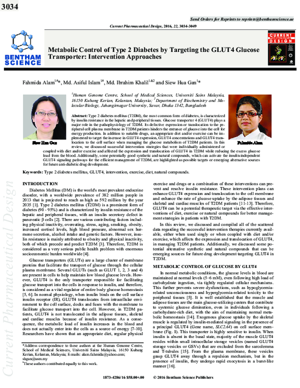
 Siew Hua Gan