ANTICANCER RESEARCH 37: 1091-1098 (2017)
doi:10.21873/anticanres.11421
Quantitative Structure–Cytotoxicity Relationship of Chalcones
HIROSHI SAKAGAMI1,2, YOSHIKO MASUDA2, MINEKO TOMOMURA2, SATOSHI YOKOSE3,
YOSHIHIRO UESAWA4, NARUHIKO IKEZOE5, DAIKI ASAHARA5, KOICHI TAKAO5, TAISEI KANAMOTO6,
SHIGEMI TERAKUBO6, HAJIME KAGAYA4, HIDEKI NAKASHIMA6 and YOSHIAKI SUGITA5
1Division
of Pharmacology, 2Meikai Pharmaco-Medical Laboratory (MPL) and
of Endodontics, Meikai University School of Dentistry, Sakado, Japan;
4Department of Clinical Pharmaceutics, Meiji Pharmaceutical University, Tokyo, Japan;
5Faculty of Pharmaceutical Sciences, Josai University, Sakado, Japan;
6Department of Microbiology, St. Marianna University School of Medicine, Kanagawa, Japan
3Division
Abstract. Background: Fifteen chalcones were subjected to
quantitative structure–activity relationship (QSAR) analysis
based on their cytotoxicity and tumor specificity, in order to find
their new biological activities. Materials and Methods:
Cytotoxicity against four human oral squamous cell carcinoma
cell lines and three oral mesenchymal cells was determined by
the 3-(4,5-dimethylthiazol-2-yl)-2,5-diphenyltetrazolium bromide
(MTT) method. Tumor specificity (TS) was evaluated by the ratio
of the mean 50% cytotoxic concentration (CC50) against normal
cells to that against tumor cell lines. Potency-selectivity
expression (PSE) value was calculated by dividing TS by CC50
against tumor cells. Apoptosis markers were detected by western
blot analysis. Physicochemical, structural and quantumchemical parameters were calculated based on the
conformations optimized by force-field minimization. Results:
Among 15 chalcone derivatives, (2E)-1-(2,4-dimethoxyphenyl)3-(4-methoxyphenyl)-2-propen-1-one had the highest TS and
PSE values, comparable with those of doxorubicin and
methotrexate, respectively. This compound also stimulated the
cleavage of poly(ADP-ribose) polymerase and caspase-3.
Chalone TS values were correlated with molecular shape and
polarization rather than the types of substituted groups. None of
the compounds had any anti-HIV activity. Conclusion: Chemical
modification of the lead compound may be a potential choice for
designing new types of anticancer drugs.
This article is freely accessible online.
Correspondence to: Hiroshi Sakagami, Division of Pharmacology,
Department of Diagnostic and Therapeutic Sciences, Meikai University
School of Dentistry, Sakado, Saitama 350-0283, Japan. Tel: +81
492792758, Fax: +81 492855171, e-mail: sakagami@dent.meikai.ac.jp
Key Words: Chalcones, QSAR analysis, cytotoxicity, tumor
selectivity, apoptosis induction, anti-HIV activity.
Chalcone has a structure of 1,3-diaryl-2-propen-1-one in
which the two aromatic rings are joined by a three-carbon
α,β-unsaturated carbonyl system, representing a class of
flavonoids that occur naturally in fruits and vegetables.
Chalcones are also metabolic precursors of some flavonoids
and isoflavonoids (1). Chalcones are promising lead
antitumor/chemopreventive drugs due to three different
activities: antioxidant, cytotoxic, and apoptosis induction (2).
Several studies with murine xenograft models have shown
that administration of chalcones significantly reduced the
tumor volume by inducing apoptosis (3-10). The tumor
specificity of chalcones has been reported in comparing
sensitivity of hepatocarcinoma HepG2 cells to normal liver
AML12 cells (11); osterosarcoma to bone marrow and smallintestinal epithelial cells (12); murine acute lymphoblastic
leukemia cells L-1210 to normal human lymphocytes (13);
and human prostate cancer cells PC3 and DU145 to normal
human prostate epithelial cells (14). Although chalcones have
been reported to induce apoptosis of human oral carcinoma
cell line (HSC-3) (15) and cultured primary and metastatic
oral cancer cell lines (16), the tumor specificity against these
has not been investigated as far as we are aware of.
In order to find new types of anticancer drugs active
against human oral cancer, we first investigated the tumor
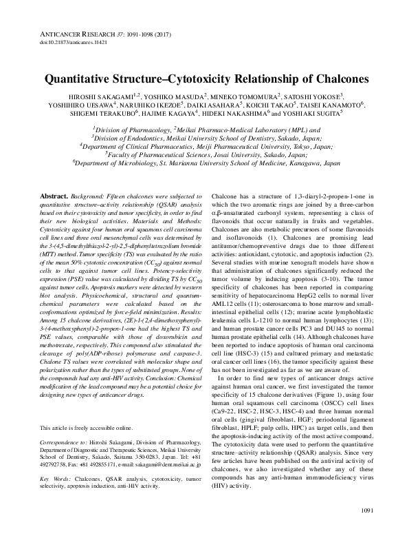
specificity of 15 chalcone derivatives (Figure 1), using four
human oral squamous cell carcinoma (OSCC) cell lines
(Ca9-22, HSC-2, HSC-3, HSC-4) and three human normal
oral cells (gingival fibroblast, HGF; periodontal ligament
fibroblast, HPLF; pulp cells, HPC) as target cells, and then
the apoptosis-inducing activity of the most active compound.
The cytotoxicity data were used to perform the quantitative
structure–activity relationship (QSAR) analysis. Since very
few articles have been published on the antiviral activity of
chalcones, we also investigated whether any of these
compounds has any anti-human immunodeficiency virus
(HIV) activity.
1091
�ANTICANCER RESEARCH 37: 1091-1098 (2017)
phenyl)-2-propen-1-one (5), (2E)-3-(4-chlorophenyl)-1-(2-hydroxyphenyl)-2-propen-1-one (6), (2E)-3-(4-bromophenyl)-1-(2-hydroxyphenyl)-2-propen-1-one (7), (2E)-1-(2-hydroxy-4-methoxyphenyl)3-(4-hydroxyphenyl)-2-propen-1-one (8), (2E)-1-(2-hydroxy-4methoxyphenyl)-3-phenyl-2-propen-1-one (9), (2E)-1-(2-hydroxy-4methoxyphenyl)-3-(4-methoxyphenyl)-2-propen-1-one (10), (2E)-3(3,4-dimethoxyphenyl)-1-(2-hydroxy-4-methoxyphenyl)-2-propen-1one (11), (2E)-3-(4-bromophenyl)-1-(2-hydroxy-4-methoxyphenyl)2-propen-1-one (12), (2E)-1-(4-methoxyphenyl)-3-(4-methoxyphenyl)-2-propen-1-one (13), (2E)-3-(2,4-dimethoxyphenyl)-1-(4methoxyphenyl)-2-propen-1-one (14), (2E)-1-(2,4-dimethoxyphenyl)-3-(4-methoxyphenyl)-2-propen-1-one (15) (structures shown
in Figure 1) were synthesized by base-catalyzed condensation of the
appropriate acetophenone with selected benzaldehyde derivatives
according to previous methods (17). All compounds were dissolved
in DMSO at 40 mM and stored at –20˚C before use.
Cell culture. Human normal oral mesenchymal cells (HGF, HPLF,
HPC), established from the first premolar tooth extracted from the
lower jaw of a 12-year-old girl (18), and human OSCC cell lines
[Ca9-22 (derived from gingival tissue); HSC-2, HSC-3, HSC-4
(derived from tongue)], purchased from Riken Cell Bank (Tsukuba,
Japan), were cultured at 37˚C in DMEM supplemented with 10%
heat-inactivated FBS, 100 units/ml, penicillin G and 100 μg/ml
streptomycin sulfate under a humidified 5% CO2 atmosphere. HGF,
HPLF and HPC cells at 10-18 population doubling levels were used
in the present study.
Figure 1. Structure of fifteen chalcones.
Materials and Methods
Materials. The following chemicals and reagents were obtained
from the indicated companies: Dulbecco’s modified Eagle’s medium
(DMEM), from GIBCO BRL, Grand Island, NY, USA; fetal bovine
serum (FBS), 3-(4,5-dimethylthiazol-2-yl)-2,5-diphenyltetrazolium
bromide (MTT), doxorubicin, azidothymidine, 2’,3’-dideoxycytidine
from Sigma-Aldrich Inc., St. Louis, MO, USA; dimethyl sulfoxide
(DMSO), dextran sulfate (molecular mass, 5 kDa) from Wako Pure
Chem. Ind., Osaka, Japan; methotrexate from Nacalai Tesque, Inc.,
Kyoto, Japan; curdlan sulfate (molecular mass: 79 kDa) from
Ajinomoto Co. Ltd., Tokyo, Japan. Culture plastic dishes and plates
(96-well) were purchased from Becton Dickinson (Franklin Lakes,
NJ, USA).
Synthesis of test compounds. (2E)-1-(2-Hydroxyphenyl)-3-phenyl-2propen-1-one (1), (2E)-1-(2-hydroxyphenyl)-3-(4-hydroxyphenyl)-2propen-1-one (2), (2E)-1-(2-hydroxyphenyl)-3-(4-methoxyphenyl)2-propen-1-one (3), (2E)-3-(3,4-dimethoxyphenyl)-1-(2-hydroxyphenyl)-2-propen-1-one (4), (2E)-3-(4-fluorophenyl)-1-(2-hydroxy-
1092
Assay for cytotoxic activity. Cells were inoculated at 2.5×10³
cells/0.1 ml in a 96-microwell plate (Becton Dickinson Labware,
Franklin Lakes, NJ, USA).After 48 h, the medium was replaced
with 0.1 ml of fresh medium containing different concentrations of
single test compounds. Cells were incubated further for 48 h and
the relative viable cell number was then determined by the MTT
method (19). The relative viable cell number was determined by the
absorbance of the cell lysate at 562 nm, using a microplate reader
(Sunrise Rainbow RC-R; TECAN, Männedorf, Switzerland).
Control cells were treated with the same amounts of DMSO and the
cell damage induced by DMSO was subtracted from that induced
by test agents. The concentration of compound that reduced the
viable cell number by 50% (CC50) was determined from the dose–
response curve and the mean value of CC50 for each cell type was
calculated from duplicate assays.
Calculation of tumor-selectivity index (TS). TS was calculated using
the following equation: TS=mean CC50 against normal cells/mean
CC50 against tumor cells [(D/B) in Table I]. Since both Ca9-22 and
HGF cells were derived from the gingival tissue (20), the relative
sensitivity of these cells was also compared [(C/A) in Table I]. We
have confirmed that the TS value thus determined reflects the
antitumor potential of test samples, although normal and tumor cells
are derived from different tissues (mesenchymal or epithelial
tissues, respectively) (21). We did not use human normal oral
keratinocytes as controls, since doxorubin and 5-fluorouracil
showed potent cytotoxicity against these epithelial cells by an as yet
unidentified mechanism (19, 22, 23).
Calculation of potency-selectivity expression (PSE). PSE was
calculated using the following equation: PSE=TS/CC50 against
tumor cells ×100 (24) [that is, (D/B2) ×100 (HGF, HPLF, HPC vs.
�Sakagami et al: QSAR of Chalcones
Table I. Cytotoxic activity of 15 chalcones against human oral malignant and non-malignant cells. Each value represents the mean of duplicate
determinations.
CC50 (μM)
Human oral squamous cell carcinoma cell lines
(A)
Compound Ca9-22
Human normal oral cells
TS
PSE
(D/B2) (C/A2)
×100
×100
HSC-2
HSC-3
HSC-4
(B)
Mean
SD
(C)
HGF
HPLF
HPC
(D)
Mean
SD
(D/B) (C/A)
1
2
3
4
5
6
7
8
9
10
11
12
13
14
15
18.2
31.2
21.7
24.2
15.1
8.7
9.5
29.6
9.8
21.5
14.6
10.3
15.8
15.8
4.2
28.6
53.0
36.2
45.7
22.2
13.2
10.0
32.7
11.2
23.1
20.9
11.0
21.8
30.0
6.6
19.2
29.4
21.0
26.0
13.4
10.1
9.9
27.8
10.7
21.6
15.4
10.1
18.9
17.9
3.9
33.2
51.0
42.0
41.1
26.6
16.4
14.3
44.6
20.5
40.2
31.8
19.4
28.7
19.8
<3.1
24.8
41.1
30.2
34.2
19.3
12.1
10.9
33.6
13.0
26.6
20.7
12.7
21.3
20.8
<4.4
7.3
12.6
10.5
10.7
6.2
3.4
2.2
7.6
5.0
9.1
7.9
4.5
5.5
6.3
1.5
41.5
115.5
56.5
83.0
34.7
21.1
22.8
73.7
31.6
47.4
64.4
38.1
32.5
30.7
23.1
72.0
281.5
145.0
153.5
69.3
35.0
47.0
151.0
51.8
234.0
97.3
98.5
52.0
68.8
50.0
71.8
167.0
69.5
112.1
46.2
30.2
36.6
153.0
55.3
197.5
73.4
81.3
70.4
70.1
40.8
61.8
188.0
90.3
116.2
50.1
28.8
35.5
125.9
46.2
159.6
78.4
72.6
51.6
56.5
37.9
17.5
85.0
47.8
35.4
17.6
7.1
12.1
45.2
12.8
98.9
17.0
31.1
19.0
22.4
13.6
2.5
4.6
3.0
3.4
2.6
2.4
3.3
3.7
3.5
6.0
3.8
5.7
2.4
2.7
>8.6
2.3
3.7
2.6
3.4
2.3
2.4
2.4
2.5
3.2
2.2
4.4
3.7
2.1
1.9
5.6
10
11
10
10
13
20
30
11
27
23
18
45
11
13
>194
13
12
12
14
15
28
26
8
33
10
30
36
13
12
134
DXR
MTX
0.089
10.7
<0.078
10.9
<0.078
<7.8
<0.078
<7.8
<0.08
<9.3
0.006
1.8
0.17
203.0
0.64
962.5
0.54
1000.0
0.45
721.8
0.25
449.7
>5.5
>77.8
1.9
18.9
>6865
>838
2102
177
HGF, Human gingival fibroblast; HPC, human pulp cells; HPLF, human periodontal ligament fibroblast; Ca9-22 (derived from gingival tissue),
HSC-2, HSC-3 and HSC-4 (derived from tongue), oral squamous cell carcinoma cell lines; CC50, 50% cytotoxic concentration; DXR, doxorubicin;
MTX, methotrexate. TS, tumor-selectivity; PSE, potency-selectivity expression.
Ca9-22, HSC-2, HSC-3, HSC-4) and (C/A2) ×100 (HGF vs. Ca922 in Table II).
Table II. Anti-HIV activity of chalcones and chemotherapeutic agents.
Each value represents the mean of triplicate determinations.
Estimation of CC50 values. Since the CC50 values had a distribution
pattern close to a logarithmic normal distribution, we used the
pCC50 (i.e., the −log CC50) for the comparison of the cytotoxicity
between the compounds. The mean pCC50 values for normal cells
and tumor cell lines were defined as N and T, respectively (19).
Compound
Calculation of chemical descriptors. The 3D-structure of each
chemical structure (drawn by Marvin) was optimized by CORINA
Classic (Molecular Networks GmbH, Germany) and force-field
calculations (amber-10: EHT) in Molecular Operating Environment
(MOE) version 2014.09 (Chemical Computing Group Inc., Quebec,
Canada). The number of structural descriptors calculated from MOE
and Dragon 7.0 (Kode srl., Pisa, Italy) after the elimination of
overlapped descriptors were 295 and 2797, respectively.
The following 12 Dragon descriptors and 4 MOE descriptors
were significantly correlated with T, N and T-N.
Dragon descriptors (25): (a) B10[O-O]: Presence/absence of O O at topological distance 10; (b) CATS3D_10_DA: CATS3D
Donor-acceptor BIN 10 (10.000-11.000 Å); (c) F10[O-O]:
Frequency of O-O at topological distance 10; (d) VE2_H2: average
coefficient of the last eigenvector (absolute values) from reciprocal
squared distance matrix (2D matrix-based descriptors); (e) L3m: 3rd
component size directional WHIM index/weighted by mass (WHIM
descriptors); (f) L3s: 3rd component size directional WHIM
1
2
3
4
5
6
7
8
9
10
11
12
13
14
15
Positive controls
Dextran sulfate (μg/ml)
Curdlan sulfate (μg/ml)
Azidothymidine
2’,3’-Dideoxycytidine
CC50 (μM)
EC50 (μM)
SI
323.627
178.21
>400
387.87
80.73
200.04
34.44
38.78
205.35
191.73
369.64
32.54
235.92
142.54
>400
>400
>400
>400
>400
>400
>400
>400
>400
>400
>400
>400
>400
>400
>400
>400
<1
<1
><1
<1
<1
<1
<1
<1
<1
<1
<1
<1
<1
<1
><1
232.68
>1000
53.004
1858.629
0.777
0.172
0.026
1.113
300
>5805
2017
1670
CC50, 50% Cytotoxic concentration; EC50, 50% effective concentration;
SI: selectivity index (=CC50/EC50).
1093
�ANTICANCER RESEARCH 37: 1091-1098 (2017)
index/weighted by atomic ionization state (WHIM descriptors); (g)
HATS6p: leverage-weighted autocorrelation of lag 6/weighted by
polarizability (GETAWAY descriptors); (h) R5v+: R maximal
autocorrelation of lag 5/weighted by van der Waals volume
(GETAWAY descriptors); (i) R6p R autocorrelation of lag
6/weighted by polarizability (GETAWAY descriptors); (j) R6v: R
autocorrelation of lag 6/weighted by van der Waal’s volume
(GETAWAY descriptors); (k) RDF010s: Radial distribution function
- 010/weighted by atomic ionization state (RDF descriptors); (l)
RDF035u: Radial distribution function - 035/unweighted (RDF
descriptors); MOE descriptors: (m) vsurf_IW6: Hydrophilic integy
moment 6 in the vsurf_ descriptors which are similar to the VolSurf
descriptors (26); (n) h_logS: Log of the aqueous solubility (mol/L)
using a 7 parameter model based on Hueckel theory (27); (o)
PEOE_VSA-6: Sum of vi where qi is less than –0.30 in the partial
equalization of orbital electronegativities (PEOE) method of
calculating atomic partial charges (27); (p) Q_VSA_PNEG: Total
negative polar van der Waals surface area (28).
Western blot analysis. The cells were washed with PBS and
processed for western blot analysis, as described previously (29).
Antibodies against cleaved caspase-3 (Cell Signaling Technology
Inc., Beverly, MP, USA), poly(ADP-ribose) polymerase (PARP)
(Cell Signaling Technology Inc.) and glyceraldehyde 3-phosphate
dehydrogenase (GAPDH; Trevigen, Gaithersburg, MD, USA) were
used as primary antibodies. As secondary antibodies, we used αrabbit IgG (DAKO Japan) antibodies which were conjugated with
horseradish peroxidase.
Assay for anti-HIV activity. HTLV-I-carrying human T-cell line MT4 cells, highly sensitive to human immunodeficiency virus-1 (HIV1), were infected with HIV-1IIIB at a multiplicity of infection of
0.01. HIV- and mock-infected (control) MT-4 cells were incubated
for 5 days with different concentrations of samples and the relative
viable cell number was determined by the MTT assay. The CC50
and 50% effective concentration (EC50) were determined from the
dose–response curve for mock-infected and HIV-infected cells,
respectively (30). All data represent the mean values of triplicate
measurements. The anti-HIV activity was evaluated by selectivity
index (SI) (=CC50/EC50).
Statistical treatment. The relation among cytotoxicity, tumor
specificity index, anti-UV activity and chemical descriptors was
investigated using simple regression analyses by JMP Pro version
12.2.0 (SAS Institute Inc., Cary, NC, USA). The significance level
was set at p<0.05.
Results
Cytotoxicity. A total of 15 chalcone derivatives (Figure 1)
generally showed higher cytotoxicity against human OSCC
lines (Ca9-22, HSC-2, HSC-3, HSC-4) (mean CC50=4.441.1 μM, mean 21.7 μM) (B) than against human
mesenchymal normal oral cells (HGF, HPLF and HPC)
(CC50=28.8-188.0 μM, mean 80.0 μM), yielding an
averaged TS of 3.7 (Table I). Among them, compounds 10,
12 and 15 had higher TS (5.7-8.6) than other compounds,
comparable to that of anticancer drugs, doxorubicin (5.5).
When tumor selectivity was calculated using cells both
1094
Figure 2. Apoptosis induction in HSC-2 human oral squamous cell
carcinoma cell line by compound 15.
derived from gingival tissue (Ca9-22 vs. HGF), 14
compounds 1-13 and 15 had much higher TS (2.1-5.6),
exceeding that of doxorubicin (1.9). Compound 15 had the
highest TS values in both cases.
In order to identify compounds which have both good
potency and are selectively toxic to neoplasms, the PSE
values for the compounds were calculated. When all three
normal cells (HGF, HPLF and HPC) and all four OSCC cell
lines (Ca9-22, HSC-2, HSC-3 and HSC-4) were used,
doxorubicin had the highest PSE value (>6,865), followed
by methotrexate > compound 15 > compounds 1-14 (Table
I). When HGF and Ca9-22 cells (both derived from gingival
tissues) were used, the same pattern was found. Compound
15 had the highest PSE value among the 15 chalcones,
approaching that of methotrexate. Western blot analysis
demonstrated that compound 15 stimulated the cleavage of
PARP and caspase-3, suggesting the induction of apoptosis
(Figure 2).
Anti-HIV activity of chalcones. In contrast to popular antiHIV agents (dextran sulfate, curdlan sulfate, azidothymidine,
2’,3’-dideoxycytidine) (SI=300-5,805), none of the chalcones
protected cells from the cytopathic effect of HIV infection
(SI<1) (Table II). Based on these data, the subsequent QSAR
analysis was focused on the cytotoxicity of chalcones.
Computational analysis. We next performed the QSAR
analysis of chalcone derivatives in regards to their
cytotoxicity against tumor cells and normal cells. Among a
total of 3,092 descriptors (295 MOE and 2797 Dragon
descriptors), 16 descriptors described below correlated well
with cytotoxicity and tumor specificity. Cytotoxicity of
chalcones against human OSCC cell lines was correlated with
HATS6p (polarizability) (r2=0.541, p=0.0018), vsurf_IW6
(hydrophilic interaction energy moment 6) (r2=0.537,
p=0.0019), R6v (van der Waal’s volume) (r2=0.524,
p=0.0023), R6p (polarizability) (r2=0.491, p=0.0036), h_logS
�Sakagami et al: QSAR of Chalcones
Figure 3. Determination of correlation coefficient between chemical descriptors and cytotoxicity of chalcones against tumor cells. The mean values
of pCC50 (i.e., the −log of the concentration causing 50% cytotoxicity) for tumor cell lines were defined as T.
(aqueous solubility) (r2=0.465, p=0.0051) and RDF035u
(spherically averaged information on the atomic correlation,
unweighted) (r2=0.455, p=0.0058) (Figure 3).
Cytotoxicity of chalcones against human normal oral
mesenchymal cells was correlated with CATS3D_10_DA
(donor-acceptor BIN) (r2=0.732, p<0.0001), RDF010s
(atomic
ionization
state)
(r2=0.696,
p=0.0001),
Q_VSA_PNEG (total negative polar van der Waals surface
area) (r2=0.633, p=0.0004), PEOE_VSA-6 (atomic partial
charges) (r2=0.633, p=0.0004), B10[O-O] (presence/absence
of O-O at topological distance 10) (r2=0.620, p=0.0005) and
F10[O-O] (frequency of O-O at topological distance 10)
(r2=0.620, p=0.0005) (Figure 4).
Tumor specificity of chalcones was correlated with R6p
(polarizability) (r2=0.601, p=0.0007), R5v+(van der Waal’s
volume) (r2=0.598, p=0.0007), L3m (mass) (r2=0.581,
p=0.0009), VE2_H2 (average coefficient of the last
eigenvector from reciprocal squared distance matrix) (r2=0.575,
p=0.0010), L3s (atomic ionization state) (r2=0.565, p=0.0012)
and HATS6p (polarizability) (r2=0.563, p=0.0013) (Figure 5).
Discussion
The present study demonstrated that 15 chalcones showed
relatively higher cytotoxicity against four OSCC cell lines
compared to that against human normal oral mesenchymal
oral cells; among them, compound 15 had the highest TS and
PSE values, although this is not a new compound. It should
be noted that the TS value of 15 was comparable with that
of doxorubicin, and the PSE value of 15 was comparable
with that of methotrexate (Table I). It is ideal to use human
epithelial cells as control normal cells in comparison with
OSCC cell lines. However, we recently found that
doxorubicin induced apoptosis in human oral keratinocytes
(i.e. loss of cell surface microvilli, chromatin condensation,
nuclear fragmentation, caspase-3 activation) at the
concentration that affected the viability of OSCC cell lines
(31). Until the mechanism of keratinocyte toxicity is clarified
and a preventive method is explored, the use of human oral
mesenchymal cells rather than normal epithelial cells may be
the only choice for us to use in comparison with tumor cells.
We calculated the possible contribution of substituted groups
to the expression of cytotoxicity against OSCC cell lines and
normal oral mesenchymal cells and tumor-specificity (Table
III). Most of the substituents listed did not affect these
activities (p=0.1067-0.9465) except for hydroxyl group
(p=0.0167) or oxygen (p=0.0248) at R5 in determining
cytotoxicity against normal cells. These data, suggest that
tumor specificity of chalcones was rather correlated with
molecular structure and polarization (Figure 5). We also
found that correlated parameters differed between tumor
cells and normal cells. For example, R6P (which represents
polarizability) is correlated with cytotoxicity against tumor
cells (Figure 3) and with tumor selectivity (Figure 5), but not
1095
�ANTICANCER RESEARCH 37: 1091-1098 (2017)
Figure 4. Determination of correlation coefficient between chemical descriptors and cytotoxicity of chalcones against normal cells. The mean values
of pCC50 (i.e., the −log of the concentration causing 50% cytotoxicity) for normal cells were defined as N.
Figure 5. Determination of correlation coefficient between chemical descriptors and tumor specificity of chalcones (defined as the difference between
the −log of the concentration causing 50% cytotoxicity in tumor cells and that for normal cells (T-N).
1096
�Sakagami et al: QSAR of Chalcones
Table III. Substituted groups that affect the cytotoxicity against OSCC
cell lines (T) and normal oral mesenchymal cells (N) and tumor
specificity (T-N).
T
T
T
T
T
T
T
T
N
N
N
N
N
N
N
N
T-N
T-N
T-N
T-N
T-N
T-N
T-N
T-N
Factor
p-Value
R1
R2Sbst
R2OH
R4
R5Sbst
R5OH
R5O
R5X
R1
R2Sbst
R2OH
R4
R5Sbst
R5OH
R5O
R5X
R1
R2Sbst
R2OH
R4
R5Sbst
R5OH
R5O
R5X
0.3433
0.7795
0.146
0.4231
0.9008
0.0851
0.2186
0.2054
0.9465
0.5165
0.2111
0.3247
0.5449
0.0167
0.0248
0.0538
0.1067
0.1729
0.6177
0.9208
0.5252
0.6722
0.3378
0.5744
so with cytotoxicity against normal cells (Figure 4). These
data indicate that an increase of polarizability of chalcones
may increase their antitumor potential.
The present study demonstrated that 15 chalcones did not
have any anti-HIV activity. This finding is not contradictory
with recent reports that chalcones exert anti-HIV activity
partially or in a very narrow range of concentrations (32, 33).
In conclusion, compound 15 is a potential lead compound
for synthesizing more potent compounds targeted to OSCC
cells.
Conflicts of Interest
We wish to confirm that there are no known conflicts of interest
associated with this publication and there has been no significant
financial support for this work that could have influenced its outcome.
Acknowledgements
This work was partially supported by KAKENHI from the Japan
Society for the Promotion of Science (JSPS) (15K08111,
16K11519). The annual license of the statistical software, JMP Pro,
was supported by the grant-in-aid of the oncology specialists
promotion program by the Ministry of Education, Culture, Sports,
Science and Technology, Japan.
References
1 Das M and Manna K: Chalcone scaffold in anticancer
armamentarium: a molecular insight. J Toxicol 2016: 7651047,
2016.
2 León-González AJ, Acero N, Muñoz-Mingarro D, Navarro I and
Martín-Cordero C: Review. Chalcones as promising lead
compounds on cancer therapy. Curr Med Chem 22(30): 34073425, 2015.
3 Wang LH, Li HH, Li M, Wang S, Jiang XR, Li Y, Ping GF, Cao
Q, Liu X, Fang WH, Chen GL, Yang JY and Wu CF: SL4, a
chalcone-based compound, induces apoptosis in human cancer
cells by activation of the ROS/MAPK signalling pathway. Cell
Prolif 48(6): 718-728, 2015.
4 Jiang C, Wang Q, Xu Z, Li WS, Chen C, Yao XQ and Liu FK:
Cyclooxygenase-2 knockdown using retinoic acid chalcone
(RAC), a promising therapeutic strategy for colon cancer. Am J
Cancer Res 5(6): 2012-2021, 2015.
5 Loch-Neckel G, Bicca MA, Leal PC, Mascarello A, Siqueira JM
and Calixto JB: In vitro and in vivo anti-glioma activity of a
chalcone-quinoxaline hybrid. Eur J Med Chem 90: 93-100,
2015.
6 Singh N, Sarkar J, Sashidhara KV, Ali S and Sinha S: Antitumour activity of a novel coumarin-chalcone hybrid is mediated
through intrinsic apoptotic pathway by inducing PUMA and
altering BAX/BCL-2 ratio. Apoptosis 19(6): 1017-1028, 2014.
7 Zhang Y, Srinivasan B, Xing C and Lü J: A new chalcone
derivative (E)-3-(4-methoxyphenyl)-2-methyl-1-(3,4,5-trimethoxyphenyl)prop-2-en-1-one suppresses prostate cancer involving
p53-mediated cell cycle arrests and apoptosis. Anticancer Res
32(9): 3689-3698, 2012.
8 Lin E, Lin WH, Wang SY, Chen CS, Liao JW, Chang HW, Chen
SC, Lin KY, Wang L, Yang HL and Hseu YC: Flavokawain B
inhibits growth of human squamous carcinoma cells:
Involvement of apoptosis and cell cycle dysregulation in vitro
and in vivo. J Nutr Biochem 23(4): 368-378, 2012.
9 Zi X and Simoneau AR: Flavokawain A, a novel chalcone from
kava extract, induces apoptosis in bladder cancer cells by
involvement of BAX protein-dependent and mitochondriadependent apoptotic pathway and suppresses tumor growth in
mice. Cancer Res 65(8): 3479-3486, 2005.
10 Hayashi A, Gillen AC and Lott JR: Effects of daily oral
administration of quercetin chalcone and modified citrus pectin
on implanted colon-25 tumor growth in Balb-c mice. Altern Med
Rev 5(6): 546-552, 2000.
11 Kuete V, Nkuete AH, Mbaveng AT, Wiench B, Wabo HK, Tane
P and Efferth T: Cytotoxicity and modes of action of 4’hydroxy-2’,6’-dimethoxychalcone and other flavonoids toward
drug-sensitive and multidrug-resistant cancer cell lines.
Phytomedicine 21(12): 1651-1657, 2014.
12 Ji T, Lin C, Krill LS, Eskander R, Guo Y, Zi X and Hoang BH:
Flavokawain B, a kava chalcone, inhibits growth of human
osteosarcoma cells through G2/M cell cycle arrest and apoptosis.
Mol Cancer 12: 55, doi:10.1186/1476-4598-12-55, 2013.
13 Pedrini FS, Chiaradia LD, Licínio MA, de Moraes AC, Curta JC,
Costa A, Mascarello A, Creczinsky-Pasa TB, Nunes RJ, Yunes
RA and Santos-Silva MC: Induction of apoptosis and cell cycle
arrest in L-1210 murine lymphoblastic leukaemia cells by (2E)3-(2-naphthyl)-1-(3’-methoxy-4’-hydroxy-phenyl)-2-propen-1one. J Pharm Pharmacol 62(9): 1128-1136, 2010.
1097
�ANTICANCER RESEARCH 37: 1091-1098 (2017)
14 Yun JM, Kweon MH, Kwon H, Hwang JK and Mukhtar H:
Induction of apoptosis and cell cycle arrest by a chalcone
panduratin A isolated from Kaempferia pandurata in androgenindependent human prostate cancer cells PC3 and DU145.
Carcinogenesis 27(7): 1454-1464, 2006.
15 Hseu YC, Lee MS, Wu CR, Cho HJ, Lin KY, Lai GH, Wang SY,
Kuo YH, Kumar KJ and Yang HL: The chalcone flavokawain B
induces G2/M cell-cycle arrest and apoptosis in human oral
carcinoma HSC-3 cells through the intracellular ROS generation
and downregulation of the AKT/p38 MAPK signaling pathway.
J Agric Food Chem 60(9): 2385-2397, 2012.
16 Lee YM, Jeong GS, Lim HD, An RB, Kim YC and Kim EC:
Isoliquiritigenin 2’-methyl ether induces growth inhibition and
apoptosis in oral cancer cells via heme oxygenase-1. Toxicol In
Vitro 24(3): 776-782, 2010.
17 Devakaram R, Black DS, Andrews KT, Fisher GM, Davis RA
and Kumar N: Synthesis and antimalarial evaluation of novel
benzopyrano[4,3-b]benzopyran derivatives. Bioorg Med Chem
19: 5199-5206, 2011.
18 Kantoh K, Ono M, Nakamura Y, Nakamura Y, Hashimoto K,
Sakagami H and Wakabayashi H: Hormetic and anti-radiation
effects of tropolone-related compounds. In Vivo 24: 843-852, 2010.
19 Sakagami H, Uesawa Y, Ishihara M, Kagaya H, Kanamoto T,
Terakubo S, Nakashima H, Takao K and Sugita Y: Quantitative
structure–cytotoxicity relationship of oleoylamides. Anticancer
Res 35: 5341-5355, 2015.
20 Horikoshi M, Kimura Y, Nagura H, Ono T and Ito H: A new
human cell line derived from human carcinoma of the gingiva.
I. Its establishment and morphological studies. Jpn J Oral
Maxillofac Surg 20: 100-106, 1974 (in Japanese).
21 Suzuki R, Matsuo S, Sakagami H, Okada Y and Shirataki Y:
Search of new cytotoxic crude materials against human oral
squamous cell carcinoma using NMR metabolomics. Anticancer
Res 34(8): 4117-4120, 2014.
22 Sakagami H, Shimada C, Kanda Y, Amano O, Sugimoto M, Ota
S, Soga T, Tomita M, Sato A, Tanuma S, Takao K and Sugita Y:
Effects of 3-styrylchromones on metabolic profiles and cell
death in oral squamous cell carcinoma cells. Toxocol Re 2:
1281-1290, 2015.
23 Uesawa Y, Sakagami H, Kagaya H, Yamashita M, Takao K and
Sugita Y: Quantitative structure-cytotoxicity relationship of 3benzylidenechromanones. Anticancer Res 36(11): 5803-5812,
2016.
1098
24 Das S, Das U, Sakagami H, Umemura N, Iwamoto S, Matsuta
T, Kawase M, Molnar J, Serly J, Gorecki DKJ and Dimmock JR:
Dimeric 3,5-bis(benzylidene)-4-piperidones : A novel cluster of
tumour-selective cytotoxins possessing multidrug-resistant
properties. Eur J Med Chem 51: 193-199, 2012.
25 https://chm.kode-solutions.net/products_dragon_descriptors.php
26 Cruciani G, Crivori P, Carrupt P-A and Testa B: Molecular
Fields In Quantitative Structure-Permeation Relationships: The
VolSurf Approach. J Mol Struct (Theochem) 503: 17-30, 2000.
27 Labute P: MOE h_mr, h_logP and h_logS Models unpublished.
Source code in $MOE/lib/svl/quasar.svl/q_eht.svl (2015).
28 Gasteiger J and Marsili M: Iterative partial equalization of
orbital electronegativity – a rapid access to atomic charges.
Tetrahedron 36: 3219, 1980.
29 Suzuki R, Matsushima Y, Okudaira N, Sakagami H and Shirataki
Y: Cytotoxic components against human oral squamous cell
carcinoma isolated from Andrographs paniculata. Anticancer
Res 36(11): 5931-5935, 2016.
30 Nakashima H, Murakami T, Yamamoto N, Sakagami H, Tanuma
S, Hatano T, Yoshida T and Okuda T: Inhibition of human
immunodeficiency viral replication by tannins and related
compounds. Antiviral Res 18(1): 91-103, 1992.
31 Sakagami H, Okudaira N, Masuda Y, Amano O, Yokose S,
Kanda Y, Suguro M, Natori T, Oizumi H and Oizumi T:
Induction of apoptosis in human oral keratinicyte by
doxorubicin. Anticancer Res 37(3): in press, 2017.
32 Pan W, Liu K, Guan Y, Tan GT, Hung NV, Cuong NM, Soejarto
DD, Pezzuto JM, Fong HH and Zhang H. Bioactive compounds
from Vitex leptobotrys. J Nat Prod 77(3): 663-667, 2014.
33 Cole AL, Hossain S, Cole AM and Phanstiel O 4th: Synthesis
and bioevaluation of substituted chalcones, coumaranones and
other flavonoids as anti-HIV agents. Bioorg Med Chem 24(12):
2768-2776, 2016.
Received December 21, 2016
Revised January 31, 2017
Accepted February 1, 2017
�
 Yoshihiro Uesawa