Neuron, Vol. 40, 643–653, October 30, 2003, Copyright 2003 by Cell Press
Functional Imaging of Perceptual Learning in Human
Primary and Secondary Somatosensory Cortex
Burkhard Pleger,1 Ann-Freya Foerster,2,5
Patrick Ragert,3,4,5 Hubert R. Dinse,3,*
Peter Schwenkreis,1 Jean-Pierre Malin,1
Volkmar Nicolas,2 and Martin Tegenthoff1
1
Department of Neurology
2
Department of Radiology
Ruhr-University Bochum
BG-Kliniken Bergmannsheil
D-44789 Bochum
Germany
3
Institute for Neuroinformatics
Department of Theoretical Biology
4
International Graduate School of Neuroscience
(IGSN)
Ruhr-University Bochum
D-44780 Bochum
Germany
Summary
Cellular mechanisms underlying synaptic plasticity are
in line with the Hebbian concept. In contrast, data linking
Hebbian learning to altered perception are rare. Combining functional magnetic resonance imaging with
psychophysical tests, we studied cortical reorganization in primary and secondary somatosensory cortex
(SI and SII) and the resulting changes of tactile perception before and after tactile coactivation, a simple type
of Hebbian learning. Coactivation on the right index finger (IF) for 3 hr lowered its spatial discrimination threshold. In parallel, blood-oxygen level-dependent (BOLD)
signals from the right IF representation in SI and SII
enlarged. The individual threshold reduction was linearly correlated with the enlargement in SI, implying
a close relation between altered discrimination and
cortical reorganization. Controls consisting of a single-site stimulation did not affect thresholds and cortical maps. Accordingly, changes within distributed cortical networks based on Hebbian mechanisms alter
the individual percept.
Introduction
Extensive use or training is paralleled by substantial
changes in cortical representations, thereby emphasizing the relevance of cortical plasticity for everyday life
(Braun et al., 2001; Buonomano and Merzenich, 1998;
Dinse and Merzenich, 2002; Elbert et al., 1995; PascualLeone and Torres, 1993; Recanzone et al., 1992; Recanzone, 2000). To study a particular form of perceptual
learning in parallel to cortical reorganization, we recently
introduced a coactivation protocol that closely followed
the idea of Hebbian learning: synchronous neural activity, necessary to drive plastic changes, is evoked by
tactile coactivation of the skin (Godde et al., 1996, 2000;
*Correspondence: hubert.dinse@neuroinformatik.ruhr-uni-bochum.de
5
These authors contributed equally to this work.
Pleger et al., 2001). From a number of animal studies,
the importance of temporally correlated inputs has been
assumed to play a key role in mediating plastic changes
(Clark et al., 1988; Fregnac et al., 1988). In fact, since
Hebb (1949), and even since James (1890), the aspect
of simultaneity has become a metaphor in neural plasticity. In the coactivation protocol, reorganization is driven
passively by manipulating the characteristics of the input statistics without invoking training of a task or cognitive factors such as attention or reinforcement.
In previous studies, we have demonstrated that a few
hours of coactivation of skin surfaces on the hindpaw
of adult rats resulted in reversible reorganization of somatosensory cortex (Godde et al., 1996): cortical maps
enlarged, thereby creating a joint representation of the
coactivated skin sites. To show that plastic changes
induced by coactivation bear relevance at the perceptual level, we assessed spatial tactile discrimination performance as a marker of plastic reorganization in human
subjects. In these experiments, discrimination thresholds were measured before and after coactivation of a
small skin region on the tip of the index finger. After
3 hr of coactivation, we found a lowering of thresholds,
an effect that was reversible within 24 hr (Godde et
al., 2000). Combining measurements of somatosensory
evoked potentials (SSEP) in primary somatosensory cortex (SI) and of tactile discrimination thresholds showed
a close correlation between the amount of coactivationinduced perceptual improvement and the degree of individual cortical reorganization (Pleger et al., 2001; Dinse
et al., 2003).
Most of our knowledge of plastic reorganization comes
from studies in primary somatosensory cortex (SI). We
therefore extended our approach of correlating psychophysical performance with cortical reorganization to the
investigation of plastic changes in secondary somatosensory cortex (SII). While in animal models maps can
be derived in great detail, evidence in humans is mostly
indirect, resulting from calculating the positions of current sources obtained from electrical or neuromagnetic
evoked potentials. We therefore used functional magnetic resonance imaging (fMRI) to assess the position
and the size of cortical finger representations in SI and
SII before and after tactile coactivation in order to demonstrate a link between cortical map changes and altered perception.
Results
Effects of Coactivation on
Discrimination Thresholds
We tested 14 right-handed subjects (4 male, 10 female;
mean age 31.6 years, SD ⫾ 3.7 years) in a 2-alternative
forced-choice simultaneous spatial 2-point discrimination task (Godde et al., 1996, 2000; Pleger et al., 2001;
P. Ragert et al., 2001, Soc. Neurosci., abstract; Dinse
et al., 2003); for experimental setup and design, see
Figure 1. Prior to coactivation, all subjects achieved a
stable 2-point discrimination performance as estimated
�Neuron
644
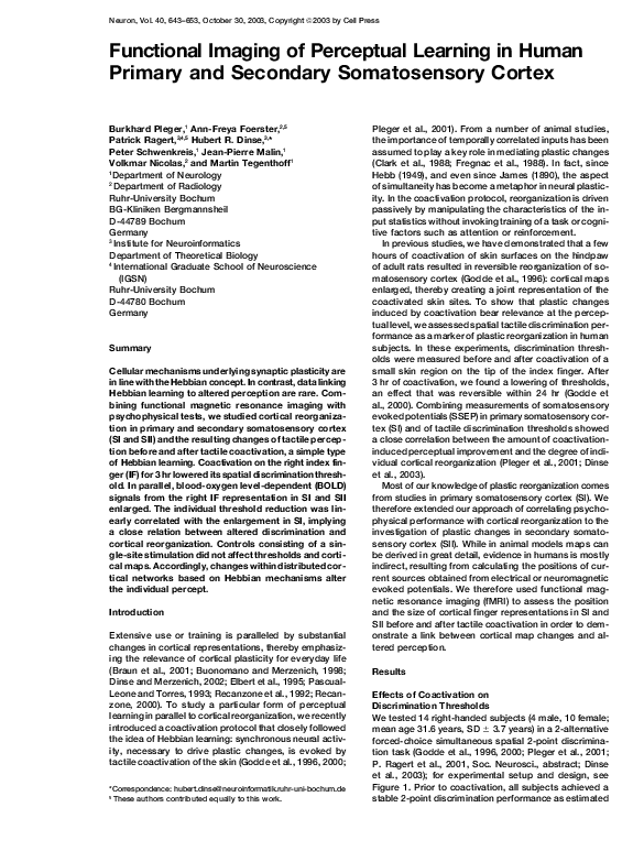
Figure 1. Coactivation Schedule and Procedure
(A) Experimental design. Sessions 1 to 5 (S1–S5) served to create a stable discrimination performance for the right IF. The left IF, serving as
control, is only tested at session S5 (precoactivation) and after coactivation (session S6, post). After S5, pre-fMRI measurements are performed,
and then coactivation of the right IF is applied for 3 hr. After the termination of coactivation and after the completion of session 6, post-fMRI
measurements are repeated. The seventh session (determination of 2-point discrimination thresholds and fMRI measurement) was performed
to assess the recovery of the coactivation-induced effect.
(B) Application of coactivation. A small solenoid with a diameter of 8 mm was mounted on the tip of the right IF to coactivate the receptive
fields representing the skin portion under the solenoid (50 mm2).
(C) Control protocol. Application of a so-called single-site stimulation: a small device consisting of only one tiny stimulator (tip diameter 0.5
mm) was mounted on the tip of the right IF to stimulate a single “point” (0.8 mm2) on the skin. Duration of stimulation was also 3 hr. The
experimental schedule was the same as described in (A) for coactivation; however, no recovery sessions were performed. Also, frequency
and duration of pulses were as described above for coactivation.
from repeated assessment of thresholds over five consecutive sessions (Figure 2A). After coactivation, discrimination thresholds were reduced from 1.58 ⫾ 0.20
mm (mean ⫾ SD) to 1.28 ⫾ 0.25 mm (repeated measures
pre versus post ANOVA: F(1,13) ⫽ 69.125; p ⬍ 0.0001,
pre-post difference t test p ⬍ 0.0001), but thresholds of
the not coactivated IF of the left hand remained unchanged (pre, 1.53 ⫾ 0.24 mm; post, 1.50 ⫾ 0.24 mm;
repeated measures ANOVA: F(1,13) ⫽ 1.128; p ⫽ 0.308,
pre-post difference t test p ⫽ 0.3, Figure 2B). We re-
investigated the recovery of coactivation in four subjects. The present findings corroborate those of earlier
studies (Godde et al., 1996, 2000; Pleger et al., 2001;
Dinse et al., 2003): 24 hr after coactivation, we found
normal 2-point discrimination thresholds similar to those
obtained prior to coactivation (1.55 ⫾ 0.26 mm [mean ⫾
SD]; Figure 2A).
To demonstrate the Hebbian nature of the coactivation protocol, we compared the effects of coactivation
with a so-called single-site stimulation in 16 subjects (7
Figure 2. Psychophysical Effects of Coactivation
(A) Effects of coactivation on discrimination
thresholds. Average data from all subjects
(n ⫽ 14). Dots represent mean thresholds,
boxes show the standard errors, and whiskers correspond to the standard deviation. Coactivation period (3 hr) of the right IF is indicated by arrows. Left: shown are results from
five consecutive sessions before coactivation
of the right index finger (IF). After the session
S5 (precondition), coactivation was applied.
After coactivation, discrimination thresholds
were significantly reduced.
(B) Discrimination thresholds obtained for the
control finger (IF of the left hand that was not
coactivated) for the pre- and postcondition. Note lack of effects after coactivation of the right IF (postcondition), indicating finger specificity
of the coactivation protocol. 24 hr after coactivation, we found normal 2-point discrimination thresholds similar to those obtained prior
to coactivation.
(C) Effects of single-site stimulation on discrimination thresholds. Average values from 16 subjects obtained for the right IF pre and post
single-site stimulation. Same conventions as in (A). We found no changes of discrimination thresholds after 3 hr of stimulation.
�Functional Imaging of Perceptual Learning
645
male, 9 female, all right-handed, mean age 26.3 ⫾ 3.66
years), where only a small “point-like” skin area was
stimulated. Otherwise, stimulation frequency and duration of stimulation period were the same as described
for coactivation. All subjects achieved a stable 2-point
discrimination performance as estimated from repeated
assessment of thresholds over five consecutive sessions. However, stimulating for 3 hr the tip of the right
IF with a single site only led to no changes of thresholds
(pre, 1.57 ⫾ 0.27 mm [mean ⫾ SD]; post, 1.58 ⫾ 0.27 mm,
repeated measures pre versus post ANOVA: F(1,15) ⫽
0.735, p ⫽ 0.408; Figure 2C). For individual subject psychometric curves, see Figure 5. These results imply that
“co”-activation is crucial for the induction of the effects.
Furthermore, we calculated the signal detection d’ value,
which is a bias-free discrimination index (Wickens,
2002), as well as the false alarm and hit rates (see Experimental Procedures). After coactivation, we found an increase of the d’ value from 2.33 ⫾ 0.29 (mean ⫾ SD) to
3.25 ⫾ 0.42, indicating enhanced discrimination abilities.
In contrast, d’ values before and after single-site stimulation remained unchanged (pre, 2.78 ⫾ 0.78; post, 2.94 ⫾
0.52). Comparing false alarm rates before and after coactivation (pre, 0.03 ⫾ 0.02; post, 0.01 ⫾ 0.01) and singlesite stimulation (pre, 0.02 ⫾ 0.03; post, 0 ⫾ 0) revealed
that they remained unchanged. However, hit rates increased after coactivation (pre, 0.66 ⫾ 0.08; post, 0.81 ⫾
0.04) but not after single-site stimulation (pre, 0.65 ⫾
0.03; post, 0.66 ⫾ 0.05).
Effects of Coactivation on BOLD Signal
For fMRI data analysis, we used the Statistical Parametric Mapping (SPM) software. The result of the group
analysis shown in the estimated statistic parametric
map of the precoactivation session revealed focused SI
activation in the postcentral gyrus. In addition, we found
a broad SII activation contralateral and ipsilateral to the
stimulated IF in the parietal operculum above the Sylvian
fissure (Figures 3A and 3B, see Figure 4A for an individual data set; for a summary of all individual fMRI data,
see Tables 1 and 2). For each subject, the coordinates
of maximal BOLD activity (see Table 1) could be assigned to either SI or SII according to the Talairach atlas
(Talairach and Tournoux, 1988). After coactivation, for
both SI and SII contralateral to the stimulated IF, we
found an enlargement of activation pattern and an enhancement of the amplitude of the BOLD signal (Figures
3C and 3D, Tables 1 and 2, Figure 4A for an individual
data set). According to the random-effect analysis, significantly activated areas were only localized in SI and
SII contralateral to the stimulated IF (Figure 3E). A lack
of changes in BOLD activity was found contralateral
to the control IF of the left hand (Figure 3F). A linear
correlation analysis between the normalized number of
activated voxels K (K ⫽ ((rightpost ⫺ rightpre) ⫺ (leftpost ⫺
leftpre))/rightpre) for SI and SII revealed a lack of relationship (r ⫽ ⫺0.55, p ⫽ 0.851).
To analyze the spatial distribution of the activated
pattern, we calculated the centers of cluster gravity
(CoG) before and after coactivation. The comparison of
the Euclidean distance between the pooled pre-post
CoG indicated a lateral shift by 11.9 mm for contralateral
SI (compare red marker position in glass brain view of
Figures 3A and 3D). Parallel to the recovery seen in
the psychophysical data, the effects of coactivation on
BOLD signals in SI and SII were reversible 24 hr after
coactivation (Figure 4, Table 2).
In six subjects, we additionally measured BOLD signals before and after single-site stimulation. For a single
subject data set together with its psychometric performance, see Figure 5. Comparing pre with post singlesite stimulation fMRI session revealed no significant
changes in BOLD signal extension and intensity in SI
and SII contralateral to the stimulated IF. These results
confirm that tactile stimulation of the tip of the IF over
several hours had no effects, either psychophysically
or cortically.
Correlation between Perceptual and BOLD
Signal Changes
To elucidate the relationship between coactivation-induced
changes in BOLD signals and the individual perceptual
changes in discrimination thresholds, we performed a
correlation analysis. This analysis showed a close link
between individual gain in performance and the degree
of reorganizational changes, which was restricted to
the contralateral SI digit representation (Figure 6A). In
contrast, no comparable correlation was found between
changes in individual performance and reorganization
within SII. These findings were corroborated by calculating Pearson’s linear correlation coefficients between
cortical enlargement (normalized number of activated
voxels K) and psychophysical threshold reduction (Figure 6B).
Discussion
Combining fMRI imaging with psychophysical assessment of discrimination thresholds demonstrates that the
involvement of somatosensory cortex in fast perceptual
learning is not restricted to SI but extends equally well
to the next hierarchical level at SII (Figures 3 and 4,
Tables 1 and 2). fMRI is widely used to study the organization of the human brain during operation by measuring
the BOLD signal, which makes use of the close link
between energy metabolism and neural activation. To
understand the relative contribution of several types of
neuronal signals to the hemodynamic response, local
field potentials and single- and multi-unit activity have
been compared to high spatiotemporal fMRI responses
recorded simultaneously in monkey visual cortex (Logothetis et al., 2001). Only local field potentials were significantly correlated with the hemodynamic response, indicating that BOLD signals primarily measure the input
and processing of neuronal information within a region
of cortex and not the output (Logothetis, 2002; Logothetis et al., 2001).
Coactivation Protocol
The coactivation is a task-free, passive stimulation protocol. It was developed to study systematically the impact of altered input statistics on plastic capacities of
cortical networks. The protocol induces cortical reorganization without invoking task training or cognitive factors such as attention or reinforcement. Many studies
have demonstrated that plastic cortical changes can be
�Neuron
646
Figure 3. Cortical Effects of Coactivation
(A–D) Fixed-effects analysis shows BOLD
signals detected pre (A and B) and post (C
and D) coactivation in the contralateral SI and
in the contralateral and ipsilateral SII. Coronar
slices are viewed from the back (LH, left hemisphere; RH, right hemisphere; y values, MNI
coordinates). Activations are projected on
coronal T1-weighted MRI slices. (A and B)
Precoactivation. S1 parameters: cluster
level ⫽ 255 v(oxels); T-score ⫽ 10.12; MNI
template coordinates (mm) ⫽ ⫺46, ⫺11, 50
(x,y,z); S2 (contralateral) parameters: 2316 v;
T-score ⫽ 12.34; ⫺48, ⫺28, 20 (x,y,z); S2
(ipsilateral) parameters (not shown): 382 v;
T-score ⫽ 11.23; 57, 2, 8 (x,y,z). (C and D)
Postcoactivation. S1 parameters: 1091 v;
T-score ⫽ 20.23; ⫺56, ⫺21, 50 (x,y,z); S2
(contralateral) parameters: 4354 v; T-score ⫽
23.34; ⫺46, ⫺30, 20 (x,y,z); S2 (ipsilateral) parameters (not shown): 562 v; T-score ⫽ 11.99;
52, 4, 8 (x,y,z). In order to obtain information
about the spatial distribution of the activated
pattern, we additionally calculated the centers of cluster gravity (CoG) before and after
coactivation: SI pre, ⫺48, ⫺11, 53 (x,y,z);
post, ⫺54, ⫺20, 48 (x,y,z); SII (contralateral)
pre, ⫺47, ⫺22, 18 (x,y,z); post, ⫺48, ⫺20, 14
(x,y,z); SII (ipsilateral) pre, 56, 2, 4 (x,y,z); post,
54, ⫺6, 8 (x,y,z). The comparison of the Euclidean distance between the pooled prepost CoG indicated a lateral shift by 11.9 mm
for contralateral SI (compare red marker position) and a medial shift of 9.17 mm for ipsilateral SII. Although contralateral SII signals
were also significantly enlarged, the Euclidean distance pre-post CoG showed a lateral
shift of only 4.58 mm.
(E) Random-effect analysis (paired t test pre
versus post, right IF) revealed significant
changes of activated patterns localized in
SI and SII contralateral to the coactivated
IF (threshold: p ⫽ 0.001; uncorrected for multiple comparisons; S1 parameters: 22 v; T-score ⫽ 5.92; ⫺44, ⫺18, 54 (x,y,z); S2 parameters:
three main patterns of activation: (1) 26 v; T-score ⫽ 4.5; ⫺60, ⫺26, 20 (x,y,z); (2) 12 v; T-score ⫽ 4.37; ⫺38, ⫺22, 14 (x,y,z); (3) 98 v; T-score ⫽
3.83; ⫺50, ⫺4, 4 (x,y,z).
(F) No changes of BOLD activity were found contralateral to the control IF of the left hand (paired t test pre versus post; threshold: p ⫽ 0.001,
uncorrected for multiple comparisons).
evoked by variation of input statistics alone, provided
the statistics are sufficiently altered (Diamond et al.,
1993; Kilgard et al., 2002; Liepert et al., 1999). For example, perceptual learning occurs even without awareness
by repetitive exposure to stimuli that are below the
threshold of visibility and that are irrelevant to the central
task (Watanabe et al., 2001).
Coactivation as used in the present study follows
closely the idea of Hebbian learning: synchronous neural
activity, which is regarded as instrumental to drive plastic changes, is generated by the simultaneous tactile
“costimulation.” Conceivably, coactivation modifies synaptic efficacy between and within the cortical neuron
pool representing the IF; for an account showing that
dendritic spikes mediate a form of synaptic potentiation
that does not require postsynaptic action potential firing
in the axon, see Golding et al. (2002). As a chain of
changes, we suggest that the simultaneous activation
on the skin alters synaptic transmission at a cortical
level, which results in an enlargement of the finger repre-
sentation in SI (Pleger et al., 2001; Dinse et al., 2003)
and SII. However, at present, little is known about how
cortical map changes translate to changes of discrimination performance. According to our data, enlargement
of cortical territory in SI is linearly correlated with a
lowering of spatial discrimination thresholds (Figure 6).
Several years ago, animal studies demonstrated a significant correlation between the enlargement of cortical
territory and the improvement in performance (Recanzone et al., 1992, 1993). Interestingly, clinical studies
showed that the degree of cortical maladaptations was
related to the degree of associated changes of behavior
(Flor et al., 1995; Traversa et al., 1997). What the present
and the previous studies have in common is the demonstration of a proportionality between performance and
cortical territory. In other words, recruitment of cortical
processing resources appears to be crucially involved
in mediating superior performance. Conceivably, small
differences in performance may not necessarily be due
to measurement artifacts or noise, but may reflect true
�Functional Imaging of Perceptual Learning
647
Figure 4. Single Subject Coactivation Effect
(A) BOLD signals detected pre, post, and 24 hr after coactivation in the contralateral SI in the postcentral gyrus and in the contralateral SII
in the parietal operculum above the Sylvian fissure. Activations are projected on an axial (left), saggital (middle), and coronar (right) T1weighted, normalized MRI slice. Comparing pre- with postcoactivation fMRI sessions revealed enlarged activation and increased BOLD signal
intensity in SI and SII contralateral to the coactivated IF. The slight increase in ipsilateral SII found in this subject and in several others did
not reach significance level in the group analysis. These changes of BOLD signal characteristics recovered 24 hr after coactivation was
applied. For individual parameters, see Table 1 (subject 1). According to the single-subject analysis, no multiple activation peaks were found
in SII, as shown in this example.
(B) Psychometric functions illustrating the coactivation-induced improvement of discrimination threshold for the subject shown in (A). Correct
responses in percent (red squares) are plotted as a function of separation distance together with the results of a logistic regression line (blue
with blue diamonds). 50% levels of correct responses are shown as well as thresholds. Top, precondition before coactivation; middle,
postcondition, immediately after coactivation; bottom, recovery after 24 hr. After coactivation there is a distinct shift in the psychometric
functions toward lower separation distances by 0.14 mm, which recovers to preconditions 24 hr later.
differences in individual brain organization. However,
in deprived animals the amount of active exploration
appeared to determine the direction of plastic changes,
suggesting that exceptions from that rule might exist
(Polley et al., 1999).
It is unlikely that unspecific factors such as overstimulation or coactivation-induced hypersensitivity might
cause systematic changes in 2-point discrimination. According to unpublished data, absolute touch thresholds
as assessed using von Frey hairs remained unchanged
after 3 hr of coactivation. As a control, to show the Hebbian
nature of the coactivation effects, we used a modified
version of the coactivation protocol consisting of a single small stimulation site instead of one large area (50
mm2 versus 0.8 mm2). When this protocol was applied
for 3 hr to the tip of the right IF, no effects, either on
discrimination or on cortical maps in SI and SII, were
found (Figure 5), indicating that “coactivation” is indeed
crucial. These findings also make it rather unlikely that
general processes such as attentional enhancement
were involved. The increase of the discrimination index
(d’ value) provides further support for the specificity of
the coactivation effect.
It should be noted that we additionally explored the
effects of a protocol of so-called uncorrelated coactivation using two independent small stimulators (separa-
tion 5 mm) driven by two independent Poisson processes. Under these conditions, simultaneity occurs by
chance. Synchronous coactivation lowered thresholds
as described here. In contrast, uncorrelated coactivation did not cause no effects, but instead revealed a
significant impairment of tactile performance (H.R.
Dinse et al., 2002, Soc. Neurosci., abstract). In parallel,
we made electrophysiological recordings in rats comparing a correlated and an uncorrelated coactivation
mode. These data showed that manipulating input correlations resulted in complementary forms of cortical
map reorganization. In addition, responsiveness of cortical neurons was potentiated after correlated, but depressed after uncorrelated, coactivation (H.R. Dinse et
al., 2002, Soc. Neurosci., abstract). According to these
data, uncorrelated coactivation is not a genuine “control,” but a new and different way to induce perceptual learning.
Due to the task-free nature of coactivation, it is reasonable to assume that coactivation alters the entire way
sensory information is processed. However, fundamental changes of tactile information processing do not rule
out that some aspects of tactile information processing
might deteriorate. According to unpublished data, tactile localization on the IF became worse after coactivation. A similar trade-off between localization and dis-
�Neuron
648
Table 1. Individual fMRI Data
SI Activity Contralateral to Coactivated Index Finger SII Activity Contralateral to Coactivated Index Finger
Height
Threshold
Subject Coactivation (T)
1
2
3
4
5
6
7
8
9
10
pre
post
pre
post
pre
post
pre
post
pre
post
pre
post
pre
post
pre
post
pre
post
pre
post
6.83
6.83
3.13
3.13
5.33
5.33
5.86
5.8
3.13
3.13
3.13
3.13
3.79
3.79
4.95
4.97
6.84
6.8
4.96
4.96
Cluster Level
of SI Activity
in Left
Hemisphere (kE)
Maximum of Activity
Summed Cluster
MNI Coordinates Voxel Level, Level of SII
MNI Coordinates Voxel Level,
(x,y,z in mm) of SI T-Score,
Activity in Left
(x,y,z in mm) of T-Score,
Pattern
SI Activity Hemisphere (kE) SII Pattern
SII Activity
51
174
13
103
15
199
40
208
61
197
19
50
20
69
142
304
238
467
37
50
⫺50,
⫺52,
⫺56,
⫺56,
⫺48,
⫺42,
⫺48,
⫺40,
⫺54,
⫺54,
⫺52,
⫺64,
⫺56,
⫺56,
⫺52,
⫺48,
⫺44,
⫺42,
⫺54,
⫺54,
⫺22, 50
⫺22, 50
⫺22, 52
⫺24, 52
⫺10, 58
⫺18, 50
⫺12, 60
⫺12, 64
⫺20, 46
⫺20, 46
⫺14, 54
⫺38, 40
⫺12, 50
⫺16, 50
⫺12, 56
⫺12, 56
⫺6, 48
⫺6, 48
⫺26, 54
⫺24, 54
12.08
12.32
4.09
8.32
6.4
8.8
7.44
13.32
4.18
4.18
4.5
4.42
4.91
6.37
9.23
9.2
13.81
12.8
8.75
9.63
372
946
139
85
219
357
60
113
42
158
70
32
477
684
689
2123
511
326
837
899
⫺36,
⫺38,
⫺38,
⫺36,
⫺44,
⫺54,
⫺52,
⫺54,
⫺56,
⫺38,
⫺50,
⫺50,
⫺52,
⫺42,
⫺44,
⫺44,
⫺52,
⫺60,
⫺52,
⫺42,
⫺20, 18
⫺18, 20
⫺26, 2
⫺10, 22
⫺38, 20
6, 2
0, 6
⫺2, 8
0, 14
⫺20, 16
⫺26, 20
⫺24, 22
⫺18, 20
⫺16, 14
⫺30, 24
⫺30, 22
4, 0
10, ⫺2
⫺30, 16
⫺26, 20
10.03
18.47
4.86
7.11
8.43
8.85
7.94
13.08
4.09
4.18
4.11
3.78
6.95
6.52
10.89
13.55
11.46
11.28
12.42
12.04
Second submaximum of Activity Third submaximum of Activity
MNI Coordinates
(x,y,z in mm) of
SII Pattern
⫺46,
⫺44,
⫺46,
⫺34,
⫺54,
⫺58,
⫺46,
⫺56,
⫺52,
⫺52,
⫺42,
⫺36,
⫺66,
⫺36,
⫺58,
⫺52,
⫺52,
⫺26, 22
⫺28, 20
⫺10, 14
⫺16, 8
⫺32, 14
⫺4, 10
⫺22, 14
0, 14
⫺10, 14
⫺8, 12
⫺14, 14
⫺10, 6
⫺32, 22
⫺22, 14
⫺14, 12
⫺26, 6
⫺30, 12
Voxel Level,
T-Score
MNI Coordinates
(x,y,z in mm) of Voxel Level,
SII Pattern
T-Score
9.87
14.25
4.04
5.52
7.02
8.02
12.98
4.09
3.95
3.55
6.32
5.03
9.1
11.35
10.3
7.2
10.16
⫺56,
⫺62,
⫺38,
⫺64,
⫺52,
⫺46,
⫺48,
⫺54,
⫺36,
⫺62,
⫺44,
⫺54,
⫺26, 20
⫺26, 20
⫺16, 12
⫺28, 14
⫺10, 14
0, 4
⫺8, 6
⫺18, 12
⫺22, 16
30, 26
⫺22, 20
⫺32, 22
8.84
13.82
3.9
6.89
7.81
12.62
3.92
3.42
7.05
10.9
9.87
9.53
This table shows the cluster level and the voxel level of activity pattern in primary (SI) and secondary (SII, cluster of maximum activity and the two main submaximums of activity) somatosensory cortex
pre- and postcoactivation.
�36
93
75
140
45
71
39
316
⫺48,
⫺40,
⫺46,
⫺44,
⫺54,
⫺58,
⫺52,
⫺54,
⫺21, 51
⫺22, 20
⫺8, 46
⫺24, 22
⫺12, 52
⫺18, 10
⫺12, 52
⫺26, 18
7.05
9.85
13.41
9.99
12.55
10.79
7.42
10.98
crimination has been shown in a study of Braille readers
(Sterr et al., 1998). Conceivably, enhanced spatial discrimination appears to emerge in parallel to impaired
localization. For a detailed discussion of the influence
of stimulus numbers, see Pleger et al. (2001).
5.21
9.22
8.47
4
3
2
SI
SII
SI
SII
SI
SII
SI
SII
1
This table shows the SI and SII activity contralateral to the coactivated index finger pre, post, and 24 hr after coactivation.
10.42
11.83
13.41
10.08
13.36
11.76
8.76
13.36
⫺18, 48
⫺24, 19
⫺6, 48
⫺22, 22
⫺6, 46
⫺22, 6
⫺22, 54
⫺24, 20
⫺50,
⫺45,
⫺46,
⫺44,
⫺54,
⫺60,
⫺52,
⫺54,
187
641
143
227
246
118
84
455
7.76
10.05
12.84
9.49
15.84
13.86
7.76
11.04
⫺20, 50
⫺22, 20
⫺6, 50
⫺24, 20
⫺10, 48
⫺42, 14
⫺24, 54
⫺32, 16
⫺48,
⫺42,
⫺44,
⫺44,
⫺56,
⫺64,
⫺52,
⫺54,
50
115
90
88
40
46
45
342
Cortex
Subject
6.83
MNI Coordinates
(x,y,z in mm) of
Activated Pattern
Cluster Level of
Activity in Left
Hemisphere (kE)
Cluster Level of
Activity in Left
Hemisphere (kE)
Height
Threshold (T)
MNI Coordinates
(x,y,z in mm) of
Activated Pattern
Voxel Level,
T-Score
Cluster Level of
Activity in Left
Hemisphere (kE)
MNI Coordinates
(x,y,z in mm) of
Activated Pattern
Voxel Level,
T-Score
(3) 24 Hours after Coactivation
(2) Postcoactivation
(1) Precoactivation
Recovery Measurements: SI and SII Activity Contralateral to the Coactivated Index Finger (Pre, Post, and 24 Hours after Coactivation)
Table 2. Recovery Measurements in Four Subjects
Voxel Level,
T-Score
Functional Imaging of Perceptual Learning
649
SI Reorganization
Cortical maps are in a permanent state of use-dependent fluctuations. According to animal studies, map size
is a reliable predictor of individual performance. Evidence in humans is mostly indirect, resulting from calculating the positions of current sources. For SI reorganization, our data revealed a linear relationship between
the degree of learning-induced perceptual improvement
and the degree of reorganization. Using the method of
fMRI imaging, we uncovered focused pattern of activation within SI, representing the correlation with the individual perceptual improvement (Figure 6). Accordingly,
little gain in discrimination abilities was associated with
small changes in BOLD signals, but subjects, who
showed a large cortical reorganization, also had a high
gain in discrimination.
These data confirm previous mapping studies in human SI using somatosensory-evoked potentials (SSEP)
(Pleger et al., 2001; Dinse et al., 2003): individually observed reorganization of SI was inferred from lateral
shifts of the N20-dipole localization of the IF, which were
linearly correlated with the parallel changes in spatial
2-point discrimination thresholds. Using fMRI, we found
a similar lateral shift for the center of gravity (Figures
3A–3D). This data demonstrates that following coactivation, a lateralization of the N20-dipole together with an
increase in dipole strength is compatible with an enlargement of cortical representational maps as described in the animal studies (Godde et al., 1996). Using
fMRI, we are now able to directly show the enlargement
of cortical territory together with a lateral shift of BOLD
signals as a consequence of coactivation, which provides further evidence for the correctness of the interpretation of dipole shifts inferred from SSEP mapping
(Pleger et al., 2001; Dinse et al., 2003). Accordingly, the
reorganization of the cortical representation of the IF in
SI after coactivation on the tip of the IF consists of an
asymmetric lateral enlargement toward the representation of the thumb, while the position of maximal activation remains at the location of the IF as mapped under
control conditions.
SII Reorganization
Besides the performance-dependent enlargement of SI,
we found that localized reorganizational changes can
be induced at the next hierarchical level at SII. While
lesion-induced reorganization has been observed outside SI (Pons et al., 1988), learning-induced plastic
changes have so far not been described for SII. In amputees, neuroelectric source imaging revealed decreased
activity in ipsilateral SII cortex associated with nonpainful phantom sensation (Flor et al., 2000). Using fMRI, an
enlarged bilateral SII representation of a telescoping
perception of the phantom limb was reported (CondesLara et al., 2000). In our study, under control conditions,
the patterns of SII activation were variable across subjects, indicating a less consistent somatotopic organization of SII (Tables 1 and 2; Del Gratta et al., 2000; Disbrow
�Neuron
650
Figure 5. Single-Site Stimulation Effects
(A) BOLD signals detected pre and post single-site stimulation in the contralateral SI and
SII in one subject. Activations are projected
on axial T1-weighted, normalized MRI slices.
Comparing pre with post single-site stimulation fMRI session revealed no substantial
changes in BOLD signal extension and intensity in SI and SII contralateral to the stimulated IF (SI pre: cluster level ⫽ 261 v(oxels),
T-score ⫽ 19.5; post: 281 v, T-score ⫽ 19.4;
SII (contralateral) pre: 236 v, T-score ⫽ 17.2;
post: 191 v, T-score ⫽ 15.9). Group data (n ⫽
6) is as follows. SI pre: mean cluster level ⫽
89 v (SEM ⫾ 36.1 v), mean T-score ⫽ 11
(⫾ 1.9); post: 96 v (⫾ 38.7 v; Student’s paired
t test pre versus post: p ⫽ 0.6), T-score ⫽ 12
(⫾ 2; pre versus post: p ⫽ 0.7); SII (contralateral) pre: 230 v (⫾ 138.9 v), T-score ⫽ 9
(⫾ 2.2); post: 176 v (⫾ 107 v; pre versus post:
p ⫽ 0.2), T-score ⫽ 9 (⫾ 2.1; pre versus post:
p ⫽ 0.17).
(B) Psychometric functions illustrating discrimination performance obtained pre and
post single-site stimulation for the subject
shown in (A). Correct responses in percent (red squares) are plotted as a function of separation distance together with the results of a logistic
regression line (blue with blue diamonds). 50% levels of correct responses are shown as well as thresholds. Top: precondition before singlesite stimulation; bottom: postcondition, immediately after single-site stimulation (prethreshold, 1.52 mm; postthreshold, 1.49 mm).
et al., 2000; Ruben et al., 2001; Simoes et al., 2001).
According to the random-effects analysis, no statistically significant pattern was found in SII ipsilaterally
(Figure 4E).
The missing correlation between SII enlargement and
the degree of discrimination improvement does not rule
out that SII is not involved in this task. According to a
recent fMRI study, no separated representations of the
index and fifth fingers were found in SII (Ruben et al.,
2001). Similarly, substantial functional overlap of finger
representations for human SII has been reported using
high-resolution MEG (Simoes et al., 2001). Conceivably,
the lack of fine-grained somatotopy together with the
substantial variability in activation might explain the lack
of correlation between individual reorganization in SII
and changes of discrimination thresholds. The enlargement of the digit representation in SII after coactivation
constitutes a recruitment of processing resources, suggesting a crucial role in processes related to improved
discrimination abilities as suggested by other studies
(Romo et al., 2002).
In conclusion, the concept of Hebbian learning has
Figure 6. Correlation Analysis
Relationship between changes in BOLD signals and coactivation induced changes of two-point discrimination thresholds (A). Results revealed
a significant correlation between perceptual and cortical changes within SI on the postcentral gyrus (see also magnified detail). In contrast,
no activated clusters were found within SII. (threshold: p ⫽ 0.001; uncorrected for multiple comparisons; v(oxel) ⫽ 2 mm3; S1 parameters:
cluster level ⫽ 14 v; T-score ⫽ 4.82; ⫺52, ⫺16, 50 (x,y,z). Linear correlation analysis between perceptual and cortical changes in SI (Pearson)
(B) corroborated these findings. To express the effect of tactile coactivation on BOLD signals of the contralateral SI, we used the corresponding
number of activated voxels per cluster: K ⫽ ((rightpost ⫺ rightpre) ⫺ (leftpost ⫺ leftpre))/rightpre. “K” was correlated with coactivation-induced changes
in psychophysical thresholds (r ⫽ 0.744; p ⫽ 0.002). We found no significant correlation between perceptual and plastic changes in SII (r ⫽
0.08; p ⫽ 0.979).
�Functional Imaging of Perceptual Learning
651
been proven to be one of the most productive paradigms
in neurosciences. While studies describing molecular
and cellular mechanisms of synaptic learning are in line
with the Hebbian ideas, data linking Hebbian learning
to altered perception are rare. Our results show that fMRI
is a valuable tool for demonstrating the implications of
fast perceptual learning based on Hebbian mechanisms
for cortical reorganizational changes within distributed
networks, which are correlated with changes of the individual percept.
Experimental Procedures
Experimental Schedule
The experiments consisted of four different components: (1) the
measurement of 2-point discrimination thresholds on the tip of the
left and right index finger (IF) as an indirect marker of cortical reorganization and as an indicator for the perceptual relevance of the
coactivation protocol; (2) fMRI measurements of BOLD images
evoked by electrical stimulation of the left and right IF; (3) the coactivation of 3 hr duration to induce cortical reorganization on the tip
of the right IF (Figure 1A); and (4) a control consisting of a singlesite stimulation.
As a rule, the IF of the right hand was used for coactivation. To
obtain a stable baseline of discrimination, we tested the subjects on
five consecutive sessions on the right IF. Sessions were statistically
analyzed for stability (ANOVA). In the fifth session, the thresholds
of the left IF were additionally measured. Previous studies had
shown that the coactivation effect to the right IF did not transfer to
the IF of the left hand (Godde et al., 1996, 2000; Pleger et al., 2001;
Dinse et al., 2003). We therefore used the IF of the left hand as a
control and for the assessment of possible unspecific side effects
of coactivation. In addition, the comparison of the thresholds of the
left IF pre-post ruled out the electrical stimulation during the fMRI
session having an effect on either the performance or the cortical
representation. Finally, the single-site stimulation on the right IF of
3 hr duration rules out that unspecific factors such as attentional
facilitation are unlikely to contribute to the coactivation effects.
After the assessment of discrimination performance of both the
test and the control finger (precondition), subjects were subjected
to fMRI measurements to obtain the pre-activation pattern (Figure
1A). Then coactivation was applied to the right IF (Figure 1B). Discrimination performance of the IF of each hand was retested starting
about 30 min after the termination of the coactivation protocol (postcondition). Then fMRI measurements were repeated to assess postactivation pattern. Previous studies had shown complete reversibility of coactivation-induced changes in discrimination performance
and in dipole localization after the termination of coactivation
(Godde et al., 1996, 2000; Pleger et al., 2001, Dinse et al., 2003). In
the present study, we investigated the recovery of coactivationinduced changes in four subjects perceptually and by using fMRI
(seventh session, Figure 1A).
Measurement of 2-Point Discrimination Thresholds
The study was performed in accordance with the Declaration of
Helsinki. The subjects gave their written informed consent, and the
protocol was approved by the local ethical committee of the RuhrUniversity Bochum.
We tested 14 right-handed subjects (4 male, 10 female; mean
age 31.6 years, SD ⫾ 3.7 years) in a 2-alternative forced-choice
simultaneous spatial 2-point discrimination task (Godde et al., 1996,
2000; Pleger et al., 2001; P. Ragert et al., 2001, Soc. Neurosci.,
abstract; Dinse et al., 2003). Seven pairs of needles were mounted on
a rotatable disc that allowed us to switch rapidly between distances.
Seven pairs of needles (diameter 200 m), separated by 0.7, 1.0,
1.3, 1.6, 1.9, 2.2, and 2.5 mm, were used and zero distance was
tested with a single needle.
To accomplish a rather uniform and standardized type of stimulation, the disc was installed in front of a plate that could be moved
up and down. The arm and fingers of the subjects were fixed on
the plate and the subjects were then asked to move the arm down.
The down movement was arrested by a stopper at a fixed position
above the needles. The test finger was held in a hollow containing
a small hole through which the finger touched the needles at approximately the same indentation in each trial. Each distance of the
needles was tested 10 times in randomized order, resulting in 80
single trials per session, which lasted about 15 min. Without being
given feedback, the subject had to decide immediately if he had
the sensation of one or two tips by answering “one” or “two.” The
subjects had been instructed that there were single needle presentations for control, but not how often. Thus, there was no knowledge
available to the subjects that could be used as a strategy to bias
the decisions.
The summed responses were plotted against distance as a psychometric function for absolute threshold, fitted by a binary logistic
regression (SPSS, SPSS Inc.). Threshold was taken from the fit at
the distance where 50% correct responses was reached. To provide
a direct experimental test for a bias-free discrimination index, we
additionally carried out psychophysical tests, where we presented
as many single needles as double needle presentations. We did this
by increasing the number of single needle presentations to 70 (70
single, 70 double needle presentations), resulting in a session with
a total of 140 presentations. From these data we calculated the
false alarm as well as the hit rates and the discrimination index (d’
value) (Wickens, 2002).
Coactivation
The coactivation protocol was the same as in our previous studies
(Godde et al., 1996, 2000; Pleger et al., 2001; P. Ragert et al., 2001,
Soc. Neurosci., abstract; Dinse et al., 2003). The basic idea behind
this design was to coactivate a large number of receptive fields in
a Hebbian manner in order to strengthen their mutual interconnectedness. Coactivation stimuli were presented at different interstimulus intervals from 100 to 3000 ms in pseudorandomized order; average stimulation frequency was 1 Hz and the duration of each pulse
was 10 ms. Pulses were recorded on tape and were played back
via portable tape recorders, allowing unrestrained mobility of the
subjects during coactivation. Subjects were instructed not to attend
the stimulation. In fact, all subjects resumed their normal day’s work.
To apply coactivation, a small solenoid with a diameter of 8 mm
was taped to the tip of the right index finger and transmitted the
tactile stimuli of the coactivation protocol to the skin. The solenoid
allowed simultaneous stimulation of the skin portions of the index
finger under the solenoid, leading to coactivation of all receptive
fields within this area, for an estimate of receptive field sizes of the
human index finger see (Figure 1B; Vega-Bermudez and Johnson,
1999). According to this data, receptive fields within 8 mm of the
tip of the index finger overlap partially or are nonoverlapping. Discrimination pre-post was always tested inside the coactivated skin
area. Coactivation stimuli were applied at suprathreshold intensities.
Duration of coactivation was 3 hr.
Single-Site Stimulation
To provide a control for the Hebbian nature of the coactivation and
to rule out that unspecific factors are unlikely to contribute to the
coactivation effects, we applied a so-called single-site stimulation.
For that purpose, we used a tiny stimulator that was also taped to
the tip of the right IF. The device consisted of a small probe (diameter
0.5 mm) that was moved up and down by means of a small relay
(Figure 1C). In that way, a single “point” (0.8 mm2) was stimulated
instead of coactivating a large area of 50 mm2. Stimulation parameters were identical to those used for coactivation: pulse duration
was 10 ms and average frequency was 1 Hz using interstimulus
intervals from 100 to 3000 ms in pseudorandomized order. Duration
of single-site stimulation was 3 hr.
fMRI Scanning
fMRI measurement was performed with a whole-body 1.5 T scanner
(Magnetom Symphony, Siemens Medical Systems, Germany) equipped
with a high-power gradient-system (30 mT/m/s; SR 125 T/m/s), using
a standard imaging head coil. Blood-oxygen level-dependent
(BOLD) images were obtained with a single-shot SpinEcho-EPI sequence (TR 1600 ms, TE 60 ms, matrix 64 ⫻ 64, field of view [FOV]
224 mm, 5 mm slice thickness, 1 mm gap between slices, voxel
3.5 ⫻ 3.5 ⫻ 5 mm). We acquired 16 transaxial slices parallel to the
�Neuron
652
AC-PC line, which covered the whole brain excluding cerebellum.
Ten subjects were examined before and after coactivation.
For finger stimulation we used a TENS stimulator (Medicommerz,
Kirchzarten, Germany) with conventional ring-electrodes (medco)
mounted on the tip of the index finger. For comparability of the
fMRI data with previously published SSEP (somatosensory evoked
potentials) data (Pleger et al., 2001; Dinse et al., 2003), we chose
an identical stimulation protocol as used in the SSEP studies: pulse
duration was 0.1 ms and repetition rate 3 Hz. Stimulation intensity
was adjusted to 2.5 times above sensory threshold. Electrodes were
removed between sessions, but position on the fingers was marked
to make sure that the same skin positions were stimulated. Each
activation study comprised nine blocks of rest and eight blocks
of stimulation, each of which contained 40 scans. Subjects were
instructed to keep their eyes closed and to concentrate on stimulation during the whole session. BOLD activation after electrical stimulation of the IF of each hand was measured in separate sessions
that were counterbalanced. Anatomical images were acquired using
an isotropic T1-3dGE (MPRAGE) sequence (TR 1790 ms, TE 388
ms, matrix 256 ⫻ 256, FOV 256 mm, 1 mm slice thickness, no gap,
voxel size: 1 ⫻ 1 ⫻ 1 mm) with 160 sagittal orientated slices covering
the whole brain.
Data Analysis
For preprocessing and statistical analysis of fMRI data, we used
the Statistical Parametric Mapping (SPM) software package, version
99 (Wellcome Department of Cognitive Neurology, London, UK) in
batch mode running under the MATLAB R12 environment (Mathworks Inc., Sherborn, MA). The first 10 images of each fMRI session
(690 images), during which the BOLD signal reaches steady state,
were discarded from further analysis. We measured both IFs in
separate sessions in pre and post conditions. First, all scans were
realigned and a mean image in the process was formed. Scans were
resliced using Sinc interpolation. Sessions in which movements exceeded 2 mm in any direction were removed from further analysis.
The individual three-dimensional data sets were normalized using
the standard template of the Montreal Neurological Institute (MNI)
(voxel size, 2 mm3) to establish an interindividual comparability
(Geyer et al., 2000). Scans were smoothed with a 6 mm (full-width
half-maximum) isotropic, three-dimensional Gaussian filter. Statistical maps were calculated using a high-pass cut-off at 256 s, a
hemodynamic response function (hrf; lowpass filter), and a threshold
of p ⫽ 0.05 (corrected for multiple comparisons). SPM contrast was
used for each subject and session separately. For the topographic
assignment of BOLD signals of the different measurements, we
coregistered the mean image formed in a realignment procedure
with the anatomic image from the T1-GE sequence scan. Statistical
parametric maps of group analysis were generated to identify localization, cluster level, center of cluster gravity (CoG), MNI coordinates, and spatial magnification of activated patterns.
Group analysis (fixed-effect model) was performed on all tested
subjects pre- and postcoactivation (Friston et al., 1999; Poldrack,
2000). Significance was determined using p ⫽ 0.001 for peak height
and corrected for multiple comparisons. To evaluate differences of
pre- and postcoactivation sessions, we performed a random-effect
analysis using Student’s paired t test of right and left sides separately. To analyze the relationship between coactivation-induced
changes in BOLD signals and the individual perceptual changes in
discrimination thresholds, we performed a simple correlation analysis. The effect of coactivation on BOLD signals was expressed by
side-corrected difference maps for each individual subject. The differences in thresholds before and after coactivation (post ⫺ pre)
were inserted as covariates. Linear correlation analysis (Pearson)
between individual pre- and post-coactivation maps were estimated
using equal thresholds for all sessions (p ⫽ 0.05; corrected for
multiple comparisons). To express the effect of tactile coactivation
on BOLD signal of contralateral SI and SII, we used the corresponding number of activated voxels per cluster which were included into
following formula: K ⫽ ((rightpost ⫺ rightpre) ⫺ (leftpost ⫺ leftpre))/rightpre.
Acknowledgments
This research was supported by the Deutsche Forschungsgemeinschaft (Di 334/10-3 to H.R.D. and M.T. and Di 334/15-1 to
H.R.D.) and by a grant from the Scientific Research Council of BGKliniken Bergmannsheil, Bochum (to V.N. and M.T.) and the Richard
Sackler Foundation (to A.-F.F.). We acknowledge excellent technical
support from M. Neef, A. Berg, M. Ziesmer, and W. Dreckmann. We
thank Dipl. Ing. Hannes Edelbrunner for his help in CoG analysis,
Dipl. Psychol. Claudia Wilimzig for her help in statistical analysis,
and R. Derrick for skilful editing of the text. We thank M. Rausch and
M. Rijntjes for critical reading of an earlier version of the manuscript.
Received: April 22, 2003
Revised: July 30, 2003
Accepted: October 7, 2003
Published: October 29, 2003
References
Braun, C., Heinz, U., Schweizer, R., Wiech, K., Birbaumer, N., and
Topka, H. (2001). Dynamic organization of the somatosensory cortex
induced by motor activity. Brain 124, 2259–2267.
Buonomano, D.V., and Merzenich, M.M. (1998). Cortical plasticity:
from synapses to maps. Annu. Rev. Neurosci. 21, 149–186.
Clark, S.A., Allard, T., Jenkins, W.M., and Merzenich, M.M. (1988).
Receptive fields in the body-surface map in adult cortex defined by
temporally correlated inputs. Nature 332, 444–445.
Condes-Lara, M., Barrios, F.A., Romo, J.R., Rojas, R., Salgado, P.,
and Sanchez-Cortazar, J. (2000). Brain somatic representation of
phantom and intact limb: a fMRI study case report. Eur. J. Pain
4, 239–245.
Del Gratta, C., Della Penna, S., Tartaro, A., Ferretti, A., Torquati, K.,
Bonomo, L., Romani, G.L., and Rossini, P.M. (2000). Topographic
organization of the human primary and secondary somatosensory
areas: an fMRI study. Neuroreport 11, 2035–2043.
Diamond, M.E., Armstrong-James, M., and Ebner, F.F. (1993). Experience-dependent plasticity in adult rat barrel cortex. Proc. Natl.
Acad. Sci. USA 90, 2082–2086.
Dinse, H.R., and Merzenich, M.M. (2002). Adaptatino of inputs in the
somatosensory system. In Perceptual Learning, M. Fahle and T.
Poggio, eds. (Cambridge, MA: MIT Press), pp. 19–42.
Dinse, H.R., Ragert, P., Pleger, B., Schwenkreis, P., and Tegenthoff,
M. (2003). Pharmacological modulation of perceptual learning and
associated cortical reorganization. Science 301, 91–94.
Disbrow, E., Roberts, T., and Krubitzer, L. (2000). Somatotopic organization of cortical fields in the lateral sulcus of Homo sapiens:
evidence for SII and PV. J. Comp. Neurol. 418, 1–21.
Elbert, T., Pantev, C., Wienbruch, C., Rockstroh, B., and Taub, E.
(1995). Increased cortical representation of the fingers of the left
hand in string players. Science 270, 305–307.
Flor, H., Elbert, T., Knecht, S., Wienbruch, C., Pantev, C., Birbaumer,
N., Larbig, W., and Taub, E. (1995). Phantom-limb pain as a perceptual correlate of cortical reorganization following arm amputation.
Nature 375, 482–484.
Flor, H., Muhlnickel, W., Karl, A., Denke, C., Grusser, S., Kurth, R.,
and Taub, E. (2000). A neural substrate for nonpainful phantom limb
phenomena. Neuroreport 11, 1407–1411.
Fregnac, Y., Shulz, D., Thorpe, S., and Bienenstock, E. (1988). A
cellular analogue of visual cortical plasticity. Nature 333, 367–370.
Friston, K.J., Holmes, A.P., and Worsley, K.J. (1999). How many
subjects constitute a study? Neuroimage 10, 1–5.
Geyer, S., Schormann, T., Mohlberg, H., and Zilles, K. (2000). Areas
3a, 3b, and 1 of human primary somatosensory cortex. Part 2. Spatial
normalization to standard anatomical space. Neuroimage 11,
684–696.
Godde, B., Spengler, F., and Dinse, H.R. (1996). Associative pairing
of tactile stimulation induces somatosensory cortical reorganization
in rats and humans. Neuroreport 8, 281–285.
Godde, B., Stauffenberg, B., Spengler, F., and Dinse, H.R. (2000).
Tactile coactivation-induced changes in spatial discrimination performance. J. Neurosci. 20, 1597–1604.
Golding, N.L., Staff, N.P., and Spruston, N. (2002). Dendritic spikes
�Functional Imaging of Perceptual Learning
653
as a mechanism for cooperative long-term potentiation. Nature
418, 326–331.
Wickens, T.D. (2002). Elementary Signal Detection Theory (New
York: Oxford University Press).
Hebb, D.O. (1949). The Organization of Behaviour (New York: Wiley
and Sons).
Watanabe, T., Nanez, J.E., and Sasaki, Y. (2001). Perceptual learning
without perception. Nature 413, 844–848.
James, W. (1890). Psychology: Brief Course (Cambridge, MA: Harvard University Press).
Kilgard, M.P., Pandya, P.K., Engineer, N.D., and Moucha, R. (2002).
Cortical network reorganization guided by sensory input features.
Biol. Cybern. 87, 333–343.
Liepert, J., Terborg, C., and Weiller, C. (1999). Motor plasticity induced by synchronized thumb and foot movements. Exp. Brain Res.
125, 435–439.
Logothetis, N.K. (2002). The neural basis of the blood-oxygen-leveldependent functional magnetic resonance imaging signal. Philos.
Trans. R. Soc. Lond. B Biol. Sci. 357, 1003–1037.
Logothetis, N.K., Pauls, J., Augath, M., Trinath, T., and Oeltermann,
A. (2001). Neurophysiological investigation of the basis of the fMRI
signal. Nature 412, 150–157.
Pascual-Leone, A., and Torres, F. (1993). Plasticity of the sensorimotor cortex representation of the reading finger in Braille readers.
Brain 116, 39–52.
Pleger, B., Dinse, H.R., Ragert, P., Schwenkreis, P., Malin, J.P.,
and Tegenthoff, M. (2001). Shifts in cortical representations predict
human discrimination improvement. Proc. Natl. Acad. Sci. USA
98, 12255–12260.
Poldrack, R.A. (2000). Imaging brain plasticity: conceptual and methodological issues—a theoretical review. Neuroimage 12, 1–13.
Polley, D.B., Chen-Bee, C.H., and Frostig, R.D. (1999). Two directions of plasticity in the sensory-deprived adult cortex. Neuron
24, 623–637.
Pons, T.P., Garraghty, P.E., and Mishkin, M. (1988). Lesion-induced
plasticity in the second somatosensory cortex of adult macaques.
Proc. Natl. Acad. Sci. USA 85, 5279–5281.
Recanzone, G. (2000). Cerebral cortical plasticity: perception and
skill acquisition. In The New Cognitive Neurosciences, M.S. Gazzaniga, ed. (Cambridge, MA: MIT Press), pp. 237–250.
Recanzone, G.H., Merzenich, M.M., Jenkins, W.M., Grajski, K.A.,
and Dinse, H.R. (1992). Topographic reorganization of the hand representation in cortical area 3b owl monkeys trained in a frequencydiscrimination task. J. Neurophysiol. 67, 1031–1056.
Recanzone, G.H., Schreiner, C.E., and Merzenich, M.M. (1993). Plasticity in the frequency representation of primary auditory cortex
following discrimination training in adult owl monkeys. J. Neurosci.
13, 87–103.
Romo, R., Hernandez, A., Zainos, A., Lemus, L., and Brody, C.D.
(2002). Neuronal correlates of decision-making in secondary somatosensory cortex. Nat. Neurosci. 5, 1217–1225.
Ruben, J., Schwiemann, J., Deuchert, M., Meyer, R., Krause, T.,
Curio, G., Villringer, K., Kurth, R., and Villringer, A. (2001). Somatotopic organization of human secondary somatosensory cortex.
Cereb. Cortex 11, 463–473.
Simoes, C., Mertens, M., Forss, N., Jousmaki, V., Lutkenhoner, B.,
and Hari, R. (2001). Functional overlap of finger representations in
human SI and SII cortices. J. Neurophysiol. 86, 1661–1665.
Sterr, A., Muller, M.M., Elbert, T., Rockstroh, B., Pantev, C., and
Taub, E. (1998). Perceptual correlates of changes in cortical representation of fingers in blind multifinger Braille readers. J. Neurosci.
18, 4417–4423.
Talairach, J., and Tournoux, P. (1988). Co-planar Stereotaxic Atlas
of the Human Brain (Stuttgart, Germany: Thieme).
Traversa, R., Cicinelli, P., Bassi, A., Rossigni, P.M., and Bernardi,
G. (1997). Mapping of motor cortical reorganization after stroke. A
brain stimulation study with focal magnetic pulses. Stroke 28,
110–117.
Vega-Bermudez, F., and Johnson, K.O. (1999). SA1 and RA receptive
fields, response variability, and population responses mapped with
a probe array. J. Neurophysiol. 81, 2701–2710.
�
 hubert dinse