Academia.edu no longer supports Internet Explorer.
To browse Academia.edu and the wider internet faster and more securely, please take a few seconds to upgrade your browser.
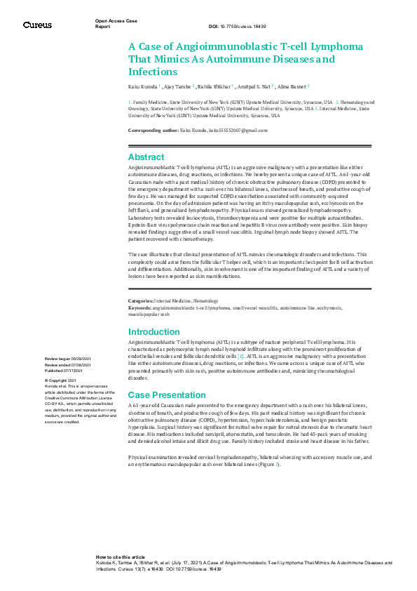
A Case of Angioimmunoblastic T-cell Lymphoma That Mimics As Autoimmune Diseases and Infections
A Case of Angioimmunoblastic T-cell Lymphoma That Mimics As Autoimmune Diseases and Infections
2021, Cureus
Related Papers
Clinical Medicine & Research
Angioimmunoblastic T-Cell Lymphoma with Polyarthritis Resembling Rheumatoid Arthritis2021 •
Angioimmunoblastic T-cell lymphoma (AITL) is an uncommon lymphoma of elderly adults with a poor prognosis. AITL patients show systemic symptoms, lymphadenopathy, and not infrequently, skin rash with various dysimmune phenomena rashes. The case is presented of a 68-year-old male with skin rash, lymphadenopathy and hypereosinophilia who, after investigations, was diagnosed with AITL. Despite the treatment used, the patient's condition gradually deteriorated and died due to heart and kidney failure. The diagnosis of AITL is often established only after several weeks or months because of transient physical findings, non-specific symptoms, and a broad range of serologic or radiologic abnormalities. Some patients with AITL experience non-specific dermatitis and eosinophilia. The presented case should raise awareness of the presentations of AITL which is important for physicians to reach an accurate diagnosis.
JAAD Case Reports
Development of a reticular rash in a febrile woman: An unusual cutaneous presentation of angioimmunoblastic T-cell lymphoma2015 •
Journal of Medical Cases
A Case of Angioimmunoblastic T-cell Lymphoma Hidden in Plain Sight: A Delay in Diagnosis and Management2022 •
Turkish journal of haematology : official journal of Turkish Society of Haematology
EBV-related Diffuse Large B-cell lymphoma in a Patient with Angioimmunoblastic T Cell Lymphoma: A Case Presentation2018 •
American Journal of Clinical Pathology
Epstein-Barr Virus-Associated B-Cell Lymphoproliferative Disorders in Angioimmunoblastic T-Cell Lymphoma and Peripheral T-Cell Lymphoma, Unspecified2002 •
Hannah Arendt - ¿Qué es la política? Comprensión y política
Hannah Arendt ¿Qué es la política? Comprensión y política2018 •
Prólogo “…La política, se dice, es una necesidad ineludible para la vida humana, tanto individual como social. Puesto que el hombre no es autárquico, sino que depende en su existencia de otros, el cuidado de ésta debe concernir a todos, sin lo cual la convivencia sería imposible.…” Hannah Arendt Procedente de una familia hebrea asentada en Hannover (Alemania), Hannah Arendt nace el 14 de octubre de 1906. Desde su infancia muestra una inteligencia precoz y un insistente interés por aprender el griego, a fin de poder leer a Kant, Kierkegaard y Karl Jaspers, quien dirigirá su tesis El concepto de amor en San Agustín, publicada en 1929, y con quien sostendrá una profunda amistad y mantendrá un constante intercambio intelectual, en toda su vida. Durante su vida académica, asistió a las cátedras impartidas por Martin Heidegger, considerado uno de los filósofos alemanes más influyentes del siglo xx, de quién absorbe gran parte de su obra y pensamiento, a tal grado que llegó a ocuparse de la traducción y publicación de sus obras en lengua inglesa. Sin embargo, a pesar de su gran admiración e influencia en sus años mozos, Arendt inicia una serie de investigaciones políticas y filosóficas que discreparán de los planteamientos de Heidegger y Jaspers. Sus investigaciones le llevan a replantear el modo en que se conducen los pensamientos filosóficos tradicionales y su reflexión sobre la política convulsiva del totalitarismo de la época. Su vivencia y reflexión personal, motivada inicialmente por ser atacada como judía, le hace buscar un sentido político y sus dimensiones universalizables, lo que quedó de manifiesto en siete de sus ensayos recopilados posteriormente en un libro titulado La tradición oculta. De ahí la importancia de poner en sus manos dos de sus obras más representativas del pensamiento filosófico-político de la autora: ¿Qué es política? y Comprensión y política (Las dificultades de la comprensión) donde, como Mary McCarthy comentaba en 1972: “…Hannah Arendt crea un espacio en el que uno puede caminar con la magnifica sensación de acceder, a través de un pórtico, a un área libre, pero, en buena parte, ocupada por definiciones…” Was ist Politik? Aus dem Nachlaß o ¿Qué es política?, nace como un proyecto para la creación de un libro que se titulará Introduction into Politics (Introducción a la política) por encargo de la editorial Klaus Piper; sin embargo, los múltiples compromisos de Arendt durante los años 1956 a 1959, como la publicación de las Walgreen Lectures y Between past and Future o Totalitarian Imperialism: Reflections on the Hungarian Revolution, así como su asistencia a Ámsterdam para pronunciar un discurso conmemorativo de la entrega Erasmus a Karl Jasper, entre otros, le impide continuar con el compromiso. Treinta cuatro años después, en 1993, es la socióloga Ursula Ludz, a solicitud de la casa Piper, quién se dedica a recopilar un sinnúmero de materiales e investigaciones escritos por Arendt, para darle forma a esta obra: ¿Qué es la política? La relevancia de estos escritos reside en que es un material único que nos ha permitido conocer lo que se denomina “ejercicios de pensamiento”, en los que la unidad de sus elementos no se presenta como la unidad de un todo, sino como secuencia de movimientos. Para Arendt, toda consideración sobre la política tiene que partir de un hecho ineludible: la pluralidad humana. Para ella la pluralidad de individuos únicos y diferenciados entre sí, es precisamente el elemento que la política debe preservar. Como consecuencia de su experiencia en la vivencia de un régimen totalitario y su exilio, a fin de escapar de las atrocidades que se cometen en Europa, concibe la creación de un espacio público mediante la acción concertada de las personas y el debate ciudadano, donde confluye la política y se manifiesta la libertad. Para Arendt, el sentido de la política es la realización de la libertad para todos los individuos que la conforman. La política es en sí un espacio donde se deben tratar los asuntos inherentes a todos los individuos que conforman la sociedad, y en ella, la política, es donde se concretarán las constituciones, leyes, estatutos e instituciones, que servirán para legislarlas, cuidarlas y hacer que todas las personas, gobernantes y gobernados, es decir, la sociedad entera, las cumplan debidamente sin manipulación alguna, para vivir en un verdadero Estado de Derecho. “…La comprensión (understanding), diferenciada de la información correcta y del conocimiento científico, es un proceso complicado que nunca produce resultados inequívocos. Es una actividad sin final, en constante cambio y variación, por medio de la cual aceptamos la realidad y nos reconciliamos con ella, esto es, intentamos sentirnos a gusto en el mundo…” En la segunda parte de este libro, el lector encontrará Comprensión y política (Las dificultades de la comprensión) «Understanding and Politics (The Difficulties of Undestanding», publicado en Partisan Review en 1953. Éste, es un compendio de artículos, conferencias, efemérides, comentarios, ensayos y artículos de fondo, basados en momentos históricos y biográficos de Arendt, en donde pone de relieve la pérdida de sentido de la sociedad occidental en el siglo pasado. Este material pretende mostrar que el “entendimiento” o comprensión aplicada a la política, debe tener como motor principal seguir buscando el sentido y la necesidad de vivir en armonía con el mundo y que, a pesar de fenómenos como el totalitarismo que abstrae el pensamiento político y los criterios de juicio moral, la comprensión debe ser el motor de la “búsqueda de sentido”, a fin de reconciliarnos con lo que hacemos y padecemos. Así, el análisis arendtiano sobre la comprensión nos refiere al juicio y a la construcción de una cultura crítica que responden a la necesidad de la acción política responsable. Para Arendt el mundo no puede interpretarse como algo relacionado exclusivamente con la naturaleza o el cosmos, sino que es el lugar de aparición de los individuos o el “espacio público” (die Offentligkeit) de encuentro entre ellos mismos y con los demás, donde la política es un espacio de relaciones humanas; lugar donde su ser coincide con su existir y sus cualidades: pluralidad, igualdad, libertad y derechos. Por otra parte, la acción es la actividad más original y libre que los hombres pueden realizar: “…una vida sin acción ni discurso está literalmente muerta para el mundo; ha dejado de ser una vida humana porque ya no la viven entre los hombres”. Así el simple hecho de la acción política es siempre esencialmente el inicio de algo nuevo, y en términos de la ciencia política, ésta es la verdadera esencia de la libertad humana. En consecuencia, es la otra cara del entendimiento que hace posible que los hombres puedan aceptar finalmente lo que ha ocurrido y reconciliarse con lo que irrevocablemente existe. «…Podemos comprender un suceso sólo como el final y culminación de todo lo que le ha precedido, como "la consumación de los tiempos”; sólo con la acción procedemos, de una forma natural, desde el conjunto de circunstancias renovadas que el acontecimiento ha creado, es decir, tratándola como un comienzo…» Manuel Cifuentes Vargas Secretario de Finanzas. CEN. PRD.
RELATED PAPERS
Il Perugino di San Pietro. La pala d'altare dell'abbazia benedettina di Perugia, a cura di L. Teza, catalogo della mostra, Perugia, Abbazia di San Pietro, 2 ottobre 2023 - 7 gennaio 2024, Milano, Cimorelli
Adorazioni di legno. Architettura lignea nella pittura di Pietro Perugino (con uno sguardo all'ultimo Raffaello)2023 •
Faculdades Integradas Rio Branco
A Rússia de Putin e o Espaço Pós-Soviético: Um Elo com o Passado2017 •
Revista Tensões Mundiais
Gaza: da Tempestade de al-Aqsa ao genocídio - Rev. Tensões MundiaisBiblio-Web para la Investigación Científica
Biblio-Web para la Investigacion Cientifica2023 •
Archives of Disease in Childhood
Continuous Ambulatory Peritoneal Dialysis in Children1985 •
Journal of Antimicrobial Chemotherapy
Impact of therapy and strain type on outcomes in urinary tract infections caused by carbapenem-resistant Klebsiella pneumoniae2014 •
The Egyptian Journal of Hospital Medicine
Rules of induction of labor, complication and benefits2018 •
International Journal of Environmental Research and Public Health
Sports Diagnostics—Maximizing the Results or Preventing Injuries2023 •
Veterinary anaesthesia and analgesia
Effects of ketamine constant rate infusions on cardiac biomarkers and cardiac function in dogs2017 •
2019 •
Experimental Neurology
Inhibition of CXCR2 signaling promotes recovery in models of multiple sclerosis2009 •
RELATED TOPICS
- Find new research papers in:
- Physics
- Chemistry
- Biology
- Health Sciences
- Ecology
- Earth Sciences
- Cognitive Science
- Mathematics
- Computer Science

 ajay tambe