Academia.edu no longer supports Internet Explorer.
To browse Academia.edu and the wider internet faster and more securely, please take a few seconds to upgrade your browser.
ถึงผลของอุณหภูมิของน้ําชะลูกตาในการลดการสูญเสียเซลล์ เย่ือบุช้ันในของกระจกตาระหว่างการผ่าตัดต้อกระจก วัลยา อุทัยสาง-ธเนศพงศ์ธรรม , วิโรจน์ ล่ิมตระการ , สมพร ร้ีพลมหา วัตถุประสงค์ : เพื่อเปรียบเทียบประสิทธิภาพของน้ําชะลูกตาชนิด balanced salt solution ( BSS ) ที่เก็บในตู้เย็น และในห้องปรับอากาศในการลด
ถึงผลของอุณหภูมิของน้ําชะลูกตาในการลดการสูญเสียเซลล์ เย่ือบุช้ันในของกระจกตาระหว่างการผ่าตัดต้อกระจก วัลยา อุทัยสาง-ธเนศพงศ์ธรรม , วิโรจน์ ล่ิมตระการ , สมพร ร้ีพลมหา วัตถุประสงค์ : เพื่อเปรียบเทียบประสิทธิภาพของน้ําชะลูกตาชนิด balanced salt solution ( BSS ) ที่เก็บในตู้เย็น และในห้องปรับอากาศในการลด
2013
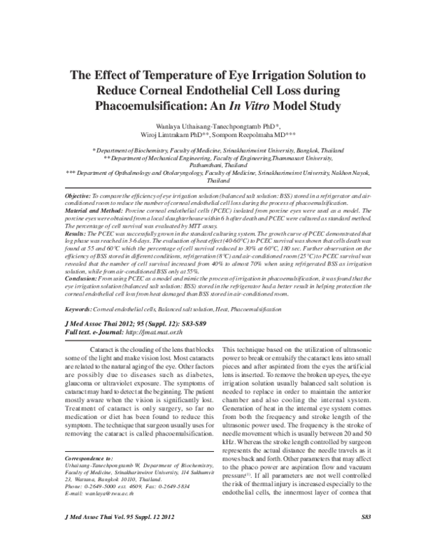
S83 Correspondence to: Uthaisang-Tanechpongtamb W, Department of Biochemistry, Faculty of Medicine, Srinakharinwirot University, 114 Sukhumvit 23, Wattana, Bangkok 10110, Thailand. Phone: 0-2649-5000 ext. 4609, Fax: 0-2649-5834 E-mail: wanlaya@swu.ac.th J Med Assoc Thai 2012; 95 (Suppl. 12): S83-S89 Full text. e-Journal: http://jmat.mat.or.th The Effect of Temperature of Eye Irrigation Solution to Reduce Corneal Endothelial Cell Loss during Phacoemulsification: An In Vitro Model Study
Related Papers
SciDoc Publishers
Effect Of Irrigating Solutions On The Corneal Endothelium Following Phacoemulsification: Balanced Salt Solution Versus Ringer Lactate Research ArticlePhacoemulsification is now considered the gold standard in cataract surgery. It provides several advantages over its counterpart SICS. Visual recovery is faster, there is minimal post-operative astigmatism and there is better overall patient satisfaction. However, the major drawback of phacoemulsification is the damage to the endothelium. The extent of damage is substantially higher than in Small Incision cataract surgery. It is also irreversible. Those patients with an already compromised endothelium like in Fuch’s Endothelial dystrophy are poor candidates for the procedure.
Journal of ophthalmology
Effect of Reformation of the Anterior Chamber by Air or by a Balanced Salt Solution (BSS) on Corneal Endothelium after Phacoemulsification: A Comparative Study2018 •
To study the effect of reformation of the anterior chamber by air or by a balanced salt solution, after smooth phacoemulsification on the corneal endothelial count and morphology. A prospective interventional nonrandomized comparative study included 500 eyes of 500 patients with age range between 50 and 60 years, prepared for cataract surgery and presented to the Ophthalmology department of Sohag University Hospital in the period from October 2016 to May 2017. Corneal endothelial morphology and count were examined, and the results were recorded for all cases before the surgery. Patients were divided into two groups, and both groups were diagnosed with grade 2 cataract and underwent uncomplicated phacoemulsification performed by well-trained surgeons. At the end of the surgery, group 1 was subjected to a reformation of the anterior chamber via a balanced salt solution (BSS) injection while group 2 was subjected to a reformation of the anterior chamber via air injection. Corneal endot...
Journal of Ocular Pharmacology and Therapeutics
Pharmacological Modification of Corneal Endothelial Intracellular pH, Intracellular Electrical Potential Difference, and Corneal Swelling and Deswelling Rates1985 •
Journal of Cataract and Refractive Surgery
Effect of hydrodynamic parameters on corneal endothelial cell loss after phacoemulsification2009 •
Journal of Cataract & Refractive Surgery
Comparison of the effect of AquaLase and NeoSoniX phacoemulsification on the corneal endothelium2008 •
Brit J Ophthalmol
Comparison of corneal changes after phacoemulsification using BSS Plus versus Lactated Ringer's irrigating solution: a prospective randomised trial2010 •
Introduction: Senile cataract has been documented to be the most significant cause of bilateral blindness in India. The aim of cataract surgery is no longer restricted to just visual restoration, but is now considered to be a refractive surgery i.e. to achieve a state of emmetropia. Preservation of corneal endothelial function is a major goal in cataract surgery. Objectives: Present study aims to highlight the importance of measurement of central corneal thickness and compares the effect of SICS vs Phacoemulsification on corneal endothelium. Material and Methods: This was a prospective study consisting of 101 patients who presented to the department of Ophthalmology, who fulfill inclusion criteria and are willing to enroll in the study. Standard uneventful small incision cataract surgery was done on 51 patients and standard uneventful clear corneal phacoemulsification was done on 50 patients. Change in central corneal thickness was observed post-surgery on day 7 th and day 30 th. This study was conducted over a period of two years. Results: Both groups showed increase in CCT values on post-operative day 7 indicating some endothelial cell disturbances, but the increase in CCT was comparable between the two groups. At day 30 there was a decrease in CCT value as compared to day 7, the decrease was more in SICS as compared to PHACO, but the difference between the two groups was statistically insignificant.
American Journal of Ophthalmology
Effects of Hydrogen in Prevention of Corneal Endothelial Damage During Phacoemulsification: A Prospective Randomized Clinical Trial2019 •
Journal of Texas Archaeology and History: Special Volume No. 6
Debitage Analysis of a Younger Dryas-age Component at Eagle Cave, Val Verde County, Texas2024 •
From 2014-2017 the Ancient Southwest Texas Project (ASWT) of Texas State University conducted excavations at Eagle Cave (41VV167), Texas. During these excavations, a discrete Paleoindian-age occupation associated with burned rock, chipped stone tools and debitage, and the scattered elements of Bison antiquus was encountered. Radiocarbon assays from the cultural component cluster between approximately 12,500 and 12,600 cal BP (Koenig et al. 2022) placing the deposit solidly in a Younger Dryas and Folsom-age time frame. While formal chipped stone artifacts from this period have received more attention, this paper addresses an artifact class often considered mundane in comparison: lithic debitage.
Alain Locke, “The Gospel for the Twentieth Century” (archival digital scan, Howard University). Source: [untitled essay], Alain Locke Papers, MSRC, Box 164-143, Folder 3 (Writings by Locke—Notes. Christianity, spirituality, religion). Permission to post on Academia granted February 25, 2019, courtesy of: Meaghan Alston Prints and Photographs Librarian Moorland-Spingarn Research Center Howard University Text edited and published in 2005: “Alain Locke in His Own Words: Three Essays.” Edited and annotated by Christopher Buck and Betty J. Fisher. World Order 36.3 (2005): 37–48. [Features four previously unpublished works by Alain Locke: “The Moon Maiden” (37); “The Gospel for the Twentieth Century” (39–42); “Peace between Black and White in the United States” (42–45); and “Five Phases of Democracy” (45–48).] See also: Christopher Buck, “Alain Locke: Race Leader, Social Philosopher, Baha’i Pluralist.” Special Issue: “Alain Locke: Dean of the Harlem Renaissance and Baha’i Race-Amity Leader.” World Order 36.3 (2005): 7–36. Winner of the 2006 DeRose-Hinkhouse Memorial Award of Excellence: Class B. Periodicals—Single Issue, Magazine, National (B-1), for excellence in religion communications and public relations. Awarded to: (1) Dr. Betty J. Fisher, Editor; (2) World Order magazine; and (3) the National Spiritual Assembly of the Bahá’ís of the United States. See http://www.religioncommunicators.org/assets/documents/derosehinkhouseawardwinners2006.pdf Excerpts: First paragraph: The gospel for the Twentieth Century rises out of the heart of its greatest problems, and few who are spiritually enlightened doubt the nature of that problem. The clashing ominous [t?]est of issues of the practical world of today, the issues of race, sect, class and nationality, all have one basic spiritual origin, and for that reason, we hope and believe one basic cure. Too long have we tried to patch these issues up and balm them over; [sic] instead of going to the heart and seat of the trouble in the limited and limiting conceptions of humanity which are alone, like a poisonous virus circulating through our whole social system, responsible for them. A change of condition will not remedy or more than temporarily ameliorate our chronic social antagonisms; only a widespread almost universal change of social heart, a new spirit of human attitudes, can achieve the social redemption that must eventually come. The finest and most practical idea of Christianity, the idea of the millenium [sic], of peace on earth, has been allowed to lapse as an illusion of the primitive Christian mind, as a mystic’s mirage of another world. And as a consequence the Brotherhood of Man, taken as a negligible corollary of the fatherhood of God, has if anything in practical effect put the truth of its own basic proposition to doubtful uncertainty. The redemption of society, social salvation, should have been sought after first, the pragmatic test and proof of the fatherhood of God is afterall [sic] whether belief in it can realize the unity of mankind; and so the brotherhood of man, as it has been inspirationally expressed, [sic] the “oneness of humanity”, [sic] must be in our day realized or religion die out gradually into ever-increasing materiality. The salvation we have sought after as individuals in an after-life and another sphere must be striven for as the practical peace and unity of the human family here in this [world]. In some very vital respects God will be rediscovered to our age if we succeed in discovering the common denominator of humanity and living in terms of it and valuing all things in accordance with it. … Last paragraph: We must begin working out the new era courageously, but it must be a revolution within the soul. How many external wars and revolutions it will make unnecessary, if it is only possible! And we must begin heroically with the great apparent irreconcilables; the East and the West, the black man and the self-arrogating Anglo-Saxon, for unless these are reconciled, the salvation of society in this world cannot be. If the world had believingly understood the full significance of Him who taught it to pray and hope “Thy Kingdom come on earth as it is in Heaven” who also said “In my Father's house are many mansions”, already we should be further toward the realization of this great millenial [sic] vision. The word of God is still insistent, and more emphatic as the human redemption delays and becomes more crucial, and we have what Dr. Elsemont [Esslemont] rightly calls Bahá’u’lláh's “one great trumpet-call to humanity”: “That all nations shall become one in faith, and all men as brothers; that the bonds of affection and unity between the sons of men should be strengthened; that diversity of religion should cease, and differences of race be annulled … These strifes and this bloodshed and discord must cease, and all men be as one kindred and family.[”]
RELATED PAPERS
Advanced Engineering Materials
Modeling of Coating Process, Phase Changes, and Damage of Plasma Sprayed Thermal Barrier Coatings on Ni-Base Superalloys2010 •
2018 •
Arquivos de Gastroenterologia
Underwater Endoscopic Mucosal Resection for Non-Pedunculated Colorectal Lesions. A Prospective Single-Arm StudyRevista Brasileira de Ciências Sociais
Companheiros servidores: o avanço do sindicalismo do setor público na CUT2001 •
Current Science
World's Tenth Largest Banyan Tree at Narora in Upper Ganga Ramsar Site, Uttar Pradesh, India2016 •
2008 Sixth Indian Conference on Computer Vision, Graphics & Image Processing
Monocular Depth by Nonlinear Diffusion2008 •
J. Mater. Chem. C
A novel paradigm for fabricating highly uniform nanowire arrays using residual stress-induced patterning2016 •
Journal of the American Society of Echocardiography
Pharmacodynamic Doppler Determination of Mitral Valve Area in Patients With Significant Aortic Regurgitation1993 •
Journal of Babol University of Medical Sciences
Essential oils as a natural additive in the edible films and coatings (active packaging system): A Review2018 •

 Wanlaya Tanechpongtamb