crossmark
THE JOURNAL OF BIOLOGICAL CHEMISTRY VOL. 290, NO. 47, pp. 28038 –28054, November 20, 2015
© 2015 by The American Society for Biochemistry and Molecular Biology, Inc. Published in the U.S.A.
Distinct Cellular Assembly Stoichiometry of Polycomb
Complexes on Chromatin Revealed by Single-molecule
Chromatin Immunoprecipitation Imaging*
⽧
Received for publication, June 8, 2015, and in revised form, August 29, 2015 Published, JBC Papers in Press, September 17, 2015, DOI 10.1074/jbc.M115.671115
Roubina Tatavosian‡, Chao Yu Zhen‡, Huy Nguyen Duc‡, Maggie M. Balas§, Aaron M. Johnson§,
and Xiaojun Ren‡1
From the ‡Department of Chemistry, University of Colorado Denver, Denver, Colorado 80217-3364 and the §Department of
Biochemistry and Molecular Genetics, University of Colorado School of Medicine, Aurora, Colorado 80045
Background: Polycomb proteins control transcription by regulating chromatin structure and dynamics.
Results: By developing and applying a novel Sm-ChIPi technique, we identified that one PRC1 binds multiple nucleosomes
within cells, although two PRC2s can bind a single nucleosome.
Conclusion: PRC1 and PRC2 complexes employ distinct mechanisms to assemble on chromatin.
Significance: The cellular assembly stoichiometry provides insight into repressive polycomb chromatin structure.
* This work was supported, in whole or in part, by National Institutes of Health
Pathway to Independence Award K99/R00 GM094291 (to A. J.). This work
was also supported by grants from the University of Colorado Denver (to
X. R.), the CU-Denver Office Research Service (to X. R.), and American Cancer Society Grant IRG 57-001-53 subaward (to X. R.). The authors declare
that they have no conflicts of interest with the contents of this article.
⽧
This article was selected as a Paper of the Week.
1
To whom correspondence should be addressed. Tel.: 303-556-5659; Fax:
303-556-4776; E-mail: xiaojun.ren@ucdenver.edu.
28038 JOURNAL OF BIOLOGICAL CHEMISTRY
could provide single-molecule insight into other epigenetic
complexes.
In the nucleus, genome organization is shaped by the nucleosome, the basic building unit of chromatin (1). The nucleosome
is formed by wrapping ⬃147 bp of DNA around a histone octamer consisting of two copies of H2A, H2B, H3, and H4 (2). The
highly conserved basic N termini and, to a lesser extent, the
globular domains of histones are extensively post-translationally modified by epigenetic regulatory complexes (3–5).
Genomic compartments and chromatin-related activities are
tightly correlated with histone modifications (1, 4, 6). These
modifications either directly organize chromatin structure or
recruit effectors that impact genome organization (1, 4, 6, 7).
However, cellular molecular details about how epigenetic complexes assemble on and spread along chromatin are incompletely understood.
Polycomb group (PcG)2 proteins are a long-standing paradigm of studying the epigenetic inheritance of transcriptional
states and are essential for the establishment and maintenance
of transcriptional profiles during normal development and in
cancer (8, 9). Two major PcG complexes, PRC1 and PRC2,
exhibit distinct enzymatic activities (8). PRC2 is a methyltransferase that catalyzes di- and trimethylation of lysine 27 on H3
(H3K27me2/3) (8). The mammalian core PRC2 is composed of
Ezh2, Suz12, Eed, and RbAp48. Ezh2 is the catalytic subunit (8),
and Eed is involved in recognition of the H3K27me3 mark (10).
PRC1 is a ubiquitin ligase that catalyzes ubiquitylation of lysine
119 on H2A (H2AK119Ub) (11). In mammals, six forms of
PRC1 have been identified, each comprising one of six Pcgf
2
The abbreviations used are: PcG, polycomb group; ChIP-Seq, chromatin
immunoprecipitation followed by high-throughput sequencing; Dox,
doxycycline; IP, immunoprecipitation; mES, mouse embryonic stem; OHT,
4-hydroxytamoxifen; PRC, polycomb repressive complex; Sm-ChIPi, singlemolecule chromatin immunoprecipitation imaging; TIRF, total internal
reflection fluorescence; EGFP, enhanced GFP; PCV, packed cell volume;
BisTris, 2-[bis(2-hydroxyethyl)amino]-2-(hydroxymethyl)propane-1,3-diol;
FCS, fluorescence correlation spectroscopy.
VOLUME 290 • NUMBER 47 • NOVEMBER 20, 2015
Downloaded from http://www.jbc.org/ by guest on May 23, 2020
Epigenetic complexes play an essential role in regulating
chromatin structure, but information about their assembly stoichiometry on chromatin within cells is poorly understood. The
cellular assembly stoichiometry is critical for appreciating the
initiation, propagation, and maintenance of epigenetic inheritance during normal development and in cancer. By combining
genetic engineering, chromatin biochemistry, and single-molecule fluorescence imaging, we developed a novel and sensitive
approach termed single-molecule chromatin immunoprecipitation imaging (Sm-ChIPi) to enable investigation of the cellular
assembly stoichiometry of epigenetic complexes on chromatin.
Sm-ChIPi was validated by using chromatin complexes with
known stoichiometry. The stoichiometry of subunits within a
polycomb complex and the assembly stoichiometry of polycomb
complexes on chromatin have been extensively studied but
reached divergent views. Moreover, the cellular assembly stoichiometry of polycomb complexes on chromatin remains unexplored. Using Sm-ChIPi, we demonstrated that within mouse
embryonic stem cells, one polycomb repressive complex (PRC) 1
associates with multiple nucleosomes, whereas two PRC2s can
bind to a single nucleosome. Furthermore, we obtained direct
physical evidence that the nucleoplasmic PRC1 is monomeric,
whereas PRC2 can dimerize in the nucleoplasm. We showed
that ES cell differentiation induces selective alteration of the
assembly stoichiometry of Cbx2 on chromatin but not other
PRC1 components. We additionally showed that the PRC2-mediated trimethylation of H3K27 is not required for the assembly
stoichiometry of PRC1 on chromatin. Thus, these findings
uncover that PRC1 and PRC2 employ distinct mechanisms to
assemble on chromatin, and the novel Sm-ChIPi technique
�Assembly Stoichiometry of Polycomb on Chromatin by Sm-ChIPi
NOVEMBER 20, 2015 • VOLUME 290 • NUMBER 47
determine the apparent molecular sizes of native protein complexes; however, these techniques cannot exclude the influence
of uncharacterized proteins and heterogeneous conformations.
Single-molecule fluorescence microscopy is a powerful technique to quantify the absolute number of subunits of the macromolecular protein complex (31–33). The quantification is
based on the photobleaching behaviors of fluorophores (32, 33)
or the ratios of the fluorescent intensities of fluorophores to the
reference fluorophores (31, 34, 35). Single-molecule techniques
have been widely applied to chromatin biology and provide a
wealth of information on nucleosome structure and dynamics
(36 – 41). Here, we combined genetic engineering, chromatin
biochemistry, and single-molecule fluorescence imaging to
develop a novel and sensitive approach termed Sm-ChIPi to
circumvent these limitations and to enable us to directly assess
the cellular assembly stoichiometry. By using Sm-ChIPi, for the
first time we present the cellular assembly stoichiometry of PcG
complexes PRC1 and PRC2 on chromatin. We have found that
PRC1 and PRC2 employ distinct mechanisms by which they
assemble on chromatin, reflecting their distinct roles in establishing and maintaining repressive polycomb domains. These
results contribute significantly to our quantitative understanding of the cellular architecture of PcG complexes, allowing us to
suggest possible molecular mechanisms for the PcG-mediated
epigenetic silencing. Sm-ChIPi is a direct and sensitive technique and could be applied to many other studies of epigenetic
complex assembly on native chromatin.
Experimental Procedures
Cell Lines and Plasmids—The Cbx2⫺/⫺ (42), Cbx7⫺/⫺ (43),
Ring1bfl/fl;Rosa26::CreERT2 (44), Bmi1⫺/⫺/Mel18⫺/⫺ (Bmi1
and Mel18 double knock-out) (45), Eed⫺/⫺ (44), Ezh2⫺/⫺ (46),
and PGK12.1 (47) mES cell lines were maintained in mES
medium (DMEM (D5796; Sigma) supplemented with 15% FBS
(SH30071.03; Hyclone), 2 mM glutamine (G7513; Life Technologies, Inc.), 100 units/ml penicillin/streptomycin (15140-122;
Life Technologies, Inc.), 55 M -mercaptoethanol (21985-023;
Life Technologies, Inc.), 103 units/ml leukemia inhibitor factor,
and 0.1 mM non-essential amino acids (11140050; Life Technologies, Inc.)) at 37 °C in 5% CO2. Medium was changed every day
unless otherwise indicated. To deplete Ring1b alleles, 4-hydroxytamoxifen (OHT; H7904; Sigma) was administered for 3
days under a concentration of 1.0 M. HEK293T cells were
maintained in DMEM supplemented with 10% FBS, 2 mM glutamine, and 100 units/ml penicillin/streptomycin at 37 °C in 5%
CO2. H3.3⫺/⫺/H3.3-EGFP DT40 cells (48) were maintained in
RPMI 1640 medium (11875093; Life Technologies, Inc.) supplemented with 5% FBS, 5% chicken serum (C5405–100ML;
Sigma), 50 M -mercaptoethanol, and 100 units/ml penicillin/
streptomycin at 37 °C in 5% CO2.
The plasmids pTRIPZ(M)-YFP-Cbx2 (49), pTRIPZ(M)YFP-Cbx4 (49), pTRIPZ(M)-YFP-Cbx7 (49), pTRIPZ(M)-YFPCbx8 (49), pTRIPZ(M)-YFP-Ring1b (49), pTRIPZ(M)-YFPMel18 (49), and pEGFP-KAP1 (50) have been described
previously. The sequences encoding Eed (Addgene) and Ezh2
(Addgene) were amplified by PCR and inserted downstream of
the coding sequence of fluorescence protein in pTRIPZ(M)
vector (49). The sequence encoding YFP was amplified by PCR
JOURNAL OF BIOLOGICAL CHEMISTRY
28039
Downloaded from http://www.jbc.org/ by guest on May 23, 2020
subunits and the E3 ligase Ring1a/b (12). Further classification
of PRC1 is determined by the mutually exclusive association of
either Rybp or Yaf2 (variant PRC1s) or one of the Cbx proteins
(canonical PRC1s) (12). Several mechanisms for the PcG-mediated gene silencing such as histone H3K27 trimethylation
(13), histone H2A monoubiquitination (11), chromatin compaction (14), and organization of higher order chromatin structure have been proposed (15); however, it is not yet clear how
PcG complexes assemble on chromatin within cells.
Although much is known about the interaction domains and
the protein identities within PRC1 complexes, far less is known
about their molecular architecture on chromatin within cells.
Several studies have shown that the PRC1 subunits and their
isolated domains self-associate in vitro (16 –20). Clearly, these
in vitro observations need to be verified within cells. In contrast
with the individual PRC1 subunits, the reconstituted Drosophila PRC1 is a monomer having one copy of each subunit (14).
Studies of the assembly stoichiometry of PRC1 on chromatin
reached varying views on how PRC1 interacts with chromatin.
The reconstituted Drosophila PRC1 packs nucleosomal arrays
with a stoichiometry of one PRC1 per tetranucleosome (14).
The reconstituted Drosophila Psc (homolog of Pcgfs) bridges
nucleosomes with a stoichiometry of one Psc per mononucleosome (21). A recent crystal structure indicated that one PRC1
ubiquitylation module binds to each disk surface of a nucleosome (22). These variations could be due to the compositions of
subunits used in the reconstitution reactions or the methods
used in the experiments. Thus, it is important to resolve these
disparities and to determine the cellular assembly stoichiometry of PRC1 complexes on chromatin.
Studies of the oligomerization status of PRC2 reached divergent opinions (23–27). The reconstituted PRC2 has been characterized as a monomer, dimer, or oligomer (23–25). By utilizing size exclusion chromatography, the endogenous PRC2
complex from both human and Drosophila was found to have a
wide range of apparent molecular masses, ranging from 300
kDa to 1 mDa or higher (26, 27), whereas gel filtration of native
complexes cannot exclude the possibility that PRC2 has
extended structures or that non-PRC2 proteins are associated.
The molecular stoichiometry of PRC2 within cells therefore
remains elusive. Electron microscopy studies suggested that
PRC2 is monomeric and may bind to a dinucleosome (25); however, whether the in vitro model recaptures the in vivo situation
remains unknown.
A few approaches have been developed to quantify the stoichiometry of epigenetic modifications at histones of nucleosomes (28, 29) or in an entire proteome (30), but addressing the
cellular assembly stoichiometry of epigenetic complexes at
chromatin has so far been hampered by the absence of adequate
techniques. Chromatin immunoprecipitation (ChIP) followed
by high throughput sequencing (ChIP-Seq) maps global patterns of histone modifications and chromatin-binding proteins,
but ChIP-Seq cannot directly reveal molecular stoichiometry.
Sequential ChIP performed on native and purified nucleosomes can reveal the co-occurrence of epigenetic proteins on
chromatin, but it is a formidable challenge to establish absolute
stoichiometry. Sedimentation velocity analytical ultracentrifugation and gel filtration chromatography are often used to
�Assembly Stoichiometry of Polycomb on Chromatin by Sm-ChIPi
28040 JOURNAL OF BIOLOGICAL CHEMISTRY
for 8 min at 37 °C. To produce polynucleosomes, chromatin
was digested with 0.7 units/ml for 8 min at 37 °C. The reaction
was stopped by 4.0 mM EGTA (pH 8.0). To purify mononucleosomes, 5–30% linear sucrose gradient was used. To purify polynucleosomes, 15– 40% linear sucrose gradient was used. Linear
sucrose gradients were prepared by dissolving sucrose in the
buffer M (10 mM HEPES, pH 7.9, 50 g/ml BSA, 10 mM KCl, 1.5
mM EDTA, 1.0 mM Na3VO4, 0.2 mM DTT and 0.5 mM PMSF).
Approximately 300 – 400 g of DNA in 0.5 ml were loaded on
the top layer of the gradient, and samples were fractionated for
18 –20 h at 200,000 ⫻ g using TH-641 Swinging Bucket Rotor
and Sorvall WX ultracentrifuge (Thermo Fisher Scientific,
Waltham, MA). 0.5 ml per fraction was collected. The DNA
fragment size of each fraction was analyzed by agarose gel
electrophoresis.
Preparation of Nucleosomes from Differentiated Cells—mES
cells were induced to differentiate as described previously (51).
Briefly, mES cell lines, Cbx2⫺/⫺/Y-Cbx2, Cbx7⫺/⫺/Y-Cbx7,
Ring1bfl/fl/Y-Ring1b, and Bmi1⫺/⫺Mel18⫺/⫺/Y-Mel18, were
cultured to reach 80 –90% confluency. Approximately 6 ⫻ 106
cells were resuspended in 10 ml of DMEM supplemented with
10% FBS, 2 mM glutamine, and 100 units/ml penicillin/streptomycin, and plated in a 10-cm polystyrene stackable Petri dish
(8609 – 0010; USA Scientific, Ocala, FL). Medium was changed
every 48 h. On day 4, a final concentration of 500 M retinoic
acid (R2625; Sigma) was administered. On day 8, cells were
changed with medium containing 2 g/ml Dox or Dox with
OHT for Ring1bfl/fl/Y-Ring1b. On day 10, chromatin was isolated, and nucleosomes were prepared as described above.
Preparation of Polynucleosomal Arrays and Their Interaction
with PRC1—Tetranucleosome reconstitution was performed as
described previously by salt dialysis (52, 53). Briefly, recombinant human histone octamer (H2A, H2B, H3.1, and H4),
assembled from Escherichia coli-expressed individual histones
as described (52), was added to DNA in approximately a 1:1
molar ratio in 10 mM Tris-HCl, pH 7.6, 2 M NaCl, 1 mM EDTA,
0.5 mg/ml BSA, 0.05% Nonidet P-40, and 5 mM -mercaptoethanol. Salt dialysis was performed at 4 °C for ⬃20 h from 2 M
NaCl buffer to 50 mM NaCl and a final dialysis step for 1 h at 50
mM NaCl. Samples were incubated at 37 °C for 1 h before storage on ice up to 4 weeks. The extent of chromatinization was
assessed by limited micrococcal nuclease digestion. The DNA
template used was an 863-bp PCR fragment amplified from a
plasmid construct3 containing two “601” nucleosome-positioning sequences (54) flanking five Gal4-binding sites and an
adenoviral E4 promoter. The PCR product was amplified with
one 5⬘-biotin-triethyleneglycol primer.
The 15-mer nucleosomal DNA template is a purified
⬃3.1-kb biotinylated PCR product from the plasmid pUC18G5cyc1G- (55). The 15-mer polynucleosome was reconstituted
through a previously developed enzymatic assembly method
(56, 57). Biotinylated DNA, human histone octamers (as
described for tetranucleosome assembly), human histone chaperone NAP1, yeast nucleosome positioning factor yIsw1a, and
an ATP regeneration system (final concentrations: 30 mM cre-
3
M. Balas and A. Johnson, unpublished data.
VOLUME 290 • NUMBER 47 • NOVEMBER 20, 2015
Downloaded from http://www.jbc.org/ by guest on May 23, 2020
and inserted into pGEX-6P-1 vector (GE Healthcare) to generate pGEX-6P-1-YFP (monomeric YFP) and pGEX-6P-1-YFPYFP (dimeric YFP). The sequences encoding fusion proteins
have been verified by DNA sequencing.
Establishing Transgenic mES Cell Lines—Establishing the
transgenic mES cell lines was performed according to the procedure described previously (49). Briefly, pseudo-viruses were
packaged in HEK293T cells by co-transfecting with 21 g of
pTRIPZ(M) containing the fusion gene, 21 g of psPAX2, and
10.5 g of pMD2.G. 60 h after transfection, medium was collected and used for transducing mES cells. Hexadimethrine
bromide (Polybrene; H9268; Sigma) was added at a concentration of 8 g/ml, and cells were seeded at ⬃15% confluence on
gelatin-coated plates. 2 days after infection, infected cells were
selected by using 1.0 –2.0 g/ml puromycin (P8833; Sigma).
The expression of transgenes was induced by doxycycline (Dox;
D9891; Sigma).
Transfection—HEK293T cells at 85–90% confluence were
transfected with pEGFP-KAP1 by calcium phosphate. After 24 h
of transfection, the medium was replaced with fresh medium. 48 h
later, cells were harvested for isolation of chromatin.
Preparation of YFP Proteins—The plasmids pGEX-6p-1-YFP
and pGEX-6p-1-YFP-YFP were transformed into the BL21competent cells, respectively. The protein expression was
induced by isopropyl -D-thiogalactopyranoside (AC121;
Omega Bio-Tek, Norcross, GA) for 5 h at 37 °C. Cell pellets
were collected, resuspended in PBS containing 0.1 mM phenylmethanesulfonyl fluoride (PMSF; 93482; Sigma) and protease
inhibitor mixture (P8340; Sigma), and sonicated using VibraCellTM sonicator (VCX130; Newtown, CT). 1% Triton X-100
was added to the mixture. After centrifugation, prewashed
GSH-Sepharose 4B beads (17-0756-01; GE Healthcare) were
added to the supernatant. The mixture was incubated for 30
min at 4 °C. After washing four times with PBS containing 1.0%
Triton X-100, the YFP proteins were eluted by 5 mM reduced
glutathione (G4251; Sigma). The purity and identity of YFP
proteins were assessed by SDS-PAGE.
Preparation of Nucleosomes from mES Cells—Approximately
5 ⫻ 108 cells were harvested by citrate saline solution (135 mM
potassium chloride and 15 mM sodium citrate) for adherent
cells or collected by centrifugation for DT40 cells, cross-linked
with 2.0% paraformaldehyde for 10 min at 4 °C, and quenched
with glycine. Cells were collected by centrifuging at 300 ⫻ g for
5 min at 4 °C, and the packed cell volume (PCV) was estimated.
Pellets were resuspended in 2.5⫻ PCV of buffer A (10 mM
HEPES, pH 7.9, 10 mM KCl, 1.5 mM MgCl2, 340 mM sucrose,
10% glycerol, 50 g/ml BSA, 1.0 mM Na3VO4, protease inhibitor mixture, and 0.1 mM PMSF). 2.5⫻ PCV of buffer B (buffer A
plus 0.2% Triton X-100) was added, and the mixture was incubated at 4 °C for 10 min. Pellets were collected by centrifuging
at 1,300 ⫻ g for 5 min at 4 °C and resuspended with 6⫻ PCV of
buffer A. The mixture was loaded to the top layer of pre-chilled
sucrose cushion (buffer A ⫹ 30% sucrose) and centrifuged at
1,300 ⫻ g for 12 min at 4 °C. Chromatin pellets were resuspended in buffer A containing 1.0 mM CaCl2 at the DNA concentration of 2.0 g/ml. To generate mononucleosomes, chromatin was digested with 1.4 units/ml micrococcal nuclease
(N5386; Sigma; the enzyme activity was defined as a Sigma unit)
�Assembly Stoichiometry of Polycomb on Chromatin by Sm-ChIPi
NOVEMBER 20, 2015 • VOLUME 290 • NUMBER 47
and background intensity was subtracted from the area surrounding the spot of interest. The photobleaching steps were
detected by Chang-Kennedy filtering (58). Histogram was constructed using data from three replicates with each measurement of over 100 individual spots analyzed. The functionalized
blank slide surface showed ⬃5 fluorescent spots per 1,000 m2.
The number of fluorescent spots was typically ⬃300 – 600 spots
per 1,000 m2 via antibody immobilization by controlling the
lysate or fraction dilution factor. At the same dilution factor and
without antibody, the number of fluorescent spots was typically
⬃10 fluorescent spots per 1,000 m2, implying that ⬃4% is
from non-specifically adsorbed proteins to the surface.
Fluorescence Correlation Spectroscopy (FCS)—FCS measurements were performed at 37 °C on a Zeiss LSM780 using a
C-Apochromat infinity color-corrected 1.2 NA 40⫻ water
objective. Cells were seeded on glass dishes the day before the
experiment. Excitation of YFP was performed with the 488-nm
line of a 20-milliwatt argon laser. For intracellular measurements,
the desired recording position was chosen in the LSM image.
Autocorrelation curves were derived from fluorescence fluctuation analysis using the ZEN2012 FCS module. Autocorrelation
curves were fit to one-component models of free diffusion in three
dimensions with triplet function of Equation 1 (59),
冉
冊
1
F 䡠 e ⫺ / F
G()⫽1⫹ 䡠 1 ⫹
䡠
N
1⫺F
冢冉
1
冊冉
1⫹
䡠 1⫹
D1
D1䡠S2
冊冣
1
2
(Eq. 1)
where D1 and F are the diffusion time and the triplet time, respectively; N and F are the number of molecules in the confocal volume
and the triplet fraction, respectively; and S ⫽ wz/wxy, where wz and
wxy represent the half height and radius of the confocal volume,
respectively. The size of the confocal volume Veff was calibrated
using a series dilution of rhodamine green dye in PBS. The concentration of YFP-Ring1b was determined by Equation 2,
YFP ⫺ Ring1b(FCS) ⫽
N
6.02 䡠 1023 䡠 Veff
(Eq. 2)
The concentration of endogenous Ring1b was determined by
Equation 3,
endogenous Ring1b ⫽ YFP ⫺ Ring1b(FCS)䡠0.90
(Eq. 3)
where 0.90 is the ratio of endogenous Ring1b to YFP-Ring1b,
which was determined by Western blotting. The number of
YFP-Ring1b molecules in single mES cell nucleus was calculated by Equation 4,
NA ⫽
冉
冊
4
abc 䡠 C Ring1b 䡠 6.02 䡠 1023
3
(Eq. 4)
where a ⫽ b ⫽ 5 m and c ⫽ 2.5 m. CRing1b is the concentration of endogenous Ring1b protein.
ChIP—ChIP was performed as described previously (43, 51).
Briefly, KO mES cells complemented with the YFP-PRC1 fusion
JOURNAL OF BIOLOGICAL CHEMISTRY
28041
Downloaded from http://www.jbc.org/ by guest on May 23, 2020
atine phosphate, 3 mM ATP, 4.1 mM MgCl2, and 6.4 g/ml
creatine kinase) were incubated at 30 °C for 5 h in buffer containing 10 mM HEPES, pH 7.5, 10 mM KCl, 1.5 mM MgCl2, 500
M EGTA, 10% glycerol, 2.5 mM -glycerophosphate, 200 M
PMSF, and 1 mM DTT.
Ring1bfl/fl/Y-Ring1b mES cells were cultured in the presence
of 0.5 g/ml doxycycline and 1.0 M OHT for 3 days. Nuclei
were purified from 5 ⫻ 107 cells and lysed in 0.5 ml of buffer
containing 20 mM Tris-HCl, pH 7.4, 0.5% Nonidet P-40, 350
mM NaCl, 0.25 mM EDTA, 10% glycerol, 0.1 mM Na3VO4, protein inhibitor mixture, and 0.1 mM PMSF. 120 l of biotinylated
nucleosomal arrays (18 nM) was incubated with 380 l of
nuclear extract at 4 °C overnight. The 0.5 ml of mixture was
loaded into 15– 40% sucrose gradient and fractionated as
described above. 0.5 ml per fraction was collected and fixed
with 0.2% of paraformaldehyde. DNAs were extracted and analyzed by agarose gel electrophoresis.
Construction and Passivation of Flow Chamber—Flow chambers were constructed as described previously with modifications (32). Two 0.75-mm holes across from each other were
drilled in a quartz slide (12-550-15; Thermo Fisher Scientific).
The slides and coverslips (48366-249; VWR, Radnor, PA) were
sonicated with Milli-Q water for 30 min and incubated with
methanol overnight. The coverslips were treated with 1.0 M
KOH for 40 min, dried, and burned for 1–2 s using a propane
torch. Then the coverslips were incubated with methanol
supplemented with 1% aminosilane (N-2-aminoethyl-3aminopropyltrimethoxysilane (A21541; Pfaltz & Bauer, Waterbury, CT)) and 5% acetic acid for 20 min in the dark at room
temperature. After washing with methanol and water, the coverslips were dried with nitrogen gas and placed in a humidified
box in the dark. To each coverslip, 70 l of the passivated solution (10 mM sodium bicarbonate, pH 8.5,16 mg of mPEG-SVA
(MPEG-SVA-5000; Laysan Bio, Arab, AL)), 0.3 mg of biotin
PEG-SVA (256-586-9004; Laysan Bio) was added and incubated in a humidified box for 3– 4 h in the dark. After washing
with Milli-Q water, the coverslips were assembled on the quartz
slide by sandwiching a piece of double-sided tape between the
slide and the coverslip in the way that it creates an ⬃6.0-mm
channel where the inlet/outlet holes are located. The edges of
the flow chambers were sealed with epoxy glue (14250; Devcon,
Danvers, MA) and stored at ⫺20 °C under nitrogen gas.
Imaging by Single-molecule Total Internal Reflection Fluorescence (TIRF) Microscopy—Samples intended for the Sm-ChIPi
analysis were incubated with biotinylated antibodies, anti-GFP
(ab6658; Abcam, Cambridge, UK), anti-histone H2B (60R1215; Fitzgerald Industries, Acton, MA), and anti-histone H3
(5748; Cell Signaling Technology, Boston, MA), at 4 °C overnight. Flow chamber was loaded with 0.2 g/ml NeutrAvidin
(31000; Thermo Fisher Scientific) and washed with TE50
buffer. After cross-linking with 0.5% paraformaldehyde for 15
min at 4 °C, 100 l of the samples were loaded into the flow
chamber. After washing with TE50 buffer, images were
acquired by using Zeiss Axio Observer D1 Manual Microscope
(Zeiss, Germany) equipped with an Alpha Plan-Apochromatic
100⫻/1.46 NA Oil Objective (Zeiss, Germany) and an Evolve
512 ⫻ 512 EMCCD camera (Photometrics, Tucson, AZ). The
fluorescent intensity of time traces was generated by ImageJ,
�Assembly Stoichiometry of Polycomb on Chromatin by Sm-ChIPi
28042 JOURNAL OF BIOLOGICAL CHEMISTRY
FIGURE 1. Development of Sm-ChIPi to assess the cellular assembly stoichiometry of PcG complexes on chromatin. a, schematic of the Sm-ChIPi
approach. Nucleosomes generated from chromatin are directly immobilized
on the surface or are separated by sucrose gradient ultracentrifugation and
then immobilized on the surface. b and c, photobleaching behavior of monomeric (b) and dimeric (c) YFPs. The YFP proteins were immobilized by biotinylated anti-GFP antibody via interaction with NeutrAvidin. A sample single
image of the acquired sequence is shown. A representative time course of
fluorescence emission of the YFP protein is shown (black line). The photobleaching steps were detected by Chung-Kennedy filter (red line). Results are
means ⫾ S.D. a.u. denotes arbitrary unit. MNase is micrococcal nuclease. Scale
bar, 5 m.
slides with ProLong antifade reagents (P7481; Life Technologies, Inc.). The images were taken and processed as described
previously (49).
Results
Development of a Novel Approach to Assess the Cellular
Assembly Stoichiometry of PcG Complexes on Chromatin—To
assess the cellular assembly stoichiometry of PcG complexes on
chromatin, we developed the Sm-ChIPi approach (Fig. 1a). A
YFP-PcG fusion gene was stably expressed in its corresponding
KO mES cells, which allows incorporating the fusion protein
into PcG complexes without the interference of the endogenous counterpart. Cells were cross-linked with paraformaldehyde to preserve complex association prior to cell lysis. After
cleaning with a sucrose cushion, chromatin extraction was subject to micrococcal nuclease digestion. The PcG䡠nucleosome
complexes were fractionated by sucrose gradient ultracentrifugation. The fractions intended to be analyzed were incubated
with biotinylated antibodies. The resultant complexes were
immobilized on a quartz slide that had been passivated and
VOLUME 290 • NUMBER 47 • NOVEMBER 20, 2015
Downloaded from http://www.jbc.org/ by guest on May 23, 2020
gene were cross-linked with 1.2% formaldehyde (28908;
Thermo Fisher Scientific) for 10 min at room temperature and
quenched by 125 mM glycine. Cells were washed sequentially
with LBI buffer (50 mM HEPES, pH 7.9, 140 mM NaCl, 1.0 mM
EDTA, 10% glycerol, 0.5% Nonidet P-40, and 0.25% Triton
X-100), LBII buffer (10 mM Tris-HCl, pH 8.0, 200 mM NaCl, and
15 mM EDTA), and LBIII buffer (10 mM Tris-HCl, 100 mM
NaCl, 1.5 mM EDTA, 0.1% sodium deoxycholate, and 0.5%
N-lauroylsarcosine). Chromatin was fragmented to the size of
200 –500 bp by Vibra-CellTM sonicator (VCX130; Sonics, Newtown, CT). One-tenth of 10% Triton X-100 was added to the
lysate. After pre-cleaning with protein G beads (101241; Life
Technologies, Inc.), antibodies, anti-Cbx7 (sc-70232; Santa
Cruz Biotechnology, Santa Cruz, CA), anti-Cbx2 (ab80044;
Abcam, Cambridge, MA), anti-Mel18 (sc-10744; Santa Cruz
Biotechnology), and anti-Ring1b (D139-1; MBL, Woburn,
MA), were incubated with lysates, respectively. The immunoprecipitated DNAs were quantified using LightCycler 4800
SYBR Green I master mix (04707516001; Roche Applied Science) with AB Applied Biosystems. Triplicate PCRs were carried out for each sample. The efficiencies of ChIP were quantified relative to a standard curve prepared using input
chromatin. The sequences of the primers used for quantitative
PCR has been described previously (43, 51).
Immunoprecipitation (IP)—IP was performed as described
previously (51). Briefly, nuclei were purified from 3 ⫻ 108 cells
and lysed using buffer containing 20 mM Tris-HCl, pH 7.4, 0.1%
Nonidet P-40, 350 mM NaCl, 0.25 mM EDTA, 20% glycerol, 0.1
mM Na3VO4, 0.1 mM PMSF, and protein inhibitor mixture.
After pre-cleaning with protein G beads, the lysate was incubated with anti-GFP mAb-agarose beads (D153-8; MBL). The
beads were washed using buffer containing 20 mM Tris-HCl,
pH 8.0, 1% Nonidet P-40, 200 mM KCl, 0.2 mM EDTA, and 0.1
mM PMSF. The proteins were resolved using NuPAGE 4 –12%
BisTris gel (NP0321BOX; Life Technologies, Inc.) and were
transferred to 0.45-m Immobilon-FL polyvinylidene fluoride
membrane (Millipore, Darmstadt, Germany). Specific proteins
were probed with anti-Phc1 (6-1-3; Active Motif, Carlsbad, CA)
and anti-Ring1b (D139-3; MBL), and detected with ECL Plus
(GE Healthcare). Membranes were imaged using a ChemiDoc
XRS system (Bio-Rad).
Immunofluorescence—Immunofluorescence was performed
as described previously (51). Wild-type, Ezh2⫺/⫺, Eed⫺/⫺,
Ezh2⫺/⫺/Y-Ezh2, and Eed⫺/⫺/Y-Eed mES cells were plated on
coverslips and cultured for 24 h. Cells were fixed using 2.0%
paraformaldehyde for 10 min. Cells were washed with PBS and
incubated with 0.2% Triton X-100 for 10 min. After washing
with basic blocking buffer (10 mM PBS, pH 7.2, 0.1% Triton
X-100, and 0.05% Tween 20), cells were incubated with blocking buffer (basic blocking buffer plus 3% goat serum and 3%
bovine serum albumin) for 1 h. Anti-H3K27me3 antibody (07449; Millipore, Billerica, MA) diluted in blocking buffer was
incubated with cells for 2 h at room temperature. After washing
with basic blocking buffer, Alexa 488-labeled goat anti-rabbit
antibody (A-11008; Life Technologies, Inc.) diluted in blocking
buffer was incubated with cells for 1 h. Cells were rinsed with
PBS and washed with basic blocking buffer. After incubating
with 0.1 g/ml Hoechst, cells were washed and mounted on
�Assembly Stoichiometry of Polycomb on Chromatin by Sm-ChIPi
NOVEMBER 20, 2015 • VOLUME 290 • NUMBER 47
Cellular Assembly Stoichiometry of YFP-PRC1 Proteins on a
Mononucleosome—To assess the cellular assembly stoichiometry of PRC1 on chromatin, the YFP-PRC1 fusion genes, Y-Cbx2,
Y-Cbx7, Y-Mel18, and Y-Ring1b, were introduced into KO mES
cells, respectively. Cbx2, Cbx7, and Mel18 are the core subunits
of canonical PRC1 complexes, and Ring1b is the core subunit of
all PRC1 complexes (8). The expression of the fusion proteins
were induced by 0.5 g/ml Dox unless otherwise indicated.
Ring1bfl/f;Rosa26::CreERT2 cells were incubated with 1.0 M
OHT for 3 days to deplete Ring1b locus (hereafter Ring1b⫺/⫺).
The YFP-PRC1䡠nucleosome complexes were isolated from cells
and fractionized by ultracentrifugation. Agarose gel electrophoresis was used to analyze the distribution of nucleosomal
DNAs extracted from ultracentrifugation fractions (Fig. 3a).
Fraction 18, the peak of mononucleosomes, was selected for the
Sm-ChIPi analysis. The YFP-PRC1䡠mononucleosome complexes were immobilized by biotinylated anti-H3 antibody (Fig.
3b). The Sm-ChIPi analysis showed that 98.6, 97.2, 97.2, and
97.2% of individual fluorescent spots had one molecule of YFPCbx2, YFP-Cbx7, YFP-Mel18, and YFP-Ring1b, respectively
(Fig. 3b), suggesting an assembly stoichiometry of PRC1 to
mononucleosome is 1:1. To rule out issues of histone epitope
accessibility, we immobilized the YFP-PRC1䡠mononucleosome
complexes by biotinylated anti-H2B antibody as well (Fig. 3c).
The immobilization by anti-H2B antibody gave the same
results as the immobilization by anti-H3 antibody. We also
immobilized the YFP-PRC1䡠mononucleosome complexes by
biotinylated anti-GFP antibody (Fig. 3d). The immobilization
produced the same results as the immobilization by antibodies
against histones. To investigate whether the protein level
affects the assembly stoichiometry, we prepared nucleosomes
from Cbx2⫺/⫺/Y-Cbx2 mES cells in the presence of a variety of
Dox concentrations. The assembly stoichiometry of YFP-Cbx2
to a mononucleosome was not affected by its protein level (Fig.
3e). Because sucrose gradient ultracentrifugation is based on
the volume and mass of particles, it is possible that the assembly
stoichiometry may be different among fractions. The Sm-ChIPi
analysis showed that fractions 22 and 23 have the same assembly stoichiometry as fraction 18 (Fig. 3f).
To seek out independent evidence for the assembly stoichiometry, we performed a single-molecule co-localization assay
(Fig. 3g). Both YFP-Cbx2 and mCherry-Cbx2 fusion genes were
co-expressed in the Cbx2⫺/⫺ mES cells. The cross-linked
mononucleosome fractions were prepared as above. The single-molecule co-localization analysis showed that 2.3% of YFPCbx2 and mCherry-Cbx2 overlap, which accounts for random
co-localization. The same analysis showed 1.4% of YFP-Cbx7
and mCherry-Cbx7 co-localize. Additionally, both YFP-Cbx2
and mCherry-Cbx7 fusion proteins were co-expressed in
Cbx7⫺/⫺ mES cells. Analysis as above showed that 2.3% of YFPCbx2 and mCherry-Cbx7 co-localize. Thus, these data suggest
that one PRC1 binds one mononucleosome.
To assess whether the YFP-PRC1 fusion proteins behave as
their endogenous counterparts, we performed biochemical
assays. Western blotting analysis indicated that the levels of
YFP-Cbx7 and YFP-Ring1b are similar to that of their endogenous counterparts at 0.5 g/ml Dox, whereas the background
expression level of YFP-Cbx2 is similar to that of its endogeJOURNAL OF BIOLOGICAL CHEMISTRY
28043
Downloaded from http://www.jbc.org/ by guest on May 23, 2020
functionalized with NeutrAvidin. Alternatively, without
sucrose gradient ultracentrifugation, the PcG䡠nucleosome
complexes were directly immobilized on the surface by biotinylated anti-histone antibodies. Image stacks were acquired by
using TIRF microscopy with laser excitation, which gives high
sensitivity and low background. Such TIRF experiments detect
surface-bound molecules as discrete spots. Individual spots
represent single PcG䡠nucleosome complexes. The time traces
of fluorescent intensity were generated from image stacks by
using ImageJ. The photobleaching steps were detected by
Chung-Kennedy filtering (58) and reflect the number of YFP
tags within a spot and thus the number of protein subunits
within a complex.
To assess the detection efficiency of Sm-ChIPi approach, we
generated monomeric and dimeric YFPs. The YFP proteins
were immobilized on the surface via biotinylated anti-GFP antibody (Fig. 1, b and c). The discrete points were observed under
TIRF microscopy, indicating individual YFP proteins on the
surface. The functionalized coverslip greatly prevents nonspecific binding of YFPs to the surface (images not shown). Analysis of fluorescence trajectories of monomeric YFPs indicated
that 97% of spots are one-step photobleaching, although 3% are
two-step photobleaching, which accounts for random co-localization (Fig. 1b). For dimeric YFPs, 70% were two-step photobleaching (Fig. 1c), which is consistent with the previous report
of a probability of p ⫽ 0.80 for an individual YFP protein to be
fluorescent (60). The 3 and 70% values were used to predict the
assembly stoichiometry of PcG complexes on chromatin, which
was reported in the text unless otherwise indicated.
To validate the Sm-ChIP approach enabling us to accurately
quantify stoichiometry of nucleosome complexes, we counted
the number of H3.3-EGFP within a nucleosome (Fig. 2, a–e).
Mononucleosomes were prepared from H3.3⫺/⫺/H3.3-EGFP
DT40 cells where both H3F3A and H3F3B have been depleted
(48). The H3.3-EGFP mononucleosomes were immobilized on
the surface by biotinylated anti-H2B antibody. The functionalized surface efficiently prevents nonspecific binding of nucleosome complexes to the surface (images not shown). Analysis of
fluorescence trajectories indicated that (75 ⫾ 2)% of spots are
two-step photobleaching. If the probability of an individual
EGFP to be fluorescent was taken into account, 100% of EGFPH3.3 nucleosome had a dimeric EGFP-H3.3.
To evaluate whether the Sm-ChIPi approach can detect oligomers of protein on a nucleosome, we analyzed oligomerization
of EGFP-KAP1 on a nucleosome (Fig. 2, f–j) because KAP1
forms trimer in solution (61). EGFP-KAP1 was transiently
expressed in HEK293T cells. The presence of endogenous
KAP1 protein prevents quantifying the exact stoichiometry of
KAP1 on nucleosome, and overexpression allows assessing its
oligomerization status. Mononucleosomes were prepared from
EGFP-KAP1-transfected HEK293T cells and immobilized on
the surface by biotinylated anti-H2B antibody. Analysis of fluorescence trajectories indicated that 4, 16, 25, 20, 17, and 18% of
spots are one- to six-step photobleaching, respectively, suggesting that one nucleosome can associate with six KAP1 proteins.
In summary, the Sm-ChIPi approach is a direct and sensitive
technique to quantitatively assess assembly stoichiometry of
epigenetic complex on chromatin.
�Assembly Stoichiometry of Polycomb on Chromatin by Sm-ChIPi
Downloaded from http://www.jbc.org/ by guest on May 23, 2020
FIGURE 2. Validation of the Sm-ChIPi approach by using chromatin complexes with known stoichiometry. a– e, H3.3⫺/⫺/H3.3-EGFP DT40 cells contain
dimer of H3.3-EGFP within a nucleosome. Agarose gel electrophoresis analysis of DNAs were extracted from nucleosomes prepared from H3.3⫺/⫺/H3.3-EGFP
DT40 cells (a). Nucleosomes were immobilized on the surface by biotinylated anti-H2B antibody (b). A sample single image of the acquired sequence is shown
(c). A representative two-step photobleaching of fluorescence trajectory (black line) detected by Chung-Kennedy filter (red line) is shown (d). The percentage
of photobleaching steps of H3.3-EGFP within a nucleosome is shown (e). f–j, EGFP-KAP1 expressed in HEK293T cells oligomerizes on a nucleosome. Agarose gel
electrophoresis analysis of DNAs extracted from nucleosomes was prepared from HEK293T/EGFP-KAP1 cells (f). Nucleosomes were immobilized on the surface
by biotinylated anti-H2B antibody (g). The arrows imply that EGFP-KAP1 oligomerizes stepwise on a nucleosome. A representative single molecule image of the
acquired sequence is shown (h). Samples of fluorescence trajectories (black line) and photobleaching steps detected by Chung-Kennedy filter (red line) are
shown (i). The percentage of photobleaching steps of EGFP-KAP1 on a nucleosome. Results are means ⫾ S.D. a.u. denotes arbitrary unit. Scale bar, 5 m.
nous counterpart (Fig. 4a). Co-IP indicated that YFP-Cbx2,
YFP-Cbx7, and YFP-Mel18 precipitate endogenous Ring1b and
Phc1, whereas YFP-Ring1b precipitates endogenous Phc1 (Fig.
4b). ChIP analysis indicated that YFP-Cbx2, YFP-Cbx7, YFPRing1b, and YFP-Mel18 are enriched at the promoters of
known PRC1 target genes (Fig. 4c). Thus, these data indicate
that the YFP fusion proteins test function as their endogenous
counterparts.
To quantify the number and the concentration of PRC1 complexes in mES cells, we performed FCS experiments (Fig. 4d).
28044 JOURNAL OF BIOLOGICAL CHEMISTRY
The concentration and the numbers of Ring1b were estimated
to be 0.12 M and 18,000 molecules, respectively. The number
of polycomb domains has been estimated to be about 16,000
(6). Thus, the number of PRC1 complexes roughly equals the
number of polycomb domains.
Cellular Assembly Stoichiometry of YFP-PRC1 Proteins on a
Polynucleosomal Array—Although fraction 23 of the sucrose
gradients typically contains both mononucleosomes and
dinucleosomes, 97.2% of individual fluorescent spots have one
YFP-Cbx2 molecule, which suggests that at least a dinucleoVOLUME 290 • NUMBER 47 • NOVEMBER 20, 2015
�Assembly Stoichiometry of Polycomb on Chromatin by Sm-ChIPi
Downloaded from http://www.jbc.org/ by guest on May 23, 2020
FIGURE 3. Cellular assembly stoichiometry of YFP-PRC1 proteins on a mononucleosome. a, agarose gel electrophoresis analysis of nucleosomal DNAs
extracted from fractions of 5–30% sucrose gradient. Nucleosomes were prepared from Cbx2⫺/⫺/Y-Cbx2, Cbx7⫺/⫺/Y-Cbx7, Ring1b⫺/⫺/Y-Ring1b, and Mel18⫺/⫺/
Y-Mel18 mES cells. A sample image of agarose gel is shown. Fraction 18 indicated by the arrow below the gel was used for single-molecule TIRF imaging. b– d,
percentage of fluorescence photobleaching steps of YFP-Cbx2, YFP-Cbx7, YFP-Ring1b, and YFP-Mel18 on a mononucleosome from fraction 18. The YFPPRC1䡠nucleosome complexes were immobilized on the surface by biotinylated antibodies directed against H3 (b), H2B (c), and GFP (d). Results are means ⫾ S.D.
e, percentage of fluorescence photobleaching steps of YFP-Cbx2 on a mononucleosome prepared from Cbx2⫺/⫺/Y-Cbx2 cells in the presence of Dox concentrations of 0 g/ml (black bar), 0.5 g/ml (red bar), or 2.0 g/ml (green bar). The YFP-Cbx2䡠nucleosome complexes were immobilized on the surface by
biotinylated anti-H3 antibody. Results are means ⫾ S.D. f, percentage of fluorescence photobleaching steps of YFP-Cbx2 on a mononucleosome from fractions
22 and 23. The YFP-Cbx2䡠nucleosome complexes were immobilized on the surface by biotinylated anti-H3 antibody. Results are means ⫾ S.D. g, singlemolecule co-localization analysis. YFP-Cbx2 and mCherry-Cbx2 were stably co-expressed in Cbx2⫺/⫺ mES cells (top). YFP-Cbx7 and mCherry-Cbx7 were stably
co-expressed in Cbx7⫺/⫺ mES cells (middle). YFP-Cbx2 and mCherry-Cbx7 were stably co-expressed in Cbx7⫺/⫺ mES cells (bottom). The PRC1䡠nucleosome
complexes from fraction 18 were immobilized by biotinylated anti-H3 antibody. YFP (left) and mCherry (center) were imaged. Overlay of the two images (right)
shows 2–3% co-localization. Scale bar, 5 m.
NOVEMBER 20, 2015 • VOLUME 290 • NUMBER 47
JOURNAL OF BIOLOGICAL CHEMISTRY
28045
�Assembly Stoichiometry of Polycomb on Chromatin by Sm-ChIPi
Downloaded from http://www.jbc.org/ by guest on May 23, 2020
FIGURE 4. Fusion proteins tested recapture the functions of their endogenous counterparts. a, Western blot analysis of protein levels using antibodies
directed against endogenous proteins. Ponceau S staining was used for the loading control. * indicates nonspecific bands. b, IP analysis of the interaction of
endogenous Ring1b and Phc1 with YFP-PRC1 fusion proteins. Extracts were precipitated by anti-GFP antibody. The precipitates were analyzed by immunoblotting using antibodies directed against Ring1b and Phc1. The input contained 5% of the extract. WT denotes PGK12.1 mES cells. * indicates nonspecific
bands. c, ChIP analysis of the binding YFP-PRC1 fusion proteins to endogenous target gene promoters. The fragmented chromatins isolated from Cbx2⫺/⫺/YCbx2, Cbx7⫺/⫺/Y-Cbx7, Ring1b⫺/⫺/Y-Ring1b, and Mel18⫺/⫺/Y-Mel18 mES cells were precipitated using antibodies directed against Cbx2, Cbx7, Ring1b, and
Mel18, respectively. Results are means ⫾ S.D. d, quantification of the number and the concentration of Ring1b䡠PRC1 complexes in mES cells. The autocorrelation curves (black dot line) were fitted with the one component model of free diffusion in three dimensions with triplet function (red line). The table shows the
ratio of endogenous to YFP-tagged Ring1b protein (En/Ex) detected by Western blotting (WB), the concentration of endogenous Ring1b and YFP-Ring1b
fusion, and the number of endogenous Ring1b proteins.
some can associate with one PRC1 (Fig. 3, a and f). To further
explore the assembly stoichiometry of PRC1 on polynucleosome, we generated a mixture of nucleosomes containing
mono-, di-, and tri-nucleosomes (Fig. 5a). Fraction 19 used for
the Sm-ChIPi analysis contained (48 ⫾ 2)%, (31 ⫾ 5)%, and
28046 JOURNAL OF BIOLOGICAL CHEMISTRY
(20 ⫾ 6)% of mononucleosomes, dinucleosomes, and trinucleosomes, respectively. The mixture of nucleosomes was immobilized by biotinylated anti-H3 antibody (Fig. 5c). The Sm-ChIPi
analysis indicated that 94.3, 94.3, 95.8, and 94.7% of individual
fluorescent spots had one molecule of YFP-Cbx2, YFP-Cbx7,
VOLUME 290 • NUMBER 47 • NOVEMBER 20, 2015
�Assembly Stoichiometry of Polycomb on Chromatin by Sm-ChIPi
Downloaded from http://www.jbc.org/ by guest on May 23, 2020
FIGURE 5. Cellular assembly stoichiometry of YFP-PRC1 proteins on a polynucleosomal array. a and b, agarose gel electrophoresis analysis of nucleosomal
DNAs extracted from fractions of 15– 40% sucrose gradient. Nucleosomes were prepared from Cbx2⫺/⫺/Y-Cbx2, Cbx7⫺/⫺/Y-Cbx7, Ring1b⫺/⫺/Y-Ring1b, and
Mel18⫺/⫺/Y-Mel18 mES cells. Representative images of agarose gel are shown. Fractions 19, 22, and 23 indicated by color-coded bars below the gels were used
for single-molecule TIRF imaging. c, schematic depiction of the immobilization of YFP-PRC1䡠nucleosome complex on the surface by biotinylated anti-H3
antibody. d, percentage of fluorescence photobleaching steps of YFP-Cbx2, YFP-Cbx7, YFP-Ring1b, and YFP-Mel18 on a polynucleosomal array. The colorcoded bars are described in a and b. The black bar indicates the percentage of photobleaching steps of YFP-PRC1 proteins on a mononucleosome, which is
replicated from Fig. 3b. Results are means ⫾ S.D. e, flow diagram describes the approach used for analyzing the assembly stoichiometry of YFP-Ring1b䡠PRC1
complex on a reconstituted tetranucleosomal array (f) and a 15-mer polynucleosomal array (g). f and g, agarose gel electrophoresis analysis of nucleosomal
DNAs extracted from the fractions indicated above the gel (left). Representative single images of the acquired sequences are shown (middle). The percentage
of fluorescence photobleaching steps for samples from fractions indicated is shown. Results are means ⫾ S.D. Scale bar, 5 m.
YFP-Mel18, and YFP-Ring1b, respectively (Fig. 5d). Thus, these
data indicate that one PRC1 can bind a trinucleosome.
To further assess the assembly stoichiometry, we generated a
mixture of nucleosomes containing nucleosomal arrays larger
than trinucleosomes (Fig. 5b). Fraction 22 contained (16 ⫾ 9)%
and (10 ⫾ 5)% of pentanucleosomes and hexanucleosomes,
respectively. The Sm-ChIPi analysis of fraction 22 showed that
NOVEMBER 20, 2015 • VOLUME 290 • NUMBER 47
7.1, 8.6, 7.1, and 5.7% of individual fluorescent spots had two
molecules of YFP-Cbx2, YFP-Cbx7, YFP-Mel18, and YFPRing1b, respectively (Fig. 5d), indicating that one PRC1 complex can associate with multiple nucleosomes. Fraction 23 contained (17 ⫾ 6)% and (12 ⫾ 5)% of hexanucleosomes and
heptanucleosomes, respectively. The Sm-ChIPi analysis of fraction 23 showed that 20, 18.6, 22.9, and 18.6% of individual fluJOURNAL OF BIOLOGICAL CHEMISTRY
28047
�Assembly Stoichiometry of Polycomb on Chromatin by Sm-ChIPi
orescent spots had two molecules of YFP-Cbx2, YFP-Cbx7,
YFP-Mel18, and YFP-Ring1b, respectively (Fig. 5d). Together,
these data suggest that one PRC1 can associate with multiple
nucleosomes.
To provide additional evidence of the PRC1 association with
multiple nucleosomes, we reconstituted tetranucleosomal and
15-mer polynucleosomal arrays from recombinant histone
octamers with biotin at one end (Fig. 5, e–g). The biotin-nucleosomal arrays were incubated with nuclear extract from
Ring1b⫺/⫺/Y-Ring1b mES cells (Fig. 5e). The mixture was subjected to sucrose gradient ultracentrifugation, and fractions
containing the nucleosomal arrays were analyzed. The YFPRing1b-PRC1䡠polynucleosome complexes were immobilized
by biotin to the surface. Analysis of fluorescence trajectories
indicated that 4.2 and 5.7% of individual fluorescent spots have
two molecules of YFP-Ring1b on a tetranucleosome for fractions 22 and 23, respectively (Fig. 5f), and 30% of individual
fluorescent spots have two molecules of YFP-Ring1b on a
15-mer polynucleosome. Notably, there were no three molecules of YFP-Ring1b on a 15-mer polynucleosome. Thus, these
data suggest that one PRC1 can potentially associate with seven
nucleosomes.
ES Cell Differentiation Selectively Alters the Assembly Stoichiometry of YFP-Cbx2 Protein on Chromatin—Features of chromatin are distinct between pluripotent and differentiated cells
(62, 63). To assess whether the ES cell differentiation alters the
PRC1-nucleosome stoichiometry, KO mES cells complemented with the YFP-PRC1 fusion gene were induced to differentiation by forming embryoid bodies. After a 10-day differentiation, the YFP-Ring1b-PRC1䡠polynucleosome complexes
were prepared and immobilized (Fig. 6, a–c). The Sm-ChIPi
analysis of the mononucleosome fraction indicated that a 1:1
assembly stoichiometry of PRC1 to mononucleosome (Fig. 6d),
28048 JOURNAL OF BIOLOGICAL CHEMISTRY
which is the same as undifferentiated mES cells. The Sm-ChIPi
analysis of fraction 20 indicated that 17.1, 7.1, 6.6, and 8.6% of
individual fluorescent spots have two molecules of YFP-Cbx2,
YFP-Cbx7, YFP-Mel18, and YFP-Ring1b, respectively (Fig. 6d),
indicating that a 2-fold larger fraction of the Cbx2-containing
PRC1 complex associates with nucleosomes that have a second
PRC1 complex bound, and that, in contrast with undifferentiated mES cells, a dinucleosome can associate with two molecules of Cbx2.
Fractions 22 and 23 contained nucleosomal array larger than
a trinucleosome (Fig. 6b). The Sm-ChIPi analysis of fraction 22
indicated that 31.0, 12.9, 11.4, and 10.0% of individual fluorescent spots have two molecules of YFP-Cbx2, YFP-Cbx7, YFPMel18, and YFP-Ring1b, respectively (Fig. 6d). The Sm-ChIPi
analysis of fraction 23 indicated that 52.8, 25.7, 27, and 31% of
individual fluorescent spots have two molecules of YFP-Cbx2,
YFP-Cbx7, YFP-Mel18, and YFP-Ring1b, respectively (Fig. 6d).
Thus, these data indicated again that a 2-fold larger fraction of
the Cbx2-containing PRC1 complexes associates with nucleosomes that have a second PRC1 complex bound. Altogether,
these data suggest that ES cell differentiation selectively alters
the assembly stoichiometry of YFP-Cbx2 protein on chromatin.
Assembly Stoichiometry Is Not Affected by the Depletion of
PRC2 Subunit Eed—Previous studies have shown that the histone tails of the nucleosome are not required for compacting
nucleosomal arrays by PRC1 (14). To test the effects of PRC2 on
the native PRC1-nucleosome stoichiometry, we took advantage
of the fact that Cbx4 and Cbx8 are not expressed in mES cells
(64). YFP-Cbx4 and YFP-Cbx8 fusion genes were expressed in
Eed KO mES cells, respectively. Nucleosomes were generated
and immobilized by anti-H3 antibody (Fig. 7, a and c). The
Sm-ChIPi analysis of the mononucleosome fraction 15 showed
that 97.0 and 97.4% of individual fluorescent spots have one
VOLUME 290 • NUMBER 47 • NOVEMBER 20, 2015
Downloaded from http://www.jbc.org/ by guest on May 23, 2020
FIGURE 6. ES cell differentiation selectively alters the assembly stoichiometry of YFP-Cbx2 protein on chromatin. a and b, agarose gel electrophoresis
analysis of nucleosomal DNAs extracted from fractions of 15 to 40% sucrose gradient. Nucleosomes were prepared from Cbx2⫺/⫺/Y-Cbx2, Cbx7⫺/⫺/Y-Cbx7,
Ring1b⫺/⫺/Y-Ring1b, and Mel18⫺/⫺/Y-Mel18 differentiated mES cells. Representative images of agarose gel are shown. Fractions 15, 20, 22, and 23 indicated by
color-coded bars below the gels were used for single-molecule TIRF imaging. c, schematic depiction of the immobilization of YFP-PRC1䡠nucleosome complex
on the surface by biotinylated anti-H3 antibody. d, percentage of fluorescence photobleaching steps of YFP-Cbx2, YFP-Cbx7, YFP-Ring1b, and YFP-Mel18 on
a nucleosome prepared from the differentiated mES cells. The color-coded bars are described in a and b. Results are means ⫾ S.D.
�Assembly Stoichiometry of Polycomb on Chromatin by Sm-ChIPi
molecule of YFP-Cbx4 and YFP-Cbx8, respectively (Fig. 7d).
The Sm-ChIPi analysis of the polynucleosome fraction 23 indicated that 26.9 and 26.3% of fluorescent spots have two molecules of YFP-Cbx4 and YFP-Cbx8, respectively (Fig. 7d). For a
control, we established wild-type mES cells that stably express
YFP-Cbx4 and YFP-Cbx8, respectively. Nucleosomes were prepared and immobilized by anti-H3 antibody as above (Fig. 7, b
and c). The Sm-ChIPi analysis of the mononucleosome fraction
15 indicated that 97.1% of individual fluorescent spots have one
molecule of both YFP-Cbx4 and YFP-Cbx8, respectively (Fig.
7e). The Sm-ChIPi analysis of the polynucleosome fraction 23
showed that 25.4 and 27.8% of individual fluorescent spots have
two molecules of YFP-Cbx4 and YFP-Cbx8, respectively (Fig.
7e). Thus, these data suggest that the PRC2 Eed protein does
not affect the cellular assembly stoichiometry of YFP-Cbx4 and
YFP-Cbx8 on chromatin.
Nucleoplasmic PRC1 Is Monomeric—To assess the stoichiometry of individual subunits of PRC1 within the nucleoplasm
of cells, we employed a recently developed single-molecule
immunoprecipitation approach (32). Nucleoplasmic fractions
were extracted from mES cell lines and cross-linked with paraformaldehyde. YFP-PRC1 was immobilized on the surface by
biotinylated anti-GFP antibody (Fig. 8a). Single-molecule
image stacks were acquired using TIRF microscopy. Analysis of
the numbers of YFP-PRC1 fusion proteins showed that 98.9,
97.9, 99.0, and 97.7% of individual fluorescent spots are one
molecule of YFP-Cbx2, YFP-Cbx7, YFP-Mel18, and YFPRing1b (Fig. 8b), respectively, indicating a stoichiometry of 1:1:
1:1 molecule for YFP-Cbx2, YFP-Cbx7, YFP-Mel18, and YFPRing1b and one copy of each subunit of PRC1.
NOVEMBER 20, 2015 • VOLUME 290 • NUMBER 47
PRC2 Is a Mixture of Monomer and Dimer and Binds to
Nucleosome in a 1:1 or 2:1 Stoichiometry—To investigate
whether PRC2 self-interacts within cells, YFP-PRC2 fusion
genes, Y-Eed and Y-Ezh2, were stably expressed in Eed⫺/⫺ and
Ezh2⫺/⫺ mES cells, respectively. Introduction of Y-Eed and
Y-Ezh2 fusions into their respective KO mES cells restored
H3K27me3 levels as demonstrated by immunofluorescence
(Fig. 9a), suggesting that the two fusion proteins function as
their endogenous counterparts. The residual H3K27me3 in
Ezh2⫺/⫺ mES cells may be generated by Ezh1. YFP-PRC2 from
the nucleoplasm was immobilized by anti-GFP antibody (Fig.
9b). Single-molecule immunoprecipitation analysis showed
that 18.6 and 15.7% of individual fluorescent spots have two
molecules of YFP-Eed and YFP-Ezh2, indicating a mixture of
monomeric and dimeric PRC2.
To assess the assembly stoichiometry of PRC2 on chromatin,
the YFP-PRC2䡠mononucleosome complexes were prepared
and immobilized by biotinylated anti-H3 antibody (Fig. 9c).
The Sm-ChIPi analysis showed that 19.5 and 19.2% of fluorescent spots have two molecules of Y-Eed and Y-Ezh2, indicating
that two PRC2 complexes can bind to a nucleosome. The
Y-Eed䡠mononucleosome complexes were also immobilized by
biotinylated anti-H2B (Fig. 9d) or anti-GFP antibodies (Fig. 9e).
The Sm-ChIPi analysis gave similar results among these antibodies. Together, these data indicate that PRC2 binds to
nucleosome in a 1:1 or 2:1 stoichiometry.
Discussion
In this study, we devised a novel approach to assess the cellular assembly stoichiometry of epigenetic complexes on chroJOURNAL OF BIOLOGICAL CHEMISTRY
28049
Downloaded from http://www.jbc.org/ by guest on May 23, 2020
FIGURE 7. Cellular assembly stoichiometry is not affected by the depletion of PRC2 subunit Eed. a and b, agarose gel electrophoresis analysis of
nucleosomal DNAs extracted from fractions of 15 to 40% sucrose gradient. Nucleosomes were prepared from Eed⫺/⫺/Y-Cbx4 and Eed⫺/⫺/Y-Cbx8 (left) and
Eed⫹/⫹/Y-Cbx4 and Eed⫹/⫹/Y-Cbx8 (right). Representative images of agarose gel are shown. Fractions 15 and 23 indicated by color-coded bars below the gels
were used for single-molecule TIRF imaging. c, schematic depiction of the immobilization of YFP-PRC1䡠nucleosome complex on the surface by biotinylated
anti-H3 antibody. d and e, percentage of fluorescence photobleaching steps of YFP-Cbx4 and YFP-Cbx8 on a nucleosome isolated from Eed⫺/⫺/Y-Cbx4 and
Eed⫺/⫺/Y-Cbx8 (d) and from Eed⫹/⫹/Y-Cbx4 and Eed⫹/⫹/Y-Cbx8 (e) is shown. The color-coded bars are described in a and b. Results are means ⫾ S.D.
�Assembly Stoichiometry of Polycomb on Chromatin by Sm-ChIPi
FIGURE 8. Nucleoplasmic PRC1 is monomeric. a, schematic depiction of the immobilization of YFP-PRC1 proteins on the surface by biotinylated anti-GFP
antibody. The YFP-PRC1 complexes extracted from Cbx2⫺/⫺/Y-Cbx2, Cbx7⫺/⫺/Y-Cbx7, Ring1b⫺/⫺/Y-Ring1b, and Mel18⫺/⫺/Y-Mel18 mES cells were pulled down
by biotinylated anti-GFP antibody via interaction with NeutrAvidin. b, percentage of fluorescence photobleaching steps of YFP-Cbx2, YFP-Cbx7, YFP-Ring1b,
and YFP-Mel18 is shown. Results are means ⫾ S.D.
28050 JOURNAL OF BIOLOGICAL CHEMISTRY
at promoters is less than 10 kb on average (6), we predict that
only a small number of PRC1s reside at the promoter of each
gene. To assess the stoichiometric relationship between PRC1
and polycomb domains, we measured the number of PRC1s in
mES cells by FCS and found that the number of Ring1bs in mES
cells roughly equals the number of polycomb domains. These
data imply that only a small number of PRC1 complexes are
decorated on chromatin to repress one gene.
The reconstituted Drosophila PRC1 packs nucleosomal
arrays where histone tails have been depleted (14). The in vitro
observations are consistent with the cellular assembly stoichiometry of PRC1 on chromatin where the depletion of the PRC2
Eed has no effect on the PRC1 assembly stoichiometry. The
mechanism by which PRC1 mediates compaction of chromatin
may be distinct from HP1 and L3MBTL1 proteins because both
require histone lysine methylation in vitro (66, 67).
Previous studies have shown that the features of chromatin
are distinct between pluripotent and differentiated cells and
reflect the importance in establishing and maintaining lineagespecific gene transcription profile (62, 63). Our observations
that the assembly stoichiometry of Cbx2 on chromatin is distinct between mES and differentiated cells suggest that the Cbx
proteins diversify their functions during cell differentiation.
Recent studies have shown that Cbx2 possesses unique characteristics during development and cell cycle progression (49, 68).
In a mouse zygote, Cbx2 targets PRC1 to constitutive heterochromatin in a parent-of-origin-dependent manner (68). In
mES cells, Cbx2 targets PRC1 to mitotic chromosomes in a
PRC2-independent manner and binds stably to mitotic chromosomes without dissociation (49). Cbx2 is the active subunit
of mammalian PRC1 for both inhibition of remodeling and
compaction of chromatin in vitro via a stretch of charged amino
acids (69). The charged domain has been proposed to interact
with a nucleosome and to create more interactions with other
nucleosomes. We propose that ES cell differentiation induces
more Cbx2 proteins to be loaded onto chromatin, which may
facilitate further chromatin compacting to establish and maintain stable epigenetic silencing. Further studies are needed to
explore the mechanisms and functional roles of the unique
Cbx2 protein.
Our single-molecule immunoprecipitation analysis indicated that the nucleoplasmic PRC1 proteins do not self-interact
within cells under their expression levels similar to endogenous
counterparts. However, several studies of the individual PRC1
VOLUME 290 • NUMBER 47 • NOVEMBER 20, 2015
Downloaded from http://www.jbc.org/ by guest on May 23, 2020
matin and provided evidence that the PcG complexes PRC1
and PRC2 employ distinct mechanisms by which they assemble
on chromatin. The cellular assembly stoichiometry reflects the
mechanism by which the PcG complexes initiate, establish, and
maintain repressive polycomb domains.
Molecular counting based on single-molecule fluorescence
microscopy is a powerful approach to quantitatively assess the
number of molecules within a macromolecular protein complex. By analyzing fluorescence photobleaching steps, Ulbrich
and Isacoff (33) counted subunit composition of membranebound proteins expressed in Xenopus laevis oocytes. By developing and applying single-molecule fluorescence two-color
coincidence detection, Balasubramanian and co-workers (31)
characterized subunit composition within a reconstituted
telomerase complex. By combining immunoprecipitation and
single-molecule imaging, Ha and co-workers (32) developed
single-molecule pull down (SiMPull) to probe how many proteins and which kinds are present in individual cellular protein
complexes. Here, by integrating genetic engineering, chromatin immunoprecipitation, and single-molecule imaging, we
developed Sm-ChIPi to quantify cellular assembly stoichiometry of epigenetic complex on chromatin. Both SiMPull and SmChIPi are based on immunoprecipitation; however, Sm-ChIPi
has been specifically developed and optimized for nucleosome
complexes.
Biological Significance of the Cellular Assembly Stoichiometry
of PRC1 on Chromatin—Although a nucleosome has 2-fold
symmetry of histone organization, histone tails have been
shown to be asymmetrically modified (28). The H3K27me3
mark has been suggested to be a dock site for the canonical
PRC1 via interaction with the Cbx proteins (65). By a biochemical principle, we should detect a mixture of nucleosomes
bound with one or two Cbx䡠PRC1 complexes. However, our
Sm-ChIPi analysis indicated that one PRC1 can potentially
associate with seven nucleosomes, suggesting that the PRC1
complex has multiple binding sites for nucleosomes or that the
nuclear environment directs the PRC1 complex assembly on
multiple nucleosomes. These data also implicate that the binding of a PRC1 to one disk surface of a nucleosome prevents
association of the second nucleosomal disk surface with an
additional PRC1. In mammalian cells, only a few large polycomb domains cover multiple neighboring genes, and the vast
majority of polycomb domains cover individual promoter
regions (6). Considering that the polycomb peaks of ChIP-Seq
�Assembly Stoichiometry of Polycomb on Chromatin by Sm-ChIPi
Downloaded from http://www.jbc.org/ by guest on May 23, 2020
FIGURE 9. PRC2 is a mixture of monomer and dimer and binds to mononucleosome in a 1:1 or 2:1 stoichiometry. a, immunostaining of H3K27me3 in
Ezh2⫹/⫹, Eed⫹/⫹, Ezh2⫺/⫺, Eed⫺/⫺, Ezh2⫺/⫺/Y-Ezh2, and Eed⫺/⫺/Y-Eed mES cells by using antibody directed against H3K27me3 (green). DNAs were stained with
Hoechst (blue). Overlay images are shown. Scale bar is 5 m. b, nucleoplasmic YFP-Eed and YFP-Ezh2 are a mixture of monomers and dimers. The YFP-PRC2
complexes extracted from Ezh2⫺/⫺/Y-Ezh2 and Eed⫺/⫺/Y-Eed mES cells were pulled down by biotinylated anti-GFP antibody via interaction with NeutrAvidin
(left). The percentage of fluorescence photobleaching steps of YFP-Eed and YFP-Ezh2 is shown as black bar (right). For a comparison, the red bar for the
monomeric YFP is replicated from Fig. 1b. Results are means ⫾ S.D. c, PRC2 binds to mononucleosome in a 1:1 or 2:1 stoichiometry. The YFPPRC2䡠mononucleosome complexes from Ezh2⫺/⫺/Y-Ezh2 and Eed⫺/⫺/Y-Eed mES cells were immobilized by biotinylated antibodies directed against H3 (left).
The percentage of fluorescence photobleaching steps of YFP-Eed and YFP-Ezh2 on a mononucleosome is shown as black bar (right). Results are means ⫾ S.D.
For comparison, the red bar for the monomeric YFP is replicated from (Fig. 1b). d and e, percentage of fluorescence photobleaching steps of YFP-Eed on a
mononucleosome. The YFP-Eed䡠PRC2䡠mononucleosome complexes were immobilized by biotinylated antibodies directed against H2B (d) and GFP (e). For a
comparison, the red bar for the monomeric YFP is replicated from Fig. 1b. Results are means ⫾ S.D.
subunits showed that they can self-associate in vitro (16 –19),
suggesting that the complex formation may prevent the selfassociation of individual subunits or that the in vitro observations do not reflect the physiological conditions. Recent studies
showed that both Drosophila Ph and mammalian homolog
NOVEMBER 20, 2015 • VOLUME 290 • NUMBER 47
Phc2 form oligomers (70, 71), and the oligomerization can be
prevented by O-GlcNAcylation (71). Such oligomerization of
Ph/Phc2 may play an architectural role in the long range organization of large polycomb domains that cover multiple neighboring genes, such as the Hox gene clusters and the inactive X
JOURNAL OF BIOLOGICAL CHEMISTRY
28051
�Assembly Stoichiometry of Polycomb on Chromatin by Sm-ChIPi
FIGURE 10. Models for the PRC1- and PRC2-mediated chromatin fibers. a,
PRC1 can potentially pack seven nucleosomes within cells. b, dimeric PRC2
facilitates trimethylation of H3K27 within a nucleosome or an adjacent
nucleosome.
28052 JOURNAL OF BIOLOGICAL CHEMISTRY
Author Contributions—X. R. conceived and designed the study,
supervised the experiments, and wrote the paper. R. T. constructed
plasmids, established transgenic mES cell lines, performed ChIP
assays, and produced data of single-molecule imaging. C. Y. Z. performed Western blotting, immunoprecipitation, immunofluorescence, and fluorescence correlation spectroscopy. H. N. D. constructed plasmids and performed transfection. M. M. B. and A. M. J.
provided reconstituted nucleosomal arrays. All authors analyzed the
results and approved the final version of the manuscript.
Acknowledgments—We thank Dr. Haruhiko Koseki for providing the
Cbx2⫺/⫺, Ring1bfl/fl;Rosa26::CreERT2, Bmi1⫺/⫺/Mel18⫺/⫺, and
Eed⫺/⫺ mES cell lines; Dr. Julian Sale for providing H3.3⫺/⫺/H3.3EGFP DT40 cell line, and Dr. Stuart Orkin and Dr. Xiaohua Shen for
providing Ezh2⫺/⫺ mES cell line. We thank the members of the Ren
laboratory for constructive criticism.
References
1. Misteli, T. (2007) Beyond the sequence: cellular organization of genome
function. Cell 128, 787– 800
2. Luger, K., Dechassa, M. L., and Tremethick, D. J. (2012) New insights into
nucleosome and chromatin structure: an ordered state or a disordered
affair? Nat. Rev. Mol. Cell Biol. 13, 436 – 447
3. Oliver, S. S., and Denu, J. M. (2011) Dynamic interplay between histone H3
modifications and protein interpreters: emerging evidence for a “Histone
Language”. Chembiochem. 12, 299 –307
4. Zentner, G. E., and Henikoff, S. (2013) Regulation of nucleosome dynamics by histone modifications. Nat. Struct. Mol. Biol. 20, 259 –266
5. Musselman, C. A., Lalonde, M. E., Côté, J., and Kutateladze, T. G. (2012)
Perceiving the epigenetic landscape through histone readers. Nat. Struct.
Mol. Biol. 19, 1218 –1227
6. Bickmore, W. A., and van Steensel, B. (2013) Genome architecture: domain organization of interphase chromosomes. Cell 152, 1270 –1284
7. Bowman, G. D., and Poirier, M. G. (2015) Post-translational modifications
of histones that influence nucleosome dynamics. Chem. Rev. 115,
2274 –2295
8. Simon, J. A., and Kingston, R. E. (2013) Occupying chromatin: polycomb
mechanisms for getting to genomic targets, stopping transcriptional traffic, and staying put. Mol. Cell 49, 808 – 824
9. Chan, K. M., Fang, D., Gan, H., Hashizume, R., Yu, C., Schroeder, M.,
Gupta, N., Mueller, S., James, C. D., Jenkins, R., Sarkaria, J., and Zhang, Z.
(2013) The histone H3.3K27M mutation in pediatric glioma reprograms
VOLUME 290 • NUMBER 47 • NOVEMBER 20, 2015
Downloaded from http://www.jbc.org/ by guest on May 23, 2020
chromosome in female cells. However, because the vast majority of polycomb domains are relatively small and usually overlap
with promoter regions, the Ph/Phc2 oligomerization may not
be required for genes with small polycomb domains. This
hypothesis is consistent with the depletion of Ph/Phc2 mainly
affecting expression of genes with larger polycomb domains
(70, 71). Clearly, it will be important to test how the oligomerization of Ph/Phc regulates long range organization of chromatin structure in vivo.
Role of a Dimeric PRC2 in the Formation of a Repressive Polycomb Domain—Here, we provide direct evidence that nucleoplasmic PRC2 is a mixture of monomers and dimers. Previous
studies of the reconstituted PRC2 reached divergent views
about states of PRC2 oligomerization (23–25). The reconstituted PRC2 with five core subunits has been identified as a
dimer (23). The reconstituted PRC2 with four subunits has
been shown to be monomeric by electron microscopy (25). The
reconstituted PRC2 with three subunits has been found to a
mixture of monomers, dimers, trimers, and higher order oligomers (24). These variations could be due to the numbers of
PRC2 subunits used in the reconstituted assay or the methods
used to characterize the oligomerization states. Gel filtration
fractionation of nuclear extracts from both mammals and Drosophila suggested that the apparent molecular weight of PRC2
is consistent with a mixture of monomers, dimers, and oligomers (26, 27). However, the gel filtration could not exclude
non-PRC2 proteins or an extended structure of PRC2. Our
observations by using ultra-sensitive single-molecule immunoprecipitation resolved these disparities.
We found that PRC2 binds to a nucleosome in a 1:1 or 2:1
stoichiometry in vivo. We suggest that a monomeric PRC2
might play a role in the initial establishment stage of polycomb
domain formation and that the subsequent assembly and
spreading of PRC2 proteins along the chromatin may require
dimeric PRC2. In this model, the initial recruitment of PRC2 to
specific loci by noncoding RNAs or sequence-specific DNA
binding factors would promote trimethylation of H3K27 on an
adjacent nucleosome (46, 72–75). This would lead to binding a
dimeric PRC2 to the nucleosome via Eed interaction with
H3K27me3 modification, which then facilitates methylation of
an adjacent nucleosome or within a nucleosome and repeats the
cycle (Fig. 10). This model is analogous to the formation of
heterochromatin by the SIR proteins (76). This model can
explain the previous discoveries that PRC2 favors di- and oligonucleosome substrates over mononucleosomes (77, 78). In a
dimeric PRC2, one Eed binding to a nucleosome would position
the second PRC2 to methylate the histone tail of H3 within the
second nucleosome.
In summary, we developed a novel approach of ChIP-coupled single-molecule fluorescence imaging to assess the cellular
assembly stoichiometry of epigenetic complexes on chromatin.
Sm-ChIPi could provide insights to other epigenetic complexes. The cellular assembly stoichiometry of the PcG complexes PRC1 and PRC2 on chromatin presented here provides
us with the first assembly stoichiometry of these two complexes
on chromatin within cells and offers invaluable in vivo data to
understand previous in vitro biochemical data. The in vivo data
of the PcG interaction with chromatin leads to novel insights
and testable hypotheses that should inspire further studies of
both PRC1 and PRC2 in the establishment and maintenance of
repressive polycomb domains.
�Assembly Stoichiometry of Polycomb on Chromatin by Sm-ChIPi
NOVEMBER 20, 2015 • VOLUME 290 • NUMBER 47
32.
33.
34.
35.
36.
37.
38.
39.
40.
41.
42.
43.
44.
45.
46.
47.
48.
49.
50.
Balasubramanian, S. (2008) Single-molecule analysis of human telomerase
monomer. Nat. Chem. Biol. 4, 287–289
Jain, A., Liu, R., Ramani, B., Arauz, E., Ishitsuka, Y., Ragunathan, K., Park,
J., Chen, J., Xiang, Y. K., and Ha, T. (2011) Probing cellular protein complexes using single-molecule pull-down. Nature 473, 484 – 488
Ulbrich, M. H., and Isacoff, E. Y. (2007) Subunit counting in membranebound proteins. Nat. Methods 4, 319 –321
Ren, X., Gavory, G., Li, H., Ying, L., Klenerman, D., and Balasubramanian,
S. (2003) Identification of a new RNA.RNA interaction site for human
telomerase RNA (hTR): structural implications for hTR accumulation and
a dyskeratosis congenita point mutation. Nucleic Acids Res. 31,
6509 – 6515
Ren, X., Li, H., Clarke, R. W., Alves, D. A., Ying, L., Klenerman, D., and
Balasubramanian, S. (2006) Analysis of human telomerase activity and
function by two color single molecule coincidence fluorescence spectroscopy. J. Am. Chem. Soc. 128, 4992–5000
Ngo, T. T., Zhang, Q., Zhou, R., Yodh, J. G., and Ha, T. (2015) Asymmetric
unwrapping of nucleosomes under tension directed by DNA local flexibility. Cell 160, 1135–1144
Lee, J. Y., Lee, J., Yue, H., and Lee, T. H. (2015) Dynamics of nucleosome
assembly and effects of DNA methylation. J. Biol. Chem. 290, 4291– 4303
Deindl, S., Hwang, W. L., Hota, S. K., Blosser, T. R., Prasad, P., Bartholomew, B., and Zhuang, X. (2013) ISWI remodelers slide nucleosomes
with coordinated multi-base-pair entry steps and single-base-pair exit
steps. Cell 152, 442– 452
Li, M., Hada, A., Sen, P., Olufemi, L., Hall, M. A., Smith, B. Y., Forth, S.,
McKnight, J. N., Patel, A., Bowman, G. D., Bartholomew, B., and Wang,
M. D. (2015) Dynamic regulation of transcription factors by nucleosome
remodeling. Elife 4, e06249
Simon, M., North, J. A., Shimko, J. C., Forties, R. A., Ferdinand, M. B.,
Manohar, M., Zhang, M., Fishel, R., Ottesen, J. J., and Poirier, M. G. (2011)
Histone fold modifications control nucleosome unwrapping and disassembly. Proc. Natl. Acad. Sci. U.S.A. 108, 12711–12716
Luo, Y., North, J. A., Rose, S. D., and Poirier, M. G. (2014) Nucleosomes
accelerate transcription factor dissociation. Nucleic Acids Res. 42,
3017–3027
Katoh-Fukui, Y., Tsuchiya, R., Shiroishi, T., Nakahara, Y., Hashimoto, N.,
Noguchi, K., and Higashinakagawa, T. (1998) Male-to-female sex reversal
in M33 mutant mice. Nature 393, 688 – 692
Cheng, B., Ren, X., and Kerppola, T. K. (2014) KAP1 represses differentiation-inducible genes in embryonic stem cells through cooperative binding with PRC1 and derepresses pluripotency-associated genes. Mol. Cell.
Biol. 34, 2075–2091
Endoh, M., Endo, T. A., Endoh, T., Fujimura, Y., Ohara, O., Toyoda, T.,
Otte, A. P., Okano, M., Brockdorff, N., Vidal, M., and Koseki, H. (2008)
Polycomb group proteins Ring1A/B are functionally linked to the core
transcriptional regulatory circuitry to maintain ES cell identity. Development 135, 1513–1524
Elderkin, S., Maertens, G. N., Endoh, M., Mallery, D. L., Morrice, N.,
Koseki, H., Peters, G., Brockdorff, N., and Hiom, K. (2007) A phosphorylated form of Mel-18 targets the Ring1B histone H2A ubiquitin ligase to
chromatin. Mol. Cell 28, 107–120
Shen, X., Kim, W., Fujiwara, Y., Simon, M. D., Liu, Y., Mysliwiec, M. R.,
Yuan, G. C., Lee, Y., and Orkin, S. H. (2009) Jumonji modulates polycomb
activity and self-renewal versus differentiation of stem cells. Cell 139,
1303–1314
Penny, G. D., Kay, G. F., Sheardown, S. A., Rastan, S., and Brockdorff, N.
(1996) Requirement for Xist in X chromosome inactivation. Nature 379,
131–137
Frey, A., Listovsky, T., Guilbaud, G., Sarkies, P., and Sale, J. E. (2014)
Histone H3.3 is required to maintain replication fork progression after UV
damage. Curr. Biol. 24, 2195–2201
Zhen, C. Y., Duc, H. N., Kokotovic, M., Phiel, C. J., and Ren, X. (2014) Cbx2
stably associates with mitotic chromosomes via a PRC2- or PRC1-independent mechanism and is needed for recruiting PRC1 complex to mitotic
chromosomes. Mol. Biol. Cell 25, 3726 –3739
Ziv, Y., Bielopolski, D., Galanty, Y., Lukas, C., Taya, Y., Schultz, D. C.,
Lukas, J., Bekker-Jensen, S., Bartek, J., and Shiloh, Y. (2006) Chromatin
JOURNAL OF BIOLOGICAL CHEMISTRY
28053
Downloaded from http://www.jbc.org/ by guest on May 23, 2020
H3K27 methylation and gene expression. Genes Dev. 27, 985–990
10. Margueron, R., Justin, N., Ohno, K., Sharpe, M. L., Son, J., Drury, W. J., 3rd,
Voigt, P., Martin, S. R., Taylor, W. R., De Marco, V., Pirrotta, V., Reinberg,
D., and Gamblin, S. J. (2009) Role of the polycomb protein EED in the
propagation of repressive histone marks. Nature 461, 762–767
11. Wang, H., Wang, L., Erdjument-Bromage, H., Vidal, M., Tempst, P., Jones,
R. S., and Zhang, Y. (2004) Role of histone H2A ubiquitination in polycomb silencing. Nature 431, 873– 878
12. Gao, Z., Zhang, J., Bonasio, R., Strino, F., Sawai, A., Parisi, F., Kluger, Y.,
and Reinberg, D. (2012) PCGF homologs, CBX proteins, and RYBP define
functionally distinct PRC1 family complexes. Mol. Cell 45, 344 –356
13. Cao, R., Wang, L., Wang, H., Xia, L., Erdjument-Bromage, H., Tempst, P.,
Jones, R. S., and Zhang, Y. (2002) Role of histone H3 lysine 27 methylation
in polycomb-group silencing. Science 298, 1039 –1043
14. Francis, N. J., Kingston, R. E., and Woodcock, C. L. (2004) Chromatin
compaction by a polycomb group protein complex. Science 306,
1574 –1577
15. Eskeland, R., Leeb, M., Grimes, G. R., Kress, C., Boyle, S., Sproul, D., Gilbert, N., Fan, Y., Skoultchi, A. I., Wutz, A., and Bickmore, W. A. (2010)
Ring1B compacts chromatin structure and represses gene expression independent of histone ubiquitination. Mol. Cell 38, 452– 464
16. Cowell, I. G., and Austin, C. A. (1997) Self-association of chromo domain
peptides. Biochim. Biophys. Acta 1337, 198 –206
17. Fujisaki, S., Ninomiya, Y., Ishihara, H., Miyazaki, M., Kanno, R., Asahara,
T., and Kanno, M. (2003) Dimerization of the Polycomb-group protein
Mel-18 is regulated by PKC phosphorylation. Biochem. Biophys. Res. Commun. 300, 135–140
18. Lo, S. M., and Francis, N. J. (2010) Inhibition of chromatin remodeling by
polycomb group protein posterior sex combs is mechanistically distinct
from nucleosome binding. Biochemistry 49, 9438 –9448
19. Czypionka, A., de los Paños, O. R., Mateu, M. G., Barrera, F. N., HurtadoGómez, E., Gómez, J., Vidal, M., and Neira, J. L. (2007) The isolated C-terminal domain of Ring1B is a dimer made of stable, well-structured monomers. Biochemistry 46, 12764 –12776
20. Kim, C. A., Gingery, M., Pilpa, R. M., and Bowie, J. U. (2002) The SAM
domain of polyhomeotic forms a helical polymer. Nat. Struct. Biol. 9,
453– 457
21. Lo, S. M., McElroy, K. A., and Francis, N. J. (2012) Chromatin modification
by PSC occurs at one PSC per nucleosome and does not require the acidic
patch of histone H2A. PLoS ONE 7, e47162
22. McGinty, R. K., Henrici, R. C., and Tan, S. (2014) Crystal structure of the
PRC1 ubiquitylation module bound to the nucleosome. Nature 514,
591–596
23. Davidovich, C., Goodrich, K. J., Gooding, A. R., and Cech, T. R. (2014) A
dimeric state for PRC2. Nucleic Acids Res. 42, 9236 –9248
24. Wu, L., Murat, P., Matak-Vinkovic, D., Murrell, A., and Balasubramanian,
S. (2013) Binding interactions between long noncoding RNA HOTAIR
and PRC2 proteins. Biochemistry 52, 9519 –9527
25. Ciferri, C., Lander, G. C., Maiolica, A., Herzog, F., Aebersold, R., and
Nogales, E. (2012) Molecular architecture of human polycomb repressive
complex 2. Elife 1, e00005
26. Cao, R., and Zhang, Y. (2004) SUZ12 is required for both the histone
methyltransferase activity and the silencing function of the EED-EZH2
complex. Mol. Cell 15, 57– 67
27. Tie, F., Prasad-Sinha, J., Birve, A., Rasmuson-Lestander, A., and Harte, P. J.
(2003) A 1-megadalton ESC/E(Z) complex from Drosophila that contains
polycomblike and RPD3. Mol. Cell. Biol. 23, 3352–3362
28. Voigt, P., LeRoy, G., Drury, W. J., 3rd, Zee, B. M., Son, J., Beck, D. B.,
Young, N. L., Garcia, B. A., and Reinberg, D. (2012) Asymmetrically modified nucleosomes. Cell 151, 181–193
29. Murphy, P. J., Cipriany, B. R., Wallin, C. B., Ju, C. Y., Szeto, K., Hagarman,
J. A., Benitez, J. J., Craighead, H. G., and Soloway, P. D. (2013) Singlemolecule analysis of combinatorial epigenomic states in normal and tumor cells. Proc. Natl. Acad. Sci. U.S.A. 110, 7772–7777
30. Baeza, J., Dowell, J. A., Smallegan, M. J., Fan, J., Amador-Noguez, D., Khan,
Z., and Denu, J. M. (2014) Stoichiometry of site-specific lysine acetylation
in an entire proteome. J. Biol. Chem. 289, 21326 –21338
31. Alves, D., Li, H., Codrington, R., Orte, A., Ren, X., Klenerman, D., and
�Assembly Stoichiometry of Polycomb on Chromatin by Sm-ChIPi
51.
52.
53.
54.
55.
56.
57.
58.
60.
61.
62.
63.
64.
65.
66.
28054 JOURNAL OF BIOLOGICAL CHEMISTRY
anism for heterochromatin assembly. Mol. Cell 41, 67– 81
67. Trojer, P., Li, G., Sims, R. J., 3rd, Vaquero, A., Kalakonda, N., Boccuni, P.,
Lee, D., Erdjument-Bromage, H., Tempst, P., Nimer, S. D., Wang, Y. H.,
and Reinberg, D. (2007) L3MBTL1, a histone-methylation-dependent
chromatin lock. Cell 129, 915–928
68. Tardat, M., Albert, M., Kunzmann, R., Liu, Z., Kaustov, L., Thierry, R.,
Duan, S., Brykczynska, U., Arrowsmith, C. H., and Peters, A. H. (2015)
Cbx2 targets PRC1 to constitutive heterochromatin in mouse zygotes in a
parent-of-origin-dependent manner. Mol. Cell 58, 157–171
69. Grau, D. J., Chapman, B. A., Garlick, J. D., Borowsky, M., Francis, N. J., and
Kingston, R. E. (2011) Compaction of chromatin by diverse Polycomb
group proteins requires localized regions of high charge. Genes Dev. 25,
2210 –2221
70. Isono, K., Endo, T. A., Ku, M., Yamada, D., Suzuki, R., Sharif, J., Ishikura,
T., Toyoda, T., Bernstein, B. E., and Koseki, H. (2013) SAM domain polymerization links subnuclear clustering of PRC1 to gene silencing. Dev. Cell
26, 565–577
71. Gambetta, M. C., and Müller, J. (2014) O-GlcNAcylation prevents aggregation of the Polycomb group repressor polyhomeotic. Dev. Cell 31,
629 – 639
72. Davidovich, C., Wang, X., Cifuentes-Rojas, C., Goodrich, K. J., Gooding,
A. R., Lee, J. T., and Cech, T. R. (2015) Toward a consensus on the binding
specificity and promiscuity of PRC2 for RNA. Mol. Cell 57, 552–558
73. Rinn, J. L., Kertesz, M., Wang, J. K., Squazzo, S. L., Xu, X., Brugmann, S. A.,
Goodnough, L. H., Helms, J. A., Farnham, P. J., Segal, E., and Chang, H. Y.
(2007) Functional demarcation of active and silent chromatin domains in
human HOX loci by noncoding RNAs. Cell 129, 1311–1323
74. Peng, J. C., Valouev, A., Swigut, T., Zhang, J., Zhao, Y., Sidow, A., and
Wysocka, J. (2009) Jarid2/Jumonji coordinates control of PRC2 enzymatic
activity and target gene occupancy in pluripotent cells. Cell 139,
1290 –1302
75. Landeira, D., Sauer, S., Poot, R., Dvorkina, M., Mazzarella, L., Jørgensen,
H. F., Pereira, C. F., Leleu, M., Piccolo, F. M., Spivakov, M., Brookes, E.,
Pombo, A., Fisher, C., Skarnes, W. C., Snoek, T., et al. (2010) Jarid2 is a
PRC2 component in embryonic stem cells required for multi-lineage differentiation and recruitment of PRC1 and RNA Polymerase II to developmental regulators. Nat. Cell Biol. 12, 618 – 624
76. Swygert, S. G., Manning, B. J., Senapati, S., Kaur, P., Lindsay, S., Demeler,
B., and Peterson, C. L. (2014) Solution-state conformation and stoichiometry of yeast Sir3 heterochromatin fibres. Nat. Commun. 5, 4751
77. Kuzmichev, A., Jenuwein, T., Tempst, P., and Reinberg, D. (2004) Different Ezh2-containing complexes target methylation of histone H1 or
nucleosomal histone H3. Mol. Cell 14, 183–193
78. Martin, C., Cao, R., and Zhang, Y. (2006) Substrate preferences of the
EZH2 histone methyltransferase complex. J. Biol. Chem. 281, 8365– 8370
VOLUME 290 • NUMBER 47 • NOVEMBER 20, 2015
Downloaded from http://www.jbc.org/ by guest on May 23, 2020
59.
relaxation in response to DNA double-strand breaks is modulated by a
novel ATM- and KAP-1 dependent pathway. Nat. Cell Biol. 8, 870 – 876
Ren, X., and Kerppola, T. K. (2011) REST interacts with Cbx proteins and
regulates polycomb repressive complex 1 occupancy at RE1 elements.
Mol. Cell. Biol. 31, 2100 –2110
Luger, K., Rechsteiner, T. J., and Richmond, T. J. (1999) Preparation of
nucleosome core particle from recombinant histones. Methods Enzymol.
304, 3–19
Johnson, A., Li, G., Sikorski, T. W., Buratowski, S., Woodcock, C. L., and
Moazed, D. (2009) Reconstitution of heterochromatin-dependent transcriptional gene silencing. Mol. Cell 35, 769 –781
Li, G., and Widom, J. (2004) Nucleosomes facilitate their own invasion.
Nat. Struct. Mol. Biol. 11, 763–769
Johnson, A., Wu, R., Peetz, M., Gygi, S. P., and Moazed, D. (2013) Heterochromatic gene silencing by activator interference and a transcription
elongation barrier. J. Biol. Chem. 288, 28771–28782
Vary, J. C., Fazzio, T. G., and Tsukiyama, T. (2004) Assembly of yeast
chromatin using ISWI complexes. Method Enzymol. 375, 88 –102
An, W., and Roeder, R. G. (2004) Reconstitution and transcriptional analysis of chromatin in vitro. Methods Enzymol. 377, 460 – 474
Chung, S. H., and Kennedy, R. A. (1991) Forward-backward non-linear
filtering technique for extracting small biological signals from noise.
J. Neurosci. Methods 40, 71– 86
Schwille, P., Meyer-Almes, F. J., and Rigler, R. (1997) Dual-color fluorescence cross-correlation spectroscopy for multicomponent diffusional
analysis in solution. Biophys. J. 72, 1878 –1886
Coffman, V. C., and Wu, J. Q. (2012) Counting protein molecules using
quantitative fluorescence microscopy. Trends Biochem. Sci. 37, 499 –506
Peng, H., Feldman, I., and Rauscher, F. J., 3rd. (2002) Hetero-oligomerization among the TIF family of RBCC/TRIM domain-containing nuclear
cofactors: a potential mechanism for regulating the switch between coactivation and corepression. J. Mol. Biol. 320, 629 – 644
Meshorer, E., and Misteli, T. (2006) Chromatin in pluripotent embryonic
stem cells and differentiation. Nat. Rev. Mol. Cell Biol. 7, 540 –546
Fisher, C. L., and Fisher, A. G. (2011) Chromatin states in pluripotent,
differentiated, and reprogrammed cells. Curr. Opin. Genet. Dev. 21,
140 –146
Morey, L., Pascual, G., Cozzuto, L., Roma, G., Wutz, A., Benitah, S. A., and
Di Croce, L. (2012) Nonoverlapping functions of the Polycomb group Cbx
family of proteins in embryonic stem cells. Cell Stem Cell 10, 47– 62
Min, J., Zhang, Y., and Xu, R. M. (2003) Structural basis for specific binding of Polycomb chromodomain to histone H3 methylated at Lys 27.
Genes Dev. 17, 1823–1828
Canzio, D., Chang, E. Y., Shankar, S., Kuchenbecker, K. M., Simon, M. D.,
Madhani, H. D., Narlikar, G. J., and Al-Sady, B. (2011) Chromodomainmediated oligomerization of HP1 suggests a nucleosome-bridging mech-
�Distinct Cellular Assembly Stoichiometry of Polycomb Complexes on Chromatin
Revealed by Single-molecule Chromatin Immunoprecipitation Imaging
Roubina Tatavosian, Chao Yu Zhen, Huy Nguyen Duc, Maggie M. Balas, Aaron M.
Johnson and Xiaojun Ren
J. Biol. Chem. 2015, 290:28038-28054.
doi: 10.1074/jbc.M115.671115 originally published online September 17, 2015
Access the most updated version of this article at doi: 10.1074/jbc.M115.671115
Alerts:
• When this article is cited
• When a correction for this article is posted
This article cites 78 references, 16 of which can be accessed free at
http://www.jbc.org/content/290/47/28038.full.html#ref-list-1
Downloaded from http://www.jbc.org/ by guest on May 23, 2020
Click here to choose from all of JBC's e-mail alerts
�
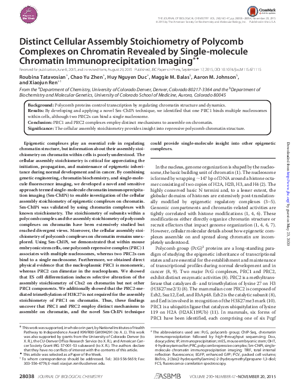
 Huy Nguyen Duc