j ocul biol dis inform (2011) 4:3–9
DOI 10.1007/s12177-011-9074-6
Interleukin-1β and mitochondria damage,
and the development of diabetic retinopathy
Renu A. Kowluru & Ghulam Mohammad &
Julia M. Santos & Shikha Tewari & Qing Zhong
Received: 6 October 2011 / Accepted: 5 December 2011 / Published online: 28 December 2011
# Springer Science+Business Media, LLC 2011
Abstract Mitochondrial dysfunction is considered to play an
important role in the development of diabetic retinopathy.
Recent evidence has also shown many similarities between
diabetic retinopathy and a low grade chronic inflammatory
disease. The aim of this study is to understand the interrelationship between proinflammtory mediator, IL-1β and mitochondrial dysfunction in the accelerated loss of capillary cells
in the retina. Using IL-1β receptor gene knockout (IL-1R1−/−)
diabetic mice, we have investigated the effect of regulation of
IL-1β on mitochondrial dysfunction and mtDNA damage,
and increased retinal capillary cell apoptosis and the development of retinopathy. Retinal mitochondrial dysfunction and
mtDNA damage were significantly ameliorated in IL-1R1−/−
mice, diabetic for ~10 months, compared to the wild-type
diabetic mice. This was accompanied by protection of accelerated capillary cell apoptosis and the development of acellular capillaries, histopathology associated with diabetic
retinopathy. Thus, mitochondrial damage could be one of the
key events via which increased inflammation contributes to
the activation of the apoptotic machinery resulting in the
development of diabetic retinopathy, and the possible mechanism via which inflammation contributes to the development
of diabetic retinopathy includes continuous fueling of the
vicious cycle of mitochondrial damage, which could be disrupted by inhibitors of inflammatory mediators.
Keywords Diabetic retinopathy . Interleukin-1β
Mitochondria
R. A. Kowluru (*) : G. Mohammad : J. M. Santos : S. Tewari :
Q. Zhong
Kresge Eye Institute, Wayne State University,
4717 St. Antoine,
Detroit, MI, USA
e-mail: rkowluru@med.wayne.edu
Introduction
Diabetic retinopathy, a chronic microvascular complication
of diabetes, remains the leading cause of acquired blindness
in working adults. In the pathogenesis of this multifactorial
disease many molecular and physiologic factors have been
implicated; however, the exact mechanism of the development of diabetic retinopathy remains elusive [1, 2]. Recent
research has shown that the retina in diabetes shows abnormalities consistent with other inflammatory diseases. The
levels of proinflammatory cytokines including interleukin1β (IL-1β), tumor necrosis factor are increased in the vitreous of the patients with proliferative diabetic retinopathy
and also in the retina from diabetic rodents [3–7]. In experimental models of diabetic retinopathy, anti-inflammatory
therapies have prevented its development, and suppression
of inflammation and regulation of tight junction proteins by
glucocorticoids have been considered as possible means to
treat/prevent this blinding disease [8–10].
Retina and its capillary cells experience increased oxidative stress in diabetes, and the mitochondria become dysfunctional and their DNA is damaged. Cytochrome c leaks
out of the mitochondria, accelerating the apoptosis of retinal
capillary cells, and ultimately, resulting in the development
of diabetic retinopathy [11–13]. Our previous work has
shown that IL-1β, via increase in oxidative stress and activation of NF-kB, increases apoptosis of retinal capillary
cells, and antioxidants, which inhibit the development of
diabetic retinopathy in rodent models, also inhibit diabetesinduced increases in retinal IL-1β [4, 14, 15]. How dysfunctional retinal mitochondria in diabetes increases IL-1β
remains unclear.
IL-1β exerts its effects by binding to the specific receptors, and there are two main homologous receptors: type I
IL-1 receptor (IL-1RI) and type II IL-1 receptor (IL-1RII).
�4
Among these two, IL-1RI is the only receptor capable of
mediating signaling, and IL-1RII acts as a ‘decoy’ receptor.
Production and activity of IL-1β are tightly regulated by IL1Ra, an endogenous IL-1β inhibitor which preferentially
binds to IL-1RI without initiating signal transduction [16].
Mohr et al., using IL-1β receptor knockout (IL-1R1−/−)
mice has shown that these mice do not present retinal
pathology associated with diabetic retinopathy and have
implicated the role of caspase-1/IL-1β signaling pathway
in the development of diabetic retinopathy [17].
The purpose of this study is to determine the interrelationship between increased IL-1β and mitochondrial
dysfunction in the accelerated apoptosis of retinal capillary cells, resulting in the development of diabetic retinopathy. Here, we have investigated the effect of diabetes
on mitochondria permeability, mtDNA damage and on
the apoptosis and the development of acellular capillaries
in the retina from IL-1R1−/− mice diabetic for over
10 months.
Methods
Mice
Wild-type (WT; C57BL/6 J) or IL-1R1−/− mice (B6.129S7Il1r1tm1Imx/J) were obtained from Jackson Laboratory (Bar
Harbor, ME). A group of WT and IL-1R1−/− mice were made
diabetic by five consecutive injections of streptozotocin
(55 mg/kg BW; WT-D and IL-D, respectively). These mice
were maintained in diabetic conditions for ~10 months, and
age-matched nondiabetic mice served as controls (WT-N and
IL-N). Blood glucose levels were four to five times higher in
diabetic mice compared to their age-matched nondiabetic
mice, but the severity of hyperglycemia was comparable in
both WT and IL-1R1−/− diabetic mice as evidenced by similar
average blood glucose values in these two groups (WT-D0
395±81 mg/dl and IL-D0409±103 mg/dl). Mice were sacrificed by carbon dioxide asphyxiation. The retina from one eye
was used to quantify biochemical and molecular parameters,
and the other eye was incubated immediately in 10% buffered
formalin for apoptosis/histopathology [18–20]. The treatment
of the animals conformed to the Association for Research in
Vision and Ophthalmology Resolution on the Use of Animals
in Research, and the Institutional Guidelines.
Mitochondria isolation
Retinal mitochondria were isolated using mitochondria isolation kit (Pierce, Rockford, IL), as routinely performed in our
laboratory [19, 20]. The mitochondria pellet, after washing
with PBS, was suspended in the mitochondria lysis buffer (2%
CHAPS in 25 mM Tris, 0.15 M NaCl, pH 7.2), and protein
j ocul biol dis inform (2011) 4:3–9
concentration was quantified by the bicinchoninic acid assay
(Sigma-Aldrich, St. Louis, MO).
Mitochondrial superoxide
Superoxide levels in the retina were quantified by incubating the freshly prepared retinal homogenate with 400 nM
MitoTracker Red CM-H2XROS (Molecular Probes, Eugene,
OR) for 30 min at 37°C. The fluorescence was measured at
599 nm emission wavelength and 579 nm excitation wavelength using LS 55 Fluorescence Spectrometer (PerkinElmer, Waltham, MA) [20].
Mitochondria damage
Mitochondria permeability was determined spectrophotometrically by quantifying calcium chloride-induced decrease in
absorbance at 540 nm [19]. In addition, the release of cytochrome c from the mitochondria into the cytosol was quantified by western blot technique [13]. Damage of mtDNA was
quantified by quantitative extended length PCR using mitochondrial genome-specific primers (Table 1), and to calculate
relative amplification, the intensity of the long product
(13.4 kb) was normalized with the short product (210 bp), as
routinely used in our laboratory [11, 12].
Quantification of IL-1β
The amount of IL-1β in the retina was quantified using ELISA
kit (R&D Systems, MN) according to the manufacturer’s
Table 1 List of primers
Target
Sequence
VEGF
Forward
Reverse
ICAM-1
Forward
5′-TGGATGTCTACCAGCGAAGC-3′
5′-ACAAGGCTCACAGTGATTTT-3′
Reverse
β-actin
Forward
Reverse
Cytochrome b
Forward
Reverse
mtDNA-long
Forward
Reverse
mtDNA-short
Forward
Reverse
5′-AGCTTGCACGACCCTTCTAA-3′
5′-AGCACCTCCCCACCTACTTT-3′
5′-CCTCTATGCCAACACAGTGC-3′
5′-CATCGTACTCCTGCTTGCTG-3′
5′-ACCCGCCCCATCCAACATCTCAT-3′
5′-TTGAGGCTCCGTTTGCGTGT-3′
5′-AAAATCCCCGCAAACAATGACCACCCC-3′
5′-GGCAATTAAGAGTGGGATGGAGCCAA-3′
5′-CCTCCCATTCATTATCGCCGCCCTTGC-3′
5′-GTCTGGGTCTCCTAGTAGGTCTGGGAA-3′
�j ocul biol dis inform (2011) 4:3–9
5
instructions, as previously reported by us [15]. Each sample
was run in duplicate to ensure reproducibility of the data, and
the sensitivity of the assay was ~3 pg/ml of IL-1β.
Gene expression
Total RNA was extracted from the retina with Trizol reagent
(Invitrogen), and cDNA was synthesized by High Capacity
cDNA Reverse Transcription Kit (Applied Biosystem). Gene
transcripts of VEGF and ICAM-1 were quantified by real time
RT-PCR using the Sybr Green assay and gene-specific primers. Internal standard used was β-actin (Table 1). Gene
expression of cytochrome b was quantified by semiquantitative conventional PCR. Change in mRNA abundance was
calculated using the ddCt method as routinely used by us
[20–22].
retinal vessel exposed to DNAse before initiation of the
TUNEL reaction were run simultaneously. After counting
the number of TUNEL-positive capillary cells, the slides
were stained with periodic acid-Schiff and hematoxylin for
histologic evaluation, and the number of acellular capillaries
was counted in multiple midretinal fields and expressed per
square millimeter of retinal area examined [18, 20, 23].
Statistical analysis
Data are reported as mean±SD and analyzed with nonparametric statistics. Experimental groups were compared using
Kruskal–Wallis test followed by Mann–Whitney test for multiple group comparison. Analyses yielded identical results
when performed using ANOVA with Fisher or Tukey.
Retinal capillary cell apoptosis and histopathology
Results
Retina was isolated from the formalin-fixed eyes and incubated with 3% crude trypsin in Tris–HCl buffer (pH 7.8)
containing 0.2 M sodium fluoride for 90 min [15, 18, 20].
Trypsin-digested retinal microvessels were evaluated for
terminal transferase dUTP nick end labeling (TUNEL) using
a commercially available kit (In Situ Cell Death Kit, Roche
Molecular Biochemicals, IN). Positive controls consisting of
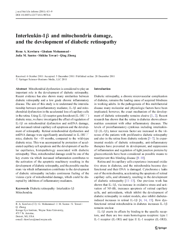
Regulation of IL-1B activation prevents diabetes-induced
damage to the retinal mitochondria
b.
Mitoch permeaility (%normal)
a.
Mitoch superoxide (%normal)
250
*
200
150
100
50
0
WT-N WT-D IL-N IL-D
c.
200
*
150
100
50
0
WT-N WT-D IL-N IL-D
Cyt c
Actin
200
Cytochrome c:actin
Fig. 1 Retinal mitochondria
damage is protected in IL1R1−/− diabetic mice: mitochondrial fraction was analyzed
for a superoxide levels by
employing fluorimetric method
using MitoTracker Red CMH2XROS, b membrane permeability by quantifying calcium
chloride-induced decrease in
absorbance at 540 nm and c
cytochrome c leakage into the
cytosol by western blot technique using β-actin as loading
control. Each measurement was
made in duplicate in five to
seven mice in each group, and
the values are represented as
mean±SD. WT and WT-D0
wild-type normal and diabetic
mice, respectively, and IL-N
and IL-D0IL-1R1−/− normal
and diabetic mice, respectively.
Asterisk indicates p<0.05 compared to WT-N
Consistent with our previous results [20, 24], diabetes in WT
mice increased mitochondrial superoxide levels in the retina by
about two fold, and mitochondria permeability by 60–70%
compared to the values obtained from WT-normal mice
*
150
100
50
0
WT-N WT-D IL-N IL-D
�6
j ocul biol dis inform (2011) 4:3–9
(Fig. 1a and b). The amount of cytochrome c into the cytosol
fraction was increased by over 50% (Fig. 1c). In the same
animals, mtDNA was damaged, as evidenced by 60% decrease
in the relative amplification of 13.4 kb and 210 bp products
(Fig. 2a). This was accompanied by 30% decrease in the gene
transcripts of mtDNA-encoded cytochrome b (Fig. 2b).
However, in IL-1R1−/− diabetic mice, mitochondria were
relatively healthy with significantly lower levels of mitochondrial superoxide, membrane that were less permeable
with decreased leakage of cytochrome c into the cytosol and
decreased damage to mtDNA compared to the values
obtained from WT diabetic mice (Figs. 1 and 2). Retina
from WT normal and IL-1R1−/− normal and diabetic groups
a.
Mice deficient in IL-1 receptor gene are protected
from diabetes-induced increase in the inflammatory cytokines
The concentration of retinal IL-1β was significantly increased
in WT-diabetes compared to WT normal. However, IL-1β
levels decreased by 30–40% in the retina from IL-1R1−/−
mice, and the values in IL-1R1−/− normal and IL-1R1−/−
diabetic mice were similar to each other (Fig. 3a). Similarly,
increase in retinal VEGF and ICAM, observed in WT-diabetic
mice, was also prevented in IL-1R1−/− diabetic mice (Fig. 3b
and c), suggesting that these mice were also protected from
diabetes-induced increase in proinflammatory mediators.
Long
Short
mtDNA damage (%normal)
had similar values for mitochondrial function, DNA damage
and the expression of cytochrome b.
150
100
50
0
*
WT-N WT-D IL-N IL-D
b.
Cytb
Actin
Capillary cell apoptosis is ameliorated in IL-1R1−/− diabetic
mice
Diabetes of ~10 months in WT mice significantly increased
the apoptosis of capillary cells, and the number of TUNELpositive cells in the trypsin digested retinal microvasculature
were approximately twofold higher compared to those from
WT-N mice (Fig. 4a). Consistent with this, the number of
acellular capillaries was also significantly increased
(Fig. 4b).
Regulation of IL-1 receptor gene protected the retinal microvasculature from diabetes-induced accelerated apoptosis,
and this was accompanied by reduced number of acellular
capillaries in IL-1R1−/− diabetic mice compared to those in
WT diabetic mice. The numbers of apoptotic capillary cells
and acellular capillaries in IL-1R1−/− diabetic mice were not
different from those obtained from IL-1R1−/− normal and WT
normal mice (Fig. 4).
Cytb mRNA (%normal)
150
Discussion
100
*
50
0
WT-N WT-D IL-N IL-D
Fig. 2 Damage to the retinal mtDNA is ameliorated in mice with IL-1
receptor gene manipulated. a Damage to mtDNA was estimated by
quantitative extended length PCR using GeneAmp XL PCR kit. b
Gene expression of mtDNA-encoded Cytb was quantified in the retina
by semiquantitative PCR using gene-specific primers. Measurements
were made in duplicate in six or more mice in each group, and the
values are represented as mean±SD. Asterisk indicates p<0.05 compared to WT-N
In the development of diabetic retinopathy, inflammatory
mediators are elevated in the retina, mitochondria become
dysfunctional, the enzyme important in scavenging superoxide is decreased and mtDNA becomes damaged [6,
11–13, 15]. The present study demonstrates that amelioration of inflammatory mediators regulates mitochondrial
damage and capillary cell apoptosis that the retina experiences in diabetes. Our data, using a mouse model of diabetic
retinopathy with genetic manipulation for interleukin 1 receptor, show that retinal mitochondria of these mice are
protected from diabetes-induced mitochondrial dysfunction
and DNA damage. Furthermore, the retinal vasculature also
escapes accelerated apoptosis, a phenomenon considered to
precede the appearance of histopathology characteristic of
diabetic retinopathy [25]. These results strongly suggest that
�j ocul biol dis inform (2011) 4:3–9
7
a.
IL-1B mRNA (% normal)
150
100
*
50
0
2
1
*
0
*
WT-N WT-D IL-N IL-D
200
VEGF mRNA (% normal)
*
200
150
100
50
*
0
*
WT-N WT-D IL-N IL-D
*
150
100
*
50
*
0
WT-N WT-D IL-N IL-D
Diabetes
WT
IL
Fig. 4 The number of apoptotic capillary cells in the retinal vasculature
is significantly lower in diabetic mice with the IL-1 receptor gene manipulated. a Trypsin-digested retinal microvasculature was analyzed for
capillary cell apoptosis by TUNEL staining. The arrow indicates
TUNEL-positive capillary cell. b After TUNEL staining, the microvessels were stained with periodic acid-Schiff–hematoxylin and examined by
light microscopy for the quantifying acellular capillaries–basement
TUNNEL+ capillary cells/retina
Diabetes
IL
Normal
*
3
d.
250
WT
b
*
WT-N WT-D IL-N IL-D
10
Acellular capillaries/ mm2retina
Normal
4
*
c.
ICAM1 mRNA (% normal)
a
b.
200
IL-1B conc (pg/mg prot)
Fig. 3 Regulation of IL-1 receptor gene protects increase in
the inflammatory cytokines in
the retina: gene transcripts of a
IL-1B, c ICAM-1 and d VEGF
were quantified in the retina by
real time RT-PCR using the Sybr
Green assay and gene-specific
primers. b The amount of IL-1β
in the retina was quantified by an
ELISA method. Each measurement was made in duplicate using retina from five to six mice in
each of the four groups, and the
values are represented as mean±
SD. Asterisk indicates p<0.05
compared to WT-N
10
*
8
#
6
#
4
2
0
WT-N WT-D IL-N IL-D
*
8
6
#
#
4
2
0
WT-N WT-D IL-N IL-D
membrane tubes lacking cell nuclei and maintaining at least one-fourth
the normal capillary caliber over their length. The arrow indicates acellular capillary. Results are expressed as mean±SD of at least six mice in
each group. WT-N and WT-D0wild-type nondiabetic and diabetic mice,
respectively, and IL-N and IL-D0IL-1R1−/− are normal and diabetic
mice, respectively. Asterisk indicates p<0.05 compared to WT-N and
number sign indicates p<0.05 compared to WT-D
�8
in diabetic environment, both inflammation and mitochondrial damage are interrelated.
Hyperglycemia elevates IL-1β in the retina and its capillary
cells; our previous work has shown that IL-1β administration
into the vitreous of normal rats increases oxidative stress and
activates redox-sensitive nuclear transcriptional factor-kB
(NF-kB), and antioxidants inhibit diabetes-induced increases
in retinal IL-1β [4, 15]. Furthermore, lipopolysaccharideinduced inflammatory response is shown to increase mitochondrial dysfunction in neuronal cells [26]. Here, our results
demonstrate that the amelioration of IL-1β activation prevents
mitochondrial dysfunction and DNA damage, and further
confirm a bidirectional mechanism between increased inflammatory cytokines and oxidative stress.
Diabetes damages mtDNA in the retina and its capillary
cells, and mitochondrial genome-encoded electron transport
chain proteins are compromised. The damage to mtDNA
initiates a vicious cycle, and the transcription of protein
encoded by mtDNA is decreased [11, 12, 22]. Protection
of the damage to mtDNA and decrease in cytochrome b (an
enzyme essential for the formation and activity of complex
III) by ameliorating IL-1β activation implies that the combination of increased inflammatory cytokine and mtDNA
damage in diabetes further fuels the vicious cycle resulting
in continued damage to the mitochondria, and decreased
flux through the subnormal electron transport chain continues to supply additional superoxide.
Apoptosis of retinal capillary is considered as a predictor
of histopathology associated with diabetic retinopathy [25].
Here, we show that IL-1R1−/− mice, maintained diabetic for
long durations, are protected from accelerated apoptosis of
capillary cells implying that IL-1β has an important role in
the apoptosis. In support, our previous studies have shown
that glucose-induced apoptosis of retinal endothelial cells is
prevented by incubating the cells with IL-1β antibody or IL1Ra [4], and administration of IL-1β in the vitreous of
normal rats accelerates capillary cell apoptosis and generates
acellular capillaries [15]. IL-1β is reported to induce apoptosis of retinal neuronal cells during reperfusion ischemia
[27], a phenomenon that is observed in the development of
diabetic retinopathy as well [28]. Furthermore, glucoseinduced apoptosis of retinal Müller cells is postulated to
depend on the increased autocrine production of IL-1β,
and via a paracrine mechanism, in later stages of diabetes,
this results in capillary cell death [29]. To make the bad
situation worse, damaged mitochondria by releasing cytochrome c and impairing mitochondrial membranes provoke
apoptosis [13]. Now, we show that the regulation of IL-1β
signalling protects retinal mitochondria from damage to
their membrane and the release of cytochrome c into the
cytosol is decreased, raising a strong possibility that the
protection of diabetes-induced retinal capillary cell apoptosis (and the histopathology) in IL-1R1−/− diabetic mice
j ocul biol dis inform (2011) 4:3–9
could be, in part, due to the protection of the integrity of
the mitochondria. In support, recent reports have shown that
the mitochondria have a central role in the activation of
inflammasomes, a crucial assembly platform which activates caspase-1, which in turn, processes IL-1β and other
inflammatory pathways [30, 31], and accumulation of damaged mitochondria is considered to contribute to increased
inflammation via initiation of inflammasomes [32]. Thus,
the possibility that the protection of the accelerated capillary
cell apoptosis and mitochondrial damage in IL-1R1−/− mice
is via regulation of inflammasomes remains to be explored.
In summary, this study, for the first time, demonstrates that
the mitochondrial damage is one of the key events via which
increased inflammation could contribute to the activation of
the apoptotic machinery resulting in the development of diabetic retinopathy. Thus, inhibition of inflammatory mediators
have potential therapeutic value in preventing the fueling of
the vicious cycle of mitochondrial damage, ultimately inhibiting the development and continued progression of diabetic
retinopathy.
Acknowledgments The authors thank Yakov Shamailov and Dug
Putt for their help in maintaining the mouse colony. This work was
supported, in part, by grants from the National Institutes of Health,
Juvenile Diabetes Research Foundation, The Thomas Foundation and
Research to Prevent Blindness.
References
1. Kowluru RA. Diabetic retinopathy: mitochondrial dysfunction and
retinal capillary cell death. Antioxid Redox Signal. 2005;7:1581–
7.
2. Frank RN. Diabetic retinopathy. N Engl J Med. 2004;350:48–58.
3. Yuuki T, Kanda T, Kimura Y, Kotajima N, Tamura J, Kobayashi I,
et al. Inflammatory cytokines in vitreous fluid and serum of
patients with diabetic vitreoretinopathy. J Diabetes Complications.
2001;15:257–9.
4. Kowluru RA, Odenbach S. Role of interleukin-1beta in the pathogenesis of diabetic retinopathy. British J Ophthalmol. 2004;88:1343–
7.
5. Demircan N, Safran BG, Soylu M, Ozcan AA, Sizmaz S. Determination of vitreous interleukin-1 (IL-1) and tumour necrosis
factor (TNF) levels in proliferative diabetic retinopathy. Eye.
2006;20:1366–9.
6. Krady JK, Basu A, Allen CM, Xu Y, LaNoue KF, Gardner TW, et
al. Minocycline reduces proinflammatory cytokine expression,
microglial activation, and caspase-3 activation in a rodent model
of diabetic retinopathy. Diabetes. 2005;54:1559–65.
7. Kern TS. Contributions of inflammatory processes to the development of the early stages of diabetic retinopathy. Exp Diabetes Res.
2007;2007:95103.
8. Felinski EA, Antonetti DA. Glucocorticoid regulation of endothelial cell tight junction gene expression: novel treatments for diabetic retinopathy. Curr Eye Res. 2005;30:949–57.
9. Kowluru RA, Kanwar M. Effect of curcumin on retinal oxidative
stress and inflammation in diabetes. Nutr Metab (Lond). 2007;4:1–
8.
�j ocul biol dis inform (2011) 4:3–9
10. Tang J, Kern TS. Inflammation in diabetic retinopathy. Prog Retin
Eye Res. 2011;30:343–58.
11. Madsen-Bouterse SA, Mohammad G, Kanwar M, Kowluru RA.
Role of mitochondrial DNA damage in the development of diabetic retinopathy, and the metabolic memory phenomenon associated
with its progression. Antioxid Redox Signal. 2010;13:797–805.
12. Madsen-Bouterse S, Zhong Q, Mohammad G, Ho YS, Kowluru
RA. Oxidative damage of mitochondrial DNA in diabetes, and its
protection by manganese superoxide dismutase. Free Rad Res.
2010;44:313–21.
13. Kowluru RA, Abbas SN. Diabetes-induced mitochondrial dysfunction in the retina. Inves Ophthalmol Vis Sci. 2003;44:5327–
34.
14. Kowluru RA, Tang J, Kern TS. Abnormalities of retinal metabolism in diabetes and experimental galactosemia. VII. Effect of
long-term administration of antioxidants on the development of
retinopathy. Diabetes. 2001;50:1938–42.
15. Kowluru RA, Odenbach S. Role of interleukin-1beta in the development of retinopathy in rats: effect of antioxidants. Invest Ophthalmol Vis Sci. 2004;45:4161–6.
16. Hallegua DS, Weisman MH. Potential therapeutic uses of interleukin 1 receptor antagonists in human diseases. Ann Rheum Dis.
2002;61:960–7.
17. Vincent JA, Mohr S. Inhibition of caspase-1/interleukin-1beta
signaling prevents degeneration of retinal capillaries in diabetes
and galactosemia. Diabetes. 2007;56:224–30.
18. Kowluru RA, Kowluru V, Ho YS, Xiong Y. Overexpression of
mitochondrial superoxide dismutase in mice protects the retina
from diabetes-induced oxidative stress. Free Rad Biol Med.
2006;41:1191–6.
19. Mohammad G, Kowluru RA. Matrix metalloproteinase-2 in the
development of diabetic retinopathy and mitochondrial dysfunction. Lab Invest. 2010;90:1365–72.
20. Kowluru RA, Mohammad G, Santos JM, Zhong Q. Abrogation of
MMP9 gene protects against the development of retinopathy in
diabetic mice by preventing mitochondrial damage. Diabetes
2011;60:3023–33.
9
21. Zhong Q, Kowluru RA. Epigenetic changes in mitochondrial superoxide dismutase in the retina and the development of diabetic
retinopathy. Diabetes. 2011;60:1304–13.
22. Santos JM, Tewari S, Goldberg AFX, Kowluru RA. Mitochondria
biogenesis and the development of diabetic retinopathy. Free Rad
Biol Med 2011;51:1849–60.
23. Mohammad G, Kowluru RA. Novel role of mitochondrial matrix
metalloproteinase-2 in the development of diabetic retinopathy.
Invest Ophthalmol Vis Sci. 2011;52:3832–41.
24. Kanwar M, Chan PS, Kern TS, Kowluru RA. Oxidative damage in
the retinal mitochondria of diabetic mice: possible protection by
superoxide dismutase. Invest Ophthalmol Vis Sci. 2007;48:3805–
11.
25. Kern TS, Tang J, Mizutani M, Kowluru R, Nagraj R, Lorenzi M.
Response of capillary cell death to aminoguanidine predicts the
development of retinopathy: comparison of diabetes and galactosemia. Invest Ophthalmol Vis Sci. 2000;41:3972–8.
26. Hunter RL, Dragicevic N, Seifert K, Choi DY, Liu M, Kim HC, et
al. Inflammation induces mitochondrial dysfunction and dopaminergic neurodegeneration in the nigrostriatal system. J Neurochem.
2007;100:1375–86.
27. Yoneda S, Tanihara H, Kido N, Honda Y, Goto W, Hara H, et al.
Interleukin-1beta mediates ischemic injury in the rat retina. Exp
Eye Res. 2001;73:661–7.
28. Zheng L, Gong B, Hatala DA, Kern TS. Retinal ischemia and
reperfusion causes capillary degeneration: similarities to diabetes.
Invest Ophthalmol Vis Sci. 2007;48:361–7.
29. Busik JV, Mohr S, Grant MB. Hyperglycemia-induced reactive
oxygen species toxicity to endothelial cells is dependent on paracrine mediators. Diabetes. 2008;57:1952–65.
30. Tschopp J. Mitochondria: sovereign of inflammation? Eur J Immunol. 2011;41:1196–202.
31. Zhou R, Yazdi AS, Menu P, Tschopp J. A role for mitochondria in
NLRP3 inflammasome activation. Nature. 2011;469:221–5.
32. Green DR, Galluzzi L, Kroemer G. Mitochondria and the
autophagy-inflammation-cell death axis in organismal aging. Science. 2011;333:1109–12.
�
 Julia MARGARIDA SANTOS