See discussions, stats, and author profiles for this publication at: https://www.researchgate.net/publication/49856215
State of the art in non-invasive imaging of
cutaneous melanoma
Article in Skin Research and Technology · February 2011
DOI: 10.1111/j.1600-0846.2011.00503.x · Source: PubMed
CITATIONS
READS
38
162
2 authors:
Louise Elizabeth Smith
Sheila Macneil
41 PUBLICATIONS 348 CITATIONS
455 PUBLICATIONS 7,923 CITATIONS
University of South Australia
SEE PROFILE
The University of Sheffield
SEE PROFILE
Some of the authors of this publication are also working on these related projects:
UK-India Centre for Advanced Technology for Minimizing Indiscriminate Use of Antibiotics View
project
All content following this page was uploaded by Louise Elizabeth Smith on 08 December 2014.
The user has requested enhancement of the downloaded file. All in-text references underlined in blue are added to the original document
and are linked to publications on ResearchGate, letting you access and read them immediately.
Skin Research and Technology 2011; 17: 257–269
Printed in Singapore All rights reserved
doi: 10.1111/j.1600-0846.2011.00503.x
r 2011 John Wiley & Sons A/S
Skin Research and Technology
Review Article
State of the art in non-invasive imaging of
cutaneous melanoma
Louise Smith and Sheila MacNeil
Department of Engineering Materials, Kroto Research Institute, University of Sheffield, Sheffield, UK
Background: This review focuses on looking at recent
developments in the non-invasive imaging of skin, in particular at how such imaging may be used at present or in the
future to detect cutaneous melanoma.
Methods: A MEDLINE search was performed for papers
using imaging techniques to evaluate cutaneous melanoma,
including melanoma metastasis.
Results: Nine different techniques were found: dermoscopy,
confocal laser scanning microscopy (including multiphoton microscopy), optical coherence tomography, high frequency ultrasound,
positron emission tomography, magnetic resonance imaging, and
Fourier, Raman, and photoacoustic spectroscopies. This review
Skin Imaging
F
imaging of skin, we want to be
able to correctly identify the epidermis and
dermis and to identify relatively large features such
as rete ridges and blood vessels. Ideally, we would
also like to be able to distinguish where the morphology of individual cells or groups of cells differs
from their immediate neighbours as is the case in
cutaneous invasive carcinomas, of which melanoma is the most difficult to diagnose and stage.
The current gold standard for morphological
investigation of the skin is achieved by taking a
small biopsy for standard histology (1). This allows
the visualisation of structures through a vertical
section of the skin, i.e. from the epidermis through
to the reticular dermis or even subcutaneous
tissue. However, this is invasive and relatively
time consuming and the accuracy of the detection
of invasive melanoma relies on biopsying the right
area at the right time.
There is a real need for non-invasive techniques for in vivo imaging of skin to assist dermaOR NON-INVASIVE
contrasts the effectiveness of these techniques when seeking to
image melanomas in skin.
Conclusions: Despite the variety of techniques available
for detecting melanoma, there remains a critical need for a
high-resolution technique to answer the question of whether
tumours have invaded through the basement membrane.
Key words: melanoma – non-invasive imaging – confocal
microscopy – dermoscopy – PET/CT – spectroscopy
& 2011 John Wiley & Sons A/S
Accepted for publication 6 November 2010
tologists and pathologists in the diagnosis of skin
conditions such as melanoma and basal cell
carcinoma. Current techniques for the diagnosis
of these conditions depend on the skill of the
clinician visually diagnosing the progression of
the disease, following the ABCD rules for melanoma detection (2) and/or by the removal of a
biopsy, which can cause pain, expense, and scarring (3). Crucially, the biopsy involves delayduring this time, the melanoma may be progressing through the basement membrane into
the dermis with its rich blood supply – a critical
stage in melanoma progression. At the time of
writing, not all melanoma are being correctly
diagnosed. Amelanotic malignant melanoma
presents a unique diagnostic challenge to physicians. While these amelanotic melanoma share
certain features with other malignant neoplasms,
namely large lesional size, asymmetrical architecture, and poor circumscription (4), the lack of
pigment can lead to the diagnosis of a less serious
cutaneous disease. According to McCalmont (5),
257
Smith and MacNeil
conventional microscopy has remained the gold
standard for melanoma diagnosis for several
decades, and a diagnosis of melanoma is optimally
based on a summation of microscopic features that
are evaluated as objectively as possible by an
experienced histopathologist. Photography, including whole-body photography, is also commonly
used in the detection and diagnosis of melanoma
and other skin cancers. However, this technique
does not allow imaging below the surface of
the stratum corneum and therefore will not be
included in this review.
Dermoscopy
Epiluminescence microscopy or dermoscopy is a
relatively new multispectral imaging technique
used to image skin. Over the past 10 years, this
non-invasive technique has been proven to be very
valuable in the in vivo evaluation of pigmented
skin lesions (6). The first dermoscopes used nonpolarised light and used a hand-held magnification device. This was placed on the skin after
the application of a liquid such as oil or alcohol gel
to reduce the diffraction of the light from the
stratum corneum. However, newer instruments
do not need the liquid layer (2, 7). All multispectral
imaging techniques have limited depth penetration and the depth of chromophores is determined
algorithmically and not optically [as it is done in
optical coherence tomography (OCT) and confocal
microscopy]. They are relatively inexpensive in
terms of components. They consist of a good
CCD camera, a multispectral source (white lamp),
and some simple intermediary optics. There have
also been efforts to combine confocal microscopy to
provide multispectral information (8). Dermoscopy
allows the visualisation of the horizontal plane of a
given skin lesion but only to the level of the
papillary dermis (9). There are several commercial
dermascope instruments available to aid clinicians
in their diagnosis such as Melafind (Mela Sciences,
Inc, Irvington, NY, USA) and SIAscope (Biocompatibles International
plc, Farnham, UK).
s
The MelaFind system consists of a hand-held
imaging device, which is comprised of several
components: an illuminator that filters 10 different
specific wavelengths of light ranging from 430 to
950 nm (10); a lens system composed of nine
elements that creates images of the light reflected
from the lesions; a photon (light) sensor; and an
image processor using proprietary algorithms to
extract many discrete characteristics or features
258
from the images. The images provided are horizontal images through the skin and images can be
obtained from as deep as the reticular dermis or
subcutaneous tissue (11). In feasibility studies,
MelaFind attained 95% sensitivity and close to
70% specificity, at 95% confidence levels (12).
Non-contact SIAscopy or spectrophotometric
intracutaneous analysis uses a digital camera to
capture cross-polarised images of a scene. A flash
gun is used as a light source, providing light over
the entire visible spectrum. The camera provides
raw information of the imaging chips’ response to
the light, which results in three pictures being
produced from the camera. Red, green, and blue
images are produced, each covering a different
region of the visible spectrum. These images are
then analysed, producing non-contact SIAscans
that represent the concentration of haemoglobin
and melanin within the skin. This technique has
been used in the imaging of pigmented skin
lesions and melanoma (13, 14), diagnosis of basal
cell carcinoma (15), and analysis of skin colour (16).
Confocal Scanning Laser Microscopy
(CSLM)
Confocal microscopy has been used in dermatology in two modes: fluorescence and reflectance
(9, 11, 17, 18). While dermatological research
tends to use fluorescence, clinical practice uses
reflectance microscopy as this does not require
fluorescent labelling of cells and tissues and
therefore is a viable technique for in vivo imaging.
Confocal microscopes consist of a light source, a
condenser, an objective lens, and a detector. The
basic premise of confocal microscopy is the selective collection of light from in focus planes in the
tissue. With the use of a low power near-infrared
(IR) laser, a beam of light is focused tightly on a
specific point in the skin. Light scattered or reflected from this illuminated point is collected
through a pinhole-sized aperture by a detector.
The light source, illuminated point, and detector
aperture are in optically conjugate focal planes (i.e.,
confocal planes). This allows for the collection of
light from the single in-focus plane and the rejection of light from all out-of-focus planes (19).
The imaging depth of a CSLM is limited by the
wavelength of the laser chosen with a 488 nm
laser imaging 50–100 mm into skin as reported by
Gareau et al. (20). However, others have been able
to image at depths of up to 300 mm (7, 9, 11, 19,
21), providing images of the basement membrane
State of the art in non-invasive imaging of melanoma
area and down into the papillary dermis. This
again provides two-dimensional horizontal images
through the skin. If a series of images are taken,
these can be stacked and a three-dimensional
rendering can be compiled. CSLM can be used
either in the reflectance or in the fluorescence
mode. The reflectance mode relies on inherent
differences in the reflectivity of the structures in
the skin. Free cytoplasmic melanin pigment and
cytoplasmic pigmented and non-pigmented melanosomes provide strong contrast for near-IR laser
light sources (11). Generally, CSLM are not considered to be portable. However, fibre-optic confocal imaging uses a single optical fibre to both
illuminate and detect the laser light in place of the
detector aperture (pinhole) in CSLM. This has
allowed miniaturisation of the system, thus producing a hand-held device more suitable for in vivo
clinical use (11). CSLM has shown promise in the
detection of both pigmented and amelanotic melanoma (7, 11, 19–21) as well as basal cell carcinoma
and pathological inflammatory conditions (19, 21).
CSLM can also potentially be used in the evaluation of topical treatments and in pharmacological
applications, especially when combined with Raman spectroscopy. However, these instruments are
generally expensive and therefore often beyond the
budget of the practicing general dermatologist (2).
Commercially, there have
been several reports
s
on the use of VivaScope (Lucid Inc., Rochester,
MN, USA) as a reflectance mode confocal microscope and Stratum (Optiscan Pty Ltd, Melbourne,
Australia) in the fluorescence mode. Reflectance
mode CSLM is a non-invasive imaging tool that
allows real-time visualisation of cells and structures with near histological resolution (0.5–1 mm
lateral and 3–5 mm axial when imaging from the
stratum corneum to the reticular dermis) (9, 21).
Initial work on reflectance mode confocal microscopy has been reported in (22). They could
achieve cellular resolution into the dermis with
a video rate laser scanning confocal microscope.
This research group followed up on this with
many studies (23–25). Contrast agents can be
used to provide clearer images in both reflectance
and fluorescence
modes (26).
s
VivaScope is a clinical confocal microscope
that allows imaging of individual cells in skin
and other living
tissues. The non-invasive nature
s
of VivaScope allows repeated imaging of a
single tissue site, which enables monitoring natural biological processes in vivo over time, and
provides real-time visualisation of processes such
as blood flow
in capillary loops. However,
the
s
s
VivaScope is not portable. VivaScope has been
used to investigate the tissue surrounding facial
pores (27) and to monitor the effectiveness of
anticancer drugs in patients with actinic keratosis
and basal cell carcinoma (28).
Figures 1–3 below illustrate the effectiveness of
CSLM in obtaining cellular resolution throughout
the epidermis and in imaging melanoma.
Multiphoton laser scanning microscopy (MPLSM)
is similar to CSLM; however, in MPLSM, excitation of fluorophores is obtained by a non-linear
multiphoton process, as opposed to the onephoton excitation used in conventional microscopy. Two-photon excitation occurs when two
photos of approximately half the one photon
energy are absorbed practically simultaneously
by the fluorescent molecule. This means that
near-infra-red radiation can be used to excite
fluorophores, either endogenous autofluorescent substrates such as the reduced coenzyme
NAD(P)H, pigmented basal cells, and collagen
(29) or fluorescent markers added to the substrate. For this to occur, high-intensity radiation
from a femtosecond pulsed laser, i.e. Ti-Sapphire,
is required (30). Figure 4 illustrates the ability of
multiphoton confocal microscopy to image with
cellular and subcellular resolution just using
the natural autofluorescence of the sample. In
the case illustrated in Fig. 4, the sample is human
skin.
While multiphoton confocal microscopy allows
the visualisation of structures with greater resolution deeper into the skin than conventional
CSLM, it can still image only relatively shallow
structures. For imaging deeper into skin tissue,
OCT is perhaps better suited. OCT can be applied
for the investigation of vertical cross sections,
instead of the horizontal sections provided by
microscopy images, up to a depth of about 2 mm.
These images are of a much lower resolution than
those given by confocal scanning laser and multiphoton confocal microscopy (31).
OCT
OCT is analogous to ultrasound B imaging, except that it uses light rather than sound waves
(11). With respect to its resolution, it is intermediate between CSLM and ultrasound, theoretically
allowing the visualisation of structures with a
resolution of approximately 1 mm. OCT is, as the
name suggests, a coherence technique based on
259
Smith and MacNeil
Fig. 1. Confocal images of normal human skin: a stratum corneum in forearm skin: (a) the corneocytes appear as bright, polygonal cells forming
islands separated by dark wrinkles and creases; (b) stratum corneum in acral (skin from the palms or soles of the feet) skin: the corneal outlines are
more clearly demarcated; (c) stratum granulosum: the nucleus of the granular cells appears as a dark oval structure surrounded by a bright, grainy rim
of cytoplasm. The granular cells are arranged in a cohesive pattern; (d) stratum spinosum: the cells look similar to the granular cells but are smaller in
size and have a less refractive cytoplasm. The cells are arranged in a honeycombed pattern; (e) stratum basale: the basal cells form bright rings around
the dark dermal papillae; (f) hair follicle: these appear as round dark whorled structures in the depth of the dermis; (g) sweat gland: sweat glands appear
as oval to round, centrally dark structures spiralling to the depth. Scale bar, 50 mm. Image from Branzan et al. (65).
the principle of Michelson interferometry. Light
from a low-coherence length light source is split
evenly between the sample and a reference mirror. By measuring the interference between light
backscattered from the tissue and from the reference mirror, the distance and magnitude of
optical scattering within the tissue can be measured with micrometre-scale precision. The axial
resolution is determined by the coherence length
260
of the light used; typically, this is 10–20 mm but
this could be improved to 2–4 mm by the use of
ultra short pulse lasers (11, 32). The images
obtained are two-dimensional vertical slices
through the tissue as shown in Fig. 5 below.
OCT is well established as a tool in ophthalmology and is being used increasingly in dermatology (1, 32–34), evaluation of wound healing
(35), and particularly in the diagnosis of mela-
State of the art in non-invasive imaging of melanoma
Fig. 2. Dermoscopic and confocal images of pigmented skin lesions and melanocytic nevus: (a) nests of bright monomorphous structures
corresponding to nevus cells and pigmented keratinocytes forming rings around the dark dermal papillae at the dermo–epidermal junction of a
compound nevus; (b) lentigo maligna: prominent dendritic refractive cells and loss of keratinocyte demarcation; (c) superficial spreading melanoma: a
nest of irregular bright structures corresponding to melanoma cells within the stratum spinosum (pagetoid spreading), with a blurred honeycombed
pattern. The dermoscopic images were taken with the Dermogenius ultrat (Rodenstock Präzisionsoptik Linus, Munich, Germany). Scale bar, 50 mm.
From Branzan et al. (65).
noma (11). Unfortunately, while the layers of the
skin, i.e. epidermis and dermis as well as adnexal
structures and blood vessels can be seen, the
basement membrane zone and cellular features
cannot be visualised and so OCT is of limited use
in the early detection of melanoma (9). OCT setups with sophisticated lasers have not yet achieved
cellular resolution images (36, 37), although their
imaging characteristics indicate that this should be
possible. For example OCT has been used successfully to detect advanced basal cell carcinoma, but it
was not able to distinguish between basal cell
carcinoma subtypes (3).
resolution (axial approaching 10 mm and lateral
o30 mm) at a depth of 1.1 mm (9, 11). Figure 6
shows a typical HFUS scan through a malignant
melanoma and the corresponding histology image.
While the major features of the sample can be
seen, HFUS does not provide cellular resolution.
HFUS can be used in the follow-up of chronic
and inflammatory skin diseases, pre- and postoperative assessments of skin tumours, especially
malignant melanoma and basal cell carcinoma
(Fig. 7), and can also be used as a guide during
surgical procedures (38).
Ultrasound
Magnetic Resonance Imaging (MRI) and
Positron Emission Tomography (PET)
High-frequency ultrasound (HFUS) uses ultrasound frequencies of between 3 and 100 MHz to
evaluate skin morphology (9, 11). Images, as in
OCT, are vertical slices through the skin, with the
depth of penetration and resolution dependent on
the frequency of the sound used. Low-frequency
(3–10 MHz) scanners are used for subcutaneous or
lymph node imaging. Twenty to 50 MHz scanners
are used for sharp focusing at superficial regions,
with 20 MHz generally considered the best compromise between resolution (axial o80 mm lateral
200 mm) and penetration ( 3.8 mm). Higher frequency scanners (50–100 MHz) have been used to
scan melanocytic lesions and have an improved
MRI and PET have also been used in the in vivo
diagnosis of skin cancer and malignant melanoma.
The application of MRI to dermatology has
become practical with the use of specialised surface coils that allow higher resolution imaging
than standard MRI coils (11, 39). Bittoun et al. (40)
have obtained images of normal skin with highly
anisotropic voxels of 70 390 3000 mm3, with
the smallest dimension being perpendicular to
the skin surface, allowing the different skin layers
to be visualised; see Fig. 8. Micro-MRI has also
been used to investigate the courses of cellulite in
humans (41) and the effects of photoageing in a
murine model (42). The use of magnetisation
261
Smith and MacNeil
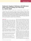
Fig. 3. The centre panel (d) shows the refined border of a lentigo maligna melanoma (LMM) on the scalp of a patient as determined
by confocal scanning laser microscopy (CSLM). The CSLM-examined foci are numbered 1–16 and are colour-coded to indicate areas
that were negative (green) and positive (purple) for LMM on CSLM images. Five pairs (marked in yellow) of these foci on either side of the border
were biopsied for histological confirmation. (a–c) show the confocal, histology, and Melan-A-immunostained sections of one representative
area of normal skin (long arrow in d). (a) Shows the epidermal layer and demonstrates the honeycomb pattern of keratinocytes and
well-defined cell-to-cell demarcations, which represent the characteristic architecture of normal skin. (b,c) The haematoxylin and eosin (H&E)stained and Melan-A-stained histological sections of normal skin, respectively. (e–g) Confocal, histology, and Melan-A immunostained
sections of one representative area of skin with LMM (long arrow in d). (e) shows the spinous layer and demonstrates pagetoid spread of atypical,
dendritic melanocytes (short arrow), loss of the normal architecture, and a grainy background – all features consistent with LMM. (f,g) H&E-stained
and Melan-A-stained histological sections of LMM, respectively. The Melan-A staining (g) shows the dendrites of the melanoma cell and correlates
with the dendritic malignant melanocyte (arrow) seen in (e). Original magnification: (b) 100; (c) 100; (f) 200; (g) 400. From
Chen et al. (66).
transfer contrast provides data enabling the evaluation of how the tissue in skin layers interacts
with the interstitial fluids. Details obtained from
high-resolution high-quality in vivo skin images
with different contrasts allowed for the differentiation of skin layers, sub-layers and excellent
correlation of MR data with known histological
features and water constituent of skin layers (43).
MRI has also been used to image melanoma
metastasis in a murine model (44) as well as the
imaging of individual skin cancers and the surrounding structures (45, 46); see Fig. 8.
PET, a whole-body imaging technique, is
widely used in the diagnosis of metastatic cancer.
A tracer is used to label the cancer cells. 18Ffluoro-deoxy-glucose (18F-FDG) is one of the
most widely applied PET tracers used to survey
cell metabolism. The metabolic turnover of tumour cells usually exceeds physiological metabolic activity. Excessive 18F-FDG uptake has
consequently been demonstrated in most cancers
in vivo, rendering whole-body 18F-FDG-PET an
excellent tool for the diagnosis of malignant skin
cancers. Non-specific uptake of 18F-FDG, how-
262
ever, has also been reported in various inflammatory conditions (47). While PET is considered a
non-invasive imaging technique, the patient does
have to take in the tracer, usually by ingestion.
The sensitivity of PET depends on the location,
and the size, of the tumour; however, a resolution
of 4–6 mm is usual. This means that PET may not
be sensitive enough to detect small nodel melanoma metastases, which are usually 1–2 mm in
size (48). PET by itself does not provide images
containing a great deal of detail and is therefore
usually combined with computerised tomography (CT) scanning (49, 50). See Fig. 9 for examples of PET, PET/CT images.
Spectroscopic Imaging Techniques
[Fourier Transform Infrared (FTIR),
Raman, and Photoacoustic
Spectroscopy]
Spectroscopic imaging techniques are not routinely used in the clinical diagnosis of melanoma
or for imaging the skin. FTIR and Raman are
State of the art in non-invasive imaging of melanoma
Fig. 4. In vivo high-resolution multiphoton imaging of human skin. The figure shows the structure of skin with complementary images of twophoton-induced autofluorescence of the different layers down to a depth of 200 mm (elastic fibres in the dermis), l 740 nm. Additional images show the
bright luminescence of pigmented cells due to melanin at the stratum basale of a nevus and the infiltration of inflammatory cells (monocytes and
granulocytes) into skin tissue at a depth of 65 mm. Image from Schenke-Layland et al. (29).
Fig. 5. Optical coherence tomographic (OCT) image over 6 mm of human finger tip skin, palm side. Image obtained using a Michelson
diagnostics EX1301 OCT system. Note the lack of cellular resolution compared with confocal and multiphoton microscopy but the increased detail
of the image in terms of the dermo–epidermal junction and the presence of rete ridges when compared with the image in Fig. 3. Image courtesy of
Dr M. Bonesi.
probably the best known of these techniques due
to their regular use in chemistry and materials
science. FTIR is based on the absorption of IR
energy. The IR radiation is split as in OCT so that
half goes to a reference arm and half to the
sample. The excitation of molecules by IR light
causes the covalent bonds in these molecules to
move. Different bonds absorb energy at different
characteristic wavelengths. When the light is
recombined in a Michelson interferometer, an
interferogram is produced. This is processed
using a Fourier transform to produce an absorbance spectrum that is unique to the compound
being analysed. This technique is rarely used in
vivo. However, FTIR has the potential to provide
information from within a sample, i.e. depth
profiling, if modes such as attenuated total reflectance or photoacoustic are used.
263
Smith and MacNeil
Fig. 6. Twenty megahertz B-scan (DUB 20, Taberna pro Medicum, Lueneburg, Germany) of malignant melanoma (a) with the corresponding
histology (b) ( 5 objective lens). E, entry echo; T, tumour; K, corneum/dermis; S, subcutis; B, blood vessel; F, fascia. Image from Marghoob et al. (11).
Fig. 7. Twenty megahertz ultrasound of a basal cell carcinoma on the nasal tip in cross sections: Discontinous entrance echo, hypoechoic tumour with
mixed echogenicity infiltrating half of the dermal depth. Lateral demarcation of tumour borders remains uncertain (yellow arrows). The cartilage (C) is
visualised as an echo-poor structure. In the transversal section (left), a small dermal vessel (V) is marked with a black arrow. The echo-rich band below
the cartilage corresponds to the endonasal skin in the vestibulum. Image from Schmid-Wendtner and Dill-Müller (38).
Fig. 8. Sagittal and axial T2-weighted images show invasion of a giant
basal cell carcinoma on the lower leg through the anterolateral compartments of the leg. Note the anterior invasion of the tibia (arrowheads) and
the osteolytic lesion of the fibular bone (*). Image from Arnaiz et al. (46).
Raman spectroscopy is based on the principle
of Raman scattering, the inelastic scattering of
electromagnetic radiation. In Raman spectro-
264
scopy, the sample is illuminated by a monochromatic visible or near IR light from a laser source
and its vibrations during the electrical polarisability changes are determined (51). Raman spectroscopy combined with confocal microscopy is
rapidly becoming a useful tool. Not only are
images obtained potentially at a depth of up
to approximately 200 mm within the skin but
chemical information is also obtained. It has
been shown that the chemical signature of
cancer cells is different from that of normal,
non-tumour, cells, e.g. basal cell carcinoma (52).
The vast majority of the dermatological studies
published using this technique focus on the
evaluation of topical agents for either cosmetic
or pharmacological applications (51, 53–58), and
the majority of these are either ex vivo or on
embedded histology sections. However, Caspers
et al. (53) obtained in vivo measurements while
State of the art in non-invasive imaging of melanoma
Fig. 9. (a,b) PET-negative/CT-positive metastases of malignant melanoma in a 44-year-old woman with a history of malignant melanoma. (a) The CT
component of PET/CT showed nodular opacities in both lungs (arrows in upper panel), which showed metastases of malignant melanoma on biopsy.
(b) A transaxial 18F-FDG PET scan (lower panel) showed normal 18F-FDG uptake in both lungs, probably because the lesions were smaller than the
resolution limit. (c,d) PET-positive/CT-negative metastases of malignant melanoma in a 53-year-old woman with a history of malignant melanoma.
(c) A transaxial 18F-FDG PET image showed increased 18F-FDG uptake in the left cerebellum (arrow in the upper panel), which was consistent with a
metastasis. (d) The CT component of PET/CT did not show any abnormality in the same area (arrow in the lower panel). (e) PET scan demonstrates
multiple sites of metastatic melanoma. (e) Whole-body PET scan showing hepatic and bilateral pulmonary metastases (arrows). Images (a–d) from
Akcali et al. (49) image E from Essner et al. (50).
Fig. 10. Left: In vivo confocal image and Raman spectroscopy of a sweat duct on the palm of the hand, 30 mm below the skin surface. The bright area
shows a sweat duct. The arrows mark the spots from which the Raman spectra were obtained. Right: The asterisk marks the prominent Raman band of
lactate at 856 cm 1. (a) Raman spectrum measured in the sweat duct. (b) Raman spectrum measured outside the sweat duct. (c) Difference spectrum
(a b). (d) Fit result of spectrum (a) with spectrum (b) and spectra of natural moisturising factor and sweat constituents. (e) In vitro Raman
spectrum of lactate. (f) In vitro Raman spectrum of urea. Image from Caspers et al. (53).
looking at the concentration of water and other
moisturising agents in the stratum corneum;
see Fig. 10. Several molecular species were measured in vivo including keratins, b-carotene, and
water, as well as exogenous materials applied
to the skin (59).
Photoacoustic spectroscopy uses the photoacoustic effect to investigate samples. As the
name suggests, this is the generation of acoustic
waves from the absorption of electromagnetic
radiation. Photoacoustic microscopy and spectroscopy have mainly been used to investigate/image
265
Smith and MacNeil
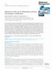
Fig. 11. In vivo non-invasive photoacoustic images of melanoma and vascular distribution in nude mouse skin. (a,b) Enface photoacoustic images for
the NIR light source (a) 5 764 nm and visible light source (b) 5 584 nm, respectively: 1, melanoma; 2, vessels perpendicular to the image plane; 3,
vessels horizontal to image plane; 4, skin. (c,d) Photoacoustic B-scan images from the NIR and visible light sources, respectively, for the dotted lines in
(a) and (b). (e) A cross-sectional histology image (H&E staining): E, epidermis; D, dermis; M, muscle. (f,g) Depthwise enface photoacoustic images
from the NIR and visible light sources, respectively; A, 0.15–0.30 mm; B, 0.30–0.45 mm; C, 0.45–0.60 mm; D, 0.60–0.75 mm from the skin surface.
Image from Oh et al. (60).
blood vessels within skin and in small animals; see
Fig. 11 (60, 61) as well as for the detection of
melanoma cells in the circulation (62–64).
Conclusion
Dermoscopy and Confocal Laser Scanning Microscopy are currently in use in the clinic, aiding in
266
the diagnosis of skin cancers, both melanoma and
non-melanoma. However, while these techniques
are useful in the evaluation of superficial skin
cancers, they are not able to image at depth.
Currently, the non-invasive techniques, OCT,
HFUS, MRI, PET, and the spectroscopic techniques do not have the resolution to detect earlystage skin cancers. These techniques can, however, be used to detect and therefore aid in the
State of the art in non-invasive imaging of melanoma
diagnosis of late-stage metastatic cancer, i.e.
when the secondary tumours become large enough to be detected. In this respect, PET and MRI
are currently used to detect advanced metastasis
of skin cancers. While both HFUS and OCT have
the potential to be used to detect local superficial
metastasis, they currently lack the resolution to
detect early-stage metastasis of melanoma. In
References
1. Gambichler T, Matip R, Moussa G,
Altmeyer P, Hoffmann K. In vivo
data of epidermal thickness evaluated by optical coherence tomography: effects of age, gender, skin
type, and anatomic site. J Dermatol
Sci 2006; 44: 145–152.
2. Menzies SW. Cutaneous melanoma:
making a clinical diagnosis, present
and future. Dermatol Ther 2006; 19:
32–39.
3. Gambichler T, Orlikov A, Vasa R,
Moussa G, Hoffmann K, Stücker M,
Altmeyer P, Bechara FG. In vivo
optical coherence tomography of
basal cell carcinoma. J Dermatol Sci
2007; 45: 167–173.
4. Adler MJ, White JCR. Amelanotic
malignant melanoma. Semin Cutan
Med Surg 1997; 16: 122–130.
5. McCalmont TH. Melanoma and
melanoma in situ: build a better
diagnosis through architecture.
Semin Cutan Med Surg 1997; 16:
97–107.
6. Ducharme EE, Silverberg NB.
Selected applications of technology
in the pediatric dermatology office.
Semin Cutan Med Surg 2008; 27:
94–100.
7. Esmaeili A, Scope A, Halpern AC,
Marghoob AA. Imaging techniques
for the in vivo diagnosis of melanoma. Semin Cutan Med Surg 2008;
27: 2–10.
8. Yaroslavsky AN, Barbosa J, Neel V,
DiMarzio C, Anderson RR. Combining multispectral polarized light
imaging and confocal microscopy
for localization of nonmelanoma
skin cancer. J Biomed Opt 2005; 10:
014011–014016.
9. Ulrich M, Stockfleth E, RoewertHuber J, Astner S. Noninvasive
diagnostic tools for nonmelanoma
skin cancer. Br J Dermatol 2007;
157 (Suppl. 2): 56–58.
10. Gutkowicz-Krusin D, Elbaum M,
Jacobs A, Keem S, Kopf AW, Kamino H, Wang S, Rubin P, Rabinovitz H, Oliviero M. Precision of
automatic measurements of pig-
11.
12.
13.
14.
15.
16.
17.
18.
19.
20.
conclusion, despite an extensive range of noninvasive imaging techniques, there is still a need
for imaging techniques with a higher resolution
to determine whether a melanoma tumour has
invaded through the basement membrane as this
is critical in the staging and treatment of these
very difficult-to-treat cancers.
mented skin lesion parameters
with a MelaFindTM multispectral
digital dermoscope. Melanoma Res
2000; 10: 563–570.
Marghoob AA, Swindle LD, Moricz
CZM S, Sanchez-Negron FA, Slueb
B, Halpern AC, Kopf AW. Instruments and new technologies for
the in vivo diagnosis of melanoma.
J Am Acad Dermatol 2003; 49: 777–
797.
Elbaum M, Kopf AW, Rabinovitz HS
et al. Automatic differentiation of
melanoma from melanocytic nevi
with multispectral digital dermoscopy: a feasibility study. J Am
Acad Dermatol 2001; 44: 207–218.
Moncrieff M, Cotton S, Claridge E,
Hall P. Spectrophotometric intracutaneous analysis: a new technique
for imaging pigmented skin lesions.
Br J Dermatol 2002; 146: 448–457.
Haniffa MA, Lloyd JJ, Lawrence
CM. The use of a spectrophotometric intracutaneous analysis device in the real-time diagnosis of
melanoma in the setting of a melanoma screening clinic. Br J Dermatol
2007; 156: 1350–1352.
Tehrani H, Walls J, Cotton S, Sassoon
E, Hall P. Spectrophotometric intracutaneous analysis in the diagnosis
of basal cell carcinoma: a pilot study.
Int J Dermatol 2007; 46: 371–375.
Matts PJ, Dykes PJ, Marks R. The
distribution of melanin in skin determined in vivo. Br J Dermatol
2007; 156: 620–628.
Meyer LE, Otberg N, Sterry W, Lademann J. In vivo confocal scanning
laser microscopy: comparison of the
reflectance and fluorescence mode
by imaging human skin. J Biomed
Opt 2006; 11–17.
Marghoob AA, Halpern AC. Confocal scanning laser reflectance microscopy: why bother? Arch Dermatol
2005; 141: 212–215.
Nehal KS, Gareau D, Rajadhyaksha
M. Skin imaging with reflectance
confocal microscopy. Semin Cutan
Med Surg 2008; 27: 37–43.
Gareau DS, Merlino G, Corless C,
Kulesz-Martin M, Jacques SL. Non-
21.
22.
23.
24.
25.
26.
27.
28.
invasive imaging of melanoma with
reflectance mode confocal scanning
laser microscopy in a murine model.
J Invest Dermatol 2007; 127: 2184–
2190.
Gonzalez S, Gilaberte-Calzada Y. In
vivo reflectance-mode confocal microscopy in clinical dermatology
and cosmetology. Int J Cosmet Sci
2008; 30: 1–17.
Rajadhyaksha M, Grossman M, Esterowitz D, Webb RH, Anderson
RR. In vivo confocal scanning laser
microscopy of human skin: melanin
provides strong contrast. J Invest
Dermatol 1995; 104: 946–952.
Rajadhyaksha M, Gonzalez S, Zavislan JM, Anderson RR, Webb RH. In
vivo confocal scanning laser microscopy of human skin II: advances in
instrumentation and comparison
with histology. J Invest Dermatol
1999; 113: 293–303.
Rajadhyaksha M, Gonzalez S, Zavislan JM. Detectability of contrast
agents for confocal reflectance imaging of skin and microcirculation. J
Biomed Opt 2004; 9: 323–331.
Rajadhyaksha M, Menaker G, Flotte
T, Dwyer PJ, Gonzalez S. Confocal
examination of nonmelanoma cancers in thick skin excisions to potentially guide mohs micrographic
surgery without frozen histopathology. J Invest Dermatol 2001; 117:
1137–1143.
Rudrabhatla SR, Petroll WM, Mahaffey CL, Mummert ME. Development of a hyaluronan targeted
contrast reagent for the demarcation
of melanoma margins in vivo. J
Invest Dermatol 2008; 128: 740–742.
Sugata K, Nishijima T, Kitahara T,
Takema Y. Confocal laser microscopic imaging of conspicuous facial pores in vivo: relation between
the appearance and the internal
structure of skin. Skin Res Technol
2008; 14: 208–212.
Astner S, Swindells K, Gonzalez S,
Stockfleth E, Lademann J. Confocal
microscopy: innovative diagnostic
tools for monitoring of noninvasive
therapy in cutaneous malignancies.
267
Smith and MacNeil
29.
30.
31.
32.
33.
34.
35.
36.
37.
38.
39.
Drug Discov Today Dis Mech 2008;
5: e81–e91.
Schenke-Layland K, Riemann I, Damour O, Stock UA, König K. Twophoton microscopes and in vivo multiphoton tomographs – Powerful diagnostic tools for tissue engineering
and drug delivery. Adv Drug Deliv
Rev 2006; 58: 878–896.
Paoli J, Smedh M, Wennberg AM,
Ericson MB. Multiphoton laser scanning microscopy on non-melanoma
skin cancer: morphologic features
for future non-invasive diagnostics.
J Invest Dermatol 2008; 128: 1248–
1255.
Lademann J, Otberg N, Richter H,
Meyer L, Audring H, Teichmann A,
Thomas S, Knüttel A, Sterry W. Application of optical non-invasive
methods in skin physiology: a comparison of laser scanning microscopy
and
optical
coherent
tomography with histological analysis. Skin Res Technol 2007; 13: 119–
132.
Fujimoto JG, Brezinski ME, Tearney
GJ, Boppart SA, Bourna B, Hee MR,
Sourthern JF, Swanson EA. Optical
biopsy and imaging using optical
coherence tomography. Nat Med
1995; 1: 970–972.
Welzel J. Optical coherence tomography in dermatology: a review.
Skin Res Technol 2001; 7: 1–9.
Gambichler T, Moussa G, Sand M,
Sand D, Altmeyer P, Hoffmann K.
Applications of optical coherence
tomography in dermatology. J Dermatol Sci 2005; 40: 85–94.
Wang Z, Pan H, Yuan Z, Liu J, Chen
W, Pan Y. Assessment of dermal
wound repair after collagen implantation with optical coherence tomography. Tissue Eng Part C Methods
2008; 14: 35–45.
Bizheva K, Povazay B, Hermann B,
Sattmann H, Drexler W, Mei M,
Holzwarth R, Hoelzenbein T, Wacheck V, Pehamberger H. Compact,
broad-bandwidth fiber laser for sub2-mm axial resolution optical coherence tomography in the 1300-nm
wavelength region. Opt Lett 2003;
28: 707–709.
Spöler F, Forst M, Marqúardt Y,
Hoeller D, Kurz H, Merk H, Abuzahra F. High-resolution optical coherence tomography as a nondestructive monitoring tool for the
engineering of skin equivalents.
Skin Res Technol 2006; 12: 261–267.
Schmid-Wendtner M-H, Dill-Müller
D. Ultrasound technology in dermatology. Semin Cutan Med Surg 2008;
27: 44–51.
Rajeswari MR, Jain A, Sharma A,
Singh D, Jagannathan NR, Sharma
268
40.
41.
42.
43.
44.
45.
46.
47.
48.
49.
50.
51.
U, Degaonkar MN. Evaluation of
skin tumors by magnetic resonance
imaging. Lab Invest 2003; 83: 1279–
1283.
Bittoun J, Querleux B, Darrasse L.
Advances in MR imaging of the
skin. NMR Biomed 2006; 19: 723–
730.
Mirrashed F, Sharp J, Krause V,
Morgan J, Tomanek B. Pilot study
of dermal and subcutaneous fat
structures by MRI in individuals
who differ in gender, BMI, and cellulite grading. Skin Res Technol
2004; 10: 161–168.
Altman AM, Bankson J, Matthias N,
Vykoukal JV, Song YH, Alt EU.
Magnetic resonance imaging as a
novel method of characterization of
cutaneous photoaging in a murine
model. Arch Dermatol Res 2008;
300: 263–267.
Mirrashed F, Sharp J. In vivo morphological characterisation of skin
by MRI micro-imaging methods.
Skin Res Technol 2004; 10: 149–160.
Foster PJ, Dunn EA, Karl KE, Snir
JA, Nycz CM, Harvey AJ, Pettis RJ.
Cellular magnetic resonance imaging: in vivo imaging of melanoma
cells in lymph nodes of mice. Neoplasia 2008; 10: 207–216.
Lanka B, Turner M, Orton C, Carrington BM. Cross-sectional imaging in non-melanoma skin cancer
of the head and neck. Clin Radiol
2005; 60: 869–877.
Arnaiz J, Gallardo E, Piedra T, SanzJimenez-Rico JR, Trillo Bohajar E,
Alonso Pena D. Giant basal cell
carcinoma on the lower leg: MRI
findings. J Plast Reconstr Aesthet
Surg 2007; 60: 1167–1168.
Hoffmann M, Vogelsang H, Kletter
K, Zettinig G, Chott A, Raderer M.
18F-fluoro-deoxy-glucose positron
emission tomography (18F-FDGPET) for assessment of enteropathy-type T cell lymphoma. Gut
2003; 52: 347–351.
Belhocine TZ, Scott AM, EvenSapir E, Urbain JL, Essner R. Role
of nuclear medicine in the management of cutaneous malignant
melanoma. J Nucl Med 2006; 47:
957–967.
Akcali C, Zincirkeser S, Erbagcý Z,
Akcali A, Halac M, Durak G, Sager
S, Sahin E. Detection of metastases
in patients with cutaneous melanoma using FDG-PET/CT. J Int
Med Res 2007; 35: 547–553.
Essner R, Belhocine T, Scott AM,
Even-Sapir E. Novel imaging techniques in melanoma. Surg Oncol
Clin N Am 2006; 15: 253–283.
Lin SY, Li MJ, Cheng WT. FT-IR and
Raman vibrational microspectrosco-
52.
53.
54.
55.
56.
57.
58.
59.
60.
61.
62.
63.
pies used for spectral biodiagnosis
of human tissues. Spectroscopy
2007; 21: 1–30.
Choi J, Choo J, Chung H, Gweon
DG, Park J, Kim HJ, Park S, Oh CH.
Direct observation of spectral differences between normal and basal cell
carcinoma (BCC) tissues using confocal Raman microscopy. Biopolymers 2005; 77: 264–272.
Caspers PJ, Lucassen GW, Puppels
GJ. Combined in vivo confocal Raman spectroscopy and confocal microscopy of human skin. Biophys J
2003; 85: 572–580.
Caspers PJ, Lucassen GW, Wolthuis
R, Bruining HA, Puppels GJ. In vitro
and in vivo Raman spectroscopy of
human skin. Biospectroscopy 1998;
4 (Suppl. 1): 31–39.
Gniadecka M, Faurskov Nielsen O,
Christensen DH, Wulf HC. Structure
of water, proteins, and lipids in intact
human skin, hair, and nail. J Invest
Dermatol 1998; 110: 393–398.
Herkenne C, Alberti I, Naik A, Kalia
YN, Mathy FX, Preat V, Guy RH. In
vivo methods for the assessment of
topical drug bioavailability. Pharm
Res 2008; 25: 87–103.
Wu J, Polefka TG. Confocal Raman
microspectroscopy of stratum corneum: a pre-clinical validation
study. Int J Cosmet Sci 2008; 30:
47–56.
Schallreuter KU, Moore J, Wood JM,
Beazley WD, Gaze DC, Tobin DJ,
Marshall HS, Panske A, Panzig E,
Hibberts NA. In vivo and in vitro
evidence for hydrogen peroxide
(H2O2) accumulation in the epidermis of patients with vitiligo and its
successful removal by a UVB-activated pseudocatalase. J Investig
Dermatol Symp Proc 1999; 4: 91–96.
Kollias N, Stamatas GN. Optical
non-invasive approaches to diagnosis of skin diseases. J Investig
Dermatol Symp Proc 2002; 7: 64–
75.
Oh JT, Li ML, Zhang HF, Maslov K,
Stoica G, Wang LV. Three-dimensional imaging of skin melanoma
in vivo by dual-wavelength photoacoustic microscopy. J Biomed Opt
2006; 11: 34032–34039.
Xu M, Wang LV. Photoacoustic imaging in biomedicine. Rev Sci Instrum 2006; 77–84.
Holan SH, Viator JA. Automated
wavelet denoising of photoacoustic
signals for circulating melanoma cell
detection and burn image reconstruction. Phys Med Biol 2008; 53: N227–
N236.
Weight RM, Viator JA, Dale PS,
Caldwell CW, Lisle AE. Photoacoustic detection of metastatic melan-
State of the art in non-invasive imaging of melanoma
oma cells in the human circulatory
system. Opt Lett 2006; 31: 2998–
3000.
64. Zharov VP, Galanzha EI, Shashkov
EV, Khlebtsov NG, Tuchin VV. In
vivo photoacoustic flow cytometry
for monitoring of circulating single
cancer cells and contrast agents. Opt
Lett 2006; 31: 3623–3625.
65. Branzan AL, Landthaler M, Szeimies RM. In vivo confocal scanning laser microscopy in derma-
tology. Lasers Med Sci 2007; 22:
73–82.
66. Chen CSJ, Elias M, Busam K,
Rajadhyaksha M, Marghoob AA.
Multimodal in vivo optical imaging,
including confocal microscopy, facilitates presurgical margin mapping for clinically complex lentigo
maligna melanoma. Br J Dermatol
2005; 153: 1031–1036.
Address:
Prof. Sheila MacNeil
Tissue Engineering Group
Kroto Research Institute
North Campus
University of Sheffield
Broad Lane, Sheffield S3 7HQ
UK
Tel: 144 0 114 222 5995
Fax: 144 0 114 222 5945
e-mail: s.macneil@sheffield.ac.uk
269
View publication stats
State of the art in non‐invasive imaging of cutaneous melanoma
Skin Research and Technology, 2011
Background: This review focuses on looking at recent developments in the non-invasive imaging of skin, in particular at how such imaging may be used at present or in the future to detect cutaneous melanoma.
Methods: A MEDLINE search was performed for papers using imaging techniques to evaluate cutaneous melanoma, including melanoma metastasis.
Results: Nine different techniques were found: dermoscopy, confocal laser scanning microscopy (including multiphoton microscopy), optical coherence tomography, high frequency ultrasound, positron emission tomography, magnetic resonance imaging, and Fourier, Raman, and photoacoustic spectroscopies. This review contrasts the effectiveness of these techniques when seeking to image melanomas in skin.
Conclusions: Despite the variety of techniques available for detecting melanoma, there remains a critical need for a high-resolution technique to answer the question of whether tumours have invaded through the basement membrane....Read more