Journal of Cellular Biochemistry 90:234–243 (2003)
PROSPECTS
Inorganic Phosphate as a Signaling Molecule
in Osteoblast Differentiation
George R. Beck, Jr.*
National Cancer Institute at Frederick, Center for Cancer Research, Basic Research Laboratory, Frederick,
Maryland 21702
Abstract
The spatial and temporal coordination of the many events required for osteogenic cells to create a
mineralized matrix are only partially understood. The complexity of this process, and the nature of the final product,
demand that these cells have mechanisms to carefully monitor events in the extracellular environment and have the ability
to respond through cellular and molecular changes. The generation of inorganic phosphate during the process of
differentiation may be one such signal. In addition to the requirement of inorganic phosphate as a component of
hydroxyapatite mineral, Ca10(PO4)6(OH)2, a number of studies have also suggested it is required in the events preceding
mineralization. However, contrasting results, physiological relevance, and the lack of a clear mechanism(s) have created
some debate as to the significance of elevated phosphate in the differentiation process. More recently, a number of studies
have begun to shed light on possible cellular and molecular consequences of elevated intracellular inorganic phosphate.
These results suggest a model in which the generation of inorganic phosphate during osteoblast differentiation may in and
of itself represent a signal capable of facilitating the temporal coordination of expression and regulation of multiple factors
necessary for mineralization. The regulation of protein function and gene expression by elevated inorganic phosphate
during osteoblast differentiation may represent a mechanism by which mineralizing cells monitor and respond to the
changing extracellular environment. J. Cell. Biochem. 90: 234–243, 2003. Published 2003 Wiley-Liss, Inc.y
Key words: inorganic phosphate; osteoblast differentiation; calcium; osteopontin; alkaline phosphatase
Biological mineralization, an ongoing process throughout life, is mainly accomplished
through the function of two cell types, osteoblasts and chondrocytes [Heinegard and
Oldberg, 1989; Karsenty and Wagner, 2002].
The molecular and cellular events required for
these two cell types to produce mineralized
bone are only partially known, while the temporal and spatial coordination of events are
even less understood. When mineralization occurs at inappropriate times and places the
consequences can be serious, resulting in conditions such as atherosclerosis and osteoarthritis among others. A more complete knowledge of
Grant sponsor: National Cancer Institute; Grant number:
CA84573.
*Correspondence to: George R. Beck, Jr., National Cancer
Institute, Center for Cancer Research, Basic Research
Laboratory, Bldg. 576, Rm. 110, Frederick MD 21702.
E-mail: gbeck@ncifcrf.gov
Received 23 June 2003; Accepted 24 June 2003
DOI 10.1002/jcb.10622
the cellular and molecular events leading to
mineralization will undoubtedly aid in understanding not only disorders of bone metabolism,
but also the inappropriate calcification of other
tissues.
Osteoblast differentiation is a complex process in which differentiation, in vitro, occurs on
the order of weeks. A number of cell culture
models have been developed and are thought to
reasonably represent the progression of events
that occur in vivo. These models, representing both primary cultures and cell lines from
various species, follow similar paths to differentiation and mineralization exhibiting a similar pattern and time frame of gene expression
[Stein et al., 1990; Aubin and Triffitt, 2002].
As an example, the murine preosteoblast cell
line MC3T3-E1 when treated with ascorbic
acid and given a source of organic phosphate,
beta-glycerophosphate (bGP) will differentiate
and mineralize within 21 days [Franceschi
and Iyer, 1992; Quarles et al., 1992]. The
differentiation process can be generally divided
into three distinct stages that are defined by:
(1) proliferation, (2) matrix maturation, and
Published 2003 Wiley-Liss, Inc. y This article is a US Government work,
and as such, is in the public domain of the United States of America.
Inorganic Phosphate and Osteoblast Differentiation
235
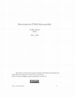
(3) mineralization (Fig. 1, top panel). A number
of genes including alkaline phosphatase, type-I
collagen, bone sialoprotein, osteopontin, and
osteocalcin have been identified that are
expressed at high levels for discrete periods
of time during differentiation. In vitro, as
differentiation proceeds, the levels of alkaline phosphatase enzyme activity rise and in
the presence organic phosphate will generate
free inorganic phosphate [Bellows et al., 1992;
Chung et al., 1992]. The result of the differentiation process is the formation of hydroxyapatite mineral that is thought to occur through
two possible mechanisms, the formation of
matrix vesicle, small vesicles that bud from
the plasma membrane and accumulate calcium
and phosphate [Anderson, 1995], and/or the
nucleation of collagen, regulated by associated
noncollagenous matrix proteins [Glimcher,
1989; Boskey, 1998].
Although inorganic phosphate is a necessary component of hydroxyapatite, a number
of studies have suggested that it is also integral
to bone remodeling in vivo [Baylink et al., 1971]
and osteoblast function during the differentiation process in vitro [Bingham and Raisz, 1974;
Gerstenfeld et al., 1987; Bellows et al., 1991;
Tenenbaum et al., 1992]. The events that define
chondrocyte maturation are somewhat different than osteoblasts, however, they do share the
common features of increased alkaline phosphatase expression, matrix vesicle formation,
hydroxyapatite mineral deposition, and the
requirement of inorganic phosphate [Fallon
Fig. 1. Temporal coordination of osteoblast differentiation and
phosphate regulated genes. Top panel: A schematic diagram of
the three general stages of osteoblast differentiation. Proliferating
osteoblasts, such as the murine preostoblast cell line MC3T3-E1,
when treated with ascorbic acid and bGP at confluency (day 0)
will go through a limited number of cell divisions and
asynchronously exit the cell cycle (approximately day 7). As
the cells exit the cell cycle, the presence of ascorbic acid
increases both collagen synthesis and alkaline phosphatase gene
expression and activity. During the collagen matrix maturation
stage collagen accumulates and non-collagenous proteins such
as bone sialoprotein (BSP), osteopontin (OPN), and osteocalcin
(OSC), among others are deposited within the matrix. This stage
also finds the formation of matrix vesicles that fill with a
nucleation core of calcium and phosphate before release to the
extracellular matrix. The final stage is marked by matrix vesicle
release and accumulation of hydroxyapatite nodules within the
collagen matrix. Bottom panel: Early in the differentiation
process alkaline phosphatase levels rise and through the
interaction with bGP produce increasing amounts of inorganic
phosphate in the extracellular environment. A number of genes
have been identified in MC3T3-E1 cells to be both positively or
negatively regulated by the increase in phosphate. A general
schematic represents the timing of the regulation of these genes
by increasing phosphate in the context of the differentiation
process.
236
Beck
et al., 1980; Wuthier, 1993]. This article will
focus on studies surrounding the significance of
inorganic phosphate generation during osteoblast differentiation as a possible signaling
mechanism that may temporally coordinate
cellular and molecular events preceding mineralization.
GENERATION AND TIMING OF
ELEVATED INORGANIC PHOSPHATE:
ALKALINE PHOSPHATASE
Alkaline phosphatase is a membrane bound
enzyme situated so that the catalytic subunit is
extracellular. Although originally identified in
1923 and theorized to be responsible for the
production of inorganic phosphate during skeletal mineralization [Robison, 1923], the function of this enzyme in osteoblast differentiation
remains a source of some debate. The importance of alkaline phosphatase in the process
of mineralization is suggested by the human
genetic disease hypophosphatasia [Rathburn,
1948; Whyte, 1994] that arises from mutations
in the alkaline phosphatase gene and is characterized by varying degrees of bone defects.
The phenotype is recapitulated in the alkaline
phosphatase knock out mouse [Fedde et al.,
1999]. Other evidence for the role of this enzyme
in mineralization comes from the ability to
transfect alkaline phosphatase cDNA into alkaline phosphatase negative cells and promote
mineralization [Yoon et al., 1989] and to inhibit
mineralization by inhibiting alkaline phosphatase activity [Tenenbaum, 1987]. Although
numerous functions have been proposed for the
enzyme [Whyte, 1994], the ability of inorganic
phosphate to substitute for both alkaline phosphatase activity and bGP supplementation
[Bellows et al., 1992; Boskey et al., 1992] in the
mineralization process argues that one critical
function is to locally increase inorganic phosphate levels.
Early in the differentiation process (days 1–
4) as osteoblasts become confluent, exit the cell
cycle, and respond to ascorbic acid with deposition of a collagen matrix, the levels of alkaline
phosphatase RNA and activity rise (Fig. 1, top
panel). As the activity of the enzyme increases
in the presence of bGP, the amount of inorganic
phosphate also rises. Studies investigating the
requirement and timing of alkaline phosphatase and bGP in the process of mineralization
have revealed that bGP, and hence elevated
levels of inorganic phosphate, are required for
the initiation of mineralization but, once the
process is initiated, mineralization will continue at non-elevated levels in both osteoblasts [Tenenbaum, 1987; Bellows et al., 1991;
Fratzl-Zelman et al., 1998] and chondrocytes
[Zimmermann et al., 1992]. The study by
Fratzl-Zelman et al., also investigated the
length of time required for the initial exposure
to 10 mM bGP to result in mineralization. These
authors found a pulse of 10 mM bGP for 24 h but
not 12 h, followed by exposure to low levels of
bGP (2 mM) still resulted in mineralization.
This agrees with the Bellows et al. [1991] study
which found mineralization was blocked when
alkaline phosphatase was inhibited within 8 h
after the addition of 10 mM bGP but was not
blocked when alkaline phosphatase was inhibited 24 h after the addition of bGP. There also
seems to be a critical time point during the
differentiation process, likely a stage of matrix
maturation, at which the generation of phosphate promotes mineralization after which no
mineralization occurs regardless of amount of
phosphate added [Tenenbaum et al., 1992;
Zimmermann et al., 1992]. Taken together, the
above studies support the notion that the
generation of inorganic phosphate may be more
important to the differentiation process than
the actual hydroxyapatite formation, and the
ability of phosphate to affect cell function may be
dependent on a particular stage of maturation.
TRANSPORT OF INORGANIC
PHOSPHATE: SODIUM DEPENDENT
PHOSPHATE TRANSPORTERS
In addition to the previously mentioned
studies other lines of research, including the
regulation of phosphate transport, have also
suggested the importance of inorganic phosphate in the differentiation process. The primary mechanism for inorganic phosphate entry
through the cell membrane is via a family of
sodium dependent phosphate transporters.
This family of transporters is subdivided into
three groups, based in part on tissue specificity
[Takeda et al., 2000]. Osteoblasts and chondrocytes express mainly the type III (NPT3)
transporters [Caverzasio and Bonjour, 1996]
which were first identified as receptors for
the gibbons ape leukemia virus (Glvr-1, Pit-1)
and amphotropic murine retrovirus (RAM,
Pit-2) [Kavanaugh and Kabat, 1996]. These
Inorganic Phosphate and Osteoblast Differentiation
transporters regulate phosphate transport
not only through the cell membrane but also
through the membrane of matrix vesicles. A
number of agents have been identified, including parathyroid hormone, insulin like growth
factor-1, platelet derived growth factor, fluoride
[Caverzasio and Bonjour, 1996], and calcium
[Schmid et al., 1998] that promote or enhance
inorganic phosphate entry into the cell or
matrix vesicles through these transporters.
The consequences following phosphate entry
into the cell are only beginning to be understood.
CELLULAR AND MOLECULAR CONSEQUENCES
OF INCREASED INTRACELLULAR PHOSPHATE:
POSITIVE REGULATION
An early demonstration that an increase
in inorganic phosphate during bone development plays an important role in addition to
mineral deposit formation came from a study by
Bingham and Raisz [1974]. Using fetal rat long
bones in organ cultures, this study examined
the effect of increasing phosphate (1.5–4.5 mM)
on bone growth and mineralization. Increasing
amounts of phosphate resulted in increased
collagen content, synthesis of labeled hydroxyproline, and calcification. The increase in collagen content and hydroxyproline synthesis
suggested that increased phosphate regulates
aspects of cell function in addition to mineralization. Although this study, and those previously mentioned in this article point to the
importance of elevated inorganic phosphate in
the differentiation process, the functional significance of increased intracellular phosphate
has been elusive.
Over the past few years, a number of studies
have begun to examine the significance of increased inorganic phosphate on osteoblast function at the cellular and molecular level. One of
the first suggestions that increased inorganic
phosphate may participate in directly regulating gene expression important in osteoblast
function came from the study of a cell line with
repressed alkaline phosphatase activity [Beck
et al., 1998]. In addition to showing repressed
alkaline phosphatase activity, this cell line also
failed to induce expression of osteopontin as
differentiation proceeded. Through a series of
experiments using exogenously added alkaline
phosphatase it was determined that osteopontin expression was regulated by increased
inorganic phosphate [Beck et al., 2000]. Use of
237
the phosphate transport inhibitor, foscarnet
(phosphonoformic acid or PFA) established
that phosphate must enter the cell to produce
changes in gene expression. The sequencing of
the human and mouse genomes and proliferation of microarray technology has made it possible to analyze thousands of genes instead of
the limited set of osteoblast marker genes
discussed thus far. A recent microarray study
has identified a discrete set of genes up and
downregulated in MC3T3-E1 osteoblasts by
treatment with 10 mM phosphate for 72 h [Beck
et al., 2003]. A number of these genes and their
protein products have been identified as regulated during osteoblast differentiation or
the mineralization process and are shown in
(Fig. 1, bottom panel). The response of these
genes to increased inorganic phosphate may
provide insight into the temporal coordination
of the differentiation process.
The identification of two important matrix
vesicle proteins, the calcium channel, annexin
V, and the phosphate transporter, Pit-1, as
genes regulated by increased inorganic phosphate suggests a potentially exciting mechanism by which phosphate may help temporally
coordinate differentiation and mineralization
[Beck et al., 2003]. The expression of these two
genes, and an additional, non-Na-dependent
phosphate transporter have been previously
associated with either bGP or inorganic phosphate induced differentiation and mineralization [Kirsch et al., 1997; Nielsen et al., 2001;
Wang et al., 2001; Garcia et al., 2002; Wu et al.,
2002]. Prior to budding and release from the
plasma membrane, matrix vesicles are supplied
with proteins from the cell including among
others, annexin V, phosphate transporters,
and alkaline phosphatase. These proteins are
thought to be necessary for the eventual
accumulation of calcium and phosphate within
the vesicle [Anderson, 1995; Caverzasio and
Bonjour, 1996]. The increased expression of
both a phosphate and calcium transporter to
increased inorganic phosphate suggests a
mechanism by which osteoblasts and chondrocytes might coordinate the initiation of matrix
vesicle formation with the generation of phosphate early in the differentiation process.
A recent study investigating the effects of
phosphate and calcium on mineralization noted
that both transcription and translation were
required for mineralization induced by either
bGP or inorganic phosphate [Chang et al.,
238
2000]. The microarray study by Beck et al.
[2003] identified a number of transcriptional
regulators that were upregulated in response
to phosphate and may provide a clue to the
regulation of phosphate responsive genes.
One such gene was Nrf2, a basic leucine zipper
transcription factor that functions in the regulation of phase II detoxifying enzymes [Itoh
et al., 1997], and has previously been identified
as a gene upregulated during osteoblast differentiation [Beck et al., 2001]. Both the demonstration that elevated phosphate will increase
expression of Nrf2 in the presence of the translation inhibitor cycloheximide and the analysis
of the Nrf2 promoter suggest that it is directly
regulated by increased phosphate and may be
considered a primary response gene [Beck et al.,
2003]. The role of Nrf2 in osteoblast differentiation remains to be elucidated, although the
knockout mice are viable and have not been
reported to have any obvious bone defects [Chan
et al., 1996]. Since Nrf2 is a member of a family
of proteins and only functions as a heterodimer,
it is possible that there are compensatory
mechanisms involved. A number of other transcription factors were identified to be upregulated by increased phosphate including two
members of the high-mobility group proteins,
HMGA1 and HMGA2, the growth arrest and
DNA damage inducible gene, Gadd153 and
Fra-2, a member of the AP-1 family of proteins.
Although Gadd153, HMGA1, and HMGA2
have not been previously associated with osteoblast differentiation, the AP-1 family has
been linked to bone development both in vivo
[Wagner, 2002] and in vitro [McCabe et al.,
1996]. Of course, as many transcription factors
are regulated post-translationally, more work
is required to determine the transcriptional
complexes responsible for regulating the phosphate-induced response.
Another possible transcriptional mediator
of the phosphate response is Cbfa1/RUNX2, a
transcription factor of the RUNT family critical
for the formation and function of osteoblasts
[Ducy, 2000]. Cbfa1 does not appear to be highly
regulated at the RNA level in response to
increased phosphate in osteoblasts [Beck et al.,
2003]. However, Fujita et al. [2001b] using
MC3T3-E1 osteoblasts and ATDC5 chondrocyte
cells identified the nuclear export of the Cbfa1 in
response to the addition of 3–10 mM inorganic
phosphate. The negative regulation of Cbfa1 by
elevated phosphate would seem to be contra-
Beck
dictory to the requirement of Cbfa1 in the
expression of osteocalcin that occurs later in
the differentiation process when phosphate
levels are high. It is possible that elevated
phosphate induces post-translational modifications of Cbfa1 and that these modifications are
transient. Clearly, more work will be required to
determine the nature of this regulation in the
context of differentiation. Although the role of
Cbfa1 in phosphate induced gene expression
remains to be determined in osteoblasts, Cecilia
Giachelli et al. have identified the elevation of
inorganic phosphate and subsequent upregulation of Cbfa1 and osteocalcin expression in
human smooth muscle cells (HSMC) as a key
factor in ectopic vascular calcification [Jono
et al., 2000]. This response is also dependent on
the presence and function of the phosphate
transporter Pit-1. The regulation of Cbfa1 at the
RNA level differs from the osteoblast model.
This may be explained by cell type specific
mechanisms and/or the possibility that basal
levels of Cbfa1 are much lower in HSMC,
making an increase more detectable. Interestingly, in this model osteopontin also plays a key
role in the response to elevated phosphate and is
thought to function as an inhibitor of ectopic
calcification [Giachelli, 2001].
Most of the mineralization studies mentioned
so far involve in vitro culture of either primary
or immortalized cells and use 4–10 mM bGP or
inorganic phosphate. The use of higher levels of
phosphate such as 10 mM bGP in these systems
has been questioned as to its physiological relevance [Gronowicz et al., 1989; Khouja et al.,
1990; Chung et al., 1992]. The findings of Beck
et al. [2003] suggest that the dose of phosphate
required to affect gene expression is related
to the amount of time the cell is exposed to
phosphate. A general curve can be constructed
illustrating the time/dose relationship related
to the ability of phosphate to upregulate gene
expression (Fig. 2). The similar changes in
gene expression in response to low doses of
phosphate at longer time points relative to those
at higher doses at shorter time points suggest
the events occurring at the 10 mM dose are
likely representative of the events that occur at
lower doses but longer exposure times. Furthermore, the demonstration that cellular exposure
to lower doses of phosphate requires longer
times to result in gene changes, and that elevated inorganic phosphate must enter the cell to
affect gene expression, suggest that it is the
Inorganic Phosphate and Osteoblast Differentiation
Fig. 2. Dose/time relationship of inorganic phosphate regulation of gene expression. Based on Northern blots described in
Beck et al. [2003] a general curve representing the dose/time
relationship of phosphate-induced changes in gene expression
can be generated. The longer the cells are exposed to elevated
phosphate the lower the dose required to produce changes in
gene expression. For example, the dose of phosphate required for
increased OPN expression within 24 h is approximately 10 mM,
but if the cells are exposed for 96 h a dose of only 2.5–5.0 mM is
required. Genes with increased expression (solid line) respond to
lower doses of phosphate at shorter time points than the genes
that are downregulated by phosphate (dashed line).
accumulated level of intracellular inorganic
phosphate that is critical not necessarily the
amount of extracellular phosphate added. In
light of the fact that various hormones and
growth factors may enhance phosphate transport and that these may differ in vitro relative
to in vivo, comparing the intracellular levels
of phosphate may be the most physiologically
relevant determinant. Additionally, hormones,
growth factors, and extracellular signals are
capable of regulating many of the phosphate
responsive genes mentioned in this article and
therefore, in vivo, lower doses of inorganic
phosphate may act in synergy with these other
factors to enhance gene expression. In this case,
relatively small changes in inorganic phosphate
may result in elevated gene expression, although this has yet to be demonstrated.
CELLULAR AND MOLECULAR CONSEQUENCES
OF INCREASED INTRACELLULAR PHOSPHATE:
NEGATIVE REGULATION
Multiple studies have suggested the possibility that increased inorganic phosphate may
represent a negative feedback loop capable of
downregulating alkaline phosphatase activity
in both chondrocytes [Genge et al., 1988] and
osteoblasts [Gerstenfeld et al., 1987; Tenenbaum, 1987; Aronow et al., 1990]. However, the
results from other studies found no decrease in
239
the level or function of the enzyme in the presence of bGP [Lee et al., 1992; Chak et al., 1995;
Anagnostou et al., 1996]. Yet another study
found an increase in enzyme activity but a
decrease in mRNA levels [Kyeyune-Nyombi
et al., 1995]. The conflicting results on the
response of alkaline phosphatase to inorganic
phosphate suggest the likelihood that phosphate acts in synergy with other signals generated during the differentiation process and
therefore the timing and amount of phosphate
present in relation to stage of differentiation
may be critical to the final response. For
example, Farley et al. [1994] demonstrate that
the level of enzyme activity is inversely proportional to calcium levels, as mineralization
proceeds, the increased amount of localized
calcium and phosphate may lead to the downregulation of alkaline phosphatase enzyme
activity, as suggested in Genge et al. [1988].
Data generated from microarray studies on
MC3T3-E1 cells also identified a number of
genes downregulated in response to treatment
with 10 mM phosphate for 72 h [Beck et al.,
2003]. The products of these genes represent
almost exclusively extracellular matrix proteins. Many of them have been previously
implicated in osteoblast differentiation and
include; collagens type I and III, decorin, perlecan (heparan sulfate proteoglycan 2), thrombospondin, and periostin (Fig. 1, bottom panel).
The downregulation of both type-I and II
collagens in response to mineralization [Gerstenfeld et al., 1987; Aronow et al., 1990;
Thomas et al., 1990; Tenenbaum et al., 1992;
Garcia et al., 2002] and inorganic phosphate
[Boskey et al., 1992; Fujita et al., 2001b] has
previously been noted in osteoblasts and chondrocytes. However, Lee et al. [1992] found no
difference in the expression of matrix-associated proteins in osteoblasts treated with
differentiation medium for 72 h, although this
may be the result of different cell culture
protocols. The downregulation of matrix proteins later in the differentiation process may
serve two purposes. Since expression and
translation require energy the downregulation
of proteins no longer required may conserve
energy stores. Additionally, the expression of
genes such as decorin, periostin, and thrombospondin are more closely associated with the
earlier stages of differentiation and may represent inhibitors of mineralization. In fact the
requirement for the downregulation of decorin
240
Beck
protein levels in the mineralization process has
previously been described [Hoshi et al., 1999].
Analysis of collagen type-I and periostin
expression revealed that downregulation occurs
only in the presence of higher levels of phosphate (4 mM) relative to upregulated genes, at
least at times tested [Beck et al., 2003]. Based on
those results a general schematic of the dose/
time relationship can be constructed (Fig. 2).
The regulation of various extracellular matrix
associated genes at higher phosphate levels and
the requirement for longer exposure times
agree with the hypothesis of a negative feedback
mechanism that would only occur at the later
stages of differentiation once matrix maturation is complete. This again emphasizes the
possible role of elevated inorganic phosphate in
the temporal coordination of the differentiation
process.
As osteoblasts and chondrocytes are responsible for the creation of bone, osteoclasts are
responsible for bone resorption. Studies suggest
that inorganic phosphate may also influence
bone formation by the inhibition of mineral
resorption [Brand and Raisz, 1972] and osteoclast differentiation [Takeyama et al., 2001;
Kanatani et al., 2003]. The Kanatani et al.
[2003] study found that increasing inorganic
phosphate concentrations (2.5–4.0 mM) inhibited osteoclast differentiation and the bone
resorbing activity of mature osteoclasts. In this
way, inorganic phosphate may not only promote
bone formation but may simultaneously block
bone loss.
CALCIUM TO PHOSPHATE RATIO
The localized concentration of both calcium
and phosphate during differentiation and the
nature of calcium and phosphate to spontaneously precipitate suggests that osteoblasts
and chondrocytes must perform a delicate balancing act to create proper hydroxyapatite
(Ca10(PO4)6(OH)2). Non-physiological precipitation must be avoided but so also must the
negative effects of the simultaneous increase in
both ions. Adams et al. [2001] investigated the
consequences of an increase of both inorganic
phosphate and calcium. The increase of relatively small amounts of calcium (0.1–1 mM),
above the 1.8 mM in the medium, in the presence of elevated inorganic phosphate caused
rapid apoptosis in both chondrocytes and osteoblasts. Similar studies were conducted using
phosphate alone and also found significant cell
death in osteoblasts [Meleti et al., 2000] and
chondrocytes [Mansfield et al., 2001]. However,
Wu et al. [2002] using chondrocyte cultures at a
different stage of development and grown in
different culture medium than the study by
Mansfield et al., did not find significant apoptosis in response to elevated phosphate. These
studies highlight the fact that a number of
factors may influence the effect of phosphate on
a given cell including, the stage of differentiation and cell type, the amount of fetal bovine
serum (FBS) present (FBS is likely to contain
factors that buffer calcium precipitation), and
the pH of inorganic phosphate used (usually a
4:1 ratio of Na2HPO4 and NaH2PO4 resulting
in a pH of 7.4). Differences such as these may
significantly alter the cell response to phosphate and may be at least partially responsible
for the conflicting results discussed throughout
this article.
Although the results in chondrocyte cultures
may still be a matter of some debate, many of the
osteoblast studies discussed thus far use 10 mM
bGP and 10% FBS and do not report significant
apoptosis during the differentiation stage. The
upregulation by inorganic phosphate of stress
related factors such as Nrf2, A170, and Gadd153
and calcium binding proteins such as osteopontin, annexin V, and calcyclin may help protect
the cell from the possible negative effects of
calcium phosphate precipitation. Furthermore,
calcium binding proteins, by balancing the
calcium to phosphate ratio in the extracellular
space or matrix vesicles, may aid in the formation of proper hydroxyapatite crystal as opposed to non-physiological mineral deposition.
The subsequent release of bound calcium at
an appropriate stage of differentiation may be
another mechanism to facilitate proper hydroxyapatite formation as proposed by Wuthier
[1977]. It is possible that once differentiation
and mineralization is complete the lack of continued expression of calcium/phosphate regulated genes results in the cells becoming more
susceptible to various bone remodeling processes that may generate unregulated increases
in both ions, eventually leading to apoptosis.
SIGNALING MECHANISMS
How can an increase in intracellular phosphate produce changes at both the transcriptional and posttranslational level? Although
Inorganic Phosphate and Osteoblast Differentiation
studies on the intracellular signaling mechanisms of increased phosphate have just begun,
some understanding is emerging. Studies using
calcium chelators and calcium channel blockers
suggest that phosphate is neither acting by
sequestering available calcium pools [Beck
et al., 2003] nor by producing an influx of
calcium through traditional calcium channels
[Adams et al., 2001; Beck et al., 2003]. However,
intracellular calcium may play a functionally
significant role in mediating phosphate-induced
changes. Adams et al. [2001] identified an increase in intracellular calcium following treatment of osteoblast cells with elevated calcium
and phosphate prior to apoptosis. Additionally,
Narayanan et al. [2003] demonstrated that
the nuclear export of the calcium binding protein dentin matrix protein 1 (DMP-1) requires
intracellular calcium and these authors speculate that the events are triggered by an influx
of inorganic phosphate.
We have recently found that phosphate
selectively activates the extracellular signalregulated kinase (ERK1/2) signaling pathway
[Beck and Knecht, submitted]. Treatment of
MC3T3-E1 cells with elevated phosphate
caused phosphorylation of ERK1/2 but did not
activate the other mitogen activated protein
kinase (MAPK) signaling proteins, p38 or the
c-jun N-terminal kinase (JNK). In response
to addition of 10 mM inorganic phosphate,
phosphorylated ERK1/2 levels rise within 10–
15 min followed by a second and more sustained
phosphorylation of ERK1/2 occurring after 10–
12 h of treatment. The timing of the second
activation closely precedes the increased transcription of osteopontin. Inhibitors of a number
of other pathways including PI3-kinase, protein kinase A, and protein kinase G do not inhibit phosphate induced osteopontin expression
suggesting a high degree of specificity in the
signaling mechanism induced by increased
inorganic phosphate. These observations agree
with a recent study demonstrating an increase
in ERK1/2 phosphorylation in response to
bisphosphonates that is further induced by
addition of 3 mM inorganic phosphate [Fujita
et al., 2001a]. These authors also did not detect
activation of either p38 or JNK in response to
phosphate. Although further investigation is
needed to fully understand the mechanism by
which increased intracellular inorganic phosphate might regulate gene expression and
ultimately cell function, it does appear that
241
specific signaling pathways exist resulting in
the possibility of manipulating these pathways
in the treatment of bone related diseases.
SUMMARY AND FUTURE
The studies discussed in this article have
begun to shed light on the significance of inorganic phosphate in osteoblast differentiation
and mineralization. The increase in inorganic
phosphate may not only represent an important
constituent of the mineral itself but also an
important signaling molecule. The elevation
of intracellular inorganic phosphate triggers a
series of cellular and molecular changes that
may transition the cell, matrix vesicles, and the
extracellular matrix to a mineralization competent state. The role of inorganic phosphate as a
signaling molecule in osteoblasts and chondrocytes is just beginning to be understood and
many challenges lie ahead. The regulation of
gene expression or protein function is usually
the sum of multiple effectors. Therefore, it will
be important to determine how signals generated by elevated inorganic phosphate are integrated with other signals generated during the
differentiation process including ascorbic acid
treatment, collagen matrix formation, cell to
cell contact, cell cycle exit, and perhaps most
importantly the accumulation of calcium. The
complexity of these interactions will first require in vitro experimentation but ultimately
will require confirmation in vivo. In the short
term, it will likely be important to establish the
mechanisms by which an increase in intracellular phosphate produces changes in gene
transcription and protein function, identifying
signaling pathways either stimulated or inhibited and the transcriptional complexes responsible for these responses.
REFERENCES
Adams CS, Mansfield K, Perlot RL, Shapiro IM. 2001.
Matrix regulation of skeletal cell apoptosis. Role of calcium and phosphate ions. J Biol Chem 276:20316–20322.
Anagnostou F, Plas C, Nefussi JR, Forest N. 1996. Role of
beta-GP-derived Pi in mineralization via ecto-alkaline
phosphatase in cultured fetal calvaria cells. J Cell Biochem 62:262–274.
Anderson HC. 1995. Molecular biology of matrix vesicles.
Clin Orthop 314:266–280.
Aronow MA, Gerstenfeld LC, Owen TA, Tassinari MS,
Stein GS, Lian JB. 1990. Factors that promote progressive development of the osteoblast phenotype in cultured
fetal rat calvaria cells. J Cell Physiol 143:213–221.
242
Aubin JE, Triffitt JT. 2002. Mesenchymal stem cells and
osteoblast differentiation. San Diego: Academic Press.
Baylink D, Wergedal J, Stauffer M. 1971. Formation, mineralization, and resorption of bone in hypophosphatemic
rats. J Clin Invest 50:2519–2530.
Beck GR, Jr., Knecht N. Osteopontin regulation by inorganic phosphate is ERK1/2, PKC, and proteasome dependent (submitted) .
Beck GR, Jr., Sullivan EC, Moran E, Zerler B. 1998.
Relationship between alkaline phosphatase levels, osteopontin expression, and mineralization in differentiating
MC3T3-E1 osteoblasts. J Cell Biochem 68:269–280.
Beck GR, Jr., Zerler B, Moran E. 2000. Phosphate is a
specific signal for induction of osteopontin gene expression. Proc Natl Acad Sci USA 97:8352–8357.
Beck GR, Jr., Zerler B, Moran E. 2001. Gene array analysis
of osteoblast differentiation. Cell Growth Differ 12:
61–83.
Beck GR, Jr., Moran E, Knecht N. 2003. Inorganic
phosphate regulates multiple genes during osteoblast
differentiation, including Nrf2. Exp Cell Res 288:288–
300.
Bellows CG, Aubin JE, Heersche JN. 1991. Initiation and
progression of mineralization of bone nodules formed
in vitro: The role of alkaline phosphatase and organic
phosphate. Bone Miner 14:27–40.
Bellows CG, Heersche JN, Aubin JE. 1992. Inorganic
phosphate added exogenously or released from betaglycerophosphate initiates mineralization of osteoid
nodules in vitro. Bone Miner 17:15–29.
Bingham PJ, Raisz LG. 1974. Bone growth in organ culture:
Effects of phosphate and other nutrients on bone and
cartilage. Calcif Tissue Res 14:31–48.
Boskey AL. 1998. Biomineralization: Conflicts, challenges,
and opportunities. J Cell Biochem Suppl 30–31:83–91.
Boskey AL, Stiner D, Doty SB, Binderman I, Leboy P. 1992.
Studies of mineralization in tissue culture: Optimal
conditions for cartilage calcification. Bone Miner 16:
11–36.
Brand JS, Raisz LG. 1972. Effects of thyrocalcitonin and
phosphate ion on the parathyroid hormone stimulated
resorption of bone. Endocrinology 90:479–487.
Caverzasio J, Bonjour JP. 1996. Characteristics and regulation of Pi transport in osteogenic cells for bone metabolism. Kidney Int 49:975–980.
Chak CW, Lee KM, Leung KS, Fung KP. 1995. No change
in bone-specific alkaline phosphatase activities in cultured rat osteoblastic cells under L-ascorbate and betaglycerophosphate-induced mineralization. Cell Biol Int
19:979–985.
Chan K, Lu R, Chang JC, Kan YW. 1996. NRF2, a member
of the NFE2 family of transcription factors, is not essential for murine erythropoiesis, growth, and development.
Proc Natl Acad Sci USA 93:13943–13948.
Chang YL, Stanford CM, Keller JC. 2000. Calcium and
phosphate supplementation promotes bone cell mineralization: Implications for hydroxyapatite (HA)-enhanced
bone formation. J Biomed Mater Res 52:270–278.
Chung CH, Golub EE, Forbes E, Tokuoka T, Shapiro
IM. 1992. Mechanism of action of beta-glycerophosphate
on bone cell mineralization. Calcif Tissue Int 51:305–
311.
Ducy P. 2000. Cbfa1: A molecular switch in osteoblast
biology. Dev Dyn 219:461–471.
Beck
Fallon MD, Whyte MP, Teitelbaum SL. 1980. Stereospecific inhibition of alkaline phosphatase by L-tetramisole
prevents in vitro cartilage calcification. Lab Invest 43:
489–494.
Farley JR, Hall SL, Tanner MA, Wergedal JE. 1994.
Specific activity of skeletal alkaline phosphatase in
human osteoblast-line cells regulated by phosphate,
phosphate esters, and phosphate analogs and release of
alkaline phosphatase activity inversely regulated by
calcium. J Bone Miner Res 9:497–508.
Fedde KN, Blair L, Silverstein J, Coburn SP, Ryan LM,
Weinstein RS, Waymire K, Narisawa S, Millan JL,
MacGregor GR, Whyte MP. 1999. Alkaline phosphatase
knock-out mice recapitulate the metabolic and skeletal
defects of infantile hypophosphatasia. J Bone Miner Res
14:2015–2026.
Franceschi RT, Iyer BS. 1992. Relationship between collagen synthesis and expression of the osteoblast phenotype in MC3T3-E1 cells. J Bone Miner Res 7:235–246.
Fratzl-Zelman N, Fratzl P, Horandner H, Grabner B, Varga
F, Ellinger A, Klaushofer K. 1998. Matrix mineralization
in MC3T3-E1 cell cultures initiated by beta-glycerophosphate pulse. Bone 23:511–520.
Fujita T, Izumo N, Fukuyama R, Meguro T, Yasutomi C,
Nakamuta H, Koida M. 2001a. Incadronate and etidronate accelerate phosphate-primed mineralization of MC4
cells via ERK1/2-Cbfa1 signaling pathway in a Rasindependent manner: Further involvement of mevalonate-pathway blockade for incadronate. Jpn J Pharmacol
86:86–96.
Fujita T, Meguro T, Izumo N, Yasutomi C, Fukuyama R,
Nakamuta H, Koida M. 2001b. Phosphate stimulates
differentiation and mineralization of the chondroprogenitor clone ATDC5. Jpn J Pharmacol 85:278–281.
Garcia T, Roman-Roman S, Jackson A, Theilhaber J,
Connolly T, Spinella-Jaegle S, Kawai S, Courtois B,
Bushnell S, Auberval M, Call K, Baron R. 2002. Behavior of osteoblast, adipocyte, and myoblast markers in
genome-wide expression analysis of mouse calvaria
primary osteoblasts in vitro. Bone 31:205–211.
Genge BR, Sauer GR, Wu LN, McLean FM, Wuthier RE.
1988. Correlation between loss of alkaline phosphatase
activity and accumulation of calcium during matrix
vesicle-mediated mineralization. J Biol Chem 263:
18513–18519.
Gerstenfeld LC, Chipman SD, Glowacki J, Lian JB. 1987.
Expression of differentiated function by mineralizing
cultures of chicken osteoblasts. Dev Biol 122:49–60.
Giachelli CM. 2001. Ectopic calcification: New concepts in
cellular regulation. Z Kardiol 90(Suppl 3):31–37.
Glimcher MJ. 1989. Mechanism of calcification: Role of
collagen fibrils and collagen–phosphoprotein complexes
in vitro and in vivo. Anat Rec 224:139–153.
Gronowicz G, Woodiel FN, McCarthy MB, Raisz LG. 1989.
In vitro mineralization of fetal rat parietal bones in defined serum-free medium: Effect of beta-glycerol phosphate. J Bone Miner Res 4:313–324.
Heinegard D, Oldberg A. 1989. Structure and biology of
cartilage and bone matrix noncollagenous macromolecules. FASEB J 3:2042–2051.
Hoshi K, Kemmotsu S, Takeuchi Y, Amizuka N, Ozawa H.
1999. The primary calcification in bones follows removal
of decorin and fusion of collagen fibrils. J Bone Miner Res
14:273–280.
Inorganic Phosphate and Osteoblast Differentiation
Itoh K, Chiba T, Takahashi S, Ishii T, Igarashi K, Katoh Y,
Oyake T, Hayashi N, Satoh K, Hatayama I, Yamamoto
M, Nabeshima Y. 1997. An Nrf2/small Maf heterodimer
mediates the induction of phase II detoxifying enzyme
genes through antioxidant response elements. Biochem
Biophys Res Commun 236:313–322.
Jono S, McKee MD, Murry CE, Shioi A, Nishizawa Y, Mori
K, Morii H, Giachelli CM. 2000. Phosphate regulation of
vascular smooth muscle cell calcification. Circ Res 87:
E10–E17.
Kanatani M, Sugimoto T, Kano J, Kanzawa M, Chihara K.
2003. Effect of high phosphate concentration on osteoclast differentiation as well as bone-resorbing activity.
J Cell Physiol 196:180–189.
Karsenty G, Wagner EF. 2002. Reaching a genetic and
molecular understanding of skeletal development. Dev
Cell 2:389–406.
Kavanaugh MP, Kabat D. 1996. Identification and characterization of a widely expressed phosphate transporter/
retrovirus receptor family. Kidney Int 49:959–963.
Khouja HI, Bevington A, Kemp GJ, Russell RG. 1990.
Calcium and orthophosphate deposits in vitro do not
imply osteoblast-mediated mineralization: Mineralization by betaglycerophosphate in the absence of osteoblasts. Bone 11:385–391.
Kirsch T, Nah HD, Shapiro IM, Pacifici M. 1997. Regulated
production of mineralization-competent matrix vesicles
in hypertrophic chondrocytes. J Cell Biol 137:1149–1160.
Kyeyune-Nyombi E, Nicolas V, Strong DD, Farley J. 1995.
Paradoxical effects of phosphate to directly regulate the
level of skeletal alkaline phosphatase activity in human
osteosarcoma (SaOS-2) cells and inversely regulate the
level of skeletal alkaline phosphatase mRNA. Calcif
Tissue Int 56:154–159.
Lee KL, Aubin JE, Heersche JN. 1992. Betaglycerophosphate-induced mineralization of osteoid does
not alter expression of extracellular matrix components
in fetal rat calvarial cell cultures. J Bone Miner Res 7:
1211–1219.
Mansfield K, Teixeira CC, Adams CS, Shapiro IM. 2001.
Phosphate ions mediate chondrocyte apoptosis through a
plasma membrane transporter mechanism. Bone 28:1–8.
McCabe LR, Banerjee C, Kundu R, Harrison RJ, Dobner
PR, Stein JL, Lian JB, Stein GS. 1996. Developmental
expression and activities of specific fos and jun proteins
are functionally related to osteoblast maturation: Role of
Fra-2 and Jun D during differentiation. Endocrinology
137:4398–4408.
Meleti Z, Shapiro IM, Adams CS. 2000. Inorganic phosphate induces apoptosis of osteoblast-like cells in culture.
Bone 27:359–366.
Narayanan K, Ramachandran A, Hao J, He G, Park KW,
Cho M, George A. 2003. Dual Functional Roles of Dentin
Matrix Protein 1. Implications in biomineralization and
gene transcription by activation of intracellular Ca2þ
store. J Biol Chem 278:17500–17508.
Nielsen LB, Pedersen FS, Pedersen L. 2001. Expression of
type III sodium-dependent phosphate transporters/retroviral receptors mRNAs during osteoblast differentiation.
Bone 28:160–166.
Quarles LD, Yohay DA, Lever LW, Caton R, Wenstrup RJ.
1992. Distinct proliferative and differentiated stages of
murine MC3T3-E1 cells in culture: An in vitro model of
osteoblast development. J Bone Miner Res 7:683–692.
243
Rathburn JC. 1948. Hypophosphatasia, a new developmental anomaly. Am J Dis Child 75:822–831.
Robison R. 1923. The possible significance of hexosephosphoric esters in ossification. Biochem J 17:286–293.
Schmid C, Keller C, Schlapfer I, Veldman C, Zapf J. 1998.
Calcium and insulin-like growth factor I stimulation of
sodium-dependent phosphate transport and proliferation
of cultured rat osteoblasts. Biochem Biophys Res Commun 245:220–225.
Stein GS, Lian JB, Owen TA. 1990. Relationship of cell
growth to the regulation of tissue-specific gene expression during osteoblast differentiation. FASEB J 4:3111–
3123.
Takeda E, Taketani Y, Morita K, Tatsumi S, Katai K, Nii T,
Yamamoto H, Miyamoto K. 2000. Molecular mechanisms
of mammalian inorganic phosphate homeostasis. Adv
Enzyme Regul 40:285–302.
Takeyama S, Yoshimura Y, Deyama Y, Sugawara Y,
Fukuda H, Matsumoto A. 2001. Phosphate decreases
osteoclastogenesis in coculture of osteoblast and
bone marrow. Biochem Biophys Res Commun 282:798–
802.
Tenenbaum HC. 1987. Levamisole and inorganic pyrophosphate inhibit beta-glycerophosphate induced mineralization of bone formed in vitro. Bone Miner 3:13–26.
Tenenbaum HC, Limeback H, McCulloch CA, Mamujee H,
Sukhu B, Torontali M. 1992. Osteogenic phase-specific
co-regulation of collagen synthesis and mineralization by
beta-glycerophosphate in chick periosteal cultures. Bone
13:129–138.
Thomas JT, Boot-Handford RP, Grant ME. 1990. Modulation of type X collagen gene expression by calcium betaglycerophosphate and levamisole: Implications for a possible role for type X collagen in endochondral bone
formation. J Cell Sci 95(Pt 4):639–648.
Wagner EF. 2002. Functions of AP1 (Fos/Jun) in bone
development. Ann Rheum Dis 61(Suppl 2):ii40–ii42.
Wang D, Canaff L, Davidson D, Corluka A, Liu H, Hendy
GN, Henderson JE. 2001. Alterations in the sensing and
transport of phosphate and calcium by differentiating
chondrocytes. J Biol Chem 276:33995–34005.
Whyte MP. 1994. Hypophosphatasia and the role of alkaline phosphatase in skeletal mineralization. Endocr Rev
15:439–461.
Wu LN, Guo Y, Genge BR, Ishikawa Y, Wuthier RE. 2002.
Transport of inorganic phosphate in primary cultures of
chondrocytes isolated from the tibial growth plate of
normal adolescent chickens. J Cell Biochem 86:475–
489.
Wuthier RE. 1977. Electrolytes of isolated epiphyseal
chondrocytes, matrix vesicles, and extracellular fluid.
Calcif Tissue Res 23:125–133.
Wuthier RE. 1993. Involvement of cellular metabolism of
calcium and phosphate in calcification of avian growth
plate cartilage. J Nutr 123:301–309.
Yoon K, Golub E, Rodan GA. 1989. Alkaline phosphatase
cDNA transfected cells promote calcium and phosphate
deposition. Connect Tissue Res 22:17–25; discussion
53–61.
Zimmermann B, Wachtel HC, Vormann J. 1992. Kinetics of
beta-glycerophosphate-induced endochondral mineralization in vitro. Calcium accumulation, alkaline phosphatase activity, and effects of levamisole. Calcif Tissue Int
51:54–61.
Keep reading this paper — and 50 million others — with a free Academia account
Used by leading Academics
Michitaka Ohtaki
Kyushu University

Dr.Yousery Sherif
Mansoura University

Rodica Zavoianu
University of Bucharest

Isidro Martínez Mira
University of Alicante / Universidad de Alicante