Exercise training increases size of hippocampus and
improves memory
Kirk I. Ericksona, Michelle W. Vossb,c, Ruchika Shaurya Prakashd, Chandramallika Basake, Amanda Szabof,
Laura Chaddockb,c, Jennifer S. Kimb, Susie Heob,c, Heloisa Alvesb,c, Siobhan M. Whitef, Thomas R. Wojcickif,
Emily Maileyf, Victoria J. Vieiraf, Stephen A. Martinf, Brandt D. Pencef, Jeffrey A. Woodsf, Edward McAuleyb,f,
and Arthur F. Kramerb,c,1
a
Department of Psychology, University of Pittsburgh, Pittsburgh, PA 15260; bBeckman Institute for Advanced Science and Technology, and fDepartment of
Kinesiology and Community Health, University of Illinois, Champaign-Urbana, IL 61801; cDepartment of Psychology, University of Illinois, Champaign-Urbana,
IL 61820; dDepartment of Psychology, Ohio State University, Columbus, OH 43210; and eDepartment of Psychology, Rice University, Houston, TX 77251
Edited* by Fred Gage, Salk Institute, San Diego, CA, and approved December 30, 2010 (received for review October 23, 2010)
aging
| brain | cognition | plasticity | MRI
Downloaded by guest on June 6, 2020
D
eterioration of the hippocampus precedes and leads to
memory impairment in late adulthood (1, 2). Strategies to
fight hippocampal loss and protect against the development of
memory impairment has become an important topic in recent
years from both scientific and public health perspectives. Physical
activity, such as aerobic exercise, has emerged as a promising lowcost treatment to improve neurocognitive function that is accessible to most adults and is not plagued by intolerable side effects
often found with pharmaceutical treatments (3). Exercise
enhances learning and improves retention, which is accompanied
by increased cell proliferation and survival in the hippocampus of
rodents (4–6); effects that are mediated, in part, by increased
production and secretion of BDNF and its receptor tyrosine kinase trkB (7, 8).
Aerobic exercise training increases gray and white matter volume in the prefrontal cortex (9) of older adults and increases the
functioning of key nodes in the executive control network (10, 11).
Greater amounts of physical activity are associated with sparing of
prefrontal and temporal brain regions over a 9-y period, which
reduces the risk for cognitive impairment (12). Further, hippocampal and medial temporal lobe volumes are larger in higher-fit
older adults (13, 14), and larger hippocampal volumes mediate
improvements in spatial memory (13). Exercise training increases
cerebral blood volume (15) and perfusion of the hippocampus
(16), but the extent to which exercise can modify the size of the
hippocampus in late adulthood remains unknown.
To evaluate whether exercise training increases the size of the
hippocampus and improves spatial memory, we designed a singleblind, randomized controlled trial in which adults were randomly
www.pnas.org/cgi/doi/10.1073/pnas.1015950108
assigned to receive either moderate-intensity aerobic exercise 3 d/
wk or stretching and toning exercises that served as a control. We
predicted that 1 y of moderate-intensity exercise would increase
the size of the hippocampus and that change in hippocampal
volume would be associated with increased serum BDNF and
improved memory function.
Results
Aerobic Exercise Training Selectively Increases Hippocampal Volume.
One hundred twenty older adults without dementia (Table 1)
were randomly assigned to an aerobic exercise group (n = 60) or
to a stretching control group (n = 60). Magnetic resonance
images were collected before the intervention, after 6 mo, and
again after the completion of the program. The groups did not
differ at baseline in hippocampal volume or attendance rates
(Table 2 and SI Results). We found that the exercise intervention
was effective at increasing the size of the hippocampus. That is,
the aerobic exercise group demonstrated an increase in volume of
the left and right hippocampus by 2.12% and 1.97%, respectively,
over the 1-y period, whereas the stretching control group displayed a 1.40% and 1.43% decline over this same interval (Fig.
1A). The moderating effect of aerobic exercise on hippocampal
volume loss was confirmed by a significant Time × Group interaction for both the left [F(2,114) = 8.25; P < 0.001; ηp2 = 0.12]
and right [F(2,114) = 10.41; P < 0.001; ηp2 = 0.15] hippocampus
(see Table 2 for all means and SDs).
As can be seen in Fig. 2, we found that aerobic exercise selectively increased the volume of the anterior hippocampus that included the dentate gyrus, where cell proliferation occurs (4, 6, 8),
as well as subiculum and CA1 subfields, but had a minimal effect
on the volume of the posterior section. Cells in the anterior hippocampus mediate acquisition of spatial memory (17) and show
more age-related atrophy compared with the tail of the hippocampus (18, 19). The selective effect of aerobic exercise on the
anterior hippocampus was confirmed by a significant Time ×
Group × Region interaction for both the left [F(2,114) = 4.05; P <
0.02; ηp2 = 0.06] and right [F(2,114) = 4.67; P < 0.01; ηp2 = 0.07]
hippocampus. As revealed by t tests, the aerobic exercise group
showed an increase in anterior hippocampus volume from baseline to after intervention [left: t(2,58) = 3.38; P < 0.001; right:
t(2,58) = 4.33; P < 0.001] but demonstrated no change in the
volume of the posterior hippocampus (both P > 0.10). In contrast,
Author contributions: K.I.E., M.W.V., R.S.P., C.B., J.A.W., E. McAuley, and A.F.K. designed
research; K.I.E., M.W.V., R.S.P., A.S., L.C., J.S.K., S.H., H.A., S.M.W., T.R.W., E. Mailey, V.J.V.,
S.A.M., B.D.P., E. McAuley, and A.F.K. performed research; K.I.E., M.W.V., and R.S.P. analyzed data; and K.I.E., M.W.V., R.S.P., and A.F.K. wrote the paper.
The authors declare no conflict of interest.
*This Direct Submission article had a prearranged editor.
1
To whom correspondence should be addressed. E-mail: a-kramer@illinois.edu.
This article contains supporting information online at www.pnas.org/lookup/suppl/doi:10.
1073/pnas.1015950108/-/DCSupplemental.
PNAS | February 15, 2011 | vol. 108 | no. 7 | 3017–3022
NEUROSCIENCE
The hippocampus shrinks in late adulthood, leading to impaired
memory and increased risk for dementia. Hippocampal and medial
temporal lobe volumes are larger in higher-fit adults, and physical
activity training increases hippocampal perfusion, but the extent to
which aerobic exercise training can modify hippocampal volume in
late adulthood remains unknown. Here we show, in a randomized
controlled trial with 120 older adults, that aerobic exercise training
increases the size of the anterior hippocampus, leading to improvements in spatial memory. Exercise training increased hippocampal
volume by 2%, effectively reversing age-related loss in volume by
1 to 2 y. We also demonstrate that increased hippocampal volume
is associated with greater serum levels of BDNF, a mediator of
neurogenesis in the dentate gyrus. Hippocampal volume declined in
the control group, but higher preintervention fitness partially
attenuated the decline, suggesting that fitness protects against
volume loss. Caudate nucleus and thalamus volumes were unaffected by the intervention. These theoretically important findings
indicate that aerobic exercise training is effective at reversing hippocampal volume loss in late adulthood, which is accompanied by
improved memory function.
hippocampal volume. To test this, we ran correlations between
change in aerobic fitness levels and change in hippocampal volume, collapsing across both groups of participants. We found that
greater improvements in aerobic fitness level over the 1-y interval
were associated with greater increases in hippocampal volume for
the left (r = 0.37; P < 0.001) and right (r = 0.40; P < 0.001)
hemispheres, suggesting that larger changes in fitness translate to
larger changes in volume (Fig. 3 A and B). This result is consistent
with several rodent studies of exercise on neurogenesis and BDNF
(20, 21). Improvements in VO2 max were correlated with increases
in both anterior (left: r = 0.28; P < 0.001; right: r = 0.51; P < 0.001)
and posterior (left: r = 0.32; P < 0.001; right: r = 0.39; P < 0.001)
hippocampal regions, indicating that changes in aerobic fitness
have a global influence on hippocampal volume. Correlations between changes in VO2 max and change in caudate nucleus and
thalamic volumes were not significant (all r < 0.14; P > 0.10).
We reasoned that if higher physical fitness is protective against
the loss of brain tissue, then higher fitness levels at baseline would
be predictive of less volume loss over the 1-y period. We examined
the participants that declined in volume in the stretching group to
test this hypothesis, because the stretching group, and not the
aerobic exercise group, showed a decline in hippocampal volume
over the 1-y interval. We found results partially consistent with
this prediction. That is, higher fitness levels at baseline were associated with less hippocampal volume loss over the 1-y interval,
for the right (r = 0.50; P < 0.002) but not for the left (r = 0.17; P <
0.30) hippocampus. Further, consistent with our expectations, it
was only the right anterior hippocampus (r = 0.48; P < 0.003) that
was protected by higher fitness levels at baseline; the posterior
hippocampus was not affected by baseline fitness (r = 0.21;
P > 0.20).
Table 1. Characteristics for the aerobic exercise and stretching
control groups
Characteristic
n
Age (y), mean (SD)
Sex (% female)
Attendance (%), mean (SD)
Fitness improvement (%), mean (SD)
Aerobic
exercise
Stretching
control
60
67.6 (5.81)
73
79.5 (13.70)
7.78 (12.7)
60
65.5 (5.44)
60
78.6 (13.61)
1.11 (13.9)
the stretching control group demonstrated a selective decline in
volume from baseline to after intervention for the anterior hippocampus [left: t(2,58) = −3.07; P < 0.003; right: t(2,58) = −2.45;
P < 0.01] but no significant change in volume for the posterior
hippocampus (both P > 0.20).
The regional specificity of the intervention was investigated
further by examining two regions that served as control: thalamus
and caudate nucleus. The volume of the thalamus increased for
both the aerobic exercise and stretching groups (Fig. 1C), but this
increase was not significant [F(2,114) = 0.65; P < 0.52]. Aerobic
exercise did not moderate the increase in thalamic volume, as
demonstrated by a nonsignificant Time × Group interaction
[F(2,114) = 0.24; P < 0.80]. The volume of both the left and right
caudate nucleus declined (Fig. 1B), but only for the stretching
group. Aerobic exercise attenuated the loss of volume, although
the Time × Group interaction was not significant for either the left
[F(2,114) = 2.25; P < 0.11; ηp2 = 0.03] or right [F(2,114) = 1.63;
P < 0.19; ηp2 = 0.02] hemispheres.
Our results demonstrate that the size of the hippocampus is
modifiable in late adulthood and that moderate-intensity aerobic
exercise is effective at reversing volume loss. Increased volume with
exercise occurred in a selective fashion, influencing the anterior
hippocampus but not the posterior hippocampus or the thalamus
or caudate nucleus.
BDNF Is Associated with Changes in Hippocampal Volume. Exercise
increases levels of BDNF in the hippocampus (5, 7, 20), which,
along with the trkB receptor, is considered to be a partial mediator of the enhancing effect of exercise on learning and memory
(7, 8). BDNF can be measured in serum, and higher serum levels
of BDNF are associated with both better memory function and
larger hippocampal volumes (22). Here, we examined whether 1 y
of aerobic exercise would change circulating levels of BDNF and
whether increased hippocampal volume would be correlated with
changes in BDNF. The aerobic exercise group did not demonstrate greater changes in serum BDNF levels compared with the
stretching group, as indicated by a nonsignificant Time × Group
interaction [F(1,97) = 1.42; P < 0.23; ηp2 = 0.01]. We reasoned,
however, that because BDNF mediates cell proliferation in the
dentate gyrus of the hippocampus, increased hippocampal vol-
Changes in Fitness Are Associated with Increased Hippocampal Volume.
The intervention was effective at increasing aerobic fitness levels.
The aerobic exercise group showed a 7.78% improvement in
maximal oxygen consumption (VO2 max) after the intervention,
whereas the stretching control group showed a 1.11% improvement in VO2 max (Table 1). This difference between the groups
was confirmed by a Time × Group interaction [F(2,111) = 4.42;
P < 0.01; ηp2 = 0.07]. We examined whether improvements in
fitness levels were associated with the magnitude of the change in
Table 2. Means (SD) for both groups at all three time points
Aerobic exercise group
Downloaded by guest on June 6, 2020
Variable
VO2 max
L hippocampus
R hippocampus
L anterior hippocampus
R anterior hippocampus
L posterior hippocampus
R posterior hippocampus
L caudate nucleus
R caudate nucleus
Thalamus
BDNF
Accuracy (%)
Baseline
21.36
4.89
5.00
2.86
2.90
2.03
2.05
4.65
5.04
14.11
21.32
85.9
(4.71)
(0.74)
(0.67)
(0.42)
(0.40)
(0.34)
(0.30)
(0.57)
(0.54)
(1.28)
(9.32)
(8.2)
6 mo
22.25 (4.66)
4.93 (0.71)
5.03 (0.63)
2.88 (0.41)
2.93 (0.38)
2.04 (0.31)
2.09 (0.27)
4.68 (0.57)
5.04 (0.52)
14.20 (1.32)
—
84.1 (17.1)
Stretching control group
After
intervention
22.61
4.98
5.09
2.93
2.99
2.05
2.09
4.67
5.05
14.16
23.77
88.2
(4.84)
(0.69)
(0.63)
(0.40)
(0.38)
(0.30)
(0.27)
(0.57)
(0.56)
(1.36)
(8.04)
(7.1)
Baseline
21.75
4.90
4.92
2.84
2.88
2.05
2.03
4.66
5.06
14.22
23.41
82.3
(4.87)
(0.80)
(0.80)
(0.48)
(0.48)
(0.33)
(0.35)
(0.57)
(0.56)
(1.41)
(9.67)
(9.9)
6 mo
21.87 (5.07)
4.86 (0.80)
4.89 (0.83)
2.82 (0.48)
2.87 (0.48)
2.03 (0.34)
2.02 (0.37)
4.63 (0.51)
5.02 (0.57)
14.33 (1.36)
—
82.5 (15.8)
After
intervention
21.87
4.83
4.86
2.78
2.84
2.03
2.01
4.63
5.02
14.26
24.04
86.0
(4.93)
(0.80)
(0.82)
(0.46)
(0.49)
(0.37)
(0.34)
(0.51)
(0.56)
(1.41)
(10.83)
(8.2)
VO2 max was measured as ml/kg per min. Brain volumes were measured as cm3. BDNF was measured as pg/mL. L, left; R, right.
3018 | www.pnas.org/cgi/doi/10.1073/pnas.1015950108
Erickson et al.
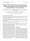
Fig. 1. (A) Example of hippocampus
segmentation and graphs demonstrating an increase in hippocampus volume
for the aerobic exercise group and
a decrease in volume for the stretching
control group. The Time × Group interaction was significant (P < 0.001) for
both left and right regions. (B) Example
of caudate nucleus segmentation and
graphs demonstrating the changes in
volume for both groups. Although the
exercise group showed an attenuation
of decline, this did not reach significance (both P > 0.10). (C) Example of
thalamus segmentation and graph
demonstrating the change in volume
for both groups. None of the changes
were significant for the thalamus. Error
bars represent SEM.
Hippocampal Volume Is Related to Improvements in Spatial Memory.
Downloaded by guest on June 6, 2020
Spatial memory (13, 22) was tested on both exercise and
stretching groups at baseline, after 6 mo, and again after the
completion of the 1-y intervention to determine whether changes
in hippocampal volume translate to improved memory. Both
groups showed improvements in memory, as demonstrated by
significant increases in accuracy between the first and last testing
sessions for the aerobic exercise [t(2,51) = 2.08; P < 0.05] and the
stretching control [t(2,54) = 4.41; P < 0.001] groups. Response
times also became faster for both groups between the baseline and
postintervention sessions (all P < 0.01), indicating that improvements in accuracy were not caused by changes in speed–accuracy
tradeoff. However, the aerobic exercise group did not improve
performance above that achieved by the stretching control group,
as demonstrated by a nonsignificant Time × Group interaction
[F(1,102) = 0.67; P < 0.40; ηp2 = 0.007]. Nonetheless, we found
that higher aerobic fitness levels at baseline (r = 0.31; P < 0.001)
and after intervention (r = 0.28; P < 0.004) were associated with
better memory performance on the spatial memory task. Change
in aerobic fitness levels from baseline to after intervention, however, was not related to improvements in memory for either the
entire sample (r = 0.15; P < 0.12) or when considering each group
separately (both P > 0.05). Furthermore, changes in BDNF were
not associated with improvements in memory function for either
group (r < 0.15; P > 0.20). On the other hand, larger left and right
hippocampi at baseline (both P < 0.005) and after intervention
(both P < 0.005) were associated with better memory performance (12). Therefore, we reasoned that increased hippocampal
Fig. 2. The exercise group showed a selective increase in
the anterior hippocampus and no change in the posterior
hippocampus. See Table 2 for Means and SDs.
Erickson et al.
PNAS | February 15, 2011 | vol. 108 | no. 7 | 3019
NEUROSCIENCE
ume could be associated with increased levels of serum BDNF.
Because the aerobic exercise group was the only group to show an
increase in volume over the 1-y period, we ran a correlation between change in BDNF and change in hippocampal volume for
the aerobic exercise group to test this hypothesis. We found that
greater changes in serum BDNF were associated with greater
increases in volume for the left (r = 0.36; P < 0.01) and for the
right (r = 0.37; P < 0.01) hippocampus (Fig. 3 C and D). Further,
these effects were selective for the left (r = 0.30; P < 0.03) and
right anterior hippocampus (r = 0.27; P < 0.04) and only marginal
with the left (r = 0.25; P < 0.06) and right (r = 0.22; P < 0.08)
posterior hippocampus. There were no associations between
changes in serum BDNF and changes in caudate nucleus or
thalamus volumes (all P > 0.50); nor were there any associations
between hippocampal volume and serum BDNF for the stretching
control group (all P > 0.40). This indicates that exercise-induced
increases in BDNF are selectively related to the changes in anterior hippocampal volume resulting from aerobic exercise.
Fig. 3. All scatterplots are of the aerobic
exercise group only because it was the only
group that showed an increase in volume
across the intervention. (A and B) Scatterplots of the association between percent
change in left and right hippocampus volume and percent change in aerobic fitness
level from baseline to after intervention.
(C and D) Scatterplots of percent change in
left and right hippocampus volume and
percent change in BDNF levels. (E and F)
Scatterplots of percent change in left and
right hippocampus and percent change in
memory performance.
Downloaded by guest on June 6, 2020
volume after the exercise intervention should translate to improved memory function. In support of this hypothesis, we found
that, in the aerobic exercise group, increased hippocampal volume
was directly related to improvements in memory performance. The
correlation between improvement in memory and hippocampal
volume reached significance for left (r = 0.23; P < 0.05) and right
(r = 0.29; P < 0.02) hemispheres (Fig. 3 E and F). This indicates
that increases in hippocampal volume after 1 y of exercise augments memory function in late adulthood. In contrast, changes in
caudate nucleus and thalamus volumes were unrelated to changes
in memory performance for either group (all P > 0.10).
Discussion
Hippocampal volume shrinks 1–2% annually in older adults
without dementia (1), and this loss of volume increases the risk for
developing cognitive impairment (2). We find results consistent
with this pattern, such that the stretching control group demonstrated a 1.4% decline in volume over the 1-y interval. With escalating health care costs and an increased proportion of people
aged >65 y, it is imperative that low-cost, accessible preventions
and treatments for brain tissue loss are discovered. In this randomized controlled study of exercise training, we demonstrate
that loss of hippocampal volume in late adulthood is not inevitable and can be reversed with moderate-intensity exercise. A
1-y aerobic exercise intervention was effective at increasing hippocampal volume by 2% and offsetting the deterioration associated with aging. Because hippocampal volume shrinks 1–2%
annually, a 2% increase in hippocampal volume is equivalent to
adding between 1 and 2 y worth of volume to the hippocampus for
this age group.
3020 | www.pnas.org/cgi/doi/10.1073/pnas.1015950108
On the basis of the several regions we examined, the effect of
exercise was rather selective, influencing only the anterior hippocampus and neither the thalamus nor the caudate nucleus. This
indicates that exercise does not influence all brain regions uniformly. In fact, research from human cognitive studies and rodents
indicates some specificity, such that exercise influences some brain
regions and behaviors but has minimal influence on others (3, 5, 9,
12, 20, 21, 23–25). Such selectivity suggests that there are regionally
dependent molecular pathways influenced by exercise. In fact, we
found here that changes in serum BDNF levels were associated
with changes in anterior hippocampal volume; an important link
because the hippocampus is rich in BDNF, and BDNF levels increase with exercise treatments in both rodents (5, 7, 20) and
humans (26, 27). BDNF is a putative mediator of neurogenesis and
contributes to dendritic expansion (28, 29) and is also critical in
memory formation (30–32). Our results suggest that cell proliferation or increased dendritic branching might explain increased
hippocampal volume and improvements in memory after exercise;
however, increased vascularization (15, 16, 33) and dendritic
complexity (34) may also be contributing to increased volume.
Aerobic exercise increased anterior hippocampal volume but
had little effect on the posterior hippocampus. Neurons in the
anterior hippocampus are selectively associated with spatial
memory acquisition (17) and show exacerbated age-related atrophy compared with the posterior hippocampus (18, 19). It is
possible that regions demonstrating less age-related decay might
also be less amenable to growth. Thus, aerobic exercise might
elicit the greatest changes in regions that show the most precipitous decline in late adulthood, such as the anterior hippoErickson et al.
Methods
Participants. Community-dwelling older adults (n = 842) were recruited, and
179 were enrolled. One hundred forty-five participants completed the intervention (81.0% of the participants originally enrolled). Five participants
were excluded because they did not attend the 6-mo MRI session, owing to
scheduling conflicts; eight participants were excluded because they did not
attend the 12-mo follow-up MRI session; and 12 participants were excluded
because they had excessive head motion that created inaccurate hippocampal, caudate nucleus, or thalamus segmentations. Therefore, 120 participants had complete MR data from all three sessions (82.7% of the
enrolled sample) and were included in the analyses.
Eligible participants had to (i) demonstrate strong right handedness (35),
(ii) be between the ages of 55 and 80 y, (iii) score ≥51 on the modified MiniMental Status Examination (36), (iv), score <3 on the Geriatric Depression
Scale to rule out possible depression (37), (v) have normal color vision, (vi)
have a corrected visual acuity of at least 20/40, (vii) have no history of neurological diseases or infarcts, including Parkinson’s disease, Alzheimer’s disease, multiple sclerosis, or stroke, (viii) have no history of major vasculature
problems, including cardiovascular disease or diabetes, (ix) obtain consent
from their personal physician, and (x) sign an informed consent form approved by the University of Illinois. In addition, all participants had to report
being currently sedentary, defined as being physically active for 30 min or less
in the last 6 mo. Participants were compensated for their participation.
After completion of the initial blood draw, MR session, and fitness assessment, participants were randomized to an aerobic walking group (n = 60)
or a stretching control group (n = 60) (Fig. 4).
Downloaded by guest on June 6, 2020
Fitness Assessments. Participants were required to obtain consent from their
personal physician before cardiorespiratory fitness testing was conducted.
Aerobic fitness (VO2 max) was assessed by graded maximal exercise testing on
a motor-driven treadmill. The participant walked at a speed slightly faster than
their normal walking pace (≈30–100 m/min), with increasing grade increments
of 2% every 2 min. A cardiologist and nurse continuously monitored oxygen
uptake, heart rate, and blood pressure (see SI Methods for more detail).
Fig. 4. Flow diagram for the randomization and assessment sessions for
both exercise and stretching control groups.
Erickson et al.
MRI Parameters and Segmentation Algorithm. MR images were collected on all
participants within 1 mo of the start of the intervention, after 6 mo, and
within 2 wk after the completion of the intervention. High-resolution (1.3
mm × 1.3 mm × 1.3 mm) T1-weighted brain images were acquired using a 3D
magnetization-prepared rapid gradient echo imaging protocol with 144
contiguous slices collected in an ascending fashion.
For segmentation and volumetric analysis of the left and right hippocampus, caudate nucleus, and thalamus we used the Oxford Centre for
Functional MRI of the Brain (FMRIB)’s Integrated Registration and Segmentation Tool in FMRIB’s Software Library version 4.1 (38–40) (see SI Methods
for more detail).
Training Protocol. Aerobic exercise condition. For the aerobic exercise program,
a trained exercise leader supervised all sessions. Participants started by walking
for 10 min and increased walking duration weekly by 5-min increments until
a duration of 40 min was achieved at week 7. Participants walked for 40 min per
session for the remainder of the program. All walking sessions started and
ended with approximately 5 min of stretching for the purpose of warming up
and cooling down. Participants wore heart rate monitors and were encouraged to walk in their target heart rate zone, which was calculated using the
Karvonen method (41) according to the resting and maximum heart rates
achieved during the baseline maximal graded exercise test. The target heart
rate zone was 50–60% of the maximum heart rate reserve for weeks 1 to 7
and 60–75% for the remainder of the program. Participants in the walking
group completed an exercise log at each exercise session. Every 4 wk, participants received written feedback forms that summarized the data from
their logs. Participants with low attendance and/or exercise heart rate were
encouraged to improve their performance in the following month.
Stretching and toning control condition. For the stretching and toning control
program, all sessions were led and monitored by trained exercise leaders. All
classes started and ended with warm-up and cool-down stretching. During
each class, participants engaged in four muscle-toning exercises using
dumbbells or resistance bands, two exercises designed to improve balance,
one yoga sequence, and one exercise of their choice. To maintain interest,
a new group of exercises was introduced every 3 wk. During the first week,
participants focused on becoming familiar with the new exercises, and during
the second and third weeks they were encouraged to increase the intensity by
using more weight or adding more repetitions. Participants in the stretching
and toning control group also completed exercise logs at each exercise session
and received monthly feedback forms. They were encouraged to exercise at
an appropriate intensity of 13–15 on the Borg Rating of Perceived Exertion
scale (42) and to attend as many classes as possible.
Spatial Memory Paradigm. To test memory function, all participants completed a computerized spatial memory task at baseline, after 6 mo, and again
after completion of the intervention (13, 22, 43).
A fixation crosshair appeared for 1 s, and participants were instructed to
keep their eyes on the crosshair. After the fixation, one, two, or three black
dots appeared at random locations on the screen for 500 ms. The dots were
removed from the display for 3 s. During this time, participants were
instructed to try and remember the locations of the previously presented
black dots. At the end of the 3-s delay, a red dot appeared on the screen in
either one of the same locations as the target dots (match condition) or at
a different location (nonmatch condition). Participants had 2 s to respond to
the red dot by pressing one of two keys on a standard keyboard—the “x” key
for a nonmatch trial and the “m” key for a match trial (Fig. 5). Forty trials
Fig. 5. Display of the spatial memory task used in this study. The spatial
memory task load was parametrically manipulated between one, two, or
three items (two-item condition shown here). Participants were asked to
remember the locations of one, two, or three black dots. After a brief delay,
a red dot appeared, and participants were asked to respond whether the
location of the red dot matched or did not match one of the locations of the
previously shown black dots. This task was administered to all participants at
baseline, after 6 mo, and again after completion of the intervention.
PNAS | February 15, 2011 | vol. 108 | no. 7 | 3021
NEUROSCIENCE
campus and prefrontal cortex (9). Overall, these data suggest that
the anterior hippocampus remains amenable to augmentation.
In sum, we found that the hippocampus remains plastic in late
adulthood and that 1 y of aerobic exercise was sufficient for enhancing volume. Increased hippocampal volume translates to
improved memory function and higher serum BDNF. We also
demonstrate that higher fitness levels are protective against loss of
hippocampal volume. These results clearly indicate that aerobic
exercise is neuroprotective and that starting an exercise regimen
later in life is not futile for either enhancing cognition or augmenting brain volume.
were presented for each set size (one, two, or three locations), with 20 trials
as match trials and 20 trials as nonmatch trials. Participants were instructed
to respond as quickly and accurately as possible. Several practice trials were
performed before the task began to acquaint the participants with the task
instructions and responses (see SI Methods for more detail).
Downloaded by guest on June 6, 2020
Serum BDNF Assay. Blood was collected at baseline before the intervention
and again immediately after the completion of the program. Blood sampling
for BDNF analysis was performed approximately 2 wk before the MR sessions.
Fasted subjects reported to the laboratory at 0800 hours, at which time blood
from the antecubital vein was collected in sterile serum separator tubes
(Becton Dickinson). The blood samples were kept at room temperature for
15 min to allow for clotting, after which the samples were centrifuged at
1,100 × g at 4 °C for 15 min. Serum was then harvested, aliquoted, and stored
at −80 °C until analysis. Serum BDNF was quantified using an enzyme-linked
immunosorbant assay (Human BDNF Quantikine Immunoassay, DBD00, R & D
Systems) according to the manufacturer’s instructions (see SI Methods for
more detail).
a between-subjects factor and Time (baseline, 6 mo, and 1 y) as a within-subject
factor. Because the distribution of men and women was slightly different
between the two groups (Table 1) we included sex as a covariate in all analyses.
In addition, as a safeguard against any residual effects of height or head size,
we included intracranial volume (ICV) as a covariate of no interest. Finally,
age was slightly different between the two groups, so we also included age
as a covariate of no interest in all models.
Correlations were calculated using percent change in VO2 max, percent
change in left and right hippocampal volumes, percent change in BDNF, and
percent change in memory performance. We also ran correlations between
absolute difference scores while controlling for variation in baseline values.
These results were identical, so the correlations from the percent change
scores are included in this report. For all correlations, we used a partial
correlation approach to control for the possible confounding effects of age,
sex, and ICV.
Analyses. All dependent variables were tested and met criteria for normality
and skew before general linear model and Pearson correlations were conducted. Effects of the intervention on VO2, BDNF, and the volume of the
hippocampus, caudate nucleus, and thalamus were examined using an ANOVA
with repeated measures with Group (aerobic exercise, stretching control) as
ACKNOWLEDGMENTS. We thank Susan Herrel, Edward Malkowski, Dawn
Epstein, Zuha Warraich, Nancy Dodge, and Holly Tracy for help with data
collection. This work was supported by National Institute on Aging, National
Institutes of Health Grants RO1 AG25667 and RO1 AG25032. K.I.E. was
supported by a Junior Scholar Award (P30 AG024827) from the Pittsburgh
Claude D. Pepper Older Americans Independence Center and a seed grant
(P50 AG005133) awarded through the University of Pittsburgh Alzheimer’s
Disease Research Center.
1. Raz N, et al. (2005) Regional brain changes in aging healthy adults: General trends,
individual differences and modifiers. Cereb Cortex 15:1676–1689.
2. Jack CR, Jr., et al.; Alzheimer’s Disease Neuroimaging Initiative (2010) Brain beta-amyloid
measures and magnetic resonance imaging atrophy both predict time-to-progression
from mild cognitive impairment to Alzheimer’s disease. Brain 133:3336–3348.
3. Hillman CH, Erickson KI, Kramer AF (2008) Be smart, exercise your heart: Exercise
effects on brain and cognition. Nat Rev Neurosci 9:58–65.
4. van Praag H, Shubert T, Zhao C, Gage FH (2005) Exercise enhances learning and
hippocampal neurogenesis in aged mice. J Neurosci 25:8680–8685.
5. Cotman CW, Berchtold NC (2002) Exercise: A behavioral intervention to enhance brain
health and plasticity. Trends Neurosci 25:295–301.
6. Creer DJ, Romberg C, Saksida LM, van Praag H, Bussey TJ (2010) Running enhances
spatial pattern separation in mice. Proc Natl Acad Sci USA 107:2367–2372.
7. Vaynman S, Ying Z, Gomez-Pinilla F (2004) Hippocampal BDNF mediates the efficacy
of exercise on synaptic plasticity and cognition. Eur J Neurosci 20:2580–2590.
8. Li Y, et al. (2008) TrkB regulates hippocampal neurogenesis and governs sensitivity to
antidepressive treatment. Neuron 59:399–412.
9. Colcombe SJ, et al. (2006) Aerobic exercise training increases brain volume in aging
humans. J Gerontol A Biol Sci Med Sci 61:1166–1170.
10. Colcombe SJ, et al. (2004) Cardiovascular fitness, cortical plasticity, and aging. Proc
Natl Acad Sci USA 101:3316–3321.
11. Rosano C, et al. (2010) Psychomotor speed and functional brain MRI 2 years after
completing a physical activity treatment. J Gerontol A Biol Sci Med Sci 65:639–647.
12. Erickson KI, et al. (2010) Physical activity predicts gray matter volume in late
adulthood: The Cardiovascular Health Study. Neurology 75:1415–1422.
13. Erickson KI, et al. (2009) Aerobic fitness is associated with hippocampal volume in
elderly humans. Hippocampus 19:1030–1039.
14. Honea RA, et al. (2009) Cardiorespiratory fitness and preserved medial temporal lobe
volume in Alzheimer’s disease. Alzheimer Dis Assoc Disord 23:188–197.
15. Pereira AC, et al. (2007) An in vivo correlate of exercise-induced neurogenesis in the
adult dentate gyrus. Proc Natl Acad Sci USA 104:5638–5643.
16. Burdette JH, et al. (2010) Using network science to evaluate exercise-associated brain
changes in older adults. Front Aging Neurosci 2:23.
17. Moser MB, Moser EI, Forrest E, Andersen P, Morris RG (1995) Spatial learning with
a minislab in the dorsal hippocampus. Proc Natl Acad Sci USA 92:9697–9701.
18. Raji CA, Lopez OL, Kuller LH, Carmichael OT, Becker JT (2009) Age, Alzheimer disease,
and brain structure. Neurology 73:1899–1905.
19. Hackert VH, et al. (2002) Hippocampal head size associated with verbal memory
performance in nondemented elderly. Neuroimage 17:1365–1372.
20. Neeper SA, Gómez-Pinilla F, Choi J, Cotman C (1995) Exercise and brain neurotrophins. Nature 373:109.
21. Holmes MM, Galea LA, Mistlberger RE, Kempermann G (2004) Adult hippocampal
neurogenesis and voluntary running activity: Circadian and dose-dependent effects. J
Neurosci Res 76:216–222.
22. Erickson KI, et al. (2010) Brain-derived neurotrophic factor is associated with agerelated decline in hippocampal volume. J Neurosci 30:5368–5375.
23. Kramer AF, et al. (1999) Ageing, fitness and neurocognitive function. Nature 400:
418–419.
24. Colcombe SJ, Kramer AF (2003) Fitness effects on the cognitive function of older
adults: A meta-analytic study. Psychol Sci 14:125–130.
25. Smith PJ, et al. (2010) Aerobic exercise and neurocognitive performance: A metaanalytic review of randomized controlled trials. Psychosom Med 72:239–252.
26. Rasmussen P, et al. (2009) Evidence for a release of brain-derived neurotrophic factor
from the brain during exercise. Exp Physiol 94:1062–1069.
27. Zoladz JA, et al. (2008) Endurance training increases plasma brain-derived
neurotrophic factor concentration in young healthy men. J Physiol Pharmacol 59
(Suppl 7):119–132.
28. Lee R, Kermani P, Teng KK, Hempstead BL (2001) Regulation of cell survival by
secreted proneurotrophins. Science 294:1945–1948.
29. Pencea V, Bingaman KD, Wiegand SJ, Luskin MB (2001) Infusion of brain-derived
neurotrophic factor into the lateral ventricle of the adult rat leads to new neurons in
the parenchyma of the striatum, septum, thalamus, and hypothalamus. J Neurosci 21:
6706–6717.
30. Figurov A, Pozzo-Miller LD, Olafsson P, Wang T, Lu B (1996) Regulation of synaptic
responses to high-frequency stimulation and LTP by neurotrophins in the
hippocampus. Nature 381:706–709.
31. Kang H, Schuman EM (1996) A requirement for local protein synthesis in
neurotrophin-induced hippocampal synaptic plasticity. Science 273:1402–1406.
32. Pang PT, et al. (2004) Cleavage of proBDNF by tPA/plasmin is essential for long-term
hippocampal plasticity. Science 306:487–491.
33. Black JE, Isaacs KR, Anderson BJ, Alcantara AA, Greenough WT (1990) Learning causes
synaptogenesis, whereas motor activity causes angiogenesis, in cerebellar cortex of
adult rats. Proc Natl Acad Sci USA 87:5568–5572.
34. Redila VA, Christie BR (2006) Exercise-induced changes in dendritic structure and
complexity in the adult hippocampal dentate gyrus. Neuroscience 137:1299–1307.
35. Oldfield RC (1971) The assessment and analysis of handedness: The Edinburgh
inventory. Neuropsychologia 9:97–113.
36. Stern Y, et al. (1987) Modified mini-mental state examination: Validity and reliability.
Neurology 37:179.
37. Sheikh JI, Yesavage JA (1986) Geriatric Depression Scale (GDS): Recent evidence and
development of a shorter version. Clinical Gerontology: A Guide to Assessment and
Intervention. (Haworth Press, New York), pp 165–173.
38. Patenaude B, et al. (2007) Bayesian Shape and Appearance Models. Technical Report
TR07BP1 (FMRIB Centre, Univ Oxford, UK).
39. Zhang Y, Brady M, Smith S (2001) Segmentation of brain MR images through
a hidden Markov random field model and the expectation-maximization algorithm.
IEEE Trans Med Imaging 20:45–57.
40. Smith SM, et al. (2004) Advances in functional and structural MR image analysis and
implementation as FSL. Neuroimage 23 (Suppl 1):S208.
41. Strath SJ, et al. (2000) Evaluation of heart rate as a method for assessing moderate
intensity physical activity. Med Sci Sports Exerc 32 (9 Suppl):S465–S470.
42. Borg G (1985) An Introduction to Borg’s RPE-Scale (Mouvement, Ithaca, NY).
43. Heo S, et al. (2010) Resting hippocampal blood flow, spatial memory and aging. Brain
Res 1315:119–127.
3022 | www.pnas.org/cgi/doi/10.1073/pnas.1015950108
Erickson et al.
Keep reading this paper — and 50 million others — with a free Academia account
Used by leading Academics

Prof.Dr. Abdulkadir Koçer
Istanbul Medeniyet University

Rommy von Bernhardi
Pontificia Universidad Catolica de Chile

John Slevin
University of Kentucky

paolo mazzarello
University of Pavia