0% found this document useful (0 votes)
212 viewsStaining
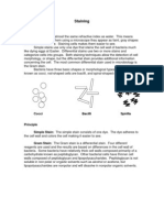
Bacteria are difficult to see under a microscope due to having a similar refractive index as water. Staining makes bacteria visible by adhering dye to their cell walls. The Gram stain is a common differential stain that categorizes bacteria based on differences in their cell wall structure. Gram positive bacteria have thick peptidoglycan cell walls that retain the primary crystal violet stain, appearing purple. Gram negative bacteria have thinner cell walls containing lipopolysaccharides that accept the safranin counterstain, appearing pink. Staining allows observation of bacterial shape and provides important clinical information.
Uploaded by
yuppie_raj2175Copyright
© Attribution Non-Commercial (BY-NC)
Available Formats
Download as PDF, TXT or read online on Scribd
0% found this document useful (0 votes)
212 viewsStaining
Bacteria are difficult to see under a microscope due to having a similar refractive index as water. Staining makes bacteria visible by adhering dye to their cell walls. The Gram stain is a common differential stain that categorizes bacteria based on differences in their cell wall structure. Gram positive bacteria have thick peptidoglycan cell walls that retain the primary crystal violet stain, appearing purple. Gram negative bacteria have thinner cell walls containing lipopolysaccharides that accept the safranin counterstain, appearing pink. Staining allows observation of bacterial shape and provides important clinical information.
Uploaded by
yuppie_raj2175Copyright
© Attribution Non-Commercial (BY-NC)
Available Formats
Download as PDF, TXT or read online on Scribd
/ 5