This document summarizes the morphology, anatomy, and life cycle of the moss species Funaria. Key points include:
- Funaria is a small, green moss that grows in dense patches in moist, shady areas.
- It has a radial body plan with an upright stem bearing spirally arranged leaves. Reproduction is both sexual and asexual.
- Sexual reproduction involves antheridia and archegonia on the same plant. Fertilization results in a diploid sporophyte that produces haploid spores.
- Upon germination, spores form a filamentous protonema that develops into upright gametophyte plants, completing the life cycle
"Equisetum" Structural development Reproduction Muhammad ArSlan
The plant body of Equisetum has an underground rhizome and an aerial shoot. The shoot has scale-like leaves arranged in whorls and ridges with stomata located in furrows. It reproduces sexually through antheridia and archegonia on separate gametophytes. Upon fertilization of the egg, the zygote develops into a four-celled embryo that grows into a new sporophyte, with multiple sporophytes potentially developing from a single gametophyte.
This document summarizes the morphology and life cycle of the moss Funaria. It belongs to the division Bryophyta, class Bryopsida, order Funariales and family Funariaceae. Funaria hygrometrica is the most common and widespread species. It grows in moist, shady places. The plant body is a gametophyte that is differentiated into a protonema and upright gametophores. It reproduces sexually through antheridia and archegonia on separate gametophores, and vegetatively through fragmentation. Fertilization occurs when sperm are released from antheridia and travel to archegonia. This results in a sporophyte called a sporogonium that
1. Marsilea is an aquatic or semi-aquatic plant commonly found in freshwater ponds and ditches in temperate regions like Pakistan and Punjab.
2. The vegetative plant is a sporophyte with a rhizome, adventitious roots, and compound leaves with four leaflets arranged in a cross-like pattern.
3. It is heterosporous, producing megaspores and microspores within sporocarps that contain sori with alternating rows of megasporangia and microsporangia. Megaspores develop into female gametophytes and microspores into male gametophytes.
Pteridophytes are classified into 4 divisions - Psilophyta, Lycophyta, Sphenophyta, and Pterophyta. Psilophyta includes the most primitive whisk ferns and Lycophyta includes club mosses and spike mosses. Sphenophyta contains the single living genus Equisetum, or horse tails. Pterophyta, or ferns, is the largest and most widely distributed division containing many families and over 10,000 living species distributed worldwide. Each division contains multiple classes that further specify characteristics such as plant body structure, leaf and spore structures, and reproductive systems.
The document describes the plant Psilotum, which is classified as a vascular plant in the division Psilophyta. It has an underground rhizome that produces aerial branches bearing sporangia in clusters of three called synangia. The plant reproduces asexually through spores produced in the synangia and sexually through an underground gametophyte that produces eggs and sperm. The rhizome and stem have anatomical features including epidermis, cortex, endodermis and a central stele of xylem and phloem. Psilotum is considered to have characteristics transitional between non-vascular bryophytes and modern vascular plants.
1. Anthoceros is a genus of hornworts that reproduces sexually as a gametophyte generation.
2. The thallus is flattened, lobed, and lacks a midrib or branches; rhizoids are present on the ventral surface.
3. Reproduction can occur vegetatively through fragmentation, gemmae, or tubers, or sexually through antheridia and archegonia developing on the upper thallus surface.
4. Fertilization results in a diploid zygote that develops into an elongated, horn-like sporophyte embedded in the gametophyte thallus.
This document summarizes the anatomy of the stem and leaf of the cycas plant. It describes that the leaf is differentiated into an upper and lower epidermis, hypodermis, mesophyll, and vascular tissue. The mesophyll contains palisade and spongy parenchyma. The stem anatomy resembles dicots, with a cortex, ring of vascular bundles, large pith, and concentric mesarch leaf traces. Each cycas leaf receives four supplies from two vascular bundles via concentric, endarch to mesarch leaf traces.
This document summarizes key details about the fern genus Pteris. It describes the systematic position of Pteris within the plant kingdom, common Indian species, and global occurrence/distribution. The morphology, anatomy, and reproductive structures of the sporophyte (fern plant) are then explained in detail, covering the rhizome, fronds, leaflets, and roots. Key anatomical features include dictyostele stele in the rhizome, vascular bundles with endodermis and pericycle, and hypostomatous leaflets. Reproduction occurs vegetatively from the rhizome as well as sexually from spores.
This document summarizes the Bennettitales, a group of fossil plants that flourished during the Mesozoic era. It describes two families of Bennettitales: Bennettitaceae and Williamsoniaceae. Bennettitaceae had deeply sunk flowers on short, thick trunks, while Williamsoniaceae had fully exposed flowers on slender stems. The document provides details on the anatomy, reproduction, and classification of these two extinct families of seed plants.
- Marsilea is a heterosporous fern that produces two types of spores, microspores and megaspores, in separate microsporangia and megasporangia structures enclosed in bean-shaped sporocarps.
- It is commonly known as water fern and has about 53 cosmopolitan species, especially in tropical regions like Africa and Australia. Species can be fully aquatic, amphibious, or terrestrial.
- The plant body has an underground rhizome and differentiated leaves and roots. Reproduction is both vegetative through rhizome tubers and sexual through the production of microspores and megaspores in sporocarps.
Bryophytes are non-vascular plants that include mosses, liverworts, and hornworts. Riccia is a genus of thallose liverworts commonly found growing terrestrially or aquatically. It has a flat, lobed gametophyte thallus with dichotomous branching and a midrib furrow containing its sexual organs. Riccia reproduces both vegetatively through thallus fragmentation and sexually through the fertilization of archegonia by antheridia-derived sperm. This results in a sporophyte embedded in the gametophyte that produces haploid spores through meiosis. The spores germinate to form new gametophyte plants,
Pteridophytes are vascular plants and have leaves (known as fronds), roots and sometimes true stems, and tree ferns have full trunks. Examples include ferns, horsetails and club-mosses. Fronds in the largest species of ferns can reach some six metres in length!
Many ferns from tropical rain forests are epiphytes, which means they only grow on other plant species; their water comes from the damp air or from rainfall running down branches and tree trunks. There are also some purely aquatic ferns such as water fern or water velvet (Salvinia molesta) and mosquito ferns (Azolla species).
Pteridophytes do not have seeds or flowers either, instead they also reproduce via spores.
There are around 13,000 species of Pteridophytes.
Gnetum: A Powerpoint Presentation on Gymnospemsshivduraigaran
The Gymnosperms are a group of seed-producing plants (spermatophytes) that includes conifers (Pinophyta), cycads, Ginkgo, and gnetophytes. The term "gymnosperm" comes from the Greek composite word γυμνόσπερμος (γυμνός gymnos, "naked" and σπέρμα sperma, "seed"), meaning "naked seeds". The name is based on the unenclosed condition of their seeds (called ovules in their unfertilized state). The non-encased condition of their seeds stands in contrast to the seeds and ovules of flowering plants (angiosperms), which are enclosed within an ovary. Gymnosperm seeds develop either on the surface of scales or leaves, which are often modified to form cones, or solitary as in Yew, Torreya, Ginkgo.
The gymnosperms and angiosperms together compose the spermatophytes or seed plants. The gymnosperms are divided into six phyla. Organisms that belong to the Cycadophyta, Ginkgophyta, Gnetophyta, and Pinophyta (also known as Coniferophyta) phyla are still in existence while those in the Pteridospermales and Cordaitales phyla are now extinct.
By far the largest group of living gymnosperms are the conifers (pines, cypresses, and relatives), followed by cycads, gnetophytes (Gnetum, Ephedra and Welwitschia), and Ginkgo biloba (a single living species). Roots in some genera have fungal association with roots in the form of micorrhiza(Pinus), while in some others(Cycas) small specialised roots called coralloid roots are associated with nitrogen fixing cyanobacteria.
Gnetum is a genus of gymnosperms, the sole genus in the family Gnetaceae and order Gnetales. They are tropical evergreen trees, shrubs and lianas. Unlike other gymnosperms, they possess vessel elements in the xylem. Some species have been proposed to have been the first plants to be insect-pollinated as their fossils occur in association with extinct pollinating scorpion flies. Molecular phylogenies based on nuclear and plastid sequences from most of the species indicate hybridization among some of the Southeast Asian species. Fossil-calibrated molecular-clocks suggest that the Gnetum lineages now found in Africa, South America and Southeast Asia are the result of ancient long-distance dispersal across seawater
Structure, reproduction, life history and systematic position of LycopodiumSankritaShankarGaonk
This document provides information on the systematic position, morphology, anatomy, reproduction, and life cycle of Lycopodium. It discusses that Lycopodium has 400 species found in varied habitats worldwide, including in India. It has dichotomously branched stems and leaves without ligules. Reproduction occurs vegetatively and via spores. Spores form in structures called strobili and develop into homothallic gametophytes that produce both antheridia and archegonia for sexual reproduction. The life cycle involves a diploid sporophyte and haploid gametophyte generation.
Equisetum popularly known a the ‘horse-tail’ or ‘scouring rush’.
It is now represented by nearly 30 species which are seen world wide except in Australia and New Zealand.
Some species prefer damp and shady places while others grow in marshes, ponds or stream banks
Some are found in xerophytic habitats
The Ranunculaceae family contains over 2000 species of annual or perennial herbs, vines, and trees that are cosmopolitan but flourish in temperate climates. Important species include buttercups, larkspurs, and monkshoods. Plants have herbaceous stems, alternate or compound leaves, and cymose or solitary inflorescences bearing numerous stamens and carpels. Fruits are achenes or follicles. Many species are used ornamentally or medicinally as they contain alkaloids and glycosides. Some are also used as condiments, while others are weeds or produce poisonous juices.
Marsilea is a genus of aquatic ferns that grow in mud or wet soil. The sporophyte has horizontal underground stems called rhizomes from which emerge roots and leaves. Leaves are arranged in pairs and each leaf has four leaflets. Sporocarps containing sporangia develop on the rhizomes. The sporocarps contain either microsporangia or megasporangia. After fertilization, the zygote develops into an embryo within the megaspore.
1) Funaria is a terrestrial moss that grows in dense green patches in moist, shady areas like damp soil, tree trunks, and walls.
2) It has both underground protonemal filaments and above-ground leafy gametophores that produce rhizoids, a stem, and spirally arranged leaves.
3) Sexual reproduction involves the production of antheridia and archegonia on separate male and female branches that lead to fertilization and the growth of a sporophyte with foot, seta, and capsule containing spores.
The topic of discussion is Pteridophytes, their general characteristics, sexual reproduction and Life cycle has been discussed along with the four different divisions that are present in Pteridophytes
1. The document describes the life cycle and morphology of the moss Funaria. It has a haplontic and diplontic life cycle with alternation of generations exhibiting heteromorphy.
2. The gametophyte generation is the dominant phase and reproduces sexually or asexually. Sexual reproduction involves antheridia and archegonia that produce gametes for fertilization.
3. Fertilization leads to formation of a sporophyte which grows from the archegonium. The sporophyte bears spores in capsules and releases them to complete the life cycle.
The document summarizes the variations in reproductive structures found among the three divisions of bryophytes (liverworts, hornworts, and mosses). It describes the key differences in antheridia, archegonia, and sporangia (capsules) between the classes within each division. For example, within the mosses (Division Bryophyta), the classes have variations in the positioning and shape of their gametangia and peristomes surrounding the capsule openings.
The plant Psilotum is a small shrub that reproduces both sexually and asexually. Its sporophyte body has underground rhizomes and aerial branches. Sporangia form in triads in the axils of leaves and contain spores that develop into subterranean gametophytes. The gametophytes are monoecious and bear both antheridia and archegonia. Fertilization occurs when sperm from the antheridia enter the archegonia. The resulting zygote divides to form an embryo sporophyte surrounded by gametophyte tissue. The sporophyte matures into a new Psilotum plant.
1. Selaginella is a heterosporous plant that produces megaspores and microspores. The spores develop into male and female gametophytes within their spore walls.
2. Microspores develop into male gametophytes containing antherozoids for fertilization. Megaspores develop into female gametophytes containing archegonia.
3. Fertilization occurs when antherozoids enter the archegonia through openings in the neck canal cells. This leads to the development of a diploid sporophyte within the megaspore.
Marchantia reproduces both sexually and asexually. Asexually, it reproduces through fragmentation, adventitious branches, and gemmae. Gemmae are multicellular structures that form in gemma cups on the thallus and contain meristematic, chlorophyll-containing, and oil cells. When released, gemmae germinate to form new plants. Sexually, male and female reproductive structures form on separate plants. Antheridiophores bear antheridia and archegoniophores bear archegonia. Fertilization occurs through splashing of sperm. After fertilization, the zygote develops into a sporophyte inside the archegonium containing a foot, set
1. Marchantia is a genus of liverworts that reproduces both sexually and asexually. It has a flat, thalloid gametophyte body that is dichotomously branched.
2. The gametophyte produces male and female sex organs called antheridia and archegonia on specialized stalks. Fertilization results in a diploid zygote that develops into a sporophyte.
3. The sporophyte produces spores through meiosis in capsules. The spores germinate to form new gametophytes, completing the life cycle with alternation between haploid and diploid generations.
The topic discussed here is the characteristic of Bryophytes with examples such as Riccia & Marchantia. The complexity in their reproductive structures and also the sporophyte is also discussed. Funaria an example of moss gives the details about the living habits of these organisms which are considered to be advanced bryophytes.
Polytrichum is a genus of moss that has worldwide distribution in cool and tropical regions. It has a life cycle that alternates between a sexual gametophyte generation and an asexual sporophyte generation. The gametophyte is the main plant body and consists of underground rhizomes and upright leafy shoots. The sporophyte develops from fertilization of an archegonium and produces spores via meiosis. Upon germination, the spores form protonema that develop into new gametophyte plants.
MARSILEA notes in detail for II year Botany.pptaigil2
This document describes the systematic position and characteristic features of the genus Marsilea. It is a heterosporous aquatic fern that belongs to the order Marsileales. It produces sporocarps that contain microsporangia and megasporangia. There are about 65 species distributed worldwide in tropical regions like Africa and Australia. The document outlines the external morphology, anatomy, reproduction and life cycle of Marsilea. It alternates between a diploid sporophytic generation and haploid gametophytic generation through the production of microspores and megaspores.
This document describes the morphology and anatomy of Adiantum, commonly known as maidenhair fern. It has nearly 200 species that are widely distributed in tropical and subtropical regions. The sporophyte has a rhizome, roots, and leaves. The rhizome is creeping or erect and covered in scales. The leaves are pinnately compound and circinate in vernation. Sporangia are borne on the undersides of pinnae in sori. Spores germinate to produce a heart-shaped, photosynthetic prothallus with antheridia and archegonia for sexual reproduction, forming a sporophyte through alternation of generations.
Marchantia is a genus of liverworts that reproduces both sexually and asexually. The plant body is a flattened thallus with dorsal midribs that bear gemmae cups for asexual reproduction. Male and female gametophytes develop on separate plants. During sexual reproduction, sperm from the male antheridia fertilize eggs in the female archegonia to form zygotes. These zygotes develop into sporophytes that produce spores through meiosis. The spores disperse and can germinate to form new gametophyte generations.
This is a Life Cycle of Shpagnum, A good content for Masters Students. (But this content is not made by me...but i thought that this will help many students who are in search for content)
Thank you 😊
- Funaria is a genus of moss that includes approximately 210 species, with 18 found in India. The most common species is Funaria hygrometrica.
- Funaria mosses are small, primitive, autotrophic plants that grow in dense patches in moist, shady areas. They reproduce both sexually through spores and asexually through fragmentation, gemmae, and bulbils.
- The life cycle involves an alternation of generations between the haploid gametophyte and diploid sporophyte phases. Fertilization occurs when sperm fertilize eggs within archegonia, forming diploid zygotes that develop into sporophytes.
The document discusses host-parasite relationships and the mechanisms of plant infection. It covers:
1. How fungi obtain food from host plants through haustoria and establish close connections.
2. The definition of host and parasite. Host-parasite relationships affect each other's growth and metabolism.
3. The mechanisms of infection include spores contacting hosts, germ tubes attaching and penetrating tissues using enzymes, and hyphae entering and spreading within host tissues. Pathogens can enter through natural openings or wounds.
The document summarizes information about the fungi group Zygomycotina. It is divided into two classes: Zygomycetes and Trichomycetes. Zygomycetes are mostly terrestrial fungi that reproduce sexually through the fusion of opposite hyphae to form spores called zygospores. Rhizopus stolonifer is a common member. Trichomycetes are typically symbiotic fungi living in arthropod guts. The document also provides details about the fungi Mucor and Rhizopus, including their structures, life cycles, and reproduction methods.
This document summarizes the life cycle of Plasmodiophora brassicae, which causes clubroot disease in cabbage. It has both a haploid and diploid phase. In the haploid phase, resting spores in soil germinate to form zoospores that infect root hairs and develop into gametangia containing gametes. Gametes fuse in pairs during the diploid phase to form zygotes that infect root cells and develop into sporangia, completing the life cycle. The sporangia cause galls or clubs to form on the roots and can remain dormant in soil for years.
Oomycetes, commonly known as water molds, are eukaryotic organisms that are closely related to algae. They include some of the most devastating plant pathogens, causing diseases like late blight of potato and downy mildew of grapevines. Oomycetes reproduce both sexually, through the formation of gametangia and fertilization leading to thick-walled oospores, and asexually via motile zoospores or non-motile sporangia. While they were long classified as fungi, genetic evidence shows they are more closely related to algae and plants. Key differences from true fungi include having cell walls composed of cellulose and lacking chitin.
- Mastigomycotina is a former taxonomic grouping of fungi that included classes like Chytridiomycetes.
- Chytridiomycetes, commonly called chytrids, are mostly aquatic fungi found in soils and aquatic habitats. They can be unicellular or filamentous.
- Synchytrium endobioticum is a chytrid fungus that causes the black wart disease of potato. It has both asexual and sexual life cycles involving zoospores, gametes and resting spores that allow it to infect and overwinter on potato plants.
This document provides an overview of Myxomycotina (slime molds). It discusses that they are fungus-like organisms characterized by an amoeboid vegetative phase without cell walls. The document outlines the key characteristics and life cycles of the four classes: Acrasiomycetes, Hydromyxomycetes, Myxomycetes, and Plasmodiophoromycetes. It also briefly discusses their economic importance in nutrient cycling and use in laboratory studies due to their protoplasm without cell walls.
This document discusses the economic importance of fungi. It describes fungi's roles in medicine, industry, and agriculture. In medicine, fungi produce important antibiotics like penicillin. In industry, fungi are used to produce alcoholic beverages, enzymes, organic acids, proteins, vitamins, and antibiotics. However, in agriculture fungi can have both positive and negative roles. Positively, soil fungi maintain soil fertility and recycle nutrients. Negatively, fungi cause many plant diseases that result in significant economic losses.
This document discusses the signs and symptoms of different types of plant diseases caused by fungi, bacteria, viruses, and phytoplasmas. It notes that around 85% of plant diseases are caused by fungi or fungal-like organisms. Fungal diseases can cause spots, wilting, rusts, mildews, and rots. Bacterial diseases cause spots, wilting, cankers, and soft rots. Viral diseases cause mosaic patterns, crinkled or malformed leaves, stunting, and yellowing. Phytoplasma diseases cause yellowing, stunted growth, witches' broom patterns, and dieback. The document provides many examples of specific diseases for each category and their characteristic signs and symptoms
General account of post harvest diseases of vegetablesvaishalidandge3
1. Losses due to postharvest diseases can occur at any point from harvest to consumption and diseased produce poses health risks. 2. Common postharvest diseases infect through wounds from mechanical, insect or physiological injuries and are caused by fungi like Penicillium, Botrytis, Colletotrichum, and Rhizopus. 3. Integrated control methods include fungicides, temperature control, hygiene, packaging, and preventing injuries to limit infection sites.
Classification of fungi proposed by Ainsworth (1971)vaishalidandge3
Ainsworth proposed a more natural system of classification of fungi in 1971 based on morphology, especially of reproductive structures. He treated fungi as a separate kingdom. The classification has seven divisions: Myxomycota, Eumycota, Mastigomycotina, Zygomycotina, Ascomycotina, Basidiomycotina, and Deuteromycotina. It provides details on the classes, orders, and characteristics used to differentiate each taxonomic level within this system.
Important fungal disease on vegetables-Brinjalvaishalidandge3
1. Fungal diseases are a major cause of plant diseases in vegetables. Fungi infect plants through natural openings or wounds and are spread by wind, water, contaminated soil/machinery.
2. Important fungal diseases of brinjal include Cercospora leaf spot, damping off caused by Pythium, Rhizoctonia, Fusarium, Alternaria rot, Anthracnose fruit rot, Fusarium wilt, Verticillium wilt, and Phytophthora blight.
3. These diseases cause symptoms like leaf spots, fruit rot, seedling damping off, stem wilting, and plant death. Management involves crop rotation, resistant varieties,
History,classification & importance of plant pathologyvaishalidandge3
This document provides a history of the field of plant pathology, beginning with early observations of plant diseases in ancient texts from 1500-500 BC in India. It discusses key figures who advanced the field such as Theophrastus in 370 BC, Anton von Leeuwenhoek in the 1600s, and Anton de Bary in 1861 who proved fungi cause diseases. The document also summarizes the development of mycology, bacteriology, virology, and classification of plant diseases. It concludes with the importance of plant pathology in developing techniques to protect crops and restrict disease spread.
Koch's postulates are four criteria developed by Robert Koch in the 19th century to establish a causative relationship between a microbe and a disease. The postulates require that 1) the microorganism must be found in all infected organisms, 2) it can be isolated and grown in pure culture, 3) the cultured microorganism causes the same disease when introduced to a healthy organism, and 4) the microorganism can be reisolated from the infected experimental host. Koch's postulates played an important role in microbiology but have limitations for diseases caused by viruses or bacteria that cannot be grown in pure culture.
Pectinases are enzymes produced by microorganisms like fungi and bacteria that break down pectin, a polysaccharide found in plant cell walls. Pectinases include pectolyase, pectozyme, and polygalacturonase. They are used in food processing to extract juices from fruits and help with winemaking. Pectinases are also used for retting plant fibers, oil extraction, improving food textures, and treating wastewater. Factors like pH, temperature, nitrogen source, and carbon source affect microbial pectinase production.
Lipases are enzymes that catalyze the hydrolysis of triglycerides. They are produced by many plants, animals, and microorganisms. Fungal sources of lipases include Aspergillus oryzae, Candida antarctica, Rhizopus, and Thermomyces lanuginosus. Lipases have a catalytic mechanism where serine performs a nucleophilic attack on the ester bond, forming an acyl-enzyme intermediate and releasing an alcohol. A second nucleophilic attack by water then hydrolyzes the intermediate. Lipases have many industrial uses including in foods, dairy, detergents, biodiesel production, and pharmaceuticals due to their ability to function under
This document discusses proteases, which are enzymes that catalyze the breakdown of proteins. It describes the seven main classes of proteases based on their catalytic residue: serine, cysteine, threonine, aspartic, glutamic, metallo, and asparagine peptide lyases. Each class is explained along with examples. The document also covers protease structure, classification based on pH, mechanisms of action, and various industrial and medical uses of proteases.
This document summarizes 26 fungal diseases that affect tomatoes. It provides the symptoms, causal organisms, and control methods for each disease. The diseases impact various tomato plant parts including leaves, stems, fruits, and roots. Common symptoms include spots, lesions, wilting, and rotting. Suggested control methods include using resistant varieties, crop rotation, sanitation, irrigation management, and fungicides. The document serves as a comprehensive reference for identifying and managing important fungal diseases of tomatoes.
This document discusses 26 fungal diseases that affect potatoes. It provides details on the symptoms and control methods for each disease. The diseases include black dot, brown spot, early blight, late blight, fusarium dry rot, pink rot, powdery mildew, and others. Control involves practices like crop rotation, using certified seed, fungicides, and resistant varieties depending on the specific disease.
This document summarizes 14 common fungal diseases that affect okra plants. It describes the symptoms and control methods for each disease. The diseases include leaf spots caused by fungi like Cercospora, powdery mildew caused by Erysiphe, and blossom blight caused by Choanephora. Control methods involve cultural practices like removing infected plant material, improving air circulation, and chemical controls like applying appropriate fungicides. Proper disease management is important to prevent yield loss and maintain okra plant health.
This document discusses 13 fungal diseases that affect cabbage: 1) Wirestem caused by Rhizoctonia solani which stunts growth, 2) Fusarium yellows caused by Fusarium oxysporum which causes yellowing and stunting, 3) Blackleg caused by Leptosphaeria maculans/ Phoma lingam which causes stem cankering and severing, 4) Leaf spot/target spot caused by Alternaria brassicae & A. brassicicola which causes circular brown leaf spots, 5) Clubroot caused by Plasmodiophora brassicae which causes root galls interfering with nutrient uptake, 6) Damping off caused by Fusarium or Py
TOPIC: INTRODUCTION TO FORENSIC SCIENCE.pptximansiipandeyy
This presentation, "Introduction to Forensic Science," offers a basic understanding of forensic science, including its history, why it's needed, and its main goals. It covers how forensic science helps solve crimes and its importance in the justice system. By the end, you'll have a clear idea of what forensic science is and why it's essential.
Hydrogen sulfide and metal-enriched atmosphere for a Jupiter-mass exoplanetSérgio Sacani
We observed two transits of HD 189733b in JWST program 1633 using JWST
NIRCam grism F444W and F322W2 filters on August 25 and 29th 2022. The first
visit with F444W used SUBGRISM64 subarray lasting 7877 integrations with 4
BRIGHT1 groups per integration. Each effective integration is 2.4s for a total effective exposure time of 18780.9s and a total exposure duration of 21504.2s (∼6 hrs)
including overhead. The second visit with F322W2 used SUBGRISM64 subarray
lasting 10437 integrations with 3 BRIGHT1 groups per integration. Each effective
integration is 1.7s for a total effective exposure time of 17774.7s and a total exposure
duration of 21383.1s (∼6 hrs) including overhead. The transit duration of HD189733
b is ∼1.8 hrs and both observations had additional pre-ingress baseline relative to
post-egress baseline in anticipating the potential ramp systematics at the beginning
of the exposure from NIRCam infrared detectors.
Search for Dark Matter Ionization on the Night Side of Jupiter with CassiniSérgio Sacani
We present a new search for dark matter (DM) using planetary atmospheres. We point out that
annihilating DM in planets can produce ionizing radiation, which can lead to excess production of
ionospheric Hþ
3 . We apply this search strategy to the night side of Jupiter near the equator. The night side
has zero solar irradiation, and low latitudes are sufficiently far from ionizing auroras, leading to a lowbackground search. We use Cassini data on ionospheric Hþ
3 emission collected three hours either side of
Jovian midnight, during its flyby in 2000, and set novel constraints on the DM-nucleon scattering cross
section down to about 10−38 cm2. We also highlight that DM atmospheric ionization may be detected in
Jovian exoplanets using future high-precision measurements of planetary spectra.
PART 1 The New Natural Principles of Electromagnetism and Electromagnetic Fie...Thane Heins
Document Summary and the History of Perpetual Motion
Every single Faraday Generator coil since 1834 has been and is currently performing Negative Work at infinite efficiency with created Electromagnetic Field Energy during electricity generation and its physical Kinetic Energy reduction or Electromagnetic Resistance of the changing magnetic field which is initially inducing Electric Current in the generator coil according to Faraday's Law of Induction.
The Work-Energy Principle confirms mathematically that the magnitude of the changing magnetic field's Kinetic Energy reduction is equal to the magnitude of Negative Work performed at infinite efficiency, which is equal to the magnitude of Energy (Electromagnetic Field Energy which is created according to Oersted's Law of Creation of Energy of 1820). Created Electromagnetic Field Energy is required in order to perform the Negative Work – because Work cannot be performed in the absence of Energy.
In 2007 Thane Heins of Almonte Ontario, Canada discovered that unlimited amounts of Positive Electromechanical Work could be performed at infinite efficiency with created and TIME DELAYED Electromagnetic Field Energy.
Every single ReGenX Generator coil since 2007 has been and is currently performing Positive Work at infinite efficiency with created Electromagnetic Field Energy during electricity generation and during its physical Kinetic Energy increase or Electromagnetic Assistance of the changing magnetic field which is initially inducing Electric Current in the generator coil according to Heins' Law of Induction.
Faraday Electric Generators all harness internally Created Electromagnetic Field Energy in order to perform Negative Work (system Kinetic Energy reduction) at infinite efficiency and ReGenX Electric Generators harness internally created and Time Delayed Electromagnetic Field Energy in order to perform Positive Work (system Kinetic Energy increase) at infinite efficiency.
Both Faraday Generators and ReGenX Generators operate as Perpetual Motion Machines of the First Kind because they both have the ability to perform both Negative or Positive Work indefinitely and at infinite efficiency without requiring any External Energy input. The unlimited Energy required to perform either the Negative or Positive Work is created at the Sub-Atomic Quantum Electron level inside the generators' Current Bearing Wires according to the Law of Creation of Energy.
Hans Christian Oersted discovered the Law of Creation of Energy in 1820 when he demonstrated the world's first Perpetual Motion Machine of the First Kind at the University of Copenhagen when he also simultaneously violated Newton's 1st, 2nd and 3rd Laws of Motion.
Michael Faraday built and demonstrated the world's second Perpetual Motion Machine of the First Kind in 1822 when he demonstrated his Electric Motor invention which harnessed created Electromagnetic Field Energy in order to perform Positive Electromechanical Work at infinite efficienc
just download it to see!
This an presentation about electrostatic force. This topic is from class 8 Force and Pressure lesson from ncert . I think this might be helpful for you. In this presentation there are 4 content they are Introduction, types, examples and demonstration. The demonstration should be done by yourself
The extremotolerant desert moss Syntrichia caninervis is a promising pioneer ...Sérgio Sacani
Many plans to establish human settlements on other planets focus on
adapting crops to growth in controlled environments. However, these settlements will also require pioneer plants that can grow in the soils and
harsh conditions found in extraterrestrial environments, such as those
on Mars. Here, we report the extraordinary environmental resilience of Syntrichia caninervis, a desert moss that thrives in various extreme environments. S. caninervis has remarkable desiccation tolerance; even after
losing >98% of its cellular water content, it can recover photosynthetic
and physiological activities within seconds after rehydration. Intact plants
can tolerate ultra-low temperatures and regenerate even after being stored
in a freezer at 80C for 5 years or in liquid nitrogen for 1 month.
S. caninervis also has super-resistance to gamma irradiation and can survive and maintain vitality in simulated Mars conditions; i.e., when simultaneously exposed to an anoxic atmosphere, extreme desiccation, low temperatures, and intense UV radiation. Our study shows that S. caninervis is
among the most stress tolerant organisms. This work provides fundamental insights into the multi-stress tolerance of the desert moss
S. caninervis, a promising candidate pioneer plant for colonizing extraterrestrial environments, laying the foundation for building biologically sustainable human habitats beyond Earth.
Prototype Implementation of Non-Volatile Memory Support for RISC-V Keystone E...LenaYu2
Handling confidential information has become an increasingly important concern among many areas of society. However, current computing environments have been still vulnerable to various threats, and we should think they are untrusted.
Trusted Execution Environments (TEEs) have attracted attention because they can execute a program in a trusted environment constructed on an untrusted platform.
Particularly, the RISC-V Keystone is one of the interesting TEEs since it is a flexibly customizable and fully open-source platform. On the other hand, as same as other TEEs, it must also delegate I/O processing, such as file accesses, to a host OS, resulting in the expensive overhead. For this problem, we thought utilizing byte-addressable non-volatile memory (NVM) modules is a useful solution to handle persistent data objects for TEEs.
In this paper, we introduce a prototype implementation of NVM support for the Keystone. Additionally, we evaluate it on the Freedom U500 built on a VC707 FPGA dev kit.
https://ken.ieice.org/ken/paper/20210720TC4K/
Science-9-Lesson-1 ang lesson 2-NLC-pptx.pptxJoanaBanasen1
just check it! just check it! just check it! just check it! just check it! just check it! just check it! just check it! just check it! just check it! just check it! just check it! just check it! just check it! just check it! just check it! just check it! just check it! just check it! just check it! just check it! just check it! just check it! just check it! just check it! just check it! just check it! just check it! just check it! just check it! just check it! just check it! just check it! just check it! just check it! just check it! just check it! just check it! just check it! just check it! just check it! just check it! just check it! just check it! just check it! just check it! just check it! just check it! just check it! just check it! just check it! just check it! just check it! just check it! just check it! just check it! just check it! just check it! just check it! just check it! just check it! just check it! just check it! just check it! just check it! just check it! just check it! just check it! just check it! just check it! just check it! just check it! just check it! just check it! just check it! just check it! just check it! just check it! just check it! just check it! just check it! just check it! just check it! just check it! just check it! just check it! just check it! just check it! just check it! just check it! just check it! just check it! just check it! just check it! just check it! just check it! just check it! just check it! just check it! just check it! just check it! just check it! just check it! just check it! just check it! just check it! just check it! just check it! just check it! just check it! just check it! just check it! just check it! just check it! just check it! just check it! just check it! just check it! just check it! just check it! just check it! just check it! just check it! just check it! just check it! just check it! just check it! just check it! just check it! just check it! just check it! just check it! just check it! just check it! just check it! just check it! just check it! just check it! just check it! just check it! just check it! just check it! just check it! just check it! just check it! just check it! just check it! just check it! just check it! just check it! just check it! just check it! just check it! just check it! just check it! just check it! just check it! just check it! just check it! just check it! just check it! just check it! just check it! just check it! just check it! just check it! just check it! just check it! just check it! just check it! just check it! just check it! just check it! just check it! just check it! just check it! just check it! just check it! just check it! just check it! just check it! just check it! just check it! just check it! just check it! just check it! just check it! just check it! just check it! just check it! just check it! just check it! just check it! just check it! just check it! just check it! just check it! just check it! just check it! just check it!
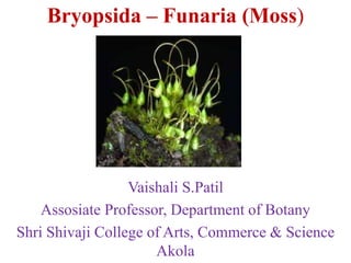
1. Bryopsida – Funaria (Moss)
Vaishali S.Patil
Assosiate Professor, Department of Botany
Shri Shivaji College of Arts, Commerce & Science
Akola
2. Division: Bryophyta
Class: Bryopsida
Subclass: Funariidae
Order: Funariales
Family: Funariaceae
Genus: Funaria
Habit-
210 species.
•Grows in dense patches or cushions
in moist shady and cool places during
the rainy seasons. These are primitive
multicellular, autotrophic, amphibious
plants. It can also be found on moist
walls and the crevices of rocks and places where recent fires have taken
place
3. External structure-
•The plant body is green, soft, and upright, about half an inch
tall (3–5 cm).
• It shows a radial symmetry with a differentiation of an axis
or stem, leaves or phylloids are multicellular colorless
branched rhizoids with oblique septa. The main axis of the
plant, which is upright, bears a set of spirally sessile leaves
having a clearly distinguishable midrib.
•The obovate leaves form a bulb-like cluster at the top of the
shortish, long stem. The leaves are long with an acute apex
and weakly serrate margins. The midrib ends just below, or
extends just beyond the leaf apex. They reproduce by spore
formation.
•They have no vascular system. Plant body consists of a long,
twisting stalk (seta) with an asymmetrical,grooved capsule
(spore-bearing part of the plant i.e.sporophyte) at the tip.
4. •At the apex of the main plant axis,the antheridium is borne.
This is the male part of the shoot. A lateral branch from the
main plant axisbears the female shoot archegonium at
its meristem.
5. Internal Structure:
1. Axis or stem
The transverse section (T. S.) of axis can be differentiated into
three distinct regions:
(i) Epidermis
(ii) Cortex
(iii) Central conducting strand or central cylinder.
(i) Epidermis:
It is the outer most single layered protective covering
consisting of small tangentially elongated chlorophyll bearing
cells. Cuticle and stomata are absent.
(ii) Cortex:
It is present between the epidermis and conducting tissue. It is
made up to parenchymatous cells. Younger part of the cortex
6. contains chloroplasts but in the older part they are lacking. At maturity
few outer layers of cortex become thick walled and are reddish brown in
colour but those of the inner layers become thin walled.
(iii) Central Conducting Strand:
It is made up of long, narrow thin walled dead cells which lack
protoplasm. These cells are now commonly called as hydroids.
Conducting strand besides providing a certain amount of mechanical
support, functions in the upward conduction of water and solutes.
7. .
2. Leaf:
Transverse section (T. S.) of ‘leaf’ shows a well-defined
midrib with two lateral wings. Except the midrib region,
the ‘leaf’ is composed of single layer of parenchymatous
polygonal cells. The cells contain many large and prominent
chloroplasts. The central part of the mid rib has narrow
conducting strand of thick walled cells which help in
conduction
8. Reproduction -
i) Vegetative Reproduction:
1. By multiplication of primary protonema:
In Funaria, spores on germination form a branched, filamentous,
multicellular structure. It’s called primary protonema. In it certain
colourless separation cells are formed by intercalary divisions. These cells
die out and break up the protonema into single cell or many celled
fragments. These fragments grow into new protonemata which bear buds.
Each bud develops into a leafy gametophore.
2. By secondary protonema:
When protonema is developed by other than the germination of spore, it is
called secondary protonema. It may be developed from any detached
living part of the gametophyte such as ‘stem’, ‘leaves’, antheridium,
archegonium paraphysis, sterile cells of capsule, seta or when the rhizoids
are exposed to sun light in moist atmosphere . It is similar to primary
protonema and develops into leafy gametophore.
10. 3. By Gemmae:
During unfavorable conditions, the terminal cells of the protonemal
branches divide by transverse, longitudinal divisions and form green
multicellular bodies of 10-30 cells. These are called gemmae. At maturity
gemmae become slightly reddish brown in colour. On the return of
favourable conditions gemmae germinate and form new plants.
4. By Bulbils:
When such gemmae like structures are produced on rhizoids inside the
substratum, these are called bulbils. These are devoid of chloroplasts but
capable of developing into leafy individuals under favourable conditions.
11. 5. Apospory:
Development of gametophyte from sporophyte without the formation of
spores is known as apospory. Any vegetative cell of the sporophyte may
form green protonemal filaments which bear lateral buds. These buds
later develop into leafy gametophores.
The gametophores thus formed are diploid. Sexual reproduction in such
gametophores results in the formation of tetraploid (4n) zygote. The
sporophytes from tetraploid are sterile because they are not capable of
bearing spores.
ii) Sexual Reproduction -
Sexual reproduction is oogamous. Male reproductive structure is known
as antheridium and female as archegonium. Funaria is monoecious
(having male and female sex organs on the same thallus) and autoicous
(antheridia and archegonia develop on separate branches of the same
thallus). Sex organs are borne on leafy gametophores in terminal clusters.
The main shoot of the leafy gametophore bears antheridia and act as
male branch. Female branch develops as a lateral outgrowth from the
base of the male branch and bears archegonia. It grows higher than the
12. male branch. Funaria is protandrous (antheridia mature before the
archegonia). It ensures the cross fertilization.
Male Branch or Antheridiophore: Longitudinal section (L. S.) of male
branch shows that its apex is expanded and convex shaped. It bears large
number of reddish brown or orange antheridia in different stages of
development. Projected antheridia are surrounded by a rosette of
spreading leaves called perigonial leaves.
The antheridial cluster with surrounding perigonial leaves is called
perigonium. The antheridia are intermingled with large number of sterile
hair like club shaped structures called paraphyses . Paraphyses store
water, protect developing antheridia, help in photosynthesis and
dehiscence of antheridia.
14. Structure of an Antheridium:
The antheridium is club shaped. It can be differentiated into two
parts:
(a) Short multicellular stalk
(b) Body of antheridium.
Body of antheridium has sterile, single layered jacket of polyhedral
flattened cells. When young the cells of the jacket contain chloroplasts
which turn orange or reddish brown at maturity. Jacket encloses a large
number of androcytes (antherozoid mother cells).
At maturity the distal end of the antheridium
bears one or two thick walled, colourless
cells called operculum.
The opercular cells become mucilaginous,
absorb water and swell, break
connections with the neighbouring cells and
form a narrow pore. Androcytes ooze out
in the form of a viscous fluid through this
pore.
15. Female Branch or Archegoniphore:
The female branch arises from the base of the male branch. Longitudinal
section (L. S.) of female branch shows that many archegonia
intermingled with paraphyses occurs at its apex. The terminal cell of
paraphyses is not swollen. The cluster of archegonia is enclosed by a
group of green foliage ‘leaves’ called perichaetial leaves. The
archegonial cluster with the surrounding perichaetial leaves is called
perichaetium.
Structure of an Archegonium:
A mature archegonium is flask shaped structure. It remains attached to
the female branch by a massive stalk. It consists upper elongated slender
neck and basal globular portion called venter .
The neck is slightly tubular, twisted, single layered and consists of six
vertical rows of neck cells, which enclose an axial row of ten or more
neck canal cells. The venter wall is two layered and encloses venter canal
cell and egg cell. Venter canal cell is situated just below the neck canal
cells.
17. Fertilization in Funaria:
Water is essential for fertilization. The opercular cells of the antheridium
rupture and releases mass of antherozoids. When archegonium reaches at
maturity, the neck canal cells and venter canal cell disintegrate to form a
mucilaginous mass. It absorbs water, swells up and comes out of the
archegonial mouth by pushing the cover cells apart. This mucilaginous
mass consists chemical substances (mainly sugars).
The cover cells of the neck separate widely from each other and form a
passage leading to the egg. Rosette like perigonial leaves serve as splash
cup from which rain drops disperse antheroziods to some distance (rain
drops falling on the archegonial cluster situated at lower level).
Many antherozoids enter the archegonial neck because of chemical
response but only one of them fuses with the egg to form the zygote.
Union of male and female nuclei is complete within 10 hours.
Fertilization ends the gametophytic phase.
18. Sporophytic Phase:
Zygote is the first cell of the sporophytic phase. Development of
sporophyte takes place within the venter of the archegonium.
Structure of Sporophyte:
The sporophyte is semi-parasitic in nature, the mature sporophyte can be
differentiated into three distinct parts—foot, seta and capsule.
(i) Foot:
It is the basal portion of the sporogonium. It is small dagger like conical
structure embedded in the apex of female branch. It functions as
anchoring and absorbing organ.
(ii) Seta:
It is long, slender, stalk like hygroscopic structure. It bears the capsule at
its tip. It raises the capsule above the apex of leafy gametophore.
19. (iii) Capsule:
It is the terminal part of the sporophyte and is developed at the apex of
the seta. It is green in colour when young but on maturity it becomes
bright orange coloured. It is covered by a cap like structure called
calyptra. (gametophytic tissue develops from the upper part of the
archegonium).
Internal Structure of the Capsule:
Longitudinal Section (L.S.) of the capsule shows that it can be
differentiated into three distinct regions-apophasis, theca and operculum.
(a) Apophysis:
It is the basal sterile part of the capsule. It is bounded by the single
layered epidermis which is interrupted by stomata. The sotmata have
single ring like guard cells. Below the epidermis is spongy parenchyma.
The central part of the apophysis is made up of elongated thin walled
cells forming a conducting strand. It is called neck of the capsule. It is
the photosynthetic region and connects seta with capsule.
(b) Theca:
It is the middle, slightly bent spore bearing region of the capsule. It lies
20. between the apophysis and operculum.
Longitudinal section (L. S.) passing through the theca shows the
following regions:(i) Epidermis:
It is the outer most layer. It is single layered with or without stomata.
(ii) Hypodermis:
It is present below the epidermis. It consists two to three layers of
compactly arranged colourless cells.
(iii) Spongy parenchyma:
It consists two to three layers of loosely arranged chlorophyllous cells. It
is present inner to hypodermis. These cells are capable to manufacture
their own food but dependent on gametophyte for water and mineral
nutrients. Therefore, the sporophyte of Funaria is partially dependent on
gametophyte.
(iv) Air spaces:
These are present just below the spongy parenchyma and outside the
spore sacs. Air spaces are traversed by green cells (chlorenchymatous
cells) called trabecular (elongated parenchymatous cells).
21. (v) Spore sac:
These are present below the air spaces on either side of the columella. It
is ‘U’ shaped and broken at the base. (It separates its both arms).
It has an outer wall (3-4 cells thick) and an inner wall (single cell in
thickness). Between the outer wall and inner wall is the cavity of the
spore sac. When young, the cavity of the spore sac is filled with many
spore mother cells. At maturity the spore mother cells divide by meiotic
divisions and form many haploid spores.
(vi) Columella:
It is the central part of the theca region. It is made up of compactly
arranged colourless parenchymatous cells. It is wide above and narrow
below, connecting the central strand of apophysis. It helps in conduction
of water and mineral nutrients.
(c) Operculum:
It is the upper region of the capsule. It is dome shaped and consists four
to five layers of cells. The outermost layer is thick walled and called
epidermis. Operculum is differentiated from theca by a well-marked
constriction. Just below the constriction there is a diaphragm (rim).
22. It is composed of two to three layers of radially elongated pitted cells.
Immediately above the rim is annulus which consists of 5-6
superimposed layers of cells. Its upper cells are thick but two lowermost
layers of cells are thin. Annulus separates the theca from the operculum.
Below the operculum lies the peristome . It is attached below to the edge
of the diaphragm. The peristome consists of two rings of radially
arranged peristomial teeth. In each ring there are sixteen teeth.
The teeth are not cellular but they are simply the strips of the cuticle. The
teeth of the outer ring are conspicuous, red with thick transverse bands
while the teeth of the inner ring are small, delicate, colourless and
without transverse bands. Inner to peristome teeth lies a mass of thin
walled parenchymatous cells.
24. Dehiscence of the Capsule:
Funaria is a stegocarpous moss (dehisce along a pre-determined line)
Dehiscence of the capsule is achieved by ‘breaking off’ of annulus. As
the capsule matures it becomes inverted due to epinasty. The thin walled
cells of the annulus break away, the operculum is thrown off and the
peristome teeth are exposed.
The outer peristomial teeth (exostome) are hygroscopic. The inner
peristomial teeth (endostome) do not show any hygroscopic movements
but act as a sieve allowing only a few spores to disperse at a time. The
lengthening and shortening of the outer peristomial teeth help in the
dispersal of spores.
In high humidity the exostome absorb water, increase in length and curve
inwards. In dry weather, the exostome teeth lose water, bend outwards
with jerky movements. It allows the dispersal of spores from the capsule
in instalments. At maturity the seta also shows jerky movements.
Twisting and swinging of seta in dry weather further aids in the dispersal
of spores.
25. Structure and Germination of Spore:
Spore is the first cell of the gametophytic phase. Each spore is spherical ,
12-20 µ in diameter and surrounded by two wall layers . The outer wall
is thick, smooth, brown and known as exosporium, while the inner wall
is thin, hyaline and called endosporium. Spore wall encloses single
nucleus, chloroplasts and many oil globules.
Under favourable conditions (sufficient moisture) spores germinate.
Exosporium ruptures and endosporium comes out in the form of one or
two germ tubes. Each germ tube is multicellular, green with oblique
septa. The germ tube grows in length, divides by septa to form green
algal filament like structure called primary protonema.
26. Primary Protonema:
It is the juvenile (young) stage of the gametophyte formed by the
germination of spore. It forms two different types of branches.
Most of the branches grow horizontally on the moist surface of the soil
and are known as chloronemal branches (positive phototrophic, thick and
rich in chloroplast) while some branches grow down in the soil and are
called rhizoidal branches (non-green, thin and possess oblique septa).
These branches can develop chlorophyll if expose to light.
Rhizoidal branches function as anchoring and absorbing organs while
chloronemal branches develop minute green buds behind the cross walls
which develop into leafy gametophores. From one primary protonema
many moss plants develop, so the moss is gregarious in habit. Primary
protonema is short lived.
According to Sirnoval (1947) development of protonema under
laboratory conditions can be differentiated into two stages—chloronemal
stage and caulonemal stage. Chloronemal stage is characterised by
irregular branching, right angle colourless cross walls, and many evenly
distributed discoid chloroplast.
27. It is positive phototropic but never produce buds. Nearly after 20 days
chloronemal stage matures into caulonemal stage. This stage is
characterised by regular branching brown cell walls, oblique cross walls
and fewer chloroplasts. It is negative phototropic and produce buds
which later develop into leafy gametophores. Rhizoids arise from the
base of a bud.