The hip joint is a ball and socket synovial joint that connects the femur to the pelvis. It has two articular surfaces: the proximal surface is the acetabulum of the pelvis, and the distal surface is the head of the femur. The acetabulum is cup-shaped and deepened by the acetabular labrum. The head of the femur fits into the acetabulum and is connected to the femoral shaft by the femoral neck. The angle of inclination and torsion of the femur can vary between individuals and abnormalities in these angles can impact joint mechanics and cause pathology.
The document discusses various pathologies that can affect the hip joint due to alterations in biomechanics. Small changes in forces or joint structure can lead to increased stress and injury over time. Common issues include arthritis/arthrosis from wear and tear, and fractures of the femoral neck which become more likely with age-related bone loss. Conditions like coxa valga/vara and torsion abnormalities of the femur can further impact forces on the joint and predispose to problems. Understanding hip biomechanics and how dysfunctions can influence other areas is important for evaluation and treatment.
1) The hip joint is a ball-and-socket joint that allows flexion, extension, abduction, adduction, and rotation. It supports the weight of the head, arms, and trunk.
2) The hip joint is made up of the femoral head articulating with the acetabulum. Several ligaments and the acetabular labrum provide stability to the joint. The angle of the femoral neck and torsion of the femur also affect biomechanics.
3) During standing and walking, forces from the body weight and ground reaction force act on the hip joint and femoral neck. A system of trabeculae in the femoral neck adapt to these forces. Muscles around the
This document discusses mechanical low back pain, defining it as pain originating from the spine that may be acute or chronic. It describes potential causes like nerve root impingement from herniated discs, musculoskeletal pain syndromes involving the muscles, and skeletal issues. Treatment options discussed include bed rest and light exercise, medications like NSAIDs, chiropractic/osteopathic manipulation, massage, and physical therapy modalities.
BIOMECHANICS & PATHOMECHANICS OF KNEE JOINT AND PATELLOFEMORAL JOINT
The document discusses the biomechanics of the knee joint, including the tibiofemoral joint and patellofemoral joint. It covers the articulating surfaces, degrees of freedom, ligaments, muscles, alignment and weight bearing forces of the knee. It also discusses normal patellar tracking in the trochlear groove during range of motion and the changing contact areas between the patella and femur through different degrees of flexion.
The document discusses hip joint anatomy and biomechanics from the perspective of total hip arthroplasty. It describes key terms like kinematics and kinetics. It provides details on normal ranges of motion for the hip. It discusses femoral head anatomy and the forces acting on the hip during single leg stance, which can be up to 4 times body weight. Factors like leg length, weight, and abductor lever arm influence joint loading.
Running requires greater balance, muscle strength, and joint range of motion compared to walking. During running, the ground reaction forces and center of pressure increase to 250% of body weight, double that of walking. The gait cycle of running consists of stance and swing phases. Key differences from walking include less time in contact with the ground, greater joint motion, and more eccentric muscle work. Running utilizes a float period where both feet are off the ground, distinguishing it from walking. Proper running form involves dorsiflexion and plantar flexion of the ankle, as well as flexion and extension of the hip and knee, to efficiently absorb impact and propel the body forward.
This document provides an overview of biomechanics concepts related to the hip joint, including:
- Forces acting on the hip joint include body weight and forces generated by hip abductor muscles. The joint reaction force is the force generated within the joint in response.
- Parameters like femoral head size, neck length, and offset impact joint stability and the required abductor muscle force. Restoring anatomy reduces joint forces.
- Gait adaptations like limping or using a cane bring the body's center of gravity closer to the hip joint, reducing forces on the joint.
This document discusses the biomechanics of the knee joint, including its structure, stability mechanisms, and kinetics. It describes the knee as a complex hinge joint made up of the femur, tibia, and patella. Key stabilizing structures include the collateral and cruciate ligaments, menisci, and surrounding muscles. The document outlines the knee's degrees of freedom and range of motion, including screw-home rotation. It also analyzes the forces acting on the knee during activities like walking, cycling, and squatting using free body diagrams and dynamic analysis.
The document discusses the biomechanics of the ankle joint. It describes the ankle's functions of stability and mobility. It details the bony structures that make up the ankle joint, including the talocrual joint and inferior tibiofibular joint. It explains the kinematics of the ankle in dorsiflexion and plantarflexion, including the axis of rotation. It also discusses the muscles, ligaments, and other factors involved in ankle stability and common mechanisms of injury.
The normal ROM for each hip motion is provided along with positioning details for accurate goniometric measurement. Precautions and common limiting factors are also outlined to ensure safe assessment.
Range of motion (ROM) measurements are performed to evaluate joint impairment, develop treatment goals, assess progress, and modify treatment. ROM is described in 3 planes and axes and measured using a goniometer. Active ROM is voluntary motion while passive ROM uses external assistance. Several factors determine ROM including joint integrity, scarring, age, gender, joint shape, and health of surrounding tissues. Common causes of limited ROM include contractures, arthritis, and pain. Precise positioning and stabilization are needed to reliably measure ROM of various joints like the shoulder, spine, and knee. Standardized testing procedures and documentation of measurements are important.
This document discusses the biomechanics of the elbow joint. It describes the bones and joints that make up the elbow complex, including the humeroulnar and humeroradial joints. It details the range of motion, ligaments, muscles, and biomechanics involved in flexion, extension, pronation and supination. Common injuries around the elbow joint like compression injuries, distraction injuries, and varus/valgus injuries are also summarized.
The document discusses the biomechanics of the hip complex, including its articulations, angles, ligaments, musculature, and kinematics. Key points include descriptions of the proximal and distal articular surfaces, angles of inclination and torsion, capsular ligaments, weight bearing structures, and osteokinematics of the femur and pelvis during motions like flexion, extension, abduction, and rotation. Gait and stance mechanics are analyzed, such as bilateral stance, unilateral stance, and trunk listing with calculations of joint torques and forces.
The document summarizes the structure and function of the hip joint. It describes the hip joint as a ball and socket joint formed by the acetabulum of the pelvis articulating with the femoral head. It has 3 degrees of freedom including flexion/extension, abduction/adduction, and medial/lateral rotation. The document outlines the bones, ligaments, and angles that make up the hip joint, as well as some common abnormalities.
The hip joint connects the femur to the pelvis and supports the weight of the upper body. It has a ball and socket structure, with the femoral head forming the ball and the acetabulum forming the socket. Several ligaments stabilize the hip joint, including the iliofemoral ligament which resembles an inverted Y shape. The hip joint allows flexion, extension, abduction, adduction, and rotation. Femoroacetabular impingement can occur if the femoral head or acetabulum have abnormal shapes that cause them to impinge upon each other.
Pelvic girdle, Femur, Sacroiliac joint and Hip Joint
The document discusses the anatomy of the pelvic girdle and femur. It describes the bones that make up the pelvic girdle - the hip bones, pubic symphysis and sacrum. It then details the individual bones, their features and articulations. This includes the sacroiliac joint and hip joint. It also outlines the ligaments supporting these joints and the movements they allow. Finally, it lists some of the muscles involved in hip joint movement.
The document describes the anatomy of the lower limb, including the pelvis, femur, patella, tibia, fibula, and hip joint. It discusses the bones that make up each part and their blood supply, fractures commonly seen in each bone, and movements at the hip joint. The lower limb consists of the gluteal region, thigh, leg, and foot and its main functions are to support body weight and enable locomotion.
The cervical spine consists of several joints including the atlanto-occipital joint and the atlanto-axial joint. It provides mobility but sacrifices stability, making it vulnerable to injury. The cervical spine can flex and extend between 15-20 degrees, side bend about 10 degrees, and rotate 50 degrees at the atlanto-axial joint. It is stabilized by muscles like the sternocleidomastoid and ligaments such as the transverse ligament of the atlas. Injuries can cause neck pain and symptoms extending into the head, arms, and shoulders.
this is a presentation on atlanto-axial and atlanto-occipital joints. after reading this, most of you will know about atlas and axis, joint type, anatomy of joint, movements allowed by joint and its clinical considerations.
The hip joint is a ball and socket joint that connects the femur to the pelvis. It is the body's largest weight bearing joint. The rounded head of the femur fits into the cup-shaped acetabulum of the pelvis. Strong ligaments and muscles provide stability to the joint. Damage to any of the hip joint components can negatively affect its range of motion and weight bearing ability, and may require hip replacement surgery. The hip allows for flexion, extension, abduction, adduction, internal and external rotation.
This document describes the anatomy of the hip joint. It discusses the bones that make up the joint, including the femoral head, femoral neck, and acetabulum. It describes the articular surfaces and cartilage in the joint. It also discusses the capsule, ligaments, blood supply, nerve supply, range of motion, muscles that act on the joint, and biomechanics of the hip joint.
This document discusses the structure and biomechanics of the hip joint. It describes the anatomy of the acetabulum and femoral head that form the ball and socket joint. It details the angles of the acetabulum, including the center edge angle and acetabular anteversion angle. It also describes the acetabular labrum and angles of the femur relative to the shaft. The primary function of the hip joint is to support weight and enable mobility through walking, running, and other activities.
The vertebral column is a complex structure composed of 33 vertebrae and intervertebral disks that meets the demanding needs of mobility and stability. It protects the spinal cord and attaches the pelvis. Each vertebra has a cylindrical vertebral body anteriorly and an irregularly shaped neural arch posteriorly. The vertebrae are arranged into five regions with variations to meet functional demands. Curves in the vertebral column provide increased resistance to compression and change throughout development. Intervertebral disks separate and cushion vertebrae. The vertebral column undergoes motions of flexion, extension, lateral flexion, and coupled rotations which place structures under varying degrees of compression and tension resisted by ligaments, disks, and facets.
The cervical spine consists of seven vertebrae that provide mobility but less stability than other regions of the spine. It has three subsystems that contribute to stability - passive (bones and ligaments), active (muscles), and neural control. Cervical instability occurs when the neutral zone between ranges of motion increases, the stabilizing subsystems can no longer compensate, and motion quality becomes poor. It can result from trauma, surgery, disease, or degeneration and often involves pain.
The temporomandibular joint (TMJ) is a hinge and gliding joint located in the skull that allows for depression, elevation, protraction, and retraction movements. It is formed by the mandibular condyle articulating with the temporal bone and is supported by ligaments and an articular disc that separates the bones. TMJ disorders can cause widespread pain in the head due to irritation of the trigeminal nerve, which innervates the face and head.
The temporomandibular joint (TMJ) is a hinge and gliding joint located in the skull that allows for depression, elevation, protraction, and retraction movements. It is formed by the mandibular condyle articulating with the temporal bone and is supported by ligaments and an articular disc that separates the bones. TMJ disorders can cause widespread pain in the head due to irritation of the trigeminal nerve, which innervates the face and scalp.
The hip joint is a ball and socket synovial joint that connects the femur to the acetabulum. It is the largest and most stable joint in the body. The hip joint allows for flexion, extension, abduction, adduction, and rotation. Several strong ligaments reinforce the hip joint capsule to provide stability, including the iliofemoral, ischiofemoral, and pubofemoral ligaments. The main muscles that act on the hip joint are the gluteal muscles, iliopsoas, quadriceps femoris, hamstrings, and adductors.
The hip joint is a ball and socket joint that connects the femur to the pelvis. It has 3 degrees of freedom and its primary function is to support the weight of the head, arms, and trunk. The hip joint has an acetabulum socket in the pelvis and a femoral head ball. It is surrounded by strong ligaments and a capsule that provide stability, especially in extension. The hip joint positioning in extension, slight abduction and medial rotation places the joint in its closest packed and most stable position.
The document provides an overview of the anatomy and examination of the hip joint. It describes the hip joint as the largest joint in the body that connects the femur to the acetabulum. It details the articular surfaces, bones, ligaments, muscles, nerves, blood supply and movements of the hip joint. The document also discusses ossification of the hip bone and bursae that can form around the joint.
This document provides an overview of the biomechanics of the knee complex. It describes the anatomy of the tibiofemoral and patellofemoral joints, including the femoral condyles, tibial plateaus, and alignment of the femur and tibia. It also discusses how weight bearing forces are distributed during static and dynamic activities, and how malalignment can increase stresses on the medial or lateral compartments.
Posture - a perquisite for functional abilities in daily life. Posture is a combination of anatomy and physiology with inherent application of bio-mechanics and kinematics. Sitting, standing, walking are all functional activities depending on the ability of the body to support that posture to carry out each activity. Injuries and pathologies either postural or structural can massively change the bio-mechanics of posture and thus affect functional abilities.
An orthosis is an external device that is applied to the body to improve function, provide support, reduce pain, correct deformities, and prevent progression of fixed deformities. Lower limb orthoses include foot orthoses, ankle-foot orthoses, knee orthoses, knee-ankle-foot orthoses, and hip-knee-ankle-foot orthoses. The goals of lower limb orthoses are to maintain or correct body segment alignment, assist or resist joint motion, provide axial loading and relieve distal weight bearing forces, and protect against injury. Orthoses can be static devices that hold body parts in position or dynamic devices that facilitate motion.
Thoracic and rib cage anatomy, biomechanics, and pathomechanicsRadhika Chintamani
The document discusses the biomechanics of the thorax and chest wall. It describes the anatomy of the rib cage including the various joints that connect the ribs to each other and to the sternum and vertebrae. It also discusses the muscles involved in respiration including the diaphragm and accessory muscles. It explains the axes of motion of the ribs during breathing and how this affects the diameters of the thorax. Finally, it covers topics such as the forces and loading on the thoracic spine during respiration and the concept of dynamic equilibrium.
The document discusses various pathologies that can affect the hip joint due to alterations in biomechanics. Small changes in forces or joint structure can lead to increased stress and injury over time. Common issues include arthritis/arthrosis from wear and tear, and fractures of the femoral neck which become more likely with age-related bone loss. Conditions like coxa valga/vara and torsion abnormalities of the femur can further impact forces on the joint and predispose to problems. Understanding hip biomechanics and how dysfunctions can influence other areas is important for evaluation and treatment.
BIOMECHANICS OF HIP JOINT BY Dr. VIKRAMVicky Vikram
1) The hip joint is a ball-and-socket joint that allows flexion, extension, abduction, adduction, and rotation. It supports the weight of the head, arms, and trunk.
2) The hip joint is made up of the femoral head articulating with the acetabulum. Several ligaments and the acetabular labrum provide stability to the joint. The angle of the femoral neck and torsion of the femur also affect biomechanics.
3) During standing and walking, forces from the body weight and ground reaction force act on the hip joint and femoral neck. A system of trabeculae in the femoral neck adapt to these forces. Muscles around the
This document discusses mechanical low back pain, defining it as pain originating from the spine that may be acute or chronic. It describes potential causes like nerve root impingement from herniated discs, musculoskeletal pain syndromes involving the muscles, and skeletal issues. Treatment options discussed include bed rest and light exercise, medications like NSAIDs, chiropractic/osteopathic manipulation, massage, and physical therapy modalities.
The document discusses the biomechanics of the knee joint, including the tibiofemoral joint and patellofemoral joint. It covers the articulating surfaces, degrees of freedom, ligaments, muscles, alignment and weight bearing forces of the knee. It also discusses normal patellar tracking in the trochlear groove during range of motion and the changing contact areas between the patella and femur through different degrees of flexion.
The document discusses hip joint anatomy and biomechanics from the perspective of total hip arthroplasty. It describes key terms like kinematics and kinetics. It provides details on normal ranges of motion for the hip. It discusses femoral head anatomy and the forces acting on the hip during single leg stance, which can be up to 4 times body weight. Factors like leg length, weight, and abductor lever arm influence joint loading.
Running requires greater balance, muscle strength, and joint range of motion compared to walking. During running, the ground reaction forces and center of pressure increase to 250% of body weight, double that of walking. The gait cycle of running consists of stance and swing phases. Key differences from walking include less time in contact with the ground, greater joint motion, and more eccentric muscle work. Running utilizes a float period where both feet are off the ground, distinguishing it from walking. Proper running form involves dorsiflexion and plantar flexion of the ankle, as well as flexion and extension of the hip and knee, to efficiently absorb impact and propel the body forward.
This document provides an overview of biomechanics concepts related to the hip joint, including:
- Forces acting on the hip joint include body weight and forces generated by hip abductor muscles. The joint reaction force is the force generated within the joint in response.
- Parameters like femoral head size, neck length, and offset impact joint stability and the required abductor muscle force. Restoring anatomy reduces joint forces.
- Gait adaptations like limping or using a cane bring the body's center of gravity closer to the hip joint, reducing forces on the joint.
This document discusses the biomechanics of the knee joint, including its structure, stability mechanisms, and kinetics. It describes the knee as a complex hinge joint made up of the femur, tibia, and patella. Key stabilizing structures include the collateral and cruciate ligaments, menisci, and surrounding muscles. The document outlines the knee's degrees of freedom and range of motion, including screw-home rotation. It also analyzes the forces acting on the knee during activities like walking, cycling, and squatting using free body diagrams and dynamic analysis.
The document discusses the biomechanics of the ankle joint. It describes the ankle's functions of stability and mobility. It details the bony structures that make up the ankle joint, including the talocrual joint and inferior tibiofibular joint. It explains the kinematics of the ankle in dorsiflexion and plantarflexion, including the axis of rotation. It also discusses the muscles, ligaments, and other factors involved in ankle stability and common mechanisms of injury.
The normal ROM for each hip motion is provided along with positioning details for accurate goniometric measurement. Precautions and common limiting factors are also outlined to ensure safe assessment.
Range of motion (ROM) measurements are performed to evaluate joint impairment, develop treatment goals, assess progress, and modify treatment. ROM is described in 3 planes and axes and measured using a goniometer. Active ROM is voluntary motion while passive ROM uses external assistance. Several factors determine ROM including joint integrity, scarring, age, gender, joint shape, and health of surrounding tissues. Common causes of limited ROM include contractures, arthritis, and pain. Precise positioning and stabilization are needed to reliably measure ROM of various joints like the shoulder, spine, and knee. Standardized testing procedures and documentation of measurements are important.
This document discusses the biomechanics of the elbow joint. It describes the bones and joints that make up the elbow complex, including the humeroulnar and humeroradial joints. It details the range of motion, ligaments, muscles, and biomechanics involved in flexion, extension, pronation and supination. Common injuries around the elbow joint like compression injuries, distraction injuries, and varus/valgus injuries are also summarized.
The document discusses the biomechanics of the hip complex, including its articulations, angles, ligaments, musculature, and kinematics. Key points include descriptions of the proximal and distal articular surfaces, angles of inclination and torsion, capsular ligaments, weight bearing structures, and osteokinematics of the femur and pelvis during motions like flexion, extension, abduction, and rotation. Gait and stance mechanics are analyzed, such as bilateral stance, unilateral stance, and trunk listing with calculations of joint torques and forces.
The document summarizes the structure and function of the hip joint. It describes the hip joint as a ball and socket joint formed by the acetabulum of the pelvis articulating with the femoral head. It has 3 degrees of freedom including flexion/extension, abduction/adduction, and medial/lateral rotation. The document outlines the bones, ligaments, and angles that make up the hip joint, as well as some common abnormalities.
The hip joint connects the femur to the pelvis and supports the weight of the upper body. It has a ball and socket structure, with the femoral head forming the ball and the acetabulum forming the socket. Several ligaments stabilize the hip joint, including the iliofemoral ligament which resembles an inverted Y shape. The hip joint allows flexion, extension, abduction, adduction, and rotation. Femoroacetabular impingement can occur if the femoral head or acetabulum have abnormal shapes that cause them to impinge upon each other.
Pelvic girdle, Femur, Sacroiliac joint and Hip JointSado Anatomist
The document discusses the anatomy of the pelvic girdle and femur. It describes the bones that make up the pelvic girdle - the hip bones, pubic symphysis and sacrum. It then details the individual bones, their features and articulations. This includes the sacroiliac joint and hip joint. It also outlines the ligaments supporting these joints and the movements they allow. Finally, it lists some of the muscles involved in hip joint movement.
Applied and clinical anatomy of lower limbdrjabirwase
The document describes the anatomy of the lower limb, including the pelvis, femur, patella, tibia, fibula, and hip joint. It discusses the bones that make up each part and their blood supply, fractures commonly seen in each bone, and movements at the hip joint. The lower limb consists of the gluteal region, thigh, leg, and foot and its main functions are to support body weight and enable locomotion.
The cervical spine consists of several joints including the atlanto-occipital joint and the atlanto-axial joint. It provides mobility but sacrifices stability, making it vulnerable to injury. The cervical spine can flex and extend between 15-20 degrees, side bend about 10 degrees, and rotate 50 degrees at the atlanto-axial joint. It is stabilized by muscles like the sternocleidomastoid and ligaments such as the transverse ligament of the atlas. Injuries can cause neck pain and symptoms extending into the head, arms, and shoulders.
this is a presentation on atlanto-axial and atlanto-occipital joints. after reading this, most of you will know about atlas and axis, joint type, anatomy of joint, movements allowed by joint and its clinical considerations.
The hip joint is a ball and socket joint that connects the femur to the pelvis. It is the body's largest weight bearing joint. The rounded head of the femur fits into the cup-shaped acetabulum of the pelvis. Strong ligaments and muscles provide stability to the joint. Damage to any of the hip joint components can negatively affect its range of motion and weight bearing ability, and may require hip replacement surgery. The hip allows for flexion, extension, abduction, adduction, internal and external rotation.
This document describes the anatomy of the hip joint. It discusses the bones that make up the joint, including the femoral head, femoral neck, and acetabulum. It describes the articular surfaces and cartilage in the joint. It also discusses the capsule, ligaments, blood supply, nerve supply, range of motion, muscles that act on the joint, and biomechanics of the hip joint.
This document discusses the structure and biomechanics of the hip joint. It describes the anatomy of the acetabulum and femoral head that form the ball and socket joint. It details the angles of the acetabulum, including the center edge angle and acetabular anteversion angle. It also describes the acetabular labrum and angles of the femur relative to the shaft. The primary function of the hip joint is to support weight and enable mobility through walking, running, and other activities.
Vertebral column... and Biomechanics.pptxsacootcbe
The vertebral column is a complex structure composed of 33 vertebrae and intervertebral disks that meets the demanding needs of mobility and stability. It protects the spinal cord and attaches the pelvis. Each vertebra has a cylindrical vertebral body anteriorly and an irregularly shaped neural arch posteriorly. The vertebrae are arranged into five regions with variations to meet functional demands. Curves in the vertebral column provide increased resistance to compression and change throughout development. Intervertebral disks separate and cushion vertebrae. The vertebral column undergoes motions of flexion, extension, lateral flexion, and coupled rotations which place structures under varying degrees of compression and tension resisted by ligaments, disks, and facets.
The cervical spine consists of seven vertebrae that provide mobility but less stability than other regions of the spine. It has three subsystems that contribute to stability - passive (bones and ligaments), active (muscles), and neural control. Cervical instability occurs when the neutral zone between ranges of motion increases, the stabilizing subsystems can no longer compensate, and motion quality becomes poor. It can result from trauma, surgery, disease, or degeneration and often involves pain.
09 Articulations Selected Articulations In Depthguest334add
The temporomandibular joint (TMJ) is a hinge and gliding joint located in the skull that allows for depression, elevation, protraction, and retraction movements. It is formed by the mandibular condyle articulating with the temporal bone and is supported by ligaments and an articular disc that separates the bones. TMJ disorders can cause widespread pain in the head due to irritation of the trigeminal nerve, which innervates the face and head.
09 Articulations Selected Articulations In DepthKevin Young
The temporomandibular joint (TMJ) is a hinge and gliding joint located in the skull that allows for depression, elevation, protraction, and retraction movements. It is formed by the mandibular condyle articulating with the temporal bone and is supported by ligaments and an articular disc that separates the bones. TMJ disorders can cause widespread pain in the head due to irritation of the trigeminal nerve, which innervates the face and scalp.
The hip joint is a ball and socket synovial joint that connects the femur to the acetabulum. It is the largest and most stable joint in the body. The hip joint allows for flexion, extension, abduction, adduction, and rotation. Several strong ligaments reinforce the hip joint capsule to provide stability, including the iliofemoral, ischiofemoral, and pubofemoral ligaments. The main muscles that act on the hip joint are the gluteal muscles, iliopsoas, quadriceps femoris, hamstrings, and adductors.
HIP JOINTS-1.pptx.........................IshaKanojiya1
The hip joint is a ball and socket joint that connects the femur to the pelvis. It has 3 degrees of freedom and its primary function is to support the weight of the head, arms, and trunk. The hip joint has an acetabulum socket in the pelvis and a femoral head ball. It is surrounded by strong ligaments and a capsule that provide stability, especially in extension. The hip joint positioning in extension, slight abduction and medial rotation places the joint in its closest packed and most stable position.
The document provides an overview of the anatomy and examination of the hip joint. It describes the hip joint as the largest joint in the body that connects the femur to the acetabulum. It details the articular surfaces, bones, ligaments, muscles, nerves, blood supply and movements of the hip joint. The document also discusses ossification of the hip bone and bursae that can form around the joint.
This document provides an overview of the biomechanics of the knee complex. It describes the anatomy of the tibiofemoral and patellofemoral joints, including the femoral condyles, tibial plateaus, and alignment of the femur and tibia. It also discusses how weight bearing forces are distributed during static and dynamic activities, and how malalignment can increase stresses on the medial or lateral compartments.
This document provides an overview of the biomechanics of the knee complex. It describes the anatomy of the tibiofemoral and patellofemoral joints, including the femoral condyles, tibial plateaus, and alignment of the femur and tibia. It also discusses how weight-bearing forces are distributed between the medial and lateral compartments during activities like standing, walking, and with conditions like genu valgum or genu varum. The complex biomechanics of the knee allow for both mobility and stability through interactions of its bones, cartilage, ligaments and muscles.
Similar to Hip joint - proximal and distal articular surface ppt (20)
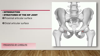
This an presentation about electrostatic force. This topic is from class 8 Force and Pressure lesson from ncert . I think this might be helpful for you. In this presentation there are 4 content they are Introduction, types, examples and demonstration. The demonstration should be done by yourself
The cryptoterrestrial hypothesis: A case for scientific openness to a conceal...Sérgio Sacani
Recent years have seen increasing public attention and indeed concern regarding Unidentified
Anomalous Phenomena (UAP). Hypotheses for such phenomena tend to fall into two classes: a
conventional terrestrial explanation (e.g., human-made technology), or an extraterrestrial explanation
(i.e., advanced civilizations from elsewhere in the cosmos). However, there is also a third minority
class of hypothesis: an unconventional terrestrial explanation, outside the prevailing consensus view of
the universe. This is the ultraterrestrial hypothesis, which includes as a subset the “cryptoterrestrial”
hypothesis, namely the notion that UAP may reflect activities of intelligent beings concealed in stealth
here on Earth (e.g., underground), and/or its near environs (e.g., the moon), and/or even “walking
among us” (e.g., passing as humans). Although this idea is likely to be regarded sceptically by most
scientists, such is the nature of some UAP that we argue this possibility should not be summarily
dismissed, and instead deserves genuine consideration in a spirit of epistemic humility and openness.
Towards Wearable Continuous Point-of-Care Monitoring for Deep Vein Thrombosis...ThrombUS+ Project
Kaldoudi E, Marozas M, Jurkonis R, Pousset N, Legros M, Kircher M, Novikov D, Sakalauskas A, Moustakidis P, Ayinde B, Moltani LA, Balling S, Vehkaoja A, Oksala N, Macas A, Balciuniene N, Bigaki M, Potoupnis M, Papadopoulou S-L, Grandone E, Gautier M, Bouda S, Schloetelburg C, Prinz T, Dionisio P, Anagnostopoulos S, Drougka I, Folkvord F, Drosatos G, Didaskalou S and the ThrombUS+ Consortium, Towards Wearable Continuous Point-of-Care Monitoring for Deep Vein Thrombosis of the Lower Limb. In: Jarm, T., Šmerc, R., Mahnič-Kalamiza, S. (eds) 9th European Medical and Biological Engineering Conference. EMBEC 2024. IFMBE Proceedings, vol 113. Springer, Cham. https://doi.org/10.1007/978-3-031-61628-0_36
Presented by Dr. Stelios Didaskalou, ThrombUS+ Project Manager
Search for Dark Matter Ionization on the Night Side of Jupiter with CassiniSérgio Sacani
We present a new search for dark matter (DM) using planetary atmospheres. We point out that
annihilating DM in planets can produce ionizing radiation, which can lead to excess production of
ionospheric Hþ
3 . We apply this search strategy to the night side of Jupiter near the equator. The night side
has zero solar irradiation, and low latitudes are sufficiently far from ionizing auroras, leading to a lowbackground search. We use Cassini data on ionospheric Hþ
3 emission collected three hours either side of
Jovian midnight, during its flyby in 2000, and set novel constraints on the DM-nucleon scattering cross
section down to about 10−38 cm2. We also highlight that DM atmospheric ionization may be detected in
Jovian exoplanets using future high-precision measurements of planetary spectra.
This an presentation about electrostatic force. This topic is from class 8 Force and Pressure lesson from ncert . I think this might be helpful for you. In this presentation there are 4 content they are Introduction, types, examples and demonstration. The demonstration should be done by yourself
SCIENTIFIC INVESTIGATIONS – THE IMPORTANCE OF FAIR TESTING.pptxJoanaBanasen1
download it
download it
download it
download it
download it
download it
download it
download it
download it
download it
download it
download it
download it
download it
download it
download it
download it
download it
download it
download it
download it
download it
download it
download it
download it
download it
download it
download it
download it
download it
download it
download it
download it
download it
download it
download it
download it
download it
download it
download it
download it
download it
download it
download it
download it
download it
download it
download it
download it
download it
download it
download it
download it
download it
download it
download it
download it
download it
download it
download it
download it
download it
download it
download it
download it
download it
download it
download it
download it
download it
download it
download it
download it
download it
download it
download it
download it
download it
download it
download it
download it
download it
download it
download it
download it
download it
download it
download it
download it
download it
download it
download it
download it
download it
download it
download it
download it
download it
download it
download it
download it
download it
download it
download it
download it
download it
download it
download it
download it
download it
download it
download it
download it
download it
download it
download it
download it
download it
download it
download it
download it
download it
download it
download it
download it
download it
download it
download it
download it
download it
download it
download it
download it
download it
download it
download it
download it
download it
download it
download it
download it
download it
download it
download it
download it
download it
download it
download it
download it
download it
download it
download it
download it
download it
download it
download it
download it
download it
download it
download it
download it
download it
download it
download it
download it
download it
download it
download it
download it
download it
download it
download it
download it
download it
download it
download it
download it
download it
download it
download it
download it
download it
download it
download it
download it
download it
download it
download it
download it
download it
download it
download it
download it
download it
download it
download it
download it
download it
download it
download it
download it
download it
download it
download it
download it
download it
download it
download it
download it
download it
download it
download it
download it
download it
download it
download it
download it
download it
download it
download it
download it
download it
download it
download it
download it
download it
download it
download it
download it
download it
download it
download it
download it
download it
download it
download it
download it
download it
download it
download it
download it
download it
download it
download it
download it
download it
download it
download it
download it
download it
Prototype Implementation of Non-Volatile Memory Support for RISC-V Keystone E...LenaYu2
Handling confidential information has become an increasingly important concern among many areas of society. However, current computing environments have been still vulnerable to various threats, and we should think they are untrusted.
Trusted Execution Environments (TEEs) have attracted attention because they can execute a program in a trusted environment constructed on an untrusted platform.
Particularly, the RISC-V Keystone is one of the interesting TEEs since it is a flexibly customizable and fully open-source platform. On the other hand, as same as other TEEs, it must also delegate I/O processing, such as file accesses, to a host OS, resulting in the expensive overhead. For this problem, we thought utilizing byte-addressable non-volatile memory (NVM) modules is a useful solution to handle persistent data objects for TEEs.
In this paper, we introduce a prototype implementation of NVM support for the Keystone. Additionally, we evaluate it on the Freedom U500 built on a VC707 FPGA dev kit.
https://ken.ieice.org/ken/paper/20210720TC4K/
Dalghren, Thorne and Stebbins System of Classification of AngiospermsGurjant Singh
The Dahlgren, Thorne, and Stebbins system of classification is a modern method for categorizing angiosperms (flowering plants) based on phylogenetic relationships. Developed by botanists Rolf Dahlgren, Robert Thorne, and G. Ledyard Stebbins, this system emphasizes evolutionary relationships and incorporates extensive morphological and molecular data. It aims to provide a more accurate reflection of the genetic and evolutionary connections among angiosperm families and orders, facilitating a better understanding of plant diversity and evolution. This classification system is a valuable tool for botanists, researchers, and horticulturists in studying and organizing the vast diversity of flowering plants.
The X‐Pattern Merging of the Equatorial IonizationAnomaly Crests During Geoma...Sérgio Sacani
A unique phenomenon—A geomagnetically quiet time merging of Equatorial IonizationAnomaly (EIA) crests, leading to an X‐pattern (EIA‐X) around the magnetic equator—has been observed in thenight‐time ionospheric measurements by the Global‐scale Observations of the Limb and Disk mission. Thepattern is also reproduced in an ionospheric model that assimilates slant Total Electron Content from GlobalNavigation Satellite System and Constellation Observing System for Meteorology, Ionosphere, and Climate 2.A free‐running whole atmospheric general circulation model simulation reproduces a similar pattern. Due to thesimilarity between measurements and simulations, the latter is used to diagnose this heretofore unexplainedphenomenon. The simulation shows that the EIA‐X can occur during geomagnetically quiet conditions and inthe afternoon to evening sector at a longitude where the vertical drift is downward. The downward vertical driftis a necessary but not sufficient condition. The simulation was performed under constant low‐solar andquiescent‐geomagnetic forcing conditions, therefore we conclude that EIA‐X can be driven by lower‐atmospheric forcing.
5. 1. ARTICULATING SURFACES:Acetabulum of pelvis and
head of femur
2. TYPE OF JOINT: Ball and socket diarthrodial synovial
joint
3. DEGREE OF FREEDOM
Flexion/ Extension in sagittal plane
Abduction/ Adduction in frontal plane
Medial/ Lateral rotation in transverse plane
7. SHOUDER JOINT
Ball and socket joint
It is made up of the head
of the humerus which rests
in the glenoid fossa of the
scapula
More mobility, less stability
More unstable
Shoulder injuries are more
such as dislocations
Main role: to provide a
stable base with a wide
range of motions . Open
change function
HIP JOINT
Ball and socket joint
It is the head of femur that fits
into the acetabulum of the
ilium (pelvic bones )
More stability, less mobility
Less unstable
Joint can suffer from
degeneration, femoral
acetabular impingement
Primary function: support
weight of the head, neck and
trunk (HAT) in static and
dynamic positions. WEIGHT
BEARING FUNCTION( more
muscles surrounding)
SHOULDER JOINT HIP JOINT
Ball and socket joint, synovial joint Ball and socket joint, synovial joint
It is made up of the head of the humerus which rests
in the glenoid fossa of the scapula
It is the head of femur that fits into the acetabulum of
the ilium (pelvic bones )
More mobility, less stability More stability, less mobility
More unstable Less unstable
Shoulder injuries are more such as dislocations Joint can suffer from degeneration, femoral
acetabular impingement
Main role: to provide a stable base with a wide range
of motions . Open chain function
Primary function: support weight of the head, arms
and trunk (HAT) in static and dynamic positions.
WEIGHT BEARING FUNCTION( more muscles
surrounding)
8. STRUCTURE OF THE HIP JOINT
COMPRISES OF TWO ARTICULAR SURFACES
• PROXIMAL ARTICULAR SURFACE (Acetabulum, Acetabular labrum )
• DISTAL ARTICULAR SURFACE ( Head of femur )
13. PROXIMAL ARTICULAR SURFACE
ACETABULUM OF PELVIC BONE FORMS PROXIMAL ARTICULAR SURFACE
1.Cuplike concave socket of the hip joint
2. Present on the lateral aspect of the pelvic bone
3. Three bones make up the pelvis that contribute to the structure of the acetabulum
a) Ischium (2/5 th of acetabulum)
b) Pubis (1/5 th of acetabulum)
c) Ilium ( remainder)
14. The periphery of the acetabulum called the lunate surface is covered with
hyaline cartilage
The acetabulum (horse shoe shaped) articulates with the head of femur
The inferior aspect of the lunate surface( ie base of horseshoe) has a notch
called acetabular notch. It is like a gap
The acetabular is spanned by a fibrous band called transverse acetabular
ligament that connects two ends of the horseshoe.
The transverse acetabular ligament creates a kind of tunnel under which
blood vessels pass beneath and reach the deepest of the acetabulum called
acetabulum fossa
The acetabular fossa ( innermost part) doesn’t take part in the articulation
The fossa contains fibroelastic fat covered with synovial membrane
The acetabular labrum deepens the acetabulum and surrounds its periphery.
15. POSITION: positioned laterally with inferior and anterior tilt
INCLINATIONS:
.50 degree laterally inclined
.20 degree anteriorly rotated (anteversion)
.20 degree anteriorly tilted in the frontal, transverse and sagittal
17. ACETABULAR ABNORMALITIES that lead to
pathology including excessive cartilage wear
ACETABULAR DYSPLASIA
COXA PROFUNDA
ACETABULAR PROTRUSIO
ANTEVERION
RETROVERSION
20. ACETABULAR DYSPLASIA:
Abnormally shallow acetabulum that results in lack of
coverage
Dysplasia is the basic mechanical abnormality for instability
and disproportionate loading of the superior acetabular rim
COXA PROFUNDA AND ACETABULAR PROTRUSIO:
Here the acetabulum excessively covers the femoral head
Acetabular over coverage can lead to limited ROM and
internal impingement (thinning) between femoral and
acetabulum junction
ABNORMALITIES IN ACETABULAR DEPTH
21. ABNORMALITIES IN ACETABULAR POSTIONING
( INCLINATION AND VERSION- abnormal positioning in
the transverse plane)
ANTEVERSION
Anteversion of acetabulum exists when the acetabulum is
positioned too far anteriorly in the transverse plane
RETROVERSION
Retroversion of the acetabulum exists when the
is positioned too far posteriorly in the transverse plane
22. ACETABULUM WITH
ANTEVERSION LESS INCLINATION LEAD TO INSTABILITY
INCLINATION/ RETROVERSION
OVER COVERAGE AND IMPINGEMENT
BETWEEN JOINT
25. CENTER EDGE ANGLE OF WIBERG
Acetabular depth can be measured using Center Edge Angle of Wiberg
It is the measure of depth of acetabulum with that of the femur head
It is formed by a line connecting the lateral rim of the acetabulum and center of the
femoral head and a vertical line from the center of the femoral head
CENTER EDGE ANGLES CLASSIFIED AS FOLLOWS:
DEFINITE DYSPLASIA: angle less than 16 degree
POSSIBLE DYSPLASIA: 16-25 degree
NORMAL: greater than 25 degree
ABNORMAL OVERCOVERAGE: greater than normal. But not well defined
EXCESSIVE ACETABULAR COVERAGE: greater than 40 degree
27. Acetabular labrum: A ring of fibrocartilage (fibrous
cartilage) that runs around the acetabulum (cup) of
the hip joint and increases its depth. The head of
the femur (the bone in the thigh) fits in
the acetabulum.
ACETABULAR LABRUM ( C SHAPED)
The labrum deepens this cavity and
effectively increases the surface (and
strength) of the hip joint. the labrum acts
like a rubber seal or gasket to help hold
the ball at the top of your thighbone
securely within your hip socket.
28. The knee meniscus and glenoid labrum are anatomically distinct yet
analogous structures. Although the gross anatomy differs, both tissues are
composed of fibrocartilage with a complex but well-organized collagen
microstructure. The meniscus and labrum function to increase congruity and
stability, decrease contact stresses, and distribute load across their
respective joints.
Meniscus of knee
Labrum of glenohumeral joint
ANALOGOUS STRUCTURES ( same function, different structure)
29. 2. ACETABULAR
LABRUM
The entire periphery of the acetabulum is rimmed by a wedge shaped
fibrocartilage called acetabular labrum
The labrum of the hip to a large extent is analogous to the meniscus of the knee
and labrum of the glenohumeral joint
The labrum is attached to the periphery of the acetabulum by a zone of
calcified cartilage
The labrum not only deepens the socket but also increases concavity of the
acetabulum through its triangular shape, grasping head of femur to maintain
contact with acetabulum
It enhances joint stability by acting as a seal to maintain intra articular pressure
It also decreases force transmitted to articular cartilage and provides
proprioceptive feedback
30. Nerve endings within the labrum not only provide proprioceptive
feedback but can also act as a source of pain
An abnormally shallow acetabulum will increase stress on the
surrounding capsule and labrum
The transverse acetabular ligament is considered to be a part of the
acetabulum labrum although unlike the labrum, it contains no
cartilage cells
Although it is positioned to protect the blood vessels travelling
beneath to reach the head of the femur, experimental data does
not support the notion of the transverse acetabular ligament as a
load bearing structure
31. The cause of a hip labral tear might be:
Trauma.
Injury to or dislocation of the hip joint
— which can occur during car accidents or from
playing contact sports such as football or hockey,
yoga not done properly
Degenerative health conditions: Osteoarthritis
is a chronic (long-term) wearing down of the
cartilage between the joints. As cartilage slowly
erodes over time, it becomes more prone to
tearing
The symptoms of a hip labral
tear include:
•Hip pain or stiffness
•Pain in the groin or buttocks
•A clicking or locking sound
hip area when you move
•Feeling unsteady on your
33. HEAD OF FEMUR
The distal articular surface is formed by the head of the femur.
The head of femur is spherical in shape covered by hyaline
cartilage
The articular area forms 2/3 rd of the sphere
Head of femur is connected to shaft by the neck
Inferior to the medial point of the head is a small depression
called fovea or fovea capitis.
The femoral neck is attached to the shaft of femur between
greater and lesser trochanters
The fovea is site of attachment of ligamentum teres.
34. ANGLE OF INCLINATION ANGLE OF TORSION
The magnitude of medial inclination and torsion of distal femur wrt head
and neck of femur depends on the embryonic growth, fetal position during
uterine life
The development of angulations continue after birth and during early
stages of development
Both normal and abnormal angles of inclination and torsion are properties
of femur alone
36. FIRST AXIS:
Passing
through the
center of
the femur
head and
neck
SECOND
AXIS:
Longitudin
al axis
passing
through
femur,
parallel to
shaft
ANGLE OF INCLINATION( femur ):
The angle formed between an axis
passing through the head and neck
of the femur and the longitudinal
axis
NORMAL VALUE OF THIS ANGLE: 125
DEGREE
It can have a variation from 110 -
144 degree
It varies between individuals and
within person
It is lesser in females ( wider pelvis )
It decreases with age ( At birth-
150, reaches 125 with maturity)
Abnormal increase in the angle -
COXA VALVA
Abnormal decrease in the angle –
COXA VARA
ANGLE OF
INCLINATION
37. COXA VALGA
It’s a condition in which angle of inclination increases
The angle is >125
The contact between articulating surfaces decreases ( exposure of
femoral head) therefore decrease in stability
Trabecular system density decreases( line of weight bearing
changes, less strain on oblique axis, decrease in density).
Therefore weakness in neck of femur/ bone
Moment arm of abductors ( lateral muscles )decreases ( as head
tilts upwards, distance decreases) , dec in muscles efficiency.. and
therefore muscle weakness ( stability )
Gravitation adduction moment is unbalanced .. leads to instability
Joint reaction force increases, leads to degenerative changes (
impingement, labral tear)
38. COXA VARA
Pathological condition in which angle of inclination of femur
decreases
Angle <120
Increase in stability ( the head of femur sits inside completely)
Increase in muscles efficiency ( increase in moment arm)
Decrease in joint reaction forces ( less degenerative chances)
But if it decreases too much then there are stability issues
Increase in bending moment, lead to fracture of neck of femur
3 types of Coxa Vera
CONGINETAL ( present since birth)
DEVELOPMENTAL ( during bone fusing .. Developmental period)
ACQUIRED ( due to diseases like rickets.. Vit D deficiency, calcium
def)
40. AXIS THROUGH
FEMORAL CONDYLES
AXIS THROUGH FEMORAL
HEAD AND NECK
ANGLE OF TORSION
ANGLE OF TORSION (femur): Angle formed between axis passing through femoral condyles and the
axis through femoral head and neck
Normal value: 10 – 20 degrees ( 15 deg in males and 18 deg in females )
Femur is slightly anteverted
30 – 40 degrees at birth
Decreases with age : About 1.5 deg until maturity till 10 to 20
Angle of torsion is similar btw both legs but angle of inclination differ
41. ANTEVERSION & RETROVERSION
◦ If the axis through femoral condyles lies in the frontal plane then the head and neck of the
femur are torsioned anteriorly, on the condyles.
◦ At birth it is 30-40 deg. This decreases about 1.5 per year until skeletal maturity
◦ Condition in which angle of anterior torsion increases is anteversion (>15 in males , >18 in
females)
◦ Articulating surfaces instable because large portion of femoral head is tilted outward, exposed
◦ When angle of torsion decrease..less than 15-20 it is called retroversion
◦ Variations in degree of Anteversion and retroversion also depends on assessing methods like CT
scan, radiograph, ultrasound to measure angle of femoral torsion
◦ Femoral anteversion is correlated with increase medial rotation ROM and decrease lateral
rotation so total hip rotation remains the same
◦ Femoral anteversion and coxa valga are commonly found togther but they function
independently
◦ can lead to dysfunction in distal and proximal parts of hip + knee +foot
◦ Other pathological angulations ( retroversion, coxa valga n coxa vara ) can also affect proximal
and distal parts of hip joint
42. RETROVERSION (AOT < 15-20)
………………………………………………………….
Retroversion refers to an abnormal backward rotation of the hip relative to the knee
This condition can affect patients of all ages and leads to abnormal stress in the low back, hip and
knee, as well as an abnormal gait (walking stance).
Femoral retroversion (also known as hip retroversion) is a rotational or torsional deformity in which
the femur (thighbone) twists backward (outward) in relation to the knee. Because the lower part of
the femur is connected to the knee, this also means that the knee is twisted outward relative to the
hip.
Femoral retroversion can occur in one or both legs
Femoral retroversion is often a congenital condition, meaning children are born with it. It also
appears to be related to the position of the baby as it grows in the womb. Torsional deformity can
also occur after a fracture, if a broken bone heals with incorrectly (called malunion).
43. Symptoms -out-toeing or "duck walk" – walking with the foot pointed outward instead of straight
ahead, learning to walk late (in children), flatfeet, difficulty with running, fatigues easily with physical
activity, poor balance or coordination ,hip and knee pain , low back pain degeneration or arthritis of
the hip
Diagnosis -The doctor will go through the developmental and family history and also observe the
patient’s gait (manner of walking) to look for signs of out-toeing or gait compensation. The physician
may also order an X ray or CT scan
Treatment - Many children born with femoral retroversion grow out it. An excessive femoral
retroversion can place stress on hip and knee joints, often leading to joint pain and abnormal wear..
labral tear. In these situations, a surgical procedure known as a femoral osteotomy may be used
44. Left: Position of an anteverted femoral head with the
foot facing straight forward. In this position, the femoral
head subluxes out of the front of the hip joint.
Right: Most patients with excessive hip anteversion
compensate by walking in-toed. This position keeps the
femoral head within the socket, which minimizes pain.
45. ANTEVERSION (AOT> 15-20)
…………………………………………………………………..
• Femoral anteversion is a forward (inward) rotation in the femur (thighbone), which connects to
the pelvis to form the hip joint. In other words the knee is excessively twisted inward relative to
the hip.
• Femoral anteversion can occur in one or both legs.
• Many children are born with femoral anteversions that they eventually grow out of. In people
who do not grow out of it, a mildly anteverted femoral head may cause no significant health
problems.
• But an excessive anteversion of the femur overloads the anterior (front) structures of the hip
joint, including the labrum and joint capsule.When the foot is positioned facing directly
forward, the femoral head may sublux (partially dislocate) from the socket of the hip joint,
called the acetabulum.This torsional malalignment places abnormal stress on both the hip and
knee joints, often leading to pain and abnormal joint wear.
•
46. • Symptoms- In-toeing, in which a person walks "pigeon-toed," with each foot
pointed slightly toward the other, Bowlegs (also called bowed legs), Keeping the
legs in this position often helps a patient maintain balance, Pain in the hips,
knees and/or ankles.
• Treatment- While many children grow out of their femoral anteversion
conditions, excessive anteversion may require surgical correction, as a
procedure known as a femoral osteotomy
Reduces hip stability
Articulating surfaces instable because large portion of femoral head is tilted outward,
exposed
Hip abductors fall more posteriorly to the joint, reducing moment arm for abduction
Affects the knee joint.When femoral head is anteverted, pressure from
capsuloligaments and ant musculature may push it into acetabulum causing entire
femur to medially rotate. Medial rotation of the condyles alters plane of knee flexion
therefore.. in toe gait. Abnormal position of knee joint axis ..medial femoral torsion.
Anteverted femur also affects biomechanics of patellofemoral joint at knee and
subtlar joint at foot
48. •INTRODUCTION- articulation, movements, comparison
between shoulder and hip joint
•ARTICULAR SURFACES
•Proximal articular surface- acetabulum( dysplasia, coxa
profunda, anteversion, retroversion), center edge angles
,labrum
•Distal articular surface- femur, angle of inclination( coxa
valga, coxa vara),angle of torsion (anteversion,
retroversion)