Eur Radiol (2010) 20: 2663–2670
DOI 10.1007/s00330-010-1851-2
INTERVENTIONAL
Jonathan L. Hart
Zaid Aldin
Philip Braude
Claire L. Shovlin
James Jackson
Embolization of pulmonary arteriovenous
malformations using the Amplatzer
vascular plug: successful treatment of 69
consecutive patients
Received: 3 March 2010
Revised: 25 April 2010
Accepted: 27 May 2010
Published online: 24 June 2010
# European Society of Radiology 2010
J. L. Hart : Z. Aldin : J. Jackson
Department of Imaging,
Imperial College Healthcare NHS Trust,
London, England
Abstract Objective The technique of
embolization of pulmonary arteriovenous malformations (PAVMs) with
the Amplatzer vascular plug (AVP)
has been reported, but no large series
has evaluated the effectiveness of this
relatively new embolic device. The
purpose of this study is to assess the
role of AVPs in the treatment of
PAVMs. Materials and methods
Sixty-nine consecutive patients
underwent embolization of
pulmonary arteriovenous
malformations between September
2006 and December 2008. Clinical,
procedural, and physiological data
were reviewed retrospectively.
Results Of 161 PAVMs, 120 (75%)
were successfully embolized
with Amplatzer vascular plugs
alone. Complete and rapid
P. Braude : C. L. Shovlin
Department of Respiratory Medicine,
Imperial College Healthcare NHS Trust,
London, England
C. L. Shovlin
National Heart and Lung Institute of
Cardiovascular Sciences,
Imperial College London, London, England
J. Jackson ())
Hammersmith Hospital,
Du Cane Road, London, W12 0HS, UK
e-mail: james.jackson@imperial.nhs.uk
Tel.: +44-208-3833485
Fax: +44-208-3833121
Introduction
Pulmonary arteriovenous malformations (PAVMs) are
high-flow, low-pressure communications between the
pulmonary arterial and venous systems allowing a rightto-left shunt and subsequent hypoxemia [1]. The morphology of these communications is variable, ranging from
complex vascular structures supplying and draining an
aneurysmal sac, to much smaller caliber telangiectatic
vessels. The majority of PAVMs occur in the context of
hereditary hemorrhagic telangiectasia (HHT), in which
there is a 50% incidence [2–4]. Sporadic PAVMs are less
common, accounting for fewer than 10% of cases. The
direct arteriovenous communication bypasses the important “filter capacity” of the pulmonary capillaries, and this
predisposes to cerebrovascular complications including
stroke, transient ischemic attack, and cerebral abscess [5].
Transcatheter embolization is established as the preferred
occlusion of feeding vessels was
easily achieved at the site of
arteriovenous communication
without complication. Particularly
small or tortuous feeding arteries
supplying 27 complex and 14 simple
PAVMs were occluded with coils.
There have been no documented
instances of recanalization on
follow-up. Conclusion Amplatzer
vascular plugs allow the rapid and
safe distal occlusion of the majority
of PAVMs.
Keywords Pulmonary .
Arteriovenous malformation .
Embolization . Technique .
Vascular plug
treatment for PAVMs in order to reduce the risk of
paradoxical embolization. The technique had previously
been recommended only for PAVMs that have one or
more feeding vessels of 3 mm or larger [6–9], but concern
was expressed about the 3 mm rule [10]. Since stroke risk
has now been shown to be independent of the size of
PAVMs [5], embolization is now recommended for all
PAVMs amenable to the procedure. Hypoxemia due to
right-to-left shunting is often well tolerated due to the
associated low pulmonary vascular resistance, but a
subjective improvement in exercise tolerance is often
reported following embolization, even in those individuals
without symptoms before treatment [1].
Embolization with MR-compatible steel or platinum
coils is the mainstay of treatment in most centers and
requires a meticulous technique; unsurprisingly, the
experience of the operator has been associated with
improved outcomes [11]. In the majority of cases,
�2664
embolization involves occlusion of the feeding vessel with
one or more coils deployed immediately proximal to the
aneurysmal sac in order to avoid the occlusion of normal
pulmonary artery branches. Occasionally the specific
anatomy of a lesion may necessitate an alternative strategy
such as packing of the aneurysm sac when the neck is
particularly short or wide or the use of occlusion balloons
to control flow during coil deployment in especially large,
high-flow feeding vessels.
The Amplatzer vascular plug (AGA Medical, Plymouth,
MN, USA) is a relatively new occlusive device made of a
self-expanding cylindrical nitinol mesh that is especially
suited for embolization of large high-flow vessels. It has
been used in the arterial and venous systemic circulation for
a number of indications including iliac artery occlusion prior
to endovascular repair of aortoiliac aneurysms or pelvic
surgery, occlusion of transjugular intrahepatic portosystemic
shunts, and testicular vein embolization [12, 13]. Several
recent reports have described its use for treatment of PAVMs
but, to date, no large series addressing its effectiveness at
treating these lesions has been published [14–18]. We report
our experience with the Amplatzer vascular plug, which has
been used for PAVM embolization at our institution since
September 2006.
Materials and methods
Study group
The study group comprised 69 consecutive patients who
underwent embolization of pulmonary arteriovenous malformations between September 2006 and December 2008.
femoral venous approach to document the anatomy of
treatable lesions. In patients in whom the MDCT had
demonstrated unilateral PAVMs, that side only was studied
at the time of angiography. The Grollman pigtail catheter
was then exchanged for a 90 cm long, 6 Fr, straight sheath
(Cordis, Ascot, Berkshire, UK) through which a 100 cm, 5
Fr Headhunter catheter (Cook Europe, Bjaeverskov, Denmark) was introduced. Selective catheterization of the
feeding vessel(s) to each treatable PAVM was performed
with this catheter sheath combination. Once a suitable
position had been achieved as distally as possible within
the feeding vessel beyond any branches to normal lung, the 5
Fr catheter was removed, and embolization was performed
through the sheath with an AVP. The size of the AVP selected
for embolization was approximately 1.5 to 2 times the
caliber of the feeding vessel, which was itself estimated by
comparing it by eye with the diameter of the sheath tip on
pre-embolization arteriograms. Satisfactory positioning of
the AVP was confirmed in most cases by repeat arteriography via the sheath before its detachment from the
introducing wire. If suboptimally positioned, the AVP was
resheathed and redeployed in a more appropriate site.
Additional feeding vessels to a PAVM were treated in the
same manner except when their small size or tortuosity
precluded the introduction of the sheath to a suitable distal
position, in which case embolization was performed through
the 5 Fr catheter with MR compatible coils.
The duration of the embolization intervention was
determined by the number and complexity of the malformations requiring embolization and patient tolerance to the
procedure. In general, procedure duration was between 90
and 120 min. All patients were discharged on the day
following the procedure. Patients were readmitted for
subsequent embolizations as required until complete occlusion of all significant lesions was achieved.
Pre-procedure assessment
Physiological tests
All patients underwent a full clinical evaluation prior to
treatment. Baseline chest radiographs were obtained and
standard blood tests included full blood count, coagulation
screen, and liver function tests (to exclude hepatopulmonary
syndrome). If recent cross-sectional imaging from the
referring hospital was not available, a non-contrast multidetector CT (MDCT) of the thorax [19] was obtained to
confirm the diagnosis of PAVM and identify the approximate
number and size of lesions prior to intervention.
Embolization procedure
All embolizations were performed by a single interventional
radiologist. Prophylactic intravenous antibiotics were
administered in all cases (1 g vancomycin) 1 h before each
procedure. The pulmonary artery pressure was measured in
all individuals at the time of angiography. Selective right
and/or left pulmonary digital subtraction arteriograms were
then obtained with a 7 French Grollman angled pigtail
catheter (Cook Europe, Bjaeverskov, Denmark) via a
Arterial oxygen saturation was measured with the patient
breathing room air using a pulse oximeter (Biox 3740;
Ohmeda, Hatfield, Hertfordshire, UK) and an ear probe.
Measurements were taken every 60 s for 10 min in the
erect and supine positions, with the result expressed as the
mean of the last four readings. The nonparametric
Wilcoxon’s matched pairs signed rank test was used to
compare the pre- and post-embolization arterial oxygen
saturation. To compensate for multiple comparison tests,
only P values of less than 0.01 were considered statistically significant. Where more than one procedure was
performed, the post-embolization value corresponds with
the recording taken after the most recent procedure.
Follow-up
Patients were invited for review in a dedicated PAVM
clinic 3–6 months following the procedure and annually
thereafter. Given the pattern of tertiary referral for patients
�2665
with PAVMs from across the UK, in the absence of any
symptoms or concerns, a small number opted for followup at the referring hospital or declined follow up. Data are
presented for all individuals who attended for a follow-up
assessment.
Results
No major complications occurred. Minor pleuritic-type
chest pain was experienced by four (6%) patients. A
single patient developed asymptomatic atrial fibrillation,
which resolved spontaneously. In one case a minor, selflimiting hemoptysis occurred following the procedure. In
two cases, the anatomy of the lesion required occlusion of
the arterial supply to a small segment of immediately
adjacent normal lung, but both patients remained asymptomatic following the procedure.
Clinical features
Physiological tests
The study group comprised 28 male and 41 female
patients, with a mean age of 44.6 years (range 16–78).
The presenting clinical features of these patients are
summarized in Table 1. A definitive diagnosis or a
possible diagnosis of HHT was made in 52 (75%) patients
and 10 (15%) patients, respectively, according to Curacao
criteria [20]. Seven (10%) individuals had isolated
PAVMs. Ten patients had undergone previous embolization of PAVMs (as part of a previously reported series
[21]): seven had recurrent lesions that had been previously
embolized with MR-compatible coils, and in three patients
pre-existing PAVMs noted at the time of their original
procedures, but not treated because of their small size, had
grown to a size amenable to embolization. A single patient
had undergone a lobectomy 19 years previously to remove
a symptomatic PAVM.
Embolization
Eighty-three procedures were performed in 69 patients.
A total of 161 PAVMs (mean 2.3 lesions per patient;
range 1–12) comprising 115 simple lesions and 46
complex lesions with feeding arteries ranging between
2 and 13 mm in diameter were embolized. A total of 120
lesions were treated with Amplatzer vascular plugs alone
(75%). Smaller additional feeding arteries to 27 complex
PAVMs required coil occlusion. Fourteen PAVMs with
small tortuous feeding arteries were treated with coils
alone. The mean pulmonary artery pressure prior to
embolization was 14.8 mmHg (range 7–26 mmHg). Pulmonary arterial hypertension, defined as a mean pulmonary
artery pressure in excess of 25 mmHg, was identified in three
patients.
Table 1 Clinical features at presentation
Symptoms
Respiratory
Dyspnea
Hemoptysis
Embolic
Cerebral abscess
Transient ischemic attack
Cerebrovascular accident
Peripheral abscess
Migraine
Asymptomatic
Number
Percentage
28
9
41
13
2
5
5
2
9
15
3
7
7
3
13
22
Statistically significant improvement in supine and erect
systemic arterial oxygen saturations was demonstrated
following completion of embolization treatments
(Table 2). Results are comparable with those from our
previous series in which MR-compatible coils were used
[6, 7, 21].
Follow-up
Clinical follow-up data were available in 51 (74%)
patients at a mean of 9.6 months (range 1–25 months).
Twenty-five out of 26 patients with impaired exercise
tolerance or dyspnea prior to the procedure reported an
improvement in symptoms following treatment (two
additional patients who had not described impaired
exercise tolerance prior to the procedure nevertheless
described an increased exercise capacity following treatment). No further events were documented in patients
with previous neurological complications attributable to
PAVMs (a single patient reported a neurological event
related to a known cerebral AVM). Eleven patients
required additional embolization sessions to achieve occlusion of all PAVMs of treatable size. Repeat angiography
prior to these subsequent treatments demonstrated that
vessels previously treated by AVPs remained occluded. In a
single patient, repeat pulmonary angiography for investigation of hemoptysis demonstrated filling of the treated
PAVM via multiple intrapulmonary collaterals that were
successfully embolized with MR-compatible coils; again,
the vessel treated with an AVP remained occluded. Five
patients underwent follow-up CT at a mean of 12.4 months
following embolization (range 2–34 months). In all of
these, there was complete occlusion of the feeding vessels
treated with AVPs with decompression of the PAVM
venous sac (Figs. 1 and 2).
Discussion
The technique of percutaneous catheter embolization of
pulmonary arteriovenous malformations involves delineation of the lesion by pulmonary angiography, followed by
super-selective catheterization and occlusion of the feeding
artery. It is established as the preferred treatment to reduce
�2666
Table 2 Systemic arterial oxygen saturation pre- and post-embolization
treatments
the risk of embolic complications [6–9]. A substantial
number of reports have demonstrated good immediate
outcomes and long-term effectiveness, with recurrence rates
ranging from 0 to 22% [10, 22–28]. Recurrences are also
amenable to retreatment by transcatheter embolotherapy
with durable results. Despite good technical outcomes in the
vast majority of cases, residual lesions remain in many
patients due to the presence of feeding vessels that are too
small to be occluded with currently available techniques.
Although the risk of infective embolic complications is
reduced, the lifelong requirement for antibiotic prophylaxis
prior to dental and surgical treatment persists.
The Amplatzer vascular plug is a relatively new
occlusive device made of a self-expanding cylindrical
nitinol mesh that can be deployed rapidly and can be
repositioned before final release. It is particularly suitable
for embolization of large high-flow vessels such as those
found in pulmonary arteriovenous malformations. AVP1,
AVP2, AVP3, and AVP4 devices are available; they vary
in their configuration and range of available diameters.
The AVP4 has only recently been introduced into clinical
practice and differs from the other devices in that it can be
introduced through a conventional diagnostic catheter
with a 0.038 inch lumen. The AVP1 device was used for
all but one PAVM in this series.
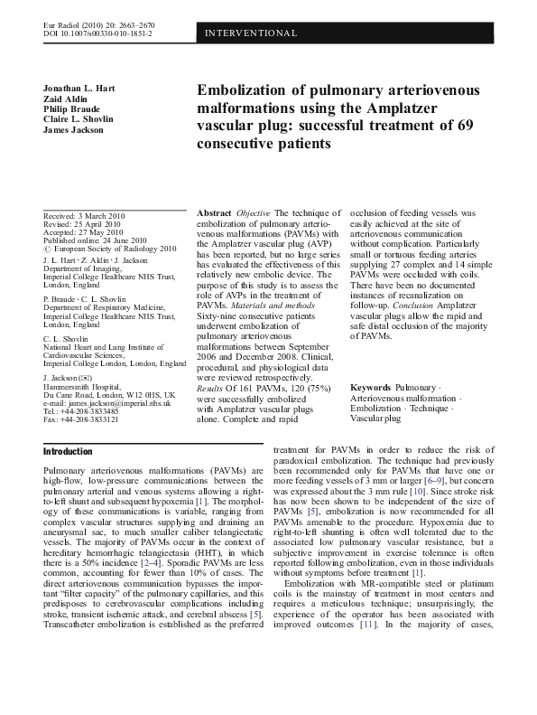
Fig. 1 A 57-year-old woman with hereditary hemorrhagic telangiectasia (HHT), several large bilateral PAVMs, and history of CVA.
a Left lower lobe pulmonary angiogram demonstrates large basal
PAVM of simple type with a feeding vessel measuring
approximately 6 mm in diameter at its point of communication
with the dilated venous sac. b Control film showing 10 mm
diameter AVP after exiting the delivery sheath but still attached to
its wire. c Selective arteriogram shows good position of AVP at the
neck of the PAVM. There is still flow through the malformation
immediately after AVP insertion. d Selective arteriogram a few
minutes later demonstrates complete vessel occlusion with preservation
of normal proximal pulmonary artery branches. e Axial CT image
through the lung bases performed before PAVM embolization
demonstrates the large venous sac in the left lower lobe. A further
PAVM is present on the right side, which was also embolized with an
AVP. f Axial CT image through the lung bases at the same level as e
performed 2 years and 10 months after PAVM embolization shows a
portion of the AVP used for vessel occlusion on the right side. The
venous sacs in both lower lobes have decompressed completely. Other
images (not shown) demonstrated only small linear markings on both
sides at the sites of the venous sacs and confirmed complete occlusion
of other embolized PAVMs
Measurement
Pre-embolization
Median
Interquartile range
Post-embolization
Median
Interquartile range
Pa
a
Systemic arterial oxygen saturation (%)
Supine (n=62)
Erect (n=62)
94
89, 96
94
92, 96
96
95, 97
<0.001
96
95, 97
<0.001
Wilcoxon’s matched pairs signed rank test
�2667
Fig. 2 A 48-year-old man with hereditary hemorrhagic telangiectasia (HHT), relatively small bilateral PAVMs, and history of CVA.
a Axial CT image through the right lung demonstrates a small
PAVM adjacent to the oblique fissure, which had a feeding vessel
measuring 4 mm in diameter. b Control film and c angiogram after
releasing a 6 mm diameter AVP demonstrate its optimal positioning
at the neck of the PAVM. d Arteriography a few minutes later
demonstrates complete vessel occlusion. e CT image 10 months
later demonstrates AVP and complete decompression of the venous
sac
The device has platinum and iridium marker bands on
either end and a stainless steel microscrew on the
proximal band attached to a 135-cm-long delivery wire.
The diameter of the AVP1 ranges from 4 to 16 mm in
2 mm increments; all sizes of AVP1 can be deployed via a
6 French sheath with a 2 mm lumen although the
introduction of the 14 mm and 16 mm plugs may be
difficult, and a 7 French sheath with a luminal diameter of
2.3 mm is recommended when these devices are used.
There are a number of important advantages of AVPs over
coils for the treatment of PAVMs; they are described below.
“anchor” technique of coil embolization, in which the first
loop of a coil is purposely deployed in a normal pulmonary
artery branch proximal to the venous sac, is utilized by some
practitioners to overcome the problem of coil migration. The
very nature of this technique, however, means that the
occlusion is performed proximal to, and often at some
distance from, the PAVM neck. In our experience with the
AVP1 device, this problem is overcome in most PAVMs; the
vascular sheath through which the plug is to be deployed can
usually be placed at the neck of the PAVM and can, in some
instances, be introduced into the venous sac itself. The plug
can then be introduced with the sheath in this position and
deployed during sheath withdrawal (Figs. 1, 2, and 3).
Ability to achieve a very distal and safe embolization in
the majority of PAVMs It is recognized by all practitioners
who regularly perform PAVM embolization that it is
desirable to achieve a very distal occlusion of the feeding
vessel to a PAVM, preferably at the neck of the venous sac.
This not only reduces the risk of occluding branches to
adjacent normal lung but may also reduce the likelihood of
persistent perfusion of the venous sac by bronchial collaterals and of pulmonary artery recanalization. Such a distal
occlusion is often very difficult to achieve with metallic
coils, particularly when the feeding vessel is large, because
of the risk of coil migration through the sac into the systemic
circulation with potentially disastrous complications. The
Complete vessel occlusion with a single device The
embolization of a feeding vessel to a PAVM, particularly
when of a large diameter, will usually require the use of
several metallic coils in order to achieve complete occlusion. In our experience complete pulmonary artery occlusion, even of feeding vessels measuring 12 mm in diameter,
is achievable with a single AVP1 (Figs. 1, 2, and 3). This has
a number of advantages:
1. It speeds up the procedure such that a larger number of
PAVMs can be embolized in a single session.
�2668
Fig. 3 A 37-year-old woman with hereditary hemorrhagic telangiectasia (HHT), single moderately sized right lower lobe PAVM,
known cerebral AVM, and history of transient ischemic attack. a
Coned image of portion of CXR demonstrates well-defined nodule
in the right lower zone consistent with a PAVM. b Right pulmonary
artery angiogram shows PAVM with 5 mm feeding vessel, an
aneurysmal venous sac, and early venous return. c Angiogram after
positioning an 8 mm diameter AVP demonstrates its optimal
positioning at the neck of the PAVM. d Arteriography a few
minutes later demonstrates complete vessel occlusion with
preservation of normal pulmonary artery branches. e Coned image
of portion of CXR 8 months after embolization demonstrates AVP
and disappearance of venous sac
2. It allows occlusion of a shorter length of vessel. When
coils are used to occlude large pulmonary feeding
arteries, a coil nest measuring several centimeters in
length is often required to achieve complete occlusion.
3. There is a theoretical reduction in the likelihood of
thrombus embolizing through the PAVM into the
systemic circulation during vessel occlusion. During
the deployment of several coils in the feeding vessel to
a PAVM, the catheter and coil manipulation adjacent
to previously positioned coils on which thrombus is
forming risks displacement of some of this thrombus.
Such manipulations are not required when using the
AVP1. A recent paper demonstrated a mean vessel
occlusion time of just over 3 min for AVPs used to
treat 12 simple PAVMs, which the authors suggested
could minimize the opportunity for systemic embolization of microthrombi from the device surface [29].
Oversizing of the device is recommended, and this means
that the choice of too small a plug is very unlikely.
Furthermore, this oversized plug does not displace the
sheath tip during deployment.
Use of the AVP to treat PAVMs has been reported by
several authors, but to our knowledge, ours is the first
report of a large series of patients successfully treated with
the device. No technical difficulties were encountered in
deploying the device, and adequate target vessel occlusion
was achieved in all cases where it was used. Fourteen
PAVMs (8.7%) with particularly small or tortuous vessels
were, however, unsuitable for treatment with the device,
and these were occluded with coils. No major complications arose, and the few that did occur were minor with no
long-term sequelae. Clinical outcomes in terms of symptom improvement and increase in systemic arterial oxygen
saturations were similar to our previous experiences using
MR-compatible coils [6, 7, 21].
The major limitation of this study is the small number
of patients who have undergone follow-up imaging of
treated PAVMs. Further follow-up will be required to fully
establish the effectiveness of the AVP in the longer term,
but on the basis of the angiographic follow-up available in
12 of our patient group (at a mean interval of 7 months),
and CT follow-up in 5 patients (at a mean interval of
12.4 months) we have had no concerns regarding its
Reduced requirement for accurate sizing of the AVP to
achieve safe vessel occlusion One of the main difficulties
with coil occlusion of PAVMs is the choice of the correct
coil size. Too small a coil risks migration through the
venous sac into the systemic circulation, whilst too large a
coil will not form the tight “nest” required for vessel
occlusion and will often displace the catheter tip from its
distal position. This is not a problem with the AVP.
�2669
effectiveness. Recanalization of PAVMs after embolization has been reported, however, by Fidelman et al. [30].
They described “spontaneous reperfusion of two PAVMs
within seven weeks of initially successful embolization
with AVPs” and suggested that “deposition of coils
proximal to the AVP may decrease the chance of PAVM
reperfusion.” As we have already documented, the additional use of coils proximal to an AVP has never been
required in our series, and we have seen no instances of
recanalization in those patients in whom follow-up
imaging has been performed. It is possible that instances
of recanalization have been missed as CT is not a part of
our routine follow-up protocol, but none of our patients
who have been followed up in clinic have shown any
decline in their oxygen saturation or suffered paradoxical
emboli. It is noteworthy that the recanalized PAVM
demonstrated in the figures in the report by Fidelman et
al. had been treated by positioning an AVP within its
feeding artery approximately 5 cm proximal to the venous
sac, and this is the most likely cause of the observed
reperfusion. The authors of this paper comment upon the
need to occlude PAVMs as close as possible to the
arteriovenous communication to prevent recurrence as it is
well recognized that recanalization of PAVMs treated with
coils is more likely the more proximally they are placed in
the feeding artery. That this malpositioned AVP was the
principal cause of the reperfusion is supported by the fact
that recanalization has not been documented in any of the
PAVMs treated in our own series in which the AVPs were
positioned at the PAVM neck.
Conclusion
The majority of PAVMs can be treated effectively with
Amplatzer vascular plugs, and distal feeding artery
occlusion can be achieved with these devices in all but
the smallest and most tortuous feeding arteries. Optimal
positioning and release of these devices is often safer and
faster than coils. As with all forms of embolotherapy, the
interventional radiologist is best served by having more
than one treatment option, including both occlusion
devices and coils. Meticulous technique, modified according
to the vascular architecture of the individual PAVM, is
required to achieve the best results.
Acknowledgments Dr. Jackson has received lecturing fees from
AGA Medical during the previous 2months.
References
1. Shovlin CL, Jackson JE (2010)
Pulmonary arteriovenous malformations
and other pulmonary vascular
abnormalities. In: Mason RJ (ed)
Murray and Nadel’s textbook of
respiratory medicine, 5th edn. Elsevier
Saunders, Philadelphia
2. Cottin V, Plauchu H, Bayle JY,
Barthelet M, Revel D, Cordier JF
(2004) Pulmonary arteriovenous
malformations in patients with
hereditary hemorrhagic telangiectasia.
Am J Respir Crit Care Med 169:994–
1000
3. Faughnan ME, Granton JT, Young LH
(2009) The pulmonary vascular
complications of hereditary
haemorrhagic telangiectasia. Eur Respir
J 33:1186–1194
4. Sabbà C, Pasculli G, Lenato GM,
Suppressa P, Lastella P, Memeo M,
Dicuonzo F, Guant G (2007) Hereditary
hemorrhagic telangiectasia: clinical
features in ENG and ALK1 mutation
carriers. J Thromb Haemost 5:1149–
1157
5. Shovlin CL, Jackson JE, Bamford KB,
Jenkins IH, Benjamin AR, Ramadan H,
Kulinskaya E (2008) Primary
determinants of ischaemic stroke/brain
abscess risks are independent of
severity of pulmonary arteriovenous
malformations in hereditary
haemorrhagic telangiectasia.
Thorax 63:259–662
6. Jackson JE, Whyte MK, Allison DJ,
Hughes JM (1990) Coil embolization
of pulmonary arteriovenous
malformations. Cor Vasa
32:191–196
7. Dutton JAE, Jackson JE, Hughes JMB,
Whyte MK, Peters AM, Ussov W,
Allison DJ (1995) Pulmonary
arteriovenous malformations: results of
treatment with embolization in 53
patients. Am J Roentgenol 165:1119–
1125
8. Haitjema TJ, Overtoom TT,
Westermann CJ, Lammers JW (1995)
Embolisation of pulmonary
arteriovenous malformations: results
and follow up in 32 patients. Thorax
50:719–723
9. White RI Jr, Pollak JS,
Wirth JA (1996) Pulmonary
arteriovenous malformations:
diagnosis and transcatheter
embolotherapy. J Vasc Interv
Radiol 7:787–804
10. Pollak JS, Saluja S, Thabet A,
Henderson KJ, White RI (2006) Clinical
and anatomical outcomes after
embolotherapy of pulmonary
arteriovenous malformations. J Vasc
Interv Radiol 17:35–45
11. Milic A, Chan RP, Cohen JH, Faughnan
ME (2005) Reperfusion of pulmonary
arteriovenous malformations after
embolotherapy. J Vasc Interv Radiol
16:1675–1683
12. Cil B, Peynircioğlu B, Canyiğit M,
Geyik S, Ciftçi T (2008) Peripheral
vascular applications of the Amplatzer
vascular plug. Diagn Interv Radiol
4:35–39
13. Rimon U, Heldenberg E, Golan G,
Shinfeld A, Garniek A (2008)
Amplatzer vascular plug: expanding the
applications spectrum. Cardiovasc
Interv Radiol 31(Suppl 2):S84–S87
14. Conti V, Fiorucci F, Serpilli M, Patrizi
A, Paone G, Neri P, Giannunzio G,
Pirozzo MG, Fiorucci C, Lucantoni G
(2008) Osler-Rendu-Weber syndrome:
congenital arteriovenous
intrapulmonary fistula treated using a
percutaneous Amplatzer plug. Eur Rev
Med Pharmacol Sci 12:213–216
�2670
15. Beck A, Dagan T, Matitiau A,
Bruckheimer E (2006) Transcatheter
closure of pulmonary arteriovenous
malformations with Amplatzer devices.
Catheter Cardiovasc Interv 67:932–937
16. Tabori NE, Love BA (2008)
Transcatheter occlusion of pulmonary
arteriovenous malformations using the
Amplatzer vascular plug II. Catheter
Cardiovasc Interv 71:940–943
17. Ferro C, Rossi UG, Bovio G, Seitun S,
Rossi GA (2007) Percutaneous
transcatheter embolization of a large
pulmonary arteriovenous fistula with an
Amplatzer vascular plug. Cardiovasc
Intervent Radiol 30:328–331
18. Anderson PE, Kieldsen AD (2007)
Occlusion of pulmonary arteriovenous
malformations by use of vascular plug.
Acta Radiol 48:496–499
19. Remy J, Remy-Jardin M, Wattinne L,
Deffontaines C (1992) Pulmonary
arteriovenous malformations: evaluation
with CT of the chest before and after
treatment. Radiology 182:809–816
20. Shovlin CL, Guttmacher AE, Buscarini
E, Faughnan M, Hyland R, Westermann
CJJ, Kjeldsen A, Plauchu H (2000)
Diagnostic criteria for hereditary
haemorrhagic telangiectasia (OslerWeber-Rendu syndrome). Am J Med
Genet 91:65–66
21. Shovlin CL, Tighe HC, Davies RJ,
Gibbs JSR, Jackson JE (2008)
Embolisation of pulmonary
arteriovenous malformations: no
consistent effect on pulmonary artery
pressure. Eur Respir J 32:162–169
22. White RI, Lynch-Nyhan A, Terry P,
Buescher PC, Farmlett EJ, Charnas L,
Shuman K, Kim W, Kinnison M,
Mitchell SE (1988) Pulmonary
arteriovenous malformations:
techniques and long-term outcome of
embolotherapy. Radiology 169:663–669
23. Remy-Jardin M, Dumont P, Brillet PY,
Dupuis P, Duhamel A, Remy J (2006)
Pulmonary arteriovenous malformations
treated with embolotherapy: helical CT
evaluation of long-term effectiveness
after 2-21-year follow-up. Radiology
239:576–585
24. Lee DW, White RI Jr, Egglin TK,
Pollak JS, Fayad PB, Wirth JA,
Rosenblatt MM, Dickey KW, Burdge
CM (1997) Embolotherapy of large
pulmonary arteriovenous
malformations: long-term results. Ann
Thorac Surg 64:930–939
25. Mager JJ, Overtoom TT, Blauw H,
Lammers JW, Westermann CJ (2004)
Embolotherapy of pulmonary
arteriovenous malformations: long-term
results in 112 patients. J Vasc Interv
Radiol 15:451–456
26. Saluja S, Sitko I, Lee DW, Pollak J,
White RI (1997) Embolization of
pulmonary arteriovenous
malformations: long term results. Ann
Thorac Surg 64:930–940
27. Brillet PY, Dumont P, Bouaziz N,
Duhamel A, Laurent F, Remy J,
Remy-Jardin M (2007) Pulmonary
arteriovenous malformation treated with
embolotherapy: systemic collateral
supply at multidetector CT angiography
after 2-20-year follow-up. Radiology
242:267–276
28. Lacombe P, Lagrange C, Beauchet A,
El Hajjam M, Chinet T, Pelage JP
(2009) Diffuse pulmonary arteriovenous
malformations in hereditary
hemorrhagic telangiectasia: long-term
results of embolization according to the
extent of lung involvement. Chest
135:1031–1037
29. Aal AKA, Hamed MF, Biosca RF,
Saddekni S, Raghuram K (2009)
Occlusion time for Amplatzer vascular
plug in the management of pulmonary
arteriovenous malformations. Am J
Roentgenol 192:793–799
30. Fidelman N, Gordon RL, Bloom AI,
LaBerge JM, Kerlan RK Jr (2008)
Reperfusion of pulmonary
arteriovenous malformations after
successful embolotherapy with
vascular plugs. J Vasc Interv Radiol
19:1246–1250
�
 Alonso Caso