5638 • The Journal of Neuroscience, May 24, 2006 • 26(21):5638 –5648
Development/Plasticity/Repair
Gas6/Axl Signaling Activates the Phosphatidylinositol 3Kinase/Akt1 Survival Pathway to Protect Oligodendrocytes
from Tumor Necrosis Factor␣-Induced Apoptosis
Sai Latha Shankar,1 Kathleen O’Guin,1 Mimi Kim,2 Brian Varnum,3 Greg Lemke,4 Celia F. Brosnan,1 and
Bridget Shafit-Zagardo1
Departments of 1Pathology and 2Epidemiology, Albert Einstein College of Medicine, Bronx, New York 10461, 3Amgen Corporation, Thousand Oaks,
California 91320, and 4Molecular Neurobiology Laboratory, The Salk Institute, La Jolla, California 92037
Growth arrest-specific protein 6 (gas6) activity is mediated through the receptor tyrosine kinase family members Axl, Rse, and Mer, all of
which are expressed in human oligodendrocytes. In this study, we examined whether recombinant human (rh) gas6 protects oligodendrocytes from growth factor (insulin) withdrawal or tumor necrosis factor-␣ (TNF␣) cytotoxicity. In addition, we examined whether the
effect was caspase-dependent, which receptor mediated the protective effect, and whether survival required Akt1 activation. Oligodendrocyte viability was assessed by O4 staining and terminal deoxynucleotidyl transferase-mediated biotinylated UTP nick end labeling.
Addition of rhgas6 to insulin-depleted cultures resulted in a significant increase in oligodendrocyte viability. Rhgas6 and caspase
inhibitors also reduced active caspase-3 immunoreactivity relative to TNF␣-only-treated cultures. In cultures treated with TNF␣ (100
ng/ml), the oligodendrocyte survival rate was 18% compared with cultures treated with TNF␣ and rhgas6 (64%) or the caspase inhibitors
IETD-fmk [z-Ile-Glu(OMe)-Thr-Asp(OMe)-fluoromethyl ketone] (65%) and zVAD-fmk (N-benzyloxycarbonyl-Val-Ala-Aspfluoromethyl ketone) (63%). Increased phosphoAkt (Ser473) immunoreactivity was detected 15 min after administration of gas6 and
TNF␣ to oligodendrocyte cultures but not in TNF␣-treated cultures. The gas6 protective effect was abrogated by the Axl decoy receptor
Axl-Fc, by the phosphatidylinositol 3 (PI3) kinase inhibitor LY294002 [2-(4-morpholinyl)-8-phenyl-1(4H)-benzopyran-4-one], and in
Akt1⫺/⫺ oligodendrocytes. Oligodendrocyte cultures established from wild-type and Rse⫺/⫺ mice, but not from Axl⫺/⫺ mice, were also
protected from TNF␣-induced cell death when maintained in rhgas6. We conclude that gas6 signaling through the Axl receptor and the
PI3 kinase/Akt1 survival pathway protects oligodendrocytes from growth factor withdrawal and TNF␣-mediated cell death.
Key words: Gas6; oligodendrocytes; TNF␣; apoptosis; Axl; Akt
Introduction
Axl, Mer, and Rse comprise a receptor tyrosine kinase family that
is activated by growth arrest-specific protein 6 (gas6). These cell
adhesion molecule-related receptors contain two Ig-like repeats
and two fibronectin repeats in the extracellular domain, a kinase
domain, and a cytoplasmic domain that, after autophosphorylation, can recruit signaling molecules (Kazlauskas, 1994; Stitt et
al., 1995; Varnum et al., 1995; Ling et al., 1996; Braunger et al.,
1997; Hasanbasic et al., 2004). gas6 is secreted by neurons and
endothelial cells and is widely expressed in the CNS, suggesting
that the interaction between gas6 and its receptors has physiological relevance (Li et al., 1996; Prieto et al., 2000). The affinity of
gas6 for the receptors is Axl ⬎ Rse ⬎ Mer with Axl activated by
gas6 in the low nanomolar range (Nagata et al., 1996). In situ
hybridization studies performed in rat CNS demonstrated that
Received Jan. 12, 2005; revised April 11, 2006; accepted April 13, 2006.
This work was supported by National Multiple Sclerosis Society Grant RG3020 (B.S.-Z) and National Institutes of
Health Grant NS11920 (C.F.B). We thank Dr. Brad Poulos, Einstein Human Fetal Tissue Repository, for material.
Correspondence should be addressed to Dr. Bridget Shafit-Zagardo, Department of Pathology, Forcheimmer 516,
1300 Morris Park Avenue, Albert Einstein College of Medicine, Bronx, NY 10461. E-mail: zagardo@aecom.yu.edu.
DOI:10.1523/JNEUROSCI.5063-05.2006
Copyright © 2006 Society for Neuroscience 0270-6474/06/265638-11$15.00/0
Axl, Mer, and Rse receptors are expressed in the white matter
during myelination (Prieto et al., 2000). We determined by reverse transcription-PCR that oligodendrocytes isolated from human fetal spinal cord express all three receptors by 20 gestational
weeks (gw). Furthermore, immunoblot analysis of brain and fetal
spinal cord homogenates confirmed the presence of all three receptors and gas6, supporting a role in human and rodent development (Shankar et al., 2003).
Growth factor-mediated survival of mammalian cells often
involves the activation of the phosphatidylinositol 3-kinase
(PI3K) pathway (Jones et al., 1999) that can promote cell survival
by activating the serine/threonine kinase Akt (Franke et al., 1997;
Datta et al., 1999). Akt protects cells of the CNS against growth
factor withdrawal and cytotoxic cytokines (Takano et al., 2000).
Oligodendrocytes require the PI3K/Akt pathway for survival, and
overexpression of constitutively active Akt, but not mitogenactivated protein kinase kinase, protected oligodendrocytes from
various cellular stresses, including tumor necrosis factor-␣
(TNF␣)-induced injury (Vemuri and McMorris, 1996; Takano et
al., 2000). Previously, we demonstrated that gas6 protects human
fetal oligodendrocytes from apoptotic cell death in the absence of
trophic support and dramatically enhanced human oligodendro-
Shankar et al. • Gas6 Protects Oligodendrocytes from Apoptosis
cyte survival in vitro. The gas6 prosurvival effect was blocked by
inhibitors of the PI3 kinase pathway but not the extracellular
signal-regulated kinase pathway (Shankar et al., 2003).
In this study, we examined the effect of insulin withdrawal or
the addition of the cytokine TNF␣ as initiators of oligodendrocyte injury. We also tested the hypothesis that recombinant human (rh) gas6 engagement of the Axl receptor activates the PI3
kinase/Akt prosurvival pathway protecting oligodendrocytes
from TNF␣-induced apoptosis. TNF␣ was studied because it is
one of the major secreted cytokines associated with white matter
injury in multiple sclerosis and periventricular leukomalacia
(Bitsch et al., 2000; Rezaie and Dean, 2002). Thus, our goal was to
determine whether gas6 would protect oligodendrocytes from
TNF␣-induced damage and to identify the receptor (Axl, Rse, or
Mer) and signaling pathways involved.
Materials and Methods
Human oligodendrocyte cultures. Human fetal spinal cord tissue was obtained from the Human Fetal Tissue Repository as approved by the Institutional Review Board of Albert Einstein College of Medicine and state
and federal laws. Human oligodendrocyte cultures were generated from
21–23 gw spinal cords as described previously (Shankar et al., 2003). Cells
(5 ⫻ 10 4) were plated on poly-L-lysine (Sigma, St. Louis, MO)-coated
24-well tissue culture wells or on 12 mm glass coverslips (Fisher Scientific, Hampton, NH). Oligodendrocyte cultures were grown for 5 d in a
chemically defined (CD) medium consisting of DMEM, N2 supplement
(5000 ng/ml insulin, 10 g/ml transferrin, 5 ng/ml selenite, 16 g/ml
putrescine, and 6 ng/ml progesterone; Invitrogen, Carlsbad, CA), penicillin/streptomycin at 100 U/ml and 0.1 mg/ml, respectively, 10 ng/ml
platelet-derived growth factor (PDGF), and 5 ng/ml neurotrophin-3
(NT-3). CD medium was supplemented with 400 ng/ml (5.6 nM) rhgas6
(Amgen, Thousand Oaks, CA) and cultured at 37°C and 5% CO2. The
purity of cultures was determined by immunostaining cells with an O4
monoclonal antibody (Sommer and Schachner, 1981; Gard and Pfeiffer,
1989) or rabbit antiserum against 2⬘,3⬘-cyclic nucleotide 3⬘phosphodiesterase (CNP) (Raible and McMorris, 1989), the astrocytespecific glial fibrillary acidic protein (GFAP), neuron-specific nuclear
protein (NeuN) antibody, CD45 and CD11b and were determined to be
71% O4 ⫹CNP ⫹ and 10% GFAP ⫹ astrocytes. Cells were negative for
NeuN and type III tubulin (neurons), CD45 and CD11b (microglia), and
vimentin (early astrocytes and endothelial cells). Thus, the remaining
19% of the cells that could not be accounted for with the standard
oligodendrocyte-specific markers were likely human glial or neuronal
progenitor cells.
Primary mouse cortical oligodendrocyte cultures. All animal studies were
approved by the Institutional Animal Care and Use Committee. Our
Animal Welfare Assurance is on file with the Office of Laboratory Animal
Welfare. Animals are maintained under barrier conditions in static microisolation cages. These colonies are tested quarterly and confirmed to
be free of adventitious murine agents. Mice lacking the Axl (Axl⫺/⫺) or
the Rse (Rse⫺/⫺) receptor were obtained from the Lemke laboratory (Lu
et al., 1999) and backcrossed to C57BL/6J wild-type (WT) mice more
than five generations. Akt⫹/⫺ mice were obtained from The Jackson
Laboratory (Bar Harbor, ME). Axl⫹/⫺, Rse⫹/⫺, and Akt⫹/⫺ heterozygotes were mated to obtain Axl⫺/⫺, Rse⫺/⫺, and Akt1⫺/⫺ homozygotes
and WT littermates. Oligodendrocyte cultures were established from
postnatal day 1 WT, homozygous knock-out mice by a modification of
previously established protocols (Knapp et al., 1987; Armstrong, 1998;
Sperber and McMorris, 2001). Mice were decapitated, and the cerebral
hemispheres were removed aseptically. The meninges were removed, and
the cortices were dissected in a dish on ice containing Ca 2⫹/Mg 2⫹ free
HBSS (Invitrogen). The minced tissue was incubated in prewarmed trypsin/EDTA solution (0.025% trypsin and 0.1 mM EDTA; Sigma) and
shaken at 110 rpm on a Clay Adams nutator (Becton Dickinson, Parsippany, NJ) at 37°C for 20 min. Trypsinization was terminated by adding a
1:1 volume of 25 g/ml soybean meal trypsin inhibitor (Sigma) for 5 min
at room temperature (RT). The tissue was spun at 1000 rpm for 5 min at
J. Neurosci., May 24, 2006 • 26(21):5638 –5648 • 5639
RT and incubated in 80 g/ml DNase in HBSS containing 3.9% MgSO4
for 10 min at 37°C to digest extracellular DNA. The tissue solution was
then mechanically triturated by pipetting up and down 15 times. The cell
suspension was centrifuged at 1000 rpm for 5 min at RT, and the cell
pellet was resuspended in 15 ml of DMEM containing 2% fetal bovine
serum. The suspended cells were transferred to a T75 flask and incubated
overnight in a 5% CO2, 37°C incubator to allow adherent cells including
microglia and type 1 astrocytes to attach to the dish. Subsequently, nonadherent cells were collected and filtered twice through a cell strainer (40
m pore size; Falcon, Franklin Lakes, NJ) and centrifuged at 1000 rpm
for 5 min at RT. The pelleted oligodendrocytes were resuspended in a
serum-free CD medium containing 10 ng/ml PDGF and 10 ng/ml basic
fibroblast growth factor (bFGF). The 5 ⫻ 10 4 cells were plated on polyL-lysine (Sigma)-coated 24-well tissue culture wells for the assessment of
oligodendrocyte survival after treatments in the presence or absence of
rhgas6 or on poly-L-lysine-coated coverslips (Fisher Scientific) for fluorescence microscopy. Cells were cultured at 37°C and 5% CO2. To allow
for the differentiation of the oligodendrocyte progenitor cells, 3 d after
plating, bFGF was excluded from the PDGF-supplemented CD medium
and replaced with 5 ng/ml NT-3 for an additional period of 2 d before all
experimentation. Immunocytochemistry performed on day 7 mouse oligodendrocyte cultures indicated that 77% of the cells were O4 ⫹CNP ⫹
oligodendrocytes, 22% were GFAP ⫹ astrocytes, and ⬍1% were CD45
and CD11b ⫹ microglia. No NeuN ⫹ neurons were detected. All subsequent treatments were performed on differentiated murine oligodendrocytes that were O4 ⫹ and CNP ⫹.
Immunocytochemistry. Oligodendrocytes were plated in CD medium
at 5 ⫻ 10 4 cells per well on poly-L-lysine-coated 24-well tissue culture
plates or coverslips. To identify the expression of O4 and TNF receptor 1
(TNFR1) on human oligodendrocytes, immunocytochemistry was performed on day 7 murine oligodendrocytes. After treatments, oligodendrocyte cultures were fixed in prewarmed 4% paraformaldehyde for 30
min. Cultures were permeabilized with 0.25% Triton X-100 in 1⫻ Trisbuffered saline [1⫻ TBS (0.14 M NaCl, 0.001 M Tris, pH 7.4)] for 30 min,
blocked in 10% goat serum in 5% nonfat dry milk or bovine serum
albumin (BSA)/1⫻ TBS for 1 h, and incubated in the primary antibodies
overnight in a humid chamber at 4°C. The 0.25% Triton X-100 permeabilization step was excluded for the immunostaining of the cell surface
markers O4 and TNFR1. The O4 antibody (Bansal et al., 1989) was
routinely used at 1:25 and CNP antibody (Raible and McMorris, 1989) at
1:500. The TNFR1 antibody (Austral Biologicals, San Ramon, CA) was
used at 1:100. Cleaved caspase-3 (Asp175) rabbit polyclonal antibody
(Cell Signaling Technology, Beverly, MA), an antibody that detects endogenous levels of the large fragment (17 kDa) of activated caspase-3
resulting from cleavage adjacent to aspartic acid 175 was used at 1:300.
After three 1⫻ TBS washes, 5 min each, the cells were incubated for 1 h at
RT in the corresponding secondary antibodies conjugated to Alexa dyes
(rabbit anti-mouse IgM or goat anti-rabbit IgG; Invitrogen), diluted
1:500 in 5% nonfat dry milk/1⫻ TBS. Coverslips were mounted on slides
with Aquamount (Biomeda, Foster City, CA) and examined with a Leica
AOBS laser scanning confocal microscope (Leica Microsystems, Bannockburn, IL).
TNF␣ treatment of oligodendrocyte cultures. Oligodendrocyte cultures
were treated with 100 ng/ml recombinant human or mouse TNF␣ (PeproTech, Rocky Hill, NJ) in the absence or presence of 400 ng/ml rhgas6.
Soluble Axl-Fc (10 g/ml) was used as a decoy to block the rhgas6 effect;
TrkA-Fc (10 g/ml) was used as a negative control (Stitt et al., 1995). The
cell-permeable pan-caspase inhibitor zVAD-fmk (N-benzyloxycarbonylVal-Ala-Asp-fluoromethyl ketone) and the caspase-8 inhibitor IETDfmk [z-Ile-Glu(OMe)-Thr-Asp(OMe)-fluoromethyl ketone] (Calbiochem, La Jolla, CA) were used at 20 M. The caspase inhibitors and TNF␣
were added simultaneously.
For the insulin withdrawal experiments, human or mouse oligodendrocytes were grown for 5 or 7 d in CD medium. On day 5 or 7, cells were
washed twice in HBSS and incubated with CD medium containing varying concentrations of insulin (25–5000 ng/ml), 10 ng/ml PDGF, and 5
ng/ml NT-3 in the absence or presence of 400 ng/ml rhgas6; oligodendrocyte survival was determined 48 h later.
Terminal deoxynucleotidyl transferase-mediated biotinylated UTP nick
Shankar et al. • Gas6 Protects Oligodendrocytes from Apoptosis
5640 • J. Neurosci., May 24, 2006 • 26(21):5638 –5648
end labeling assay and the assessment of oligodendrocyte cell survival. To
assess apoptotic cell death in the TNF␣-treated oligodendrocytes incubated in the presence and absence of rhgas6 terminal deoxynucleotidyl
transferase-mediated biotinylated UTP nick end labeling (TUNEL),
staining was performed using the Fluorescein In Situ Cell Death Detection kit (Roche Molecular Biochemicals, Indianapolis, IN). The TUNEL
reaction preferentially labels cleaved genomic DNA generated during
apoptosis by the addition of fluorescein dUTP at strand breaks. At 48 h
after treatment, oligodendrocytes were fixed and permeabilized as described above for immunocytochemistry. O4 immunostaining was performed first, washed, and subsequently incubated in the TUNEL reaction
mixture, prepared according to the recommendations manufacturer, for
1 h at 37°C. Omission of the terminal deoxynucleotidyl transferase in the
label solution served as a negative control. Cells were examined with an
Olympus Optical (Melville, NY) IX70 inverted microscope. For each
treatment, 20 random, 200⫻ magnification microscopic fields consisting
of ⬃400 – 450 cells in total were examined in duplicate wells of 24-well
tissue culture plates. O4 ⫹TUNEL ⫹ nuclei were expressed as a percentage of the total number of O4 ⫹ cells per individual field [% oligodendrocyte survival ⫽ 100 ⫺ %(O4 ⫹TUNEL ⫹/total O4 ⫹)].
Western blot analysis. Total protein homogenates were prepared from
whole mouse brains at postnatal day 30 or for time course experiments at
postnatal day 10 –160, analyzed by SDS-PAGE on 10% gels, and electrophoretically transferred to nitrocellulose, blocked with 5% nonfat dry
milk in 1⫻ TBS (Shankar et al., 2003). Blots were incubated overnight at
4°C with affinity-purified rabbit polyclonal antibodies generated against
Axl (Amgen) or Gas6 (Amgen). Visualization was by enhanced chemiluminescence (ECL) (Amersham Biosciences, Arlington Heights, IL).
O4/phosphoAkt (Ser473) double-label immunofluorescence microscopy
of oligodendrocyte cultures and fluorescence intensity measurements. Human or mouse oligodendrocyte cultures were grown for 5 d on poly-Llysine-coated coverslips as described above. By day 5 in culture, the oligodendrocytes exhibited morphological and antigenic differentiation
(O4 ⫹CNP ⫹). Cultures were washed and placed overnight in DMEM
only without any trophic support. The following day, cultures were
washed with HBSS and incubated in 100 ng/ml TNF␣ in DMEM (without insulin, PDGF, or NT-3) in the absence or presence of rhgas6 (400
ng/ml) for 15 min. Cells were fixed and blocked with 10% goat serum in
5% nonfat dry milk and incubated with the O4 monoclonal antibody
(1:25) overnight at 4°C, washed three times in 1⫻ TBS, and incubated
with biotinylated anti-mouse IgM secondary antibody (1:200), followed
by streptavidin Alexa 568. The secondary antibody and streptavidin Alexa 568 were diluted in 5% nonfat dry milk/1⫻ TBS, and each incubation
was performed for 1 h at RT. After O4 labeling, cells were permeabilized
with 0.25% Triton X-100 in 1⫻ TBS for 30 min at RT, blocked with 10%
goat serum in 5% BSA, and incubated with anti-phosphoAkt1/PKB␣
(Ser473), clone 11E6 antibody (1:200 dilution in 5% BSA/1⫻ TBS; Upstate Biotechnology, Lake Placid, NY) overnight at 4°C. Cells were
washed three times and incubated with anti-mouse IgG1 Alexa 488 secondary antibody (1:500) for 1 h at RT. Coverslips were mounted with
Aquamount (Biomeda). Images of random fields of O4 and phosphoAkt
Ser473 immunostaining were collected using a Leica AOBS laser scanning confocal microscope at 600⫻ magnification. Controls included
complete labeling of either O4 or phosphoAkt antibody, followed by the
secondary antibody of the second label and revealed no cross-reactivity.
To ensure no crosstalk from one fluorescent channel to another, each
channel was collected in the lambda scanning sequential mode with independent excitation and emission detection through its own narrow
band filter.
To quantitate the phosphoAkt Ser473 immunofluorescence, lowmagnification photomicrographs were obtained with an Olympus Optical 1X70 with a 20⫻, 1.0 numerical aperture planapo optics inverted
microscope with a Photometrics (Roper Scientific, Tuscon, AZ) Censys
cooled CCD camera; images were collected on a personal computer using
IP Lab Spectrum software (Scanalytics, Fairfax, VA). Immunofluorescence measurements for phosphoAkt Ser473 and O4 were obtained from
⬃80 cells in 15 random 200⫻ magnification microscopic fields by NIH
ImageJ software. phosphoAkt Ser473/O4 immunofluorescence quantification data were expressed as relative fluorescence intensity ⫾ SD. Con-
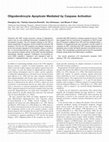
Figure 1. rhgas6 protects human and murine oligodendrocytes from insulin withdrawal
induced-apoptosis. Human fetal oligodendrocytes (a) or murine oligodendrocytes (b) were
grown in CD medium supplemented with insulin, PDGF, NT-3, with rhgas6 (a) or without rhgas6
(b) for 5 d on poly-L-lysine-coated plastic tissue culture wells in duplicate. On day 5 in culture,
cells were washed twice and incubated in CD medium containing PDGF, NT-3, and the absence
(Minus) or presence (Plus) of rhgas6 (400 ng/ml; 5.6 nM) with varying concentrations of insulin
(25–5000 ng/ml) for 48 h. Apoptotic oligodendrocytes were detected by immunofluorescence
staining with the oligodendrocyte-specific O4 antibody, followed by TUNEL staining. The number of O4 ⫹TUNEL ⫹ double-labeled oligodendrocytes was counted from duplicate wells in 20
random, 200⫻ microscopic fields and expressed as a percentage of the total O4 ⫹ cell numbers
per field. Representative data are shown as percentage oligodendrocyte survival (mean ⫾ SD)
with ⬃400 O4 ⫹ cells counted per treatment condition; three independent experiments were
performed with similar results. ANOVA results indicated a significant effect of rhgas6 treatment
(**p ⬍ 0.0001).
firmation of data were obtained by repeating the experiment three times
on independent material.
PI3 kinase/Akt inhibition using LY294002. Human oligodendrocyte
cultures grown for 5 d in CD medium in the presence of rhgas6 were
incubated with 100 ng/ml TNF␣ for 48 h in the presence or absence of the
PI3 kinase inhibitor LY294002 [2-(4-morpholinyl)-8-phenyl-1(4 H)benzopyran-4-one] (10 M; Promega, Madison, WI). After 48 h, oligodendrocytes were fixed and immunofluorescence staining for O4 was
performed followed by TUNEL staining. For each treatment, 20 random,
200⫻ magnification fields consisting of ⬃400 cells were examined in
duplicate wells of 24-well tissue culture plates, and the percentage oligodendrocyte survival was determined.
Data analysis. Data from each of the random microscopic fields, which
were examined for O4 ⫹TUNEL ⫹ oligodendrocytes, were analyzed as
independent observations in an ANOVA model using Prism 2.01 software (GraphPad Software, San Diego, CA) to evaluate the effect of treatment. To validly combine data from multiple experiments in the analysis,
the ANOVA model also included a factor to control for experiment to
experiment variability, in addition to the main effect for treatment.
Therefore, all p values corresponding to the effect of treatment are adjusted for between-experiment effects. Data are expressed as mean ⫾ SD.
Results
rhgas6 is a survival factor for human oligodendrocytes after
insulin withdrawal
The addition of rhgas6 to CD medium containing insulin at a
final concentration of 5000 ng/ml significantly enhanced the survival of cultured human oligodendrocytes and reduced the number of TUNEL ⫹ oligodendrocytes relative to CD medium lacking
rhgas6 (Shankar et al., 2003). Here, we tested whether rhgas6
would maintain oligodendrocyte survival in CD medium with
reduced insulin. Human (Fig. 1a) or mouse (Fig. 1b) oligoden-
Shankar et al. • Gas6 Protects Oligodendrocytes from Apoptosis
drocytes were washed twice in HBSS and incubated with CD
medium containing rhgas6 and varying concentrations of insulin
(25–5000 ng/ml). Oligodendrocyte survival was determined 48 h
later and compared with cultures similarly maintained in the
absence of rhgas6. As demonstrated in Figure 1, a and b, the
presence of rhgas6 in the medium (open circles) protected
against insulin withdrawal with ⬎60 –75% oligodendrocyte survival observed in the cultures with lower insulin concentrations
(25–100 ng/ml) for 48 h. When compared with the cultures
grown in CD medium in the absence of rhgas6 (filled circles),
rhgas6 significantly increased oligodendrocyte survival over the
entire range of insulin concentrations examined. In the absence
of rhgas6, oligodendrocyte survival was dramatically reduced in
cultures with ⬍100 ng/ml insulin in the medium. These data
demonstrate that rhgas6 exerts its antiapoptotic effects independent of insulin-activated IGF-1 receptor (IGF1R) and that rhgas6
significantly enhances oligodendrocyte survival over that induced by PDGF, NT-3, and low insulin concentrations.
Variation in the concentration of rhgas6 and TNF␣ alters
oligodendrocyte survival
We next examined whether rhgas6 would protect human oligodendrocytes against cell death induced by the cytotoxic cytokine
TNF␣. As illustrated in Figure 2, a and b, we observed numerous
O4 ⫹TUNEL ⫹ apoptotic oligodendrocytes 48 h after the addition
of 100 ng/ml TNF␣. In contrast, the coadministration of 100
ng/ml TNF␣ and 400 ng/ml rhgas6 significantly reduced the
numbers of apoptotic oligodendrocytes (Fig. 2c). As shown in
Figure 2d, addition of varying doses of TNF␣ (10 –100 ng/ml)
induced apoptosis in a concentration-dependent manner. In the
absence of rhgas6 and in the presence of 100 ng/ml TNF␣, 18.7 ⫾
4.4% oligodendrocytes survived, whereas the addition of 400
ng/ml rhgas6 led to a dramatic increase in cell survival to 64.3 ⫾
6.1%. Whereas the addition of varying concentrations of TNF␣
induced a dose-dependent decrease in oligodendrocyte survival,
the coadministration of 10 –100 ng/ml TNF␣ with rhgas6 significantly protected against oligodendrocyte apoptosis relative to
TNF␣ alone.
We examined whether varying concentrations of rhgas6 (25–
400 ng/ml; 0.35–5.6 nM) would protect human oligodendrocytes
from cell death 48 h after the administration of TNF␣ (100 ng/
ml). As shown in Figure 2e (open bar), in the absence of rhgas6,
40.7 ⫾ 6.0% of the oligodendrocytes survived. After administration of TNF␣ (black bar), there was an additional ⬃20% reduction in oligodendrocyte survival (20.2 ⫾ 5.3%) after 48 h.
Addition of 25– 400 ng/ml rhgas6 significantly increased oligodendrocyte survival at all doses in both the absence of TNF␣
(white bar with black dots) and the presence of TNF␣ (black bar
with white dots). Approximately 35% of the oligodendrocytes
survived TNF␣ cytotoxicity in rhgas6 concentrations of 25 and 50
ng/ml. When 100 – 400 ng/ml rhgas6 was added to the medium,
52–74% of the oligodendrocytes survived TNF␣ toxicity, demonstrating that rhgas6 protected human oligodendrocytes from
TNF␣-induced apoptosis in a concentration-dependent manner.
The specificity of the rhgas6 protective response from TNF␣induced apoptosis was examined by simultaneously administering TNF␣ and rhgas6 with the Axl decoy receptor Axl-Fc or a
control decoy receptor TrkA-Fc. The Axl-Fc binds rhgas6 and,
therefore, is a control to demonstrate that rhgas6 activity is a
direct response. Figure 2f demonstrates that the addition of
Axl-Fc eliminated the rhgas6-induced survival effect to oligodendrocytes in both the presence of rhgas6 alone and the presence of
rhgas6 and TNF␣. The percentage oligodendrocyte survival in
J. Neurosci., May 24, 2006 • 26(21):5638 –5648 • 5641
the presence of the Axl-Fc was equivalent to the percentage survival observed after TNF␣ treatment in the absence of rhgas6.
TrkA-Fc did not alter the rhags6 response, indicating that rhgas6
significantly protects oligodendrocytes from TNF␣ cytotoxicity.
Because TNF␣-mediated apoptosis in oligodendrocytes is a
result of TNF␣ binding to the TNFR1 (Hsu et al., 1996), we
confirmed that the receptor was expressed on O4 ⫹ oligodendrocytes that were grown for 6 d in CD medium. As shown in Figure
2g, the O4 ⫹ oligodendrocytes (red) express the TNFR1 (green).
TNFR1 membrane expression was primarily seen on the O4 ⫹ cell
surface with fainter staining on the processes; robust O4 immunoreactivity was observed on both the oligodendrocyte cell body
and the processes.
TNF␣-induced caspase activation is inhibited by the addition
of rhgas6
Figure 3a demonstrates that, 48 h after the addition of 100 ng/ml
TNF␣, there is an increase in active caspase-3 (green) detected
within the O4 ⫹ (red) oligodendrocyte cultures. Quantification of
the data showed that 68% of the oligodendrocytes were O4 ⫹/
cleaved caspase-3 ⫹ (Fig. 3c). In contrast, cultures treated with
rhgas6 and TNF␣ for the identical period of time had significantly fewer active caspase-3 ⫹ cells (Fig. 3b). Addition of rhgas6
and TNF␣ to the medium significantly decreased the numbers of
O4 ⫹ oligodendrocytes with active caspase-3 to 43% (Fig. 3c). To
determine whether the inhibition of caspases would enhance oligodendrocyte survival as effectively as rhgas6, day 5 human oligodendrocytes were washed twice and incubated in CD medium
minus rhgas6 (Fig. 3d, open bar) plus 20 M zVAD-fmk (stripes),
IETD-fmk (diagonal), or rhgas6 (dots) and examined 48 h later.
In the absence of TNF␣, rhgas6 was most effective at supporting
oligodendrocyte cell viability. In the presence of TNF␣, rhgas6
was as effective as the caspase inhibitors in protecting the cultures
from TNF␣ cytotoxicity. These results demonstrate that TNF␣
induces caspase-8 and downstream caspase activation in human
fetal oligodendrocytes and that rhgas6 and caspase inhibitors can
reduce TUNEL ⫹ nuclei resulting from TNF␣-induced cytotoxicity. These experiments not only show that caspase inhibitors
can block TNF␣-induced oligodendrocyte cytotoxicity but also
stress the ability of rhgas6 to inhibit TNF␣-induced caspase activation as effectively as the chemical caspase inhibitors.
Rhgas6 protects human oligodendrocytes from cell death via
the PI3 kinase/Akt pathway
To determine whether rhgas6 signaling protects against TNF␣induced toxicity via the PI3 kinase/Akt pathway, we examined
whether the addition of TNF␣ alone or rhgas6 and TNF␣ would
activate and hence phosphorylate the prosurvival kinase Akt. Human oligodendrocytes were placed overnight in DMEM, washed,
treated with TNF␣ plus and minus rhgas6 for 15 min, and immediately fixed with 4% paraformaldehyde. Double-label immunofluorescence was performed with O4 and a phosphoAkt Ser473
antibody (Fig. 4a), and the relative phosphoAkt Ser473/O4 fluorescence intensity was compared with DMEM-only-treated cells
arbitrarily set as 1.0 (Fig. 4b). TNF␣ alone did not significantly
activate phosphoAkt. However, the presence of rhgas6 in the
TNF␣-treated cultures activated Akt relative to TNF␣ alone (Fig.
4b). When the PI3 kinase inhibitor LY294002 was simultaneously
added to the medium with TNF␣ plus rhgas6, phosphoAkt immunoreactivity was reduced similar to the TNF␣-only-treated
cultures (Fig. 4b). To address whether rhgas6-induced Akt activation is enhanced in the presence of other survival-promoting
factors, oligodendrocyte cultures were treated with insulin,
5642 • J. Neurosci., May 24, 2006 • 26(21):5638 –5648
Shankar et al. • Gas6 Protects Oligodendrocytes from Apoptosis
IGF-1, PDGF␣, or NT-3 in the presence
and absence of rhgas6. rhgas6-induced Akt
activation was neither additive nor synergistic in the presence of any of the survivalpromoting factors (Fig. 4c).
To determine whether Akt activation
was transient or sustained after rhgas6
stimulation, oligodendrocytes were placed
overnight in DMEM only without any trophic support, washed with HBSS, and
treated with DMEM plus rhgas6 for 5 min,
15 min, 30 min, 1 h, and 24 h time points.
Relative phosphoAkt Ser473/O4 fluorescence intensities were obtained and compared with DMEM-only-treated cells. As
shown in Figure 4d, a significant increase
in relative phosphoAkt Ser473/O4 fluorescence intensity was observed 5 min after
rhgas6 treatment; the maximal increase in
phosphoAkt Ser473/O4 was observed at 15
and 30 min after rhgas6 treatment. By 1 h,
Akt activation was reduced, but elevated
levels of phosphoAkt persisted at 24 h.
Thus, rhgas6 treatment of oligodendrocytes results in sustained Akt activation.
We examined whether inhibiting the
rhgas6-induced Akt activation with
LY294002 would influence oligodendrocyte survival 48 h after treatment with
TNF␣. Figure 4e demonstrates that oligodendrocyte survival was compromised in
the rhgas6 plus TNF␣-treated cultures after the addition of LY294002. Thus, the
rhgas6-dependent increase in oligodendrocyte survival is dependent on the PI3
kinase/Akt pathway, and LY294002 eliminated the rhgas6-induced survival effect.
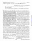
Figure 2. rhgas6 prevents dose-dependent TNF␣-induced oligodendrocyte apoptosis. Human fetal oligodendrocytes grown
in CD medium plus rhgas6 for 5 d were washed twice and incubated in CD medium containing 5000 ng/ml insulin in the absence
of rhgas6 (a, b) or the presence of rhgas6 (c) (400 ng/ml) and 100 ng/ml human recombinant TNF␣ for 48 h. Cells were fixed and
immunostained with O4 (red), followed by TUNEL (green). A marked increase in O4 ⫹TUNEL ⫹ oligodendrocytes was observed in
cultures incubated with TNF␣ in the absence of rhgas6 (a). b shows a representative O4 ⫹TUNEL ⫹ double-labeled oligodendrocyte at high magnification. Scale bars: a, c, 40 m; b, 10 m. d, TNF␣ (10 –100 ng/ml) was administered in the absence or
presence of rhgas6. The number of O4 ⫹TUNEL ⫹ double-labeled oligodendrocytes was counted from duplicate wells in 20
random, 200⫻ microscopic fields containing ⬃350 – 400 O4 ⫹ oligodendrocytes in total. Data are shown as percentage oligodendrocyte survival (mean ⫾ SD) of one of three independent experiments with similar results. ANOVA results indicated a
significant effect on oligodendrocyte survival of TNF␣ alone or rhgas6 treatment alone or when rhgas6 was administered with
TNF␣ (**p ⬍ 0.0001) when compared with minus gas6 or TNF␣ alone. e, Human oligodendrocytes were grown in CD medium
plus 400 ng/ml rhgas6 for 5 d and washed with HBSS. Cultures were incubated for 48 h in CD medium containing 5000 ng/ml
insulin with varying concentrations of rhgas6 (25– 400 ng/ml; 0.36 –5.6 nM) in the presence and absence of 100 ng/ml TNF␣. Cells
were fixed and stained with the O4 antibody, followed by TUNEL labeling. The numbers of O4 ⫹/TUNEL ⫹ double-labeled oligodendrocytes were counted from 20 random, 200⫻ microscopic fields from duplicate wells containing ⬃500 O4 ⫹ oligodendrocytes in total. Data are shown as percentage oligodendrocyte survival (mean ⫾ SD) obtained from one of two independent
experiments with a second experiment yielding similar results. Significance relative to untreated or TNF␣ alone controls for all
rhgas6 and rhgas6 plus TNF␣ treatments was **p ⬍ 0.0001 (ANOVA). f, Axl-Fc but not the TrkA-Fc decoy receptor blocks the
rhgas6 protective effect. Human oligodendrocytes were grown as above and treated for 48 h with rhgas6 (400 ng/ml), Axl-Fc (10
Rhgas6 protection against TNF␣induced apoptosis is mediated through
the Axl receptor
Immunoblot analysis of various developmental time points determined that Axl
and gas6 are expressed throughout rodent
brain development. Both Axl and gas6
were expressed at high levels in whole
mouse brain protein homogenates at post4
g/ml), or TrkA-Fc (10 g/ml) in the absence and presence
of TNF␣ (100 ng/ml). Data were obtained from 20 random,
200⫻ microscopic fields from duplicate wells consisting of
⬃500 O4 ⫹ oligodendrocytes in total. Data are expressed as
percentage oligodendrocyte survival (mean ⫾ SD) obtained
from a single experiment (**p ⬍ 0.0001, unpaired Student’s
t test; ns, not significant; p ⱖ 0.05). g, Expression of TNFR1 in
primary cultures of human oligodendrocytes. Top, A representative low-magnification field of O4 ⫹ (red) oligodendrocytes expressing TNFR1 (green). Nuclei were labeled with
4⬘,6⬘-diamidino-2-phenylindole (DAPI) (blue). Scale bar, 10
m. Bottom, O4 ⫹ oligodendrocyte with membrane expression of TNFR1 primarily at the cell surface with fainter staining in processes. Scale bar, 8 m.
Shankar et al. • Gas6 Protects Oligodendrocytes from Apoptosis
J. Neurosci., May 24, 2006 • 26(21):5638 –5648 • 5643
100 ng/ml TNF␣ plus and minus 400
ng/ml rhgas6 and a range of insulin concentrations (25–5000 ng/ml). After 48 h,
the cultures were fixed and stained with
O4 and TUNEL, and the percentage oligodendrocyte survival was quantified. When
compared with TNF␣ alone, rhgas6 significantly increased oligodendrocyte survival
at both high and low insulin concentrations (Fig. 5e). This demonstrated that rhgas6 could protect murine oligodendrocytes from TNF␣ toxicity at reduced
insulin concentrations (25–100 ng/ml).
Because gas6 signaling to Axl protects
oligodendrocytes from TNF␣-induced
cell death, we examined whether rhgas6
would activate Akt in the Axl⫺/⫺ mice and
whether rhgas6-treated oligodendrocytes
from Akt1⫺/⫺ mice would be protected
from TNF␣-induced cell death. Murine
oligodendrocytes were placed overnight in
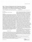
Figure 3. rhgas6 is as effective as the pan-caspase inhibitor zVAD-fmk and the caspase-8 inhibitor IETD-fmk at protecting
DMEM, washed, and simultaneously
human oligodendrocytes against TNF␣-induced apoptosis. a, TNF␣ treatment of human oligodendrocytes induces the activation
of caspase-3. Human oligodendrocytes were grown in CD medium plus rhgas6 for 5 d, washed with HBSS, and incubated for 48 h treated for 15 min in growth factor-free
in CD medium containing 5000 ng/ml insulin and TNF␣ (100 ng/ml) in the absence (a) and presence (b) of rhgas6 (400 ng/ml). DMEM with TNF␣ plus or minus rhgas6.
Cells were fixed and immunostained with O4 antibody (red), followed by cleaved caspase-3 antibody (green). c, Quantitation of Cultures were fixed, double-label immuO4 ⫹/cleaved caspase-3 ⫹ oligodendrocytes after TNF␣ alone or with rhgas6 treatment. O4 ⫹/cleaved caspase-3 ⫹ double- nofluorescence was performed with O4
labeled oligodendrocytes were counted from 20 random, 200⫻ microscopic fields from duplicate wells containing ⬃350 O4 ⫹ and the phosphoAkt serine 473 antibodies,
oligodendrocytes in total. Data are shown as percentage O4 ⫹/cleaved caspase-3 ⫹ oligodendrocytes (mean ⫾ SD) obtained and the relative phosphoAkt and O4 fluofrom one of two independent experiments with a second experiment yielding similar results (**p ⬍ 0.0001, ANOVA). d, Human rescence intensities were quantified. Figoligodendrocyte cultures grown in rhgas6 for 5 d were washed with HBSS and incubated for 48 h in TNF␣ (100 ng/ml) in the ure 6a shows that, in the WT cultures, rhpresence or absence of rhgas6 (400 ng/ml), with and without the cell-permeable pan-caspase inhibitor zVAD-fmk or the
gas6 alone (white stippled bars) or rhgas6
caspase-8 inhibitor IETD-fmk (20 M). Apoptotic oligodendrocytes were detected by immunofluorescence staining with the O4
plus TNF␣ (black stippled bars) induced
⫹
⫹
antibody, followed by TUNEL labeling. O4 /TUNEL double-labeled oligodendrocytes were counted from 20 random, 200⫻
⫹
microscopic fields from duplicate wells containing ⬃400 O4 oligodendrocytes in total. Data are shown as percentage oligo- an approximate threefold increase in
phosphoAkt Ser473 relative to untreated
dendrocyte survival (mean ⫾ SD) obtained from one of two independent experiments (*p ⬍ 0.001, **p ⬍ 0.0001, ANOVA).
cultures or cultures treated with TNF␣
alone. However, in the Axl⫺/⫺ cultures, no
natal days 10 –13 (Fig. 5a), and Axl remained high through day
increase in phosphoAkt was observed. To determine whether the
30. Although gas6 and Axl levels decreased by day 60, protein
Axl⫺/⫺ oligodendrocytes were capable of Akt phosphorylation in
levels were detectable in the adult mouse brain at day 160.
response to other survival-promoting factors, we treated Axl⫺/⫺
To determine whether the loss of the Axl or the Rse receptor
cultures for 15 min in growth factor-free DMEM or medium
would compromise murine oligodendrocyte survival,
containing rhgas6, IGF-1, NT-3, or PDGF. Whereas rhgas6
O4 ⫹CNP ⫹ oligodendrocyte cultures (Fig. 5b) prepared from
lacked the ability to induce Akt phosphorylation in Axl⫺/⫺ oligodendrocytes (Fig. 6b), a robust Akt activation was observed in
postnatal day 1 mouse brains were challenged with TNF␣ and
rhgas6. In the absence of rhgas6, the percentage oligodendrocyte
cultures treated with IGF-1 (4.3-fold), NT-3 (4.2-fold), and
survival in the WT, Axl⫺/⫺, and Rse⫺/⫺ cultures was ⬎74% (Fig.
PDGF (3.6-fold).
Akt1 is the predominant Akt isoform in oligodendrocytes.
5d, white bars). When rhgas6 was administered to each of the
Therefore, we treated oligodendrocyte cultures prepared from
cultures (stippled bars), there was a statistically significant inAkt1⫺/⫺ mouse brains with TNF␣ plus and minus rhgas6 and
crease in the percentage oligodendrocyte survival in the WT and
⫺/⫺
⫺/⫺
Rse
examined whether rhgas6 protected against TNF␣-induced cell
mice but not in the Axl
mice, suggesting that, in the
death. Treatments were performed with 100 ng/ml insulin in the
presence of exogenous rhgas6, oligodendrocyte survival can be
medium to determine whether the effect of rhgas6 on Akt1⫺/⫺
enhanced only when the Axl receptor is present. Furthermore,
oligodendrocyte survival was independent of insulin-activated
when 100 ng/ml TNF␣ was administered to the cultures in the
absence of rhgas6 (black bars), there was a dramatic decrease in
IGF1R. As shown in Figure 6c, treatment of the Akt1⫺/⫺ cultures
oligodendrocyte viability that could be recovered when rhgas6 was
with rhgas6 (white stippled bars) or rhgas6 plus TNF␣ (black
administered to the WT and Rse⫺/⫺ cultures but not the Axl⫺/⫺
stippled bars) did not alter oligodendrocyte survival relative to
cultures (Fig. 5c,d, black stippled bars). These results suggest that the
the untreated cultures or cultures treated with only TNF␣. These
presence of the Axl receptor is crucial for rhgas6-induced oligodenresults demonstrate that rhgas6/Axl signaling protects oligodendrocyte survival after TNF␣ administration.
drocytes from TNF␣-induced apoptosis by activating the PI3 kiTo address whether rhgas6 protected murine oligodendronase/Akt1 signaling pathway.
cytes from TNF␣-induced apoptosis independent of the survival
Discussion
induced by high insulin concentrations activating IGF1R, oligodendrocyte cultures were washed thrice to remove trophic facOur studies demonstrate that rhgas6 protects oligodendrocytes
tors, and the medium was replaced with CD medium containing
against growth factor withdrawal and TNF␣-induced cytotoxic-
5644 • J. Neurosci., May 24, 2006 • 26(21):5638 –5648
Shankar et al. • Gas6 Protects Oligodendrocytes from Apoptosis
ity by ligand activation of the receptor tyrosine kinase Axl. Human oligodendrocytes are more dependent on rhgas6 for
their survival than murine oligodendrocytes, but, when oligodendrocytes are
challenged with TNF␣, rhgas6 is highly effective in maintaining survival in both species. The inability of rhgas6 to protect
Axl⫺/⫺ oligodendrocytes from TNF␣induced toxicity further supports our conclusion that Axl is the predominant receptor through which rhgas6 signals in
oligodendrocytes. Axl receptor activation
was shown to be more responsive to low
doses of rhgas6 (Nagata et al., 1996), and
our observation that as little as 0.35 nM
rhgas6 protected oligodendrocytes from
TNF␣-induced cytotoxicity supports that
study.
In a previous study, we showed that two
independent inhibitors of PI3 kinase
blocked the protective effect of rhgas6 and
speculated that rhgas6 was signaling to Akt
(Shankar et al., 2003). In this study, we
demonstrate that rhgas6 signaling through
the Axl receptor results in increased Akt
phosphorylation at serine 473 in both human and murine oligodendrocytes and
that the Akt activation was maintained
when oligodendrocyte cultures were cotreated with TNF␣ and rhgas6. In addition, the rhgas6 protective effect was abrogated in oligodendrocytes from both
Axl⫺/⫺ and Akt1⫺/⫺ mice, further demonstrating the importance of the Axl receptor
and Akt1 in protection against TNF␣. Several growth factors, including PDGF, high
doses of insulin, and NT-3, increase cell
survival by activating Akt. Doses of insulin
above 250 ng/ml activate the IGF1R and
lead to sustained activation of Akt (Ness
and Wood, 2002). In our studies, the absence of insulin, or insulin concentrations
4
Figure 4. The rhgas6 protective effect against TNF␣ is mediated by Akt activation and is inhibited by treatment with the PI3
kinase inhibitor LY294002. All human oligodendrocytes were grown for 5 d in CD medium plus rhgas6. a, Human oligodendrocytes
were washed twice with HBSS, incubated in DMEM (without insulin, PDGF, or NT-3) and 100 ng/ml TNF␣ in the absence or
presence of rhgas6 (400 ng/ml) for 15 min, and fixed with 4% paraformaldehyde. Oligodendrocytes were double-labeled sequentially with the O4 (red) and phosphoAkt Ser473 (green) antibodies. Nuclei were labeled with DAPI (blue). Confocal microscopy was
performed, and images were collected in Z-series. Scale bar, 8 m. Low-magnification images (red/green merge) were obtained
with a 20⫻ objective. Scale bar, 10 m. b, phosphoAkt Ser473 and O4 immunofluorescence measurements were obtained from
80 cells in 15 random, 200⫻ magnification microscopic fields by NIH ImageJ software. Data are expressed as relative fluorescence
intensity (mean ⫾ SD) from one of three independent experiments with similar results (**p ⬍ 0.0001, ANOVA). Data are shown
relative to minus gas6 (set as 1.0); TNF␣ alone did not significantly (ns) activate phosphoAkt at 15 min. c, Human oligodendrocytes grown in rhgas6 for 5 d were placed overnight in DMEM, washed twice with HBSS, treated with minus or plus gas6
supplemented with 10 ng/ml IGF-1, 5 ng/ml NT-3, or 10 ng/ml PDGF for 15 min, and then stopped by the addition of 4%
paraformaldehyde. PhosphoAkt Ser473 and O4 immunofluorescence measurements were obtained from 32 cells in 10 random,
200⫻ magnification microscopic fields by NIH ImageJ software, and data are expressed as relative fluorescence intensity
(mean ⫾ SD) from one of two independent experiments
with similar results. d, Human oligodendrocytes placed overnight in DMEM, washed twice with HBSS, and treated with
minus or plus gas6 for 5 min, 15 min, 30 min, 1 h, and 24 h.
PhosphoAkt Ser473 and O4 immunofluorescence measurements were obtained from 32 cells in 10 random, 200⫻
magnification microscopic fields by NIH ImageJ software, and
data are expressed as relative fluorescence intensity
(mean ⫾ SD) from one of two independent experiments
with similar results (**p ⬍ 0.0001, ANOVA). e, Human oligodendrocytes grown as above were washed with HBSS and
incubated for 48 h in TNF␣ (100 ng/ml) in the presence or
absence of rhgas6 (400 ng/ml) with or without the PI3 kinase/Akt inhibitor LY294002 (10 M). Data are shown as percentage oligodendrocyte survival (mean ⫾ SD with ⬃400
O4 ⫹ cells counted from 20 random, 200⫻ microscopic fields
per treatment condition from duplicate wells) obtained from
one of two independent experiments (**p ⬍ 0.0001,
ANOVA).
Shankar et al. • Gas6 Protects Oligodendrocytes from Apoptosis
J. Neurosci., May 24, 2006 • 26(21):5638 –5648 • 5645
below 100 ng/ml, dramatically decreased
oligodendrocyte survival in both human
and murine oligodendrocyte cultures. In
contrast, oligodendrocytes maintained in
rhgas6 with low concentrations (25–100
ng/ml) of insulin in the CD medium significantly reduced the numbers of
TUNEL ⫹ apoptotic cells. This demonstrates that rhgas6 potently increases oligodendrocyte survival independent of the
insulin/IGF1R signaling.
Oligodendrocytes are sensitive to
TNF␣-mediated cytotoxicity in vitro and
in vivo. In vitro administration of TNF␣
induced apoptotic cell death of primary
oligodendrocytes (Selmaj and Raine, 1988;
D’Souza et al., 1996; Scurlock and Dawson, 1999; Hisahara et al., 2000; Takano et
al., 2000). In vivo, elevated levels of TNF␣
are found in multiple sclerosis plaques and
in experimental autoimmune encephalomyelitis lesions (Powell et al., 1990;
Genain et al., 1995; Brosnan and Raine,
1996). Transgenic mice expressing TNF␣
in the CNS showed increased oligodendrocyte apoptosis and primary demyelination (Akassoglou et al., 1997). TNF␣mediated apoptosis occurs as a result of
TNF␣ binding to the TNFR1 receptor,
which recruits FAS-associated death domain FADD, receptor-interacting protein
RIP1, and TNF receptor-associated factor
TRAF2/5 via the adaptor protein TNFRassociated death domain-containing protein TRADD (Hsu et al., 1996). FADD recruits and activates caspase-8, resulting in
the cleavage and activation of caspase-3
and caspase-7 and subsequent apoptosis.
Our studies indicate that the addition of
TNF␣ to human and murine oligodendro4
Figure 5. rhgas6 protects oligodendrocytes from WT and Rse⫺/⫺ but not oligodendrocytes from Axl⫺/⫺ mice against TNF␣induced apoptotic cell death. a, Developmental expression of Axl and Gas6 in mouse brain. A total of 50 g of whole mouse brain
protein homogenate was loaded per lane. Blots were incubated with affinity-purified antibodies Axl polyclonal antibodies (1:500;
Amgen), Gas6 polyclonal antibodies (1:1000; Amgen), or -tubulin (1:1000; Sigma). Axl migrates at 140 kDa, Gas6 at 86 kDa, and
-tubulin at 55 kDa. Visualization is by ECL. b, Mouse cortical oligodendrocytes grown for 7 d in culture were triple labeled with
O4 (red), CNP (green), or GFAP (green) and DAPI (blue). Middle panel shows membrane extensions typical of mouse oligodendrocytes in vitro. Merged images show healthy oligodendrocytes with minimal astrocyte contamination. Scale bar, 16 m. c, d,
Rhgas6 protects oligodendrocytes from Axl⫹/⫹ and Rse⫺/⫺ but not oligodendrocytes from Axl⫺/⫺ mice against TNF␣-induced
apoptosis. Oligodendrocytes isolated from postnatal day 1
WT Axl⫺/⫺, and Rse⫺/⫺ mice were grown for 5 d, washed
with HBSS, and incubated for 96 h in DMEM (with 5000 ng/ml
insulin) plus TNF␣ (100 ng/ml) in the presence or absence of
rhgas6 (400 ng/ml). O4 and TUNEL labeling was performed
sequentially, and low-magnification images (200⫻) were
obtained on an Olympus Optical 1X70 microscope with a
20⫻ objective (c). Scale bar, 10 m. Results in d are based
on duplicate wells of 20 random, 200⫻ microscopic fields
containing ⬃600 oligodendrocytes in total. Data are shown
as percentage oligodendrocyte survival (mean ⫾ SD) of one
of three independent experiments with similar results
(**p ⬍ 0.0001, ANOVA; ns, not significant). e, Rhgas6 protects murine cortical oligodendrocytes from TNF␣-induced
cell death independent of insulin-activated IGFR. Oligodendrocytes were washed and placed in CD medium in the absence (minus) or presence (plus) of rhgas6 (400 ng/ml; 5.6
nM) with varying concentrations of insulin (25–5000 ng/ml)
and in the presence and absence of 100 ng/ml TNF␣ for 48 h.
Data are shown as percentage oligodendrocyte survival
(mean ⫾ SD) of one of three independent experiments with
similar results (**p ⬍ 0.0001, ANOVA).
Shankar et al. • Gas6 Protects Oligodendrocytes from Apoptosis
5646 • J. Neurosci., May 24, 2006 • 26(21):5638 –5648
cytes induces apoptosis. In our cultures, TNF␣ activated
caspase-8 and caspase-3 and increased the number of active
cleaved caspase-3 ⫹TUNEL ⫹ oligodendrocytes. The effect was
blocked with either the caspase-8 inhibitor IETD-fmk or the pancaspase inhibitor zVAD-fmk. The presence of rhgas6 in TNF␣treated oligodendrocyte cultures significantly protected against
caspase activation and cell death. There was an additional reduction in TUNEL ⫹ oligodendrocytes when the caspase inhibitors
were added to the rhgas6, TNF␣-containing medium. However,
rhgas6 was more effective at maintaining oligodendrocyte survival than the addition of the caspase inhibitors alone. This suggests that rhgas6 does not act to directly inhibit caspase activation
but works upstream to activate signaling pathways such as PI3K/
Akt. This was supported by our observation that increased phosphoAkt Ser473 was observed in both human and murine oligodendrocytes 15 min after the addition of TNF␣ and rhgas6 but
not TNF␣ alone. Furthermore, the addition of the PI3 kinase
inhibitor LY294002 blocked the protective effect of rhgas6 and
the observed increase in phosphoAkt. Our observation of Akt
activation in the rhgas6- and TNF␣-treated human and murine
oligodendrocyte cultures is supported by an in vitro study that
demonstrated that, after gas6 stimulation, the Axl receptor undergoes autophosphorylation, resulting in tyrosine phosphorylation at three sites (Braunger et al., 1997). Two of the tyrosines
phosphorylated form part of a consensus sequence pYXXM that
may recruit PI3 kinase by direct binding of the Src homology 2
domain of the p85 subunit of PI3 kinase to Axl.
Axl, Rse, and Mer single, double, and triple knock-out mice
have been generated and are viable but have varying degrees of
impairments in cell viability and homeostasis (Lu et al., 1999;
D’Cruz et al., 2000). The triple knock-out mice have neurologic
abnormalities, major organ defects, physiological deficits, and
autoimmune defects. Altered histology, increased apoptosis, and
cellular degeneration are apparent in the CNS, and male mice
produce no sperm (Lu et al., 1999; Lu and Lemke, 2001). To gain
an understanding of the contribution of Axl or Rse receptors to
4
Figure 6. rhgas6 activates Akt in WT but not in Axl⫺/⫺ murine oligodendrocytes; the survival effect requires activation of Akt1. a, Murine oligodendrocyte cultures (day 7) were placed
overnight in DMEM, washed with HBSS, and incubated in 100 ng/ml TNF␣ in DMEM (without
insulin, PDGF, or NT-3), in the absence or presence of rhgas6 (400 ng/ml) for 15 min. Oligodendrocytes were triple labeled with the O4, phosphoAkt Ser473, and DAPI. phosphoAkt Ser473
and O4 immunofluorescence measurements were obtained from 35 cells in 10 random, 200⫻
magnification microscopic fields by NIH ImageJ software. Data are expressed as relative fluorescence intensity (mean ⫾ SD) from one of three independent experiments with similar results. Data are shown relative to minus gas6 (set as 1.0). rhgas6 treatment significantly increased phosphoAkt Ser473 expression in TNF␣-treated wild-type Axl⫹/⫹ oligodendrocytes
(**p ⬍ 0.0001, ANOVA; ns, not significant). b, IGF-1, NT-3, and PDGF but not rhgas6 induce Akt
phosphorylation in Axl⫺/⫺ murine oligodendrocytes. Day 5 murine Axl⫺/⫺ oligodendrocytes
were placed overnight in DMEM, washed twice with HBSS, and treated with DMEM only or
DMEM with either 10 ng/ml IGF-1, 5 ng/ml NT-3, 10 ng/ml PDGF, or 400 ng/ml rhgas6 for 15
min. phosphoAkt Ser473 and O4 immunofluorescence measurements were obtained from 35
cells in 10 random, 200⫻ magnification microscopic fields by NIH ImageJ software. Data are
expressed as relative fluorescence intensity (mean ⫾ SD) from one of two independent experiments with similar results (**p ⬍ 0.0001, ANOVA). c, rhgas6 does not protect against TNF␣induced cell death in Akt1⫺/⫺ oligodendrocytes. Oligodendrocytes isolated from postnatal day
1 Akt1⫺/⫺ mouse brains were grown for 5 d, washed with HBSS, and incubated for 48 h in CD
medium containing 100 ng/ml insulin plus TNF␣ (100 ng/ml) in the presence or absence of
rhgas6 (400 ng/ml). O4 and TUNEL labeling was performed sequentially, and lowmagnification images (200⫻) obtained on an Olympus Optical 1X70 microscope with a 20⫻
objective. Results are based on duplicate wells of 20 random, 200⫻ microscopic fields containing ⬃300 oligodendrocytes in total. Data are shown as percentage oligodendrocyte survival
(mean ⫾ SD) of one of two independent experiments with similar results (ns, not significant).
Shankar et al. • Gas6 Protects Oligodendrocytes from Apoptosis
oligodendrocyte survival, we generated oligodendrocyte cultures
from Axl and Rse single knock-outs. Oligodendrocyte cultures
from the Axl⫺/⫺ or Rse⫺/⫺ mouse brains appeared morphologically similar to the WT and exhibited normal survival and differentiation in culture medium supplemented with insulin, PDGF,
and NT-3. However, when oligodendrocytes were challenged
with TNF␣, the absence of signaling through the Axl receptor
prevented rhgas6 from exerting its survival effect, supporting an
important role for Axl in cell survival.
The data obtained from the TNF␣-treated differentiated oligodendrocytes from the Axl null mice strongly support our hypothesis that Axl is the primary receptor activated in response to
low doses of rhgas6. Although we cannot rule out a role for the
Rse or Mer receptors, we did not observe a significant change in
cell survival in the Rse null oligodendrocytes expressing functional Axl and Mer receptors. Furthermore, examination of total
brain homogenates from the Axl⫹/⫹ and Axl⫺/⫺ mice did not
indicate the upregulation of either the Rse or Mer receptor in the
null animals (data not shown).
Western blot analysis of mouse brain homogenates prepared
at several development ages determined that gas6 expression paralleled Axl expression, peaking early in development and leveling
off in the mature animal. Several studies have demonstrated that
gas6 is expressed and secreted by both neurons and endothelial
cells and that gas6 is abundantly present in the CNS (Crosier and
Crosier, 1997; Prieto et al., 2000). The expression of gas6 and Axl
in adult animals suggests that the gas6/Axl signaling pathway may
be able to protect cells from injury. Furthermore, the downstream Akt signaling pathway can be activated to protect against
stress induced by insufficient trophic support or the release of
cytotoxic cytokines. In summary, our results demonstrate that
rhgas6 potently inhibits TNF␣-induced oligodendrocyte apoptosis and identifies the gas6/Axl/PI3 kinase/Akt1 signaling pathway
as an important mediator of oligodendrocyte survival after cellular stress and cytokine challenge.
References
Akassoglou K, Probert L, Kontogeorgos G, Kollias G (1997) Astrocytespecific but not neuron- specific transmembrane TNF triggers inflammation and degeneration in the central nervous system of transgenic mice.
J Immunol 158:438 – 445.
Armstrong RC (1998) Isolation and characterization of immature oligodendrocyte lineage cells. Methods 16:282–292.
Bansal R, Warrington AE, Gard AL, Ranscht B, Pfeiffer SE (1989) Multiple
and novel specificities of monoclonal antibodies O1, O4, and R-mAb used
in the analysis of oligodendrocyte development. J Neurosci Res
24:548 –557.
Bitsch A, Kuhlmann T, Da Costa C, Bunkowski S, Polak T, Bruck W (2000)
Tumor necrosis factor alpha mRNA expression in early multiple sclerosis
lesions: correlation with demyelinating activity and oligodendrocyte pathology. Glia 29:366 –375.
Braunger J, Schleithoff L, Schulz AS, Kessler H, Lammers R, Ullrich A, Bartram CR, Janssen JW (1997) Intracellular signaling of the Ufo/Axl receptor tyrosine kinase is mediated mainly by a multi-substrate dockingsite. Oncogene 14:2619 –2631.
Brosnan CF, Raine CS (1996) Mechanisms of immune injury in multiple
sclerosis. Brain Pathol 6:243–257.
Crosier KE, Crosier PS (1997) New insights into the control of cell growth;
the role of the Axl family. Pathology 29:131–135.
Datta SR, Brunet A, Greenberg ME (1999) Cellular survival: a play in three
Akts. Genes Dev 13:2905–2927.
D’Cruz PM, Yasumura D, Weir J, Matthes MT, Abderrahim H, LaVail MM,
Vollrath D (2000) Mutations of the receptor tyrosine kinase gene Mertk
in the retinal dystrophic RCS rat. Hum Mol Genet 9:645– 651.
D’Souza SD, Alinauskas KA, Antel JP (1996) Ciliary neurotrophic factor
J. Neurosci., May 24, 2006 • 26(21):5638 –5648 • 5647
selectively protects human oligodendrocytes from tumor necrosis factormediated injury. J Neurosci Res 43:289 –298.
Franke TF, Kaplan DR, Cantley LC (1997) PI3K: downstream AKTion
blocks apoptosis. Cell 88:435– 437.
Gard AL, Pfeiffer SE (1989) Oligodendrocyte progenitors isolated directly
from developing telencephalon at a specific phenotypic stage: myelinogenic potential in a defined environment. Development 106:119 –132.
Genain CP, Roberts T, Davis RL, Nguyen MH, Uccelli A, Faulds D, Li Y,
Hedgpeth J, Hauser SL (1995) Prevention of autoimmune demyelination in non-human primates by a cAMP-specific phosphodiesterase inhibitor. Proc Natl Acad Sci USA 92:3601–3605.
Hasanbasic I, Cuerquis J, Varnum B, Blostein MD (2004) Intracellular signaling pathways involved in Gas6-Axl-mediated survival of endothelial
cells. Am J Physiol Heart Circ Physiol 287:H1207–H1213.
Hisahara S, Araki T, Sugiyama F, Yagami K, Suzuki M, Abe K, Yamamura K,
Miyazaki J, Momoi T, Saruta T, Bernard CCA, Okano H, Miura M
(2000) Targeted expression of baculovirus p35 caspase inhibitor in oligodendrocytes protects mice against autoimmune-mediated demyelination. EMBO J 19:341–348.
Hsu H, Shu HB, Pan MG, Goeddel D (1996) TRADD-TRAF2 and TRADDFADD interactions define two distinct TNF receptor 1 signal transduction
pathways. Cell 84:299 –308.
Jones SM, Klinghoffer R, Prestwich GD, Toker A, Kazlauskas (1999) A
PDGF induces an early and a late wave of PI 3-kinase activity, and only the
late wave is required for progression through G1. Curr Biol 9:512–521.
Kazlauskas A (1994) Receptor tyrosine kinases and their targets. Curr Opin
Genet Dev 4:5–14.
Knapp PE, Bartlett WP, Skoff RP (1987) Cultured oligodendrocytes mimic
in vivo phenotypic characteristics: cell shape, expression of myelinspecific antigens, and membrane production. Dev Biol 120:356 –365.
Li R, Chen J, Hammonds G, Phillips H, Armanini M, Wood P, Bunge R,
Godowski PJ, Sliwkowski MX, Mather JP (1996) Identification of
Gas6 as a growth factor for human Schwann cells. J Neurosci
16:2012–2019.
Ling L, Templeton D, Kung HJ (1996) Identification of the major autophosphorylation sites of Nyk/Mer, an NCAM-related receptor tyrosine kinase.
J Biol Chem 271:18355–18362.
Lu Q, Lemke G (2001) Homeostatic regulation of the immune system by
receptor tyrosine kinases of the Tyro 3 family. Science 293:306 –311.
Lu Q, Gore M, Zhang Q, Camenisch T, Boast S, Casagranda F, Lai C, Skinner
MK, Klein R, Matsushima GK, Earp HS, Goff SP, Lemke G (1999)
Tyro-3 family receptors are essential regulators of mammalian spermatogenesis. Nature 398:723–728.
Nagata K, Ohashi K, Nakano T, Arita H, Zong C, Hanafusa H, Mizuno K
(1996) Identification of the product of growth arrest-specific gene 6 as a
common ligand for Axl, Sky, and Mer receptor tyrosine kinases. J Biol
Chem 271:30022–30027.
Ness JK, Wood TL (2002) Insulin-like growth factor I, but not
neurotrophin-3, sustains Akt activation and provides long-term protection of immature oligodendrocytes from glutamate-mediated apoptosis.
Mol Cell Neurosci 20:476 – 488.
Powell MB, Mitchell D, Lederman J, Buckmeier J, Zamvil SS, Graham M,
Ruddle NH, Steinman L (1990) Lymphotoxin and tumor necrosis
factor-alpha production by myelin basic protein-specific T cell clones
correlates with encephalitogenicity. Int Immunol 2:539 –544.
Prieto A, Weber JL, Lai C (2000) Expression of the receptor protein tyrosine
kinase Tyro3, Axl and Mer in the developing rat central nervous system.
J Comp Neurol 425:295–314.
Raible DW, McMorris FA (1989) Cyclic AMP regulates the rate of differentiation of oligodendrocytes without changing the lineage commitment of
their progenitors. Dev Biol 133:437– 446.
Rezaie P, Dean A (2002) Periventricular leukomalacia, inflammation and
white matter lesions within the developing nervous system. Neuropathology 22:106 –132.
Scurlock B, Dawson G (1999) Differential responses of oligodendrocytes to
TNF and other pro-apoptotic agents: role of ceramide in apoptosis. J Neurosci Res 55:514 –522.
Selmaj KW, Raine CS (1988) Tumor necrosis factor mediates myelin and
oligodendrocyte damage in vitro. Ann Neurol 23:339 –346.
Shankar SL, O’Guin K, Cammer M, McMorris FA, Stitt TN, Basch RS,
Varnum B, Shafit- Zagardo B (2003) The growth arrest-specific gene
5648 • J. Neurosci., May 24, 2006 • 26(21):5638 –5648
product Gas6 promotes the survival of human oligodendrocytes via a
phosphatidylinositol 3-kinase-dependent pathway. J Neurosci
23:4208 – 4218.
Sommer I, Schachner M (1981) Monoclonal antibodies (O1 to O4) to oligodendrocyte cell surfaces: an immunocytological study in the central
nervous system. Dev Biol 83:311–327.
Sperber BR, McMorris FA (2001) Fyn tyrosine kinase regulates oligodendroglial cell development but is not required for morphological differentiation of oligodendrocytes. J Neurosci Res 63:303–312.
Stitt TN, Conn G, Gore M, Lai C, Bruno J, Radziejewski C, Mattsson K, Fisher
J, Gies DR, Jones PF, Masiakowski P, Ryan TE, Tobkes NJ, Chen DH,
DiStefano PS, Long GL, Basilico C, Goldfarb MP, Lemke G, Glass DJ,
Yancopoulos GD (1995) The anticoagulation factor protein S and its
Shankar et al. • Gas6 Protects Oligodendrocytes from Apoptosis
relative, Gas6, are ligands for the Tyro 3/Axl family of receptor tyrosine
kinases. Cell 80:661– 670.
Takano R, Hisahara S, Namikawa K, Kiyama, Okano H, Miura M (2000)
NGF protects oligodendrocytes from TNF-alpha induced injury through
Akt-mediated signaling mechanisms. J Biol Chem 275:16360 –16365.
Varnum BC, Young C, Elliott G, Garcia A, Bartley TD, Fridell YW, Hunt RW,
Trail G, Clogston C, Toso RJ, Yanagihara D, Bennett L, Sylber M,
Merewether LA, Tseng A, Escobar E, Liu ET, Yamane HK (1995) Axl
receptor tyrosine kinase stimulated by the vitamin K-dependent protein
encoded by growth-arrest-specific gene 6. Nature 373:623– 626.
Vemuri GS, McMorris FA (1996) Oligodendrocytes and their precursors
require phosphatidylinositol 3-kinase signaling for survival. Development 122:2529 –2537.
Keep reading this paper — and 50 million others — with a free Academia account
Used by leading Academics

Elif Karlık
Istanbul University
Monica Ballarino
Università degli Studi "La Sapienza" di Roma

Prof. Dr. Rasime Kalkan
European University of Lefke

Branka Vasiljevic
University of Belgrade