molecules
Article
Fast Isolation of Flavonoids from the Endemic Species
Nolana ramosissima I.M. Johnst and Its
Endothelium-Independent Relaxation Effect in
Rat Aorta
Fredi Cifuentes 1 , Javier Palacios 2, * , Jorge Bórquez 3 , Adrián Paredes 3 , Claudio Parra 4 ,
Alejandra Bravo 1,3 and Mario J. Simirgiotis 5, *
1
2
3
4
5
*
Laboratorio de Fisiología Experimental (EPhyL), Instituto Antofagasta (IA), Universidad de Antofagasta,
Casilla 170, Antofagasta 1271155, Chile; fredi.cifuentes@uantof.cl (F.C.); alejandrabravo.bq@gmail.com (A.B.)
Laboratorio de Bioquímica Aplicada, Instituto de EtnoFarmacología, Facultad de Ciencias de la Salud,
Universidad Arturo Prat, Iquique 1110939, Chile
Departamento de Química, Facultad de Ciencias Básicas, Universidad de Antofagasta, Casilla 170,
Antofagasta 1271155, Chile; jorge.borquez@uantof.cl (J.B.); adrian.paredes@uantof.cl (A.P.)
Laboratorio de Productos Naturales y Química Medica, Facultad de Ciencias Agronómicas, Universidad de
Tarapacá, Arica 1000000, Chile; cparra@cihde.cl
Instituto de Farmacia, Facultad de Ciencias, Universidad Austral de Chile, Valdivia 5110566, Chile
Correspondence: clpalaci@unap.cl (J.P.); mario.simirgiotis@gmail.com (M.J.S.); Tel.: +56-57-2526910 (J.P.);
+56-55-2637229 (M.J.S.)
Received: 24 December 2019; Accepted: 19 January 2020; Published: 24 January 2020
Abstract: The infusion of the desertic plant Nolana ramosissima I.M. Johnst showed vascular
smooth muscle relaxation in rat aorta and the presence of several phenolic compounds, which
were detected by high resolution UHPLC-Orbitrap-HESI-MS. In addition, five flavonoids were
rapidly isolated from a methanolic extract using high-performance counter-current chromatography
(HPCCC). The N. ramosissima extract showed endothelium-independent relaxation effect in rat
aorta. Sixty-one compounds were detected in the infusion, mainly glycosylated flavonoids,
flavanones and several oxylipins, suggesting that a synergistic effect between the compounds
in the extracts could be responsible for the relaxation activity. Vascular activity experiments
were done in isolated organ bath. In rat aorta, a nitric oxide inhibitor did not prevent the
relaxation effects of the extract; however, a selective guanylyl cyclase inhibitor partially blunted this
effect. The compound 5,3′ -dihydroxy-4′ 7-dimethoxyflavone presented higher relaxation effect than
100 µg/mL of N. ramosissima extract. The extract and the isolated metabolites from N. ramosissima can
show relaxation effects on rat aorta by a mechanism that is independent of the endothelium.
Keywords: mass spectrometry; high-performance countercurrent chromatography; flavonoids;
vasodilation; Chilean plants
1. Introduction
Nolana species (Solanaceae) are one of the most important genera of the Atacama Desert represented
by about 85 species and subspecies [1,2]. So far, a few species of Nolana were investigated regarding
the chemical constituents by High-Resolution Liquid Chromatography-Mass Spectrometry (HPLC-MS)
fingerprints. However, mostly labdane diterpenoids were reported until now in Nolanas. From Nolana
elegans (Phil.) Reiche, new labdane diterpenoids were reported long time ago [3], and two labdanes were
reported from Nolana rostrate (Lindl.) Miers ex Dunal [4] and two more labdanes were reported from
Nolana filifolia I.M. Johnst [5]. In addition, from Nolana coelestis Miers ex Dunal four sesquiterpenoids
Molecules 2020, 25, 520; doi:10.3390/molecules25030520
www.mdpi.com/journal/molecules
Molecules 2020, 25, 520
2 of 17
were also reported [6]. Nolana sedifolia Poepp. was shown to produce polyphenolics which are
responsible for the antifungal activity against the fungus Botrytis cinerea [7]. We have reported few
years ago the phenolic constituents and antioxidant activity of three Nolanas species, but the detection
methodology employed was only Low Resolution Ion Trap-Mass Spectrometry (LR-ESI-MS) [8] and
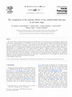
the study was incomplete, since we studied only ethyl acetate extracts. N. ramosissima is a species with
blue bellflowers (Figure 1) which grows in the Chilean coastal area of Paposo valley at an altitude of
500–2000 m. High-performance counter-current chromatography (HPCCC) is a special liquid-liquid
separation method which uses two immiscible phases, one is a stationary phase retained in a coil
by a high centrifugal force, and the other is a mobile phase which is pumped through the stationary
phase using an HPLC pump [9]. This methodology was broadly used to separate the flavonoids
from plants and fruits [10–13]. HSCCC offer important advantages in separation of natural products:
lower consumption of solvents, use of green chemistry solvents, such as water and ethyl acetate, no
absorption on solid surfaces such as conventional column chromatography, very higher amounts of
processing sample, introduction of crude extracts, and full recovery of natural products [14–18]. In this
work we have applied this technique for the fast detection of flavonoids, from the methanolic extract
of N. ramosissima for the testing of their relaxation activity in rat aorta. In addition, we discuss in this
paper the relaxation activities of the polar extracts, (namely herbal tea or infusion and methanolic
extract) of N. ramosissima plus their metabolite composition by UHPLC high-resolution orbitrap mass
spectrometry. HPLC hyphenated with high resolution mass spectrometry with the help of diode array
UV detection such as PDA Q-TOF-MS or PDA-HESI-Q-orbitrap HR-MS are outstanding techniques for
the fast and accurate untargeted small metabolite analysis of plant samples [19,20]. This technique
has been successfully used in the last few years by our group to analyze several endemic Chilean
species [21–24].
Figure 1. Picture of N. ramosissima I.M. Johnst.
in October 2015.
Collected in Paposo Valley, Atacama Desert,
2. Results and Discussion
2.1. Identification of the Compounds in N. ramosissima Methanol and Herbal Tea
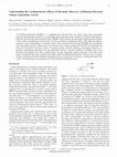
Sixty-one compounds (56 in the methanolic extract and 42 in the infusion) were identified or
tentatively identified by means of high resolution orbitrap mass spectrometry and PDA detection (Figure 2,
Table S1 Supplementary Materials). The fast identification of the compounds is explained below.
Molecules 2020, 25, 520
3 of 17
Figure 2. Photodiode array (PDA) chromatograms (UHPLC-PDA) of N. ramosissima extracts (a) methanol
extract; (b) aqueous extract, at 280 nm.
2.1.1. Flavonoids
Several compounds were identified as flavanones (Figures 3 and 4) and among them, some tentatively
identified as naringenin derivatives [25]. Peak 7 with a [M − H]− ion at m/z 461.14371 was identified as the
flavanone glycoside naringenin-4′ ,7-dimethoxyl-3-O-rhamnoside (C23 H25 O10 − ), while peak 9 with a
pseudo-molecular ion at m/z 489.13876 was identified as the related glycosylated and acylated compound
naringenin-4′ -acetyl-7-methoxyl-3-O-rhamnoside (C24 H25 O11 − ), peak 15 with a [M − H]− ion at m/z
517.17004 was identified as naringenin 3-hydroxyl-8-(3-methyl-2-butenyl)-7-O-glucoside (C26 H29 O11 − )
and peak 21 as the related naringenin derivative naringenin 3,4′ -dimethoxyl-8-(3-methyl-2-butenyl)
7-O-glucoside ([M − H]− ion at m/z 545.20111).
Figure 3. Structure of isolated flavonoids by high-performance counter-current chromatography
(HPCCC).
Molecules 2020, 25, 520
4 of 17
Figure 4. Biosynthetic relationship among flavonoids detected in N. ramosissima.
Peak 56 with a pseudomolecular ion at m/z 271.06161 was identified as naringenin by co-elution
experiments with an authentic compound and peak 22 with a [M − H]− ion at m/z 285.07687 as its O-methylated
derivative 7-methoxynaringenin (C16H13O5 −). Peak 19 ([M − H]− ion at m/z 623.16132) was identified as
isorhamnetin 3-O-rutinoside (C28H31O16 −). Peak 17 with a [M − H]− ion at m/z 531.18567 was identified as
naringenin-3-hydroxyl-4′ -methoxyl-8-O-(3-methyl-2-butenyl)-7-O-glucoside. Peak 11 with a [M − H]−
ion at m/z 503.15442 was identified as eriodictyol-5-acetyl-3′ ,4′ -dimethoxyl-7-O-glucoside (C25 H27 O11 − )
and peak 4 as 7-methoxy-3-glucose-flavanone (C22 H23 O10 − ). Regarding flavonol compounds, peak 12
with a [M − H]− ion at m/z 609.14606 was identified as rutin [25], identity confirmed using co-injection
of the standard (C27 H29 O16 − ), while peak 13 with a [M − H]− ion at m/z 593.15119 as kaempferol
3-O-rutinoside (C27 H29 O15 − ) [26] and peak 23 as quercetin-3-O-glucoside (C27 H29 O16 − ) [25]. Peak
39 with a [M − H]− ion at m/z 283.06158 was identified as 7-methoxyapigenin (C16 H11 O5 − ) and peak
38 with a [M − H]− ion at m/z 255.06653 was identified as 5,4′ -dihydroxyflavanone, while peak 32
with a [M − H]− ion at m/z 255.06656 was identified as its isomer 5,7-dihydroxyflavanone (C15 H11 O4 − )
and peak 40 as 5,7-dihydroxyflavone (C15 H9 O4 − ). Peak 49 with a [M − H]− ion at m/z 269.08224 was
identified as pinostrobin by using co-spiking experiments with authentic pinostrobin (C16 H13 O4 − ),
peak 50 was identified as the aglycone apigenin (C15 H9 O5 − ) and peak 44 as quercetin (C15 H9 O7 − ).
Molecules 2020, 25, 520
5 of 17
Peaks 30, 42 and 43 were identified as 7,3′ -dimethoxyquercetin, 7,4′ -dimethoxykaempferol and
7,4′ -dimethoxyapigenin (C17 H13 O7 − , C17 H13 O6 − and C17 H13 O5 − ), respectively.
2.1.2. Fatty Acids
Several compounds were identified the important dietary antioxidant fatty acids known as
oxylipins [26,27] (Figure 5).
Figure 5. Biosynthetic relationship among fatty acids detected in N. ramosissima.
Thus, Peak 51 with a [M − H]− ion at m/z 315.25458 was identified as dihydroxy-octadecanoic
acid (C18 H35 O4 − ); peak 45 with a [M − H]− ion at m/z 355.32230 was identified as hydroxy-docosanoic
acid (C22 H43 O3 − ) and peak 20 as 10-hydroxy-6-oxodecanoic acid (C10 H17 O4 − ). We could detect
also some glycosylated fatty acids; thus, peak 25 with a [M − H]− ion at m/z 489.27121 was
identified as dihydroxy-octadecadienoic acid–O-glucoside (C24 H41 O10 − ), while peak 34 with a
[M − H]− ion at m/z 447.2599 was identified as hydroxy-hexadecadienoic acid-O-glucoside (C22 H39 O9 − ).
Peak 37 with a [M − H]− ion at m/z 311.22330 was identified as dihydroxy-octadecadienoic acid
(C18 H31 O4 − ) [26], peak 58 with a [M − H]− ion at m/z 313.23889 as 9,10-dihydroxy-octadecenoic
Molecules 2020, 25, 520
6 of 17
acid (C18 H33 O4 − ) [26] and peak 53 as tetrahydroxy-eicosadienoic acid (C20 H35 O6 − ). In a similar
manner, Peak 20 (ion at m/z 201.11302) was identified as 10-hydroxy-6-oxodecanoic acid (C10 H17 O4 − ),
and peak 24 (ion at m/z 327.2181) was identified as trihydroxy-octadecadienoic acid (C18 H31 O5 − ).
In the same way, peak 25 was identified as the glycosyl derivative dihydroxy-octadecadienoic
acid-O-glucoside (C24 H41 O10 − ) and peak 33 as hydroxy-eicosaenoic acid glucoside (ion at m/z 503.32269).
Peaks 28 and 27 were identified as 9,10-dihydroxy-hexadecanoic acid and 9,10-tetradecanoic acid
(C16 H31 O4 − and C14 H27 O4 − ), respectively, while peak 17 with a [M − H]− ion at m/z 217.10805
was identified as 9,10-dihydroxy-6-oxodecanoic acid (C10 H17 O5 − ) [26], and peak 16 with a [M −
H]− ion at m/z 343.21320 was identified as trihydroxy-octadecadienoic acid (C18 H31 O5 − ). Peak 59
with a [M − H]− ion at m/z 369.30176 was identified as dihydroxydocosenoic acid (C22 H41 O4 − )
while isomer peaks 52 and 55 were identified as tetrahydroxy-eicosatrienoic acids (C20 H33 O6 − ).
Peak 26 with a [M − H]− ion at m/z 329.23386 was identified as trihydroxy-octadecaenoic acid
(C18 H33 O5 − ) and peak 29 as its isomer (C18 H33 O5 − ) as previously reported by us to occur in the
mesocarp of Keule fruits [27]. Peak 24 with 2 a.m.u of difference (327.21817) was identified as
trihydroxy-octadecadienoic acid (C18 H31 O5 − ) [27]. Peak 57 with a [M − H]− ion at m/z 309.20761
was identified as dihydroxy-octadecatrienoic acid (C18 H29 O4 − ), in contrast to a compound detected
in Asparagus, which was identified as 15-hydroperoxy-octadecatrienoic acid [26]. Peak 33 with a
[M − H]− ion at m/z 517.17004 was identified as hydroxy-hexadecaenoic acid-O-glucoside (C22 H39 O9 − ).
The saturated fatty acids Hydroxypalmitic (C16 H31 O3 − , peak 61), hydroxymiristic acid (C14 H27 O3 − ,
peak 60) and its glucoside derivative miristic acid-11-O-glucoside (C20 H37 O8 − , peak 31) were also
detected (Table S1, Supplementary Materials). Peak 1 with a [M − H]− ion at m/z 209.06636 was
identified as 2,4,5,6,7-pentahydroxypentanoic acid (C7 H13 O7 − ).
2.1.3. Coumarins
Peak 18 with a [M − H]− ion at m/z 191.03490 was identified as the simple coumarin scopoletin
(C10 H7 O4 − )[27]. Peak 3 with a molecular ion at m/z 339.07178 was identified as the glucoside coumarin
derivative esculetin-6-O-glucoside (sculin C15 H15 O9 − ) [28], while peak 2 with a [M − H]− ion at m/z
377.08542 was identified as sculetin-5-hydroxy-7-methoxy-6-O-glucoside (C18 H17 O9 − ), and peak 6
with a molecular ion at m/z 369.08267 was identified as 5-hydroxy-7-methoxyesculin (C16 H17 O10 − ).
2.1.4. Phenolic Acids
Peak 5 with [M − H]− ion at m/z 153.01881 was identified as 2,4-dihydroxybenzoic acid
(C7 H5 O4 − ) [29], peak 8 as salicylic acid (C7 H5 O3 − ) and peak 10 as free caffeic acid (C9 H7 O4 − ) [30].
2.2. Fast HPCCC Isolation of Major Compounds in N. ramosissima Methanol Extract
The employment of immiscible solvent systems in our HPCCC machine allowed the fast isolation
of the main five components (compounds 1–5) from a crude methanol extract of N. ramosissima. Major
isolated compounds 1–5 were identified by their ESI-MS, GC-MS data and mono and bidimentional
NMR spectra. (Please see Supplementary Materials, Figures S4–S32). Furthermore, from the extrusion
partition of the HSCCC run, only hydroxypalmitic acid (Peak 61) and inseparable mixtures of fatty
acids were isolated. Other potential approaches to pre-treat and extract the active compounds are
nowadays employed, including experimental approaches for the identification after elimination of the
effect of matrix on quantitative analyses by HPLC−MS, such as CO2 extraction, pretreatment with
ammonia and hydrogen peroxide, pressurized solvents; however, infusion is the typical edible form,
and methanol extraction at ambient temperature proved to be good solvent to extract all the flavonoids
and phenolics in our Nolana species for purification purposes [31–33].
Compound 1: 3,5-dihydroxy-7-methoxyflavanone (peak 22). Colourless crystals, m.p. 178.8–179.0 ◦ C.
[M − H]− : 285.0765, MS2 : 267 [M − H2 O]− , 251 [M − H2 O − CH3 ]− . 1 H NMR (300 MHz, CDCl3 ) δ
ppm: 3.65 (s, OCH3 ), 4.59 (1H, d, J = 12 Hz), 5.10 (1H, d, J = 12 Hz), 6.10 (1H, d, J = 17.3 Hz), 6.12 (1H,
d, J = 17,3 Hz), 7.52 (3H, m), 7.69 (2H, m), 11.24 (1H, s, OH). 13 C NMR (300 MHz, CDCl3 ) δ ppm: 56.60
Molecules 2020, 25, 520
7 of 17
(OCH3 ), 72.21 (C-2), 82.61 (C-3), 194.95 (C-4), 162.46 (C-5), 96.07 (C-6), 168.85 (C-7), 93.82 (C-8), 163.46
(C-9), 100.55 (C-10), 136.01 (C-1′ ), 127.56 (C-2′ ), 128.89 (C-3′ ), 127.56 (C-4′ ), 128.59 (C-5′ ), 127.56 (C-6′ ).
These data are in agreement with the literature [34–36].
Compound 2: 5,3′ -dihydroxy-4′ , 7-dimethoxyflavanone (Peak 48). Colourless crystals, m.p.
123.0–125.0 ◦ C [M − H]− : 315.0874, MSn : 283.8 [M – H − CH3 ]− , 254.7 [M – H − CH3 − CO]− . 1 H NMR
(300 MHz, CDCl3 ) δ ppm: 2.65 (1H, dd, J = 3.1 and 16.4Hz), 3.05 (1H, dd, J = 16.4 and 12.5 Hz), 3.75 (s,
OCH3 ), 3.80 (s, OCH3 ), 5.54 (1H, dd, J = 12.5 and 3.1 Hz), 6.21 (1H, d, J = 2,3 Hz), 6.23 (1H, d, J = 2,3
Hz), 7.4 (1H, m), 7.5 (2H, m), 11.24 (1H, s, OH).13 C NMR (300 MHz, CDCl3 ) δ ppm: 56.32 (OCH3 ),
56.20 (OCH3 ), 78.63 (C-2), 45.23 (C-3), 188.07 (C-4), 164.68 (C-5), 94.17 (C-6), 165.89 (C-7), 93.35 (C-8),
162.11 (C-9), 105.83 (C-10), 139.40 (C-1′ ), 126.96 (C-2′ ), 128.90 (C-3′ ), 162.00 (C-4′ ), 128.90 (C-5′ ), 126.96
(C-6′ ). These data are in agreement with the literature [35].
Compound 3: 5-hydroxy-3,4′ ,7-trimethoxyflavone (peak 41). Yellow crystals, m.p. 148.0–149.0 ◦ C.
[M − H]− : 327.0874, MSn : 312 [M – H − CH3 ]− , 314 [M + H − CH3 ]+ , 297 [M – H − 2CH3 ]- , 299 [M +
H − 2CH3 ]+ . 1 H NMR (300 MHz, CDCl3 ) δ ppm: 3.87 (s, OCH3 ), 3.89 (s, OCH3 ) 3.92 (s, OCH3 ), 6.38
(1H, d, J = 2,3 Hz), 6.47 (1H, d, J = 2,3 Hz), 7.04 (1H, d, J = 9.2 Hz), 8.10 (1H, d, J = 9.2 Hz), 11.24 (1H,
s, OH).13 C NMR (300 MHz, CDCl3 ) δ ppm: 55.10 (OCH3 ), 56.02 (OCH3 ), 60.49 (OCH3 ), 155.0 (C-2),
122.9 (C-3), 194.15 (C-4), 161.70 (C-5), 98.31 (C-6), 164.88 (C-7), 92.91 (C-8), 161.22 (C-9), 105.45 (C-10),
138.27 (C-1′ ), 114.06 (C-2′ ), 129.30 (C-3′ ), 157.62 (C-4′ ), 129.30 (C-5′ ), 114.06 (C-6′ ). These data are in
agreement with the literature [35,37,38].
Compound 4: 3-acetyl-5-hydroxy-7-methoxyflavanone (peak 47). Colourless crystals, m.p. 96–97 ◦ C
[M − H]− : 327.0874. MSn : 268.9 [M + H − CH3 COOH]+ , 255.0 [M + H − CH3 COOH − CH3 ]+ . 1 H
NMR (300 MHz, CDCl3 ) δ ppm: 2.05 (s, OCOCH3 ), 3.83 (s, OCH3 ), 5.39 (1H, d, J = 11.8 Hz), 5.84 (1H,
d, J = 11.8 Hz), 6.09 (1H, d, J = 2,3 Hz), 6.14 (1H, d, J = 2,3 Hz), 7.45 (5H, m),11.51 (1H, s, OH).13 C NMR
(300 MHz, CDCl3 ) δ ppm: 20.33 (OCOCH3 ), 55.87 (OCH3 ), 81.36 (C-2), 72.42 (C-3), 191.70 (C-4), 164.13
(C-5), 95.69 (C-6), 169.27 (C-7), 94.73 (C-8), 168.53 (C-9), 101.88 (C-10), 135.16 (C-1′ ), 128.91 (C-2′ ), 129.59
(C-3′ ), 127.38 (C-4′ ), 129.59 (C-5′ ), 128.91 (C-6′ ). These data are in agreement with the literature [39].
Compound 5: 5-hydroxy-7-methoxy-flavanone (peak 49), Pinostrobin. Colourless crystals, m.p.
119.7–120.0 ◦ C. [M − H]− : 269.0818, MS2 : 241.9, 178.3, 161.8, 153.2. 1 H NMR (300 MHz, CDCl3 ) δ ppm:
2.84 (1H, dd, J = 17.2 and 3.1 Hz), 3.11 (1H, dd, J = 13.0 and 17.2 Hz), 3.80 (s, OCH3 ), 5.44 (1H, dd,
J = 3.1 and 13. Hz), 6.10 (1H, d, J = 2.3 Hz), 6.09 (1H, d, J = 2,3 Hz), 7.47 (5H, m), 12.11 (1H, s, OH).
13 C NMR (300 MHz, CDCl ) δ ppm: 55.91 (OCH ), 79.14 (C-2), 42.75 (C-3), 196.16 (C-4), 162.44 (C-5),
3
3
94.62 (C-6), 167.94 (C-7), 94.13 (C-8), 163.87 (C-9), 102.77 (C-10), 137.86 (C-1’), 126.96 (C-2′ ), 129.16
(C-3’), 126.96 (C-4′ ), 129.16 (C-5’), 126.96 (C-6′ ). These data are in agreement with the literature [35,40].
Furthermore the X-ray crystal structure of this compound was already published by us [41].
2.3. N. ramosissima Induced Relaxation in Aortic Ring of Rat, Endothelium-Independent Activity
We found that N. ramosissima could have a potential antihypertensive effect, since it caused a
relaxation effect on rat aortic rings pre-contracted with PE (Figure 6).
Molecules 2020, 25, 520
8 of 17
Figure 6. Original record of the relaxation effects of N. ramosissima in intact rat aorta. Rat aorta was
pre-contracted with 10−6 M phenylephrine (PE) for 10 min, and then, cumulative concentrations of N.
ramosissima (0.01 to 1000 g/mL) were added in organ bath at 7 min intervals (B) to compare with the
control (A).
N. ramosissima produced the concentration-dependent relaxation in intact aortic rings (34 ± 5%
with 2 [log µg/mL] or 100 µg/mL versus 91 ± 8% with 3 [log µg/mL] or 1000 µg/mL; p < 0.001) and
denuded (29 ± 4% with 2 [log µg/mL] or 100 µg/mL versus 77 ± 1% with 3 [log µg/mL] or 1000 µg/mL;
p < 0.001; Figure 7A).
Although relaxation effect observed with N. ramosissima did not involve endothelial nitric oxide
synthase, the soluble guanylate cyclase pathway it was (Figure 7). The pre-incubation with an inhibitor
of nitric oxide synthase (10−4 M, N(ω)-nitro-L-arginine methyl ester (L-NAME) did not reduce the
relaxation to N. ramosissima in intact aorta (Figure 7B).
Figure 7. Relaxation effect of N. ramosissima in rat aorta. Arteries were pre-contracted with 10−6 M PE.
Concentration-response curves for N. ramosissima extract in intact and denuded-endothelium aortic
rings (A) in absence (Control) or in presence of 10−4 M L-NAME (B), 10−6 M methylene blue (C) or 10−6
M 1H-(1,2,4) oxadiazolo [4 ,3-a]quinoxalin-1-one (ODQ) (D) in rat aorta. Methylene blue and ODQ are
non-selective and selective inhibitors of soluble guanylyl cyclase, respectively. Data are the average ±
standard error of the mean (SEM) of 5 independent experiments. *p < 0.05, **p < 0.01 vs. Control.
Molecules 2020, 25, 520
9 of 17
However, compared with control (34 ± 5% with 2 [log µg/mL] or 100 µg/mL), the pre-incubation
with methylene blue, a nonspecific soluble guanylyl cyclase inhibitor, significantly decreased the
relaxation in intact aorta (5 ± 3% with 2 [log µg/mL] or 100 µg/mL; p < 0.01; Figure 7c). Methylene blue
has been used extensively to inhibit soluble guanylyl cyclase, the effector that mediates the vasodilator
effect of nitric oxide [42]. The pre-incubation with 10-6 M 1H-(1,2,4) oxadiazolo[4,3-a]quinoxalin-1-one
(an inhibitor of soluble guanylyl cyclase; ODQ) reduced the relaxation and confirmed that soluble
guanylyl cyclase is involved on vascular relaxation by the extract (12 ± 4% with 2 [log µg/mL] or 100
µg/mL; p < 0.01; Figure 7D). The log half-maximal inhibitor concentration (Log IC50 ) of ODQ was
significantly (p < 0.05) different in the presence of extract versus control (Table 1).
These findings suggest that N. ramosissima produced relaxation in intact and endothelium-denuded
rat aorta when they were exposed to cumulative concentrations of the extract. Commonly, vasodilators
substances induced the activation of soluble guanylyl cyclase, but a few can stimulate nitric
oxide-independent soluble guanylate cyclase activity and thus produce relaxation [43–45]. Thus, it is
possible that N. ramosissima caused relaxation of vascular smooth muscle by directly stimulating the
nitric oxide-independent soluble guanylate cyclase (sGC) and cGMP pathway [46].
Table 1. Effect of N. ramosissima (Nr) on the vascular response to different vasoactive substances on the
nitric oxide pathway in rat aorta.
Drugs
Log (IC50 ) (g/mL)
Control
Endo-denuded
L-NAME
Methylene blue
ODQ
2.38 ± 0.12
2.37 ± 0.06
2.35 ± 0.10
10−4
3.51 ± 0.38 *
10−6
Vasoactive substances:
M L-NAME,
M methylene blue and 10−6 M 1H-(1,2,4) oxadiazolo[4,3-a]
quinoxalin-1-one (ODQ). Log (IC50 ) represent the logarithm half-maximal inhibitory concentration. The values are
mean ± standard error of the mean (S.E.M.) and n = 5. Statistically significant difference * p < 0.05 vs. Control.
2.4. N. ramosissima Reduced the Contractile Response to KCl and Phenylephrine
To study whether the effect of N. ramosissima on vascular response is mediated by the membrane
depolarization or pharmacological stimulation, the contractile response to KCl and PE was evaluated.
The pre-incubation with the extract significantly reduced the maximal contractile response to
KCl (144 ± 4% control vs. 31 ± 3%; p < 0.001; Figure 8A) and to PE (139 ± 8% control vs. 75 ± 8%;
p < 0.001; Figure 8B). The log half-maximal effective concentration (Log EC50 ) to KCl and PE was
not significantly different in the presence of extract versus control, indicating that the extract did not
modify the sensitivity of K+ channels or alpha-adrenergic receptor (Table 2).
Figure 8. N. ramosissima decreases the vascular contractile response to KCl and PE. The vascular tissue
was pre-incubated in absence (control) or presence of 100 µg/mL of extract or nimodipine (10-6 M) for
20 min before adding KCl (10–60 mM) (A) or PE (10−9 − 10−5 M) (B). Data are the average ± SEM of 5
independent experiments. Statistically significant differences: *** p < 0.001 vs control.
Molecules 2020, 25, 520
10 of 17
Table 2. Effect of N. ramosissima (Nr.; 100 µg/mL) on the contractile response to KCl (10–60 mM),
and phenylephrine (PE; 10-9 to 10-5 M) in intact rat aorta.
Drugs
KCl (mM)
Control
Nr
Nimodipine
PE (nM)
Control
Nr
Nimodipine
Log (EC50 )
1.57 ± 0.15
1.63 ± 0.50
−7.47 ± 0.10
−7.18 ± 0.26
−7.05 ± 0.14
Log (EC50 ) represents the logarithm of half-maximal effective concentration of drug. The values are mean ± SEM,
representing the mean of 5 independent experiments.
Interestingly, the blockage of L-type voltage-gated Ca2+ channels (Cav1.2) with 10-6 M nimodipine
decreased the contractile response to KCl (5 ± 2%) and PE (57 ± 7%) in a similar way than N. ramosissima
(Figure 8B). Therefore, this comparison with nimodipine suggests that extract-induced vascular effect
is major by blocking of Ca2+ influx through the plasmatic membrane [24].
2.5. Pure Compounds of N. ramosissima Induced Relaxation
Isolated compounds 1–4 showed different vascular relaxation in rat aorta pre-contracted with
10-6 M PE. The relaxation effect was compared with the extract of N. ramosissima and an agonist
dependent drug on endothelial nitric oxide, acetylcholine. As shown in Figure 9, only the isolated
compound 2 (115 ± 2%; 10−4 M) and 4 (77 ± 5%; 10−4 M) possessed an important relaxation effect
in intact aortic rings. Interestingly, compound 2 presented a higher relaxation than 100 µg/mL
N. ramosissima extract (91 ± 8%). Compound 2 is 5,3′ -dihydroxy-4′ 7-dimethoxyflavone and compound
4 is 3-acetyl-5-hydroxy-7-methoxyflavone. Apparently the free OH groups in position 5 and 3′ is
important for the increase of this activity.
Figure 9. Screening test of 4 pure compounds of N. ramosissima on vascular response in rat aorta.
Relaxation effect of extract 100 µg/mL N. ramosissima (black bar; Nr), 4 pure compounds isolated (1–4;
10−4 M) and acetylcholine (ACh; 10−4 M) in intact aortic rings pre-constricted with 10−6 M PE. The
compound 2 and 4 presented a similar relaxation than 100 µg/mL N. ramosissima extract. Values are
mean ± standard error of the mean of 4 experiments. Statistically significant differences: *p < 0.05,
***p < 0.001 vs. Nr.
3. Materials and Methods
3.1. Chemicals
HPLC-MS solvents and Gradient Grade (GR) acetonitrile, methanol, hexane and ethyl acetate were
from Merck (Santiago, Chile). Ultrapure water was obtained from a Millipore water purification system
(Milli-Q Merck Millipore, Chile). HPLC standards, quercetin, isorhamnetin, kaempferol, naringenin,
Molecules 2020, 25, 520
11 of 17
eriodictyol, hesperetin, rhamnetin, linoleic acid, (all standards with purity higher than 95% by HPLC)
were purchased either from Extrasynthèse (Genay, France), Sigma Aldrich (Saint Louis, Missouri,
MO, USA) or ChromaDex (Santa Ana, California, CA, USA). TLC: Silica gel 60 F254 plates (Merck,
Darmstadt, Germany). Column chromatography: Sephadex LH-20, MeOH as solvent. A Quattro
semi preparative MK-7 HSCCC instrument (AECS inc., Bridgend, UK) with a total capacity of 437 mL
(Figure S1, Supplementary Materials) with two bobbins, each of them bearing two stainless steel coils
(one bobbin with two coils, an analytical of 27 mL, 1 mm tubing bore, one preparative of 205 mL
and 2.1 mm i.d. and the other bobbin bearing two 116 mL, 2.1 mm tubing bore preparative coils).
The mobile phase pumped using two Series II SSI model HPLC pumps (LabAlliance, Pennsyivania,
PA, USA) and fractions collected with a Gilson FC 203B model fraction collector (Middleton, MI,
USA). The effluent was monitored using a UV–visible-ECOM Flash 06 S single 254 nm wavelength
detector governed by Ecomac software (Ecom, Prague, Czech Republic). Nuclear Magnetic Resonance
(NMR) spectroscopy: 1 H-, and 13 C- and 2D NMR spectra: Bruker Avance 400 or Bruker Avance II
600 UltraShield spectrometers: δ in ppm relative to Me4 Si as internal standard, J in Hz. The melting
point was measured in a Stuart Scientific apparatus SMP3 (Bibby, London UK). L-phenylephrine
hydrochloride (PE), acetylcholine chloride (ACh), 1H-(1,2,4)oxadiazolo[4,3-a]quinoxalin-1-one (ODQ),
Nω -nitro-L-arginine methyl ester (L-NAME) were purchased to Sigma-Aldrich (St Luis, MO, USA).
Nimodipine was obtained from Merck (Darmstadt, Germany). Several drugs were dissolved in
distilled deionized water (deionized water Millipore) and kept at 4 ◦ C. The stock solution of ODQ and
nimodipine was dissolved in dimethyl sulfoxide (DMSO; 0.1% final concentration) (Merck, Germany).
The extract of N. ramosissima was dissolved in physiological Krebs–Ringer bicarbonate buffer (KRB) in
all vascular experiments.
3.2. Plant Material
N. ramosissima was collected in Paposo Valley, northern Chile in April 2011 and was identified by
the botanist Alicia Marticorena (University of Concepción, Chile). A voucher specimen is deposited at
the Natural Products’ laboratory, University of Antofagasta, Chile, with the number Nr-111004-1.
3.3. Extraction
Approximately 100 g of the dried plant was pulverized in a mortar and then extracted with
500 mL of HPLC-MS grade methanol in the dark in an ultrasonic bath for one hour (three times);
the extracts were combined, filtered and evaporated in vacuo in the dark (40 ◦ C) to give 7.83 g of N.
ramosissima methanolic extract. For the preparation of the herbal tea, 2 g of the pulverized plant was
added distilled water (250 mL) at 45 ◦ C and left to stand for 12 h. The plant material was then filtered
and the solution lyophilized to give 0.63 g of lyophilized material.
3.4. UHPLC-PDA-MS Instrument
For UHPLC Photodiode-Array–Mass-Spectrometry (UHPLC-PDA-MS) analysis, 5 mg of the
methanol extract and lyophilized herbal tea (infusion) were individually dissolved in 2 mL of methanol;
filtered (using a PTFE 200 m filter) and 10 L were injected in the instrument. A Thermo Scientific
Ultimate 3000 UHPLC system equipped with a quaternary Series RS pump and a Thermo Scientific
Dionex Ultimate 3000 Series TCC-3000RS column compartments with Ultimate 3000 Series WPS-3000RS
autosampler and a rapid separations photodiode array (PDA) detector controlled by Chromeleon
7.2 Software (Thermo Fisher Scientific, Darmstadt, Germany) hyphenated with a Thermo high
resolution Q-Exactive focus mass spectrometer (Thermo, Bremen, Germany) were used for analysis.
The chromatographic system was coupled to the MS with a Heated Electrospray Ionization Source
II (HESI II). Nitrogen (purity > 99.999%) obtained from a Genius NM32LA nitrogen generator (Peak
Scientific, Billerica, Massachusetts, MA, USA) was employed as both the collision and damping gas.
Mass calibration for Orbitrap was performed once a day, in both (−) and (+) modes, to ensure a
working mass accuracy lowers than or equal to 5 ppm. N-butylamine and Cafeine, (Sigma Aldrich,
Molecules 2020, 25, 520
12 of 17
Saint Louis, Missouri, Mo, USA) were the calibration standards for positive ions and taurocholic acid
sodium salt, buspirone hydrochloride, sodium dodecyl sulfate, (Sigma Aldrich, Saint Louis, Missouri,
MO, USA) were used as negative standards to calibrate the spectrometer. These compounds were
added to a mixture of acetonitrile, acetic acid, water, and methanol (Merck Darmstadt, Germany)
and afterwards infused using a Chemyx Fusion 100 syringe pump (Thermo Fisher Scientific, Bremen,
Germany). XCalibur 2.3 and Trace Finder 3.2 software (Thermo Fisher Scientific, San José, California,
CA, USA) were used for UHPLC control and data processing, respectively. Q Exactive 2.0 SP 2 from
Thermo Fisher Scientific was used to control the mass spectrometer.
3.5. LC Parameters
The separations were done using an UHPLC C18 column (Acclaim, 150 mm × 4.6 mm ID, 2.5 m,
Thermo Fisher Scientific, Bremen, Germany) at 25 ◦ C. The detection wavelengths were 254, 280 and
320 nm, and PDA from 200 to 800 nm was recorded. Mobile phases were 1% formic aqueous solution
(A) and acetonitrile (B). The gradient program (time (min), % B) was: (0.00, 7); (5.00, 7); (10.00, 35);
(15.00, 40); (20.00, 70); (25.00, 70); (35.00, 7) and 12 min for column equilibration before each injection.
The flow rate was 1.00 mL min−1 , and the injection volume was 10 L. Standards and extracts dissolved
in methanol were kept at 10 ◦ C during storage in the autosampler.
3.6. MS Parameters
The HESI II parameters were sheath gas flow rate, 75 units; aux. gas unit flow rate, 20; capillary
temperature, 400 ◦ C; aux gas heater temperature, 500 ◦ C; spray voltage, 2500 V (for ESI-); and S lens RF
level, 30. Full scan data in negative mode was acquired at a resolving power of 70,000 full width half
maximum (FWHM) at m/z 200. For the compounds of interest, a scan range of m/z 100–1000 was chosen;
the automatic gain control (AGC) was set at 3e6 , and the injection time was set to 200 ms. Scan-rate
was set at 2 scans s−1 . External calibration was performed using a calibration solution in positive
and negative modes before each sample series. In addition to the full scan acquisition method, for
confirmations purposes, a targeted MS/MS analysis was performed using the mass inclusion list and
expected retention times of the target analytes, with a 30 s time window, with the Orbitrap spectrometer
operating both in positive and negative mode at 17,500 FWHM (m/z 200). The AGC target was set
to 2e5 , with the maximum injection time of 20 ms. The precursor ions filtered by the quadrupole
operates at an isolation window of m/z 2. The fore vacuum, high vacuum and ultrahigh vacuum were
maintained at approximately 2 mbar, 105 and below 1010 mbar, respectively. Collision energy (HCD
cell) operated at 30 kv. Detection was based on calculated exact mass and on retention time of target
compounds. The mass tolerance window was set to 5 ppm.
3.7. Selection of the Solvent System for HPCCC
According to the requirements for solvent systems in HPCCC [47], the selection was performed by a
partition experiment of the crude extract using several solvent systems including (1) hexane:acetonitrile
(stationary phase poorly retained in the coil), (2) hexane: methanol, (3) HEMWAT (n-hexane: ethyl
acetate: methanol: water) and (4) n-hexane: ethanol: water at different volume ratios. The measurement
of K values of target flavonoids from crude sample was as follows: A portion of the crude methanol
extract (2 mg) was weighed into a 5 mL glass tube and added 1 mL of each phase of a pre-equilibrated
two-phase solvent system. The glass tube was capped and then placed in a vortex mixer for 5 min to
equilibrate the sample between the two-phases. After settling, the two phases were separated and
evaporated to dryness. The residues were diluted with 1 mL methanol and 20 µL of the resulting
solution was injected into the UHPLC system. Then, the quantitative UHPLC-PDA was performed by
UHPLC. The K value was expressed as the peak area of target compounds in the upper phase (stationary
phase) divided by that in the lower phase (mobile phase). The best liquid–liquid separation system,
in our opinion, for the UV active compounds from N. ramosissima petroleum ether extract was the
Molecules 2020, 25, 520
13 of 17
biphasic non-aqueous solvent system: n-hexane: ethanol: water 6:5:1 v/v/v (Table S2, Supplementary
Materials).
3.8. HSCCC Separation of N. ramosissima Methanol Extract
After equilibration of the two solvents in a separating funnel, the two resulting phase layers were
separated shortly before use and degassed in an ultrasonic bath (for 15 min). The upper phase was then
used as stationary phase and the lower phase as mobile phase in the ‘head-to-tail’ mode. The separation
was performed using temperature control during the separation (approx. 25 ◦ C) with a rotation velocity
of 800 rpm. The columns of the HSCCC were then filled with upper phase, and the lower mobile
phase was pumped at a flow rate of 5.0 mL/min using the ‘head-to-tail’ mode. After the mobile phase
front emerged and the hydrodynamic equilibrium was established in the columns, the percentage
of the retention of the stationary phase (75%) was recorded. Then the dried methanol extract of
N. ramosissima (500 mg) was dissolved in 5 mL each of upper and lower phase, filtered through a 0.45 m
micropore membrane (PTFE, Waters), introduced via a plastic syringe to a 10 mL sample loop and then
directly injected into the separation column through a manual low-pressure sample injection valve
(Rheodyne, Cotati, CA, USA). For recovery of all existing N. ramosissima metabolites, a two column
volume with elution and extrusion steps was applied [48]. The effluent from the outlet of the column
was collected (10 mL/tube, 5 mL/min), and 32 fractions were collected in the elution mode (numbered
F1–F32). Then, the system was changed to the extrusion mode with pumping of stationary phase at a
lower spinning velocity (400 rpm) and the same flow rate. Every 2 min the extrusion-fractions were
collected (F33 until F 50). Component detection of the effluent was performed with UV-light (λ = 254
nm, Figure S2, Supplementary Materials), and visualization of the spots in a TLC plate (Figure S3,
Supplementary Materials) of each collected tube (Silica gel F254 , Merck Darmstadt, Germany, developed
with n-hexane:Ethyl acetate 8:2, v/v) with the universal spray reagent p-anisaldehyde-concentrated
sulphuric acid-glacial acid (1:2:97, v/v/v), and flash heating (110 ◦ C) on a hot plate [11].
3.9. Isolation and Identification of Compounds
The HSCCC fractions I–IV (Figures S2 and S3, Supplementary Materials) collected in the elution
mode were refined by Gel-permeation chromatography on Sephadex LH-20 (5 cm x 25 cm, 100 g, eluted
with HPLC grade methanol) to yield flavonoids 1–5 (peaks 22, 48, 41, 47 and 49). From fraction I: tubes
12–14 (96–112 mL), 32 mg of compound 1 were obtained, from fraction II: tubes 15–16 (120–128 mL),
15.3 mg of compound 2, were obtained, from fraction III: tubes 17–20 (130–160 mL), 18 mg of compound
3 and 12 mg of compound 4 were obtained and from fraction IV: tubes 21–26 (168–208 mL), 73 mg of
compound 5 were isolated. From the extrusion fractions, (tubes 32–40) (Figure S3, Supplementary
Materials) inseparable mixtures of fatty acids could be detected by TLC analysis.
3.10. Animals
For vascular reactivity experiments, female Sprague–Dawley rats (6–8 weeks of age, 170–200 g)
from the breeding colony at the Antofagasta University were used. All animals were housed in a
temperature-controlled, light-cycled (08:00–20:00 h) room with ad libitum access to drinking water
and standard rat chow (Champion, Santiago). The investigation conformed to the Guide for the Care
and Use of Laboratory Animals published by the U. S. National Institutes of Health (NIH Publication
revised 2013), and the local animal research committee approved the experimental procedure used in
the present study (number 135/2018).
3.11. Isolation of Aortic Rings
The procedure of these experiments was realized in accordance to described by Cifuentes et al. [24].
Rats were sacrificed through cervical dislocation. The thoracic aorta was quickly excised and placed in
physiological Krebs-Ringer bicarbonate buffer (KRB) containing (mM): 4.2 KCl, 1.19 KH2 PO4 , 120 NaCl,
25 NaHCO3 , 1.2 MgSO4 , 1.3 CaCl2 , and 5 d-glucose (pH 7.4). Rings (3-5 mm and 2-4 mg) were
Molecules 2020, 25, 520
14 of 17
prepared after connective tissue was cleaned out from the aorta, taking special care to avoid endothelial
damage. Aortic rings were equilibrated for 40 min in KRB at 37 ◦ C by constant bubbling with 95% O2
and 5% CO2 .
3.12. Vascular Reactivity Experiments
Aortic rings from the same animal were studied in duplicate, using different vasoactive substances
(phenylephrine [PE], KCl and acetylcholine [ACh]). The rings were mounted on two 25-gauge stainless
steel wires; the lower one was attached to a stationary glass rod and the upper one was attached to an
isometric transducer (Radnoti, Monrovia, California, CA, USA). The transducer was connected to a
PowerLab 8/35 (Colorado Springs, Colorado, CO, USA) for continuous recording of vascular tension
using the LabChart Pro v8.1.2 computer program (ADInstrument). After the equilibration period for
40 min, the aortic rings were stabilized by 3 successive near-maximum contractions with KCl (60 mM)
for 10 min. The passive tension on aorta was 1.0 g, which was determined to be the resting tension for
obtaining maximum active tension induced by 60 mM KCl. To study the effect of methanolic extract
or pure compounds on vascular reactivity in rat aorta, we performed different protocols. In the first
protocol, the aortic rings were pre-contracted with 10−6 M PE, and then increasing concentrations
of N. ramosissima or pure compounds were added to the bath. In the second protocol, the rat aorta
was pre-incubated in presence of N. ramossima for 20 min, followed by a contraction with 10−6 M PE.
A stock solution in DMSO (10−3 M) was prepared with pure compounds, and then, dilutions in KRB
were added in the bath. In some experiments, the endothelium removal was by gently rubbing it off
using a small piece of cotton. To evaluate the vascular function of the endothelium, the relaxation to
10−6 M acetylcholine (muscarinic agonist) in pre-contracted aortic rings with 10−6 M PE was tested.
According to the general use of rat aorta as a pharmacological tool for in vitro, the aortic rings were
considered with a functional endothelial response if relaxation was up to 70% to 80% [49].
4. Conclusions
Five major flavones were quickly isolated by HSCCC from a methanol extract of the endemic
species N. ramosissima, and four of them showed relaxation activity. Besides, some 61 compounds were
detected in both N. ramosissima polar extracts by UHPLC-MS. Of those, four were coumarins (peaks 2,
3, 6 and 18), 15 flavanones (peaks 4, 7, 9, 11, 15, 17, 21, 22, 32, 38, 46–49 and 56), 12 flavones (peaks 12,
13, 19, 23, 30, 39, 40–44 and 50), 3 phenolic acids (peaks 5,8 and 10) and 22 oxylipins/fatty acids (peaks
1, 14, 16, 20, 24, 29, 31, 33–37, 51-55, 57–61). Compound 2 presented higher relaxation effects than N.
ramosissima extract. Moreover, since the methanolic extract and infusion of the plant showed higher
relaxation effect than the isolated compounds (1, 3, 4); it can be assumed that these compounds present
in the extract could have a synergistic effect and boost the hypotensive or antihypertensive activity.
Furthermore, N. ramosissima caused relaxation through an endothelium-independent mechanism;
this effect could be exerted by pure compounds 2 and 4. Regarding these results, N. ramosissima could
be used as a natural medicine to lower blood pressure. However, more research is needed to support
the use of this plant as an antihypertensive agent.
Supplementary Materials: The following are available online.
Author Contributions: J.B. and M.J.S. isolated the compound; M.J.S., F.C., J.P. and A.P. conceived and designed of
the research study; J.B., A.B., A.P., J.P. and F.C. performed the experiments; M.J.S., A.P., F.C., C.P. and J.P. analyzed
data; M.J.S. and J.P. drafted the manuscript; F.C., J.P., M.J.S. and A.P. edited and revised the manuscript. All
authors have read and agreed to the published version of the manuscript.
Funding: Financial support was provided by FONDECYT 1180059 to M.J.S., FONDECYT 1200610 to J.P.,
the Network for Extreme Environments Research Project (NEXER, Project ANT1756, Universidad de Antofagasta,
Chile).
Acknowledgments: The authors wish to express their gratitude to the Rectoría of Universidad de Antofagasta for
their financial support.
Conflicts of Interest: The authors declare no conflict of interest.
Molecules 2020, 25, 520
15 of 17
Data Availability: The datasets generated during and/or analyzed during the current study are available from
the corresponding author on reasonable request.
References
1.
2.
3.
4.
5.
6.
7.
8.
9.
10.
11.
12.
13.
14.
15.
16.
17.
18.
Jewell, C.; Papineau, A.D.; Freyre, R.; Moyle, L.C. Patterns of reproductive isolation in Nolana (Chilean
bellflower). Evolution 2012, 66, 2628–2636. [CrossRef] [PubMed]
Ossa, P.G.; Pérez, F.; Armesto, J.J. Phylogeography of two closely related species of Nolana from the coastal
Atacama Desert of Chile: Post-glacial population expansions in response to climate fluctuations. J. Biogeogr.
2013, 40, 2191–2203. [CrossRef]
Chamy, M.C.; Piovano, M.; Garbarino, J.A. Diterpenoids from Nolana elegans. Bol. Soc. Chil. Quim. 2002, 47,
367–370. [CrossRef]
Chamy, M.C.; Garbarino, J.A.; Piovano, E.; Lopez-Perez, J.L.; Nicoletti, M.; Gandolfo, R.; Feliciano, A.S.
9-epi-labdane diterpenoids from Nolana rostrata var. rostrata. Phytochemistry 1997, 45, 797–800. [CrossRef]
Garbarino, J.A.; Chamy, M.C.; Piovano, M.; Gambaro, V. Labdane diterpenoids from Nolana filifolia.
Phytochemistry 1988, 27, 1795–1796. [CrossRef]
Garbarino, J.A.; Chamy, M.C.; Montagna, M.P.; Gambaro, V. Sesquiterpenoids in Nolana coelestis.
Phytochemistry 1993, 32, 987–989. [CrossRef]
Vio-Michaelis, S.; Apablaza-Hidalgo, G.; Gómez, M.; Peña-Vera, R.; Montenegro, G. Antifungal activity of
three Chilean Plant extracts on Botritis Cinerea. Bot. Sci. 2012, 90, 179–183. [CrossRef]
Simirgiotis, M.J.; Benites, J.; Areche, C.; Sepúlveda, B. Antioxidant capacities and analysis of phenolic
compounds in three endemic Nolana species by HPLC-PDA-ESI-MS. Molecules 2015, 20, 11490–11507.
[CrossRef]
Ito, Y. Golden rules and pitfalls in selecting optimum conditions for high-speed counter-current
chromatography. J. Chromatogr. A 2005, 1065, 145–168. [CrossRef]
Peng, J.; Yang, G.; Fan, G.; Wu, Y. Preparative isolation and separation of a novel and two known flavonoids
from Patrinia villosa Juss by high-speed counter-current chromatography. J. Chromatogr. A 2005, 1092,
235–240. [CrossRef]
Rodriguez-Rivera, M.P.; Lugo-Cervantes, E.; Winterhalter, P.; Jerz, G. Metabolite profiling of polyphenols in
peels of Citrus limetta Risso by combination of preparative high-speed countercurrent chromatography and
LC-ESI-MS/MS. Food Chem. 2014, 158, 139–152. [CrossRef] [PubMed]
Xiao, X.H.; Si, X.X.; Tong, X.; Li, G.K. Preparation of flavonoids and diarylheptanoid from Alpinia katsumadai
hayata by microwave-assisted extraction and high-speed counter-current chromatography. Sep. Purif.
Technol. 2011, 81, 265–269. [CrossRef]
Pan, S.B.; Wang, X.; Duan, W.J.; Yu, Z.Y.; Zhang, L.; Liu, W. Preparative isolation and purification of flavonoids
from Cuscuta chinensis Lam. by high speed countercurrent chromatography. J. Liq. Chromatogr. Relat.
Technol. 2014, 37, 2162–2171. [CrossRef]
Schafer, K.; Winterhalter, P. Application of high speed countercurrent chromatography (HSCCC) to the
isolation of kavalactones. J. Liq. Chromatogr. Relat. Technol. 2005, 28, 1703–1716. [CrossRef]
Cheel, J.; Hajek, J.; Kuzma, M.; Saurav, K.; Smykalova, I.; Ondrackova, E.; Urajova, P.; Vu, D.L.; Faure, K.;
Kopecky, J.; et al. Application of HPCCC Combined with Polymeric Resins and HPLC for the Separation of
Cyclic Lipopeptides Muscotoxins A-C and Their Antimicrobial Activity. Molecules 2018, 23, 17. [CrossRef]
[PubMed]
Simirgiotis, M.J.; Schmeda-Hirschmann, G.; Borquez, J.; Kennelly, E.J. The Passiflora tripartita (Banana
Passion) Fruit: A Source of Bioactive Flavonoid C-Glycosides Isolated by HSCCC and Characterized by
HPLC-DAD-ESI/MS/MS. Molecules 2013, 18, 1672–1692. [CrossRef] [PubMed]
Di, D.L.; Zheng, Y.Y.; Chen, X.F.; Huang, X.Y.; Feng, S.L. Advance of Application of High Speed
Counter-current Chromatography in Separation and Purification of Flavonoids. Chinese J. Anal. Chem. 2011,
39, 269–275. [CrossRef]
Yi, T.; Zhu, L.; Zhu, G.Y.; Tang, Y.N.; Xu, J.; Fan, J.Y.; Zhao, Z.Z.; Chen, H.B. HSCCC-based strategy for
preparative separation of in vivo metabolites after administration of an herbal medicine: Saussurea laniceps,
a case study. Sci. Rep. 2016, 6, 8. [CrossRef]
Molecules 2020, 25, 520
19.
20.
21.
22.
23.
24.
25.
26.
27.
28.
29.
30.
31.
32.
33.
34.
35.
36.
37.
16 of 17
Yi, T.; Tang, Y.N.; Zhang, J.Y.; Zhao, Z.Z.; Yang, Z.J.; Chen, H.B. Characterization and determination of six
flavonoids in the ethnomedicine “Dragon’s Blood” by UPLC-PAD-MS. Chem. Cent. J. 2012, 6, 7. [CrossRef]
Chen, Q.L.; Zhu, L.; Tang, Y.N.; Kwan, H.Y.; Zhao, Z.Z.; Chen, H.B.; Yi, T. Comparative evaluation of chemical
profiles of three representative ‘snow lotus’ herbs by UPLC-DAD-QTOF-MS combined with principal
component and hierarchical cluster analyses. Drug Test. Anal. 2017, 9, 1105–1115. [CrossRef]
Brito, A.; Areche, C.; Sepulveda, B.; Kennelly, E.J.; Simirgiotis, M.J. Anthocyanin Characterization, Total
Phenolic Quantification and Antioxidant Features of Some Chilean Edible Berry Extracts. Molecules 2014, 19,
10936–10955. [CrossRef] [PubMed]
Brito, A.; Ramirez, J.E.; Areche, C.; Sepulveda, B.; Simirgiotis, M.J. HPLC-UV-MS Profiles of Phenolic
Compounds and Antioxidant Activity of Fruits from Three Citrus Species Consumed in Northern Chile.
Molecules 2014, 19, 17400–17421. [CrossRef] [PubMed]
Borquez, J.; Bartolucci, N.L.; Echiburu-Chau, C.; Winterhalter, P.; Vallejos, J.; Jerz, G.; Simirgiotis, M.J. Isolation
of cytotoxic diterpenoids from the Chilean medicinal plant Azorella compacta Phil from the Atacama Desert
by high-speed counter-current chromatography. J. Sci. Food Agric. 2016, 96, 2832–2838. [CrossRef] [PubMed]
Cifuentes, F.; Palacios, J.; R Nwokocha, C.; Bórquez, J.; Simirgiotis, M.J.; Norambuena, I.; Chiong, M.;
Paredes, A. Polyphenolic Composition and Hypotensive Effects of Parastrephia quadrangularis (Meyen)
Cabrera in Rat. Antioxidants (Basel) 2019, 8. [CrossRef]
Simirgiotis, M.J.; Quispe, C.; Bórquez, J.; Areche, C.; Sepúlveda, B.x. Fast Detection of Phenolic Compounds
in Extracts of Easter Pears (Pyrus communis) from the Atacama Desert by Ultrahigh-Performance Liquid
Chromatography and Mass Spectrometry (UHPLC-Q/Orbitrap/MS/MS). Molecules 2016, 21, 92. [CrossRef]
Jiménez-Sánchez, C.; Lozano-Sánchez, J.; Rodríguez-Pérez, C.; Segura-Carretero, A.; Fernández-Gutiérrez, A.
Comprehensive, untargeted, and qualitative RP-HPLC-ESI-QTOF/MS2 metabolite profiling of green
asparagus (Asparagus officinalis). J. Food Compos. Anal. 2016, 46, 78–87. [CrossRef]
Simirgiotis, M.J.; Ramirez, J.E.; Schmeda Hirschmann, G.; Kennelly, E.J. Bioactive coumarins and
HPLC-PDA-ESI-ToF-MS metabolic profiling of edible queule fruits (Gomortega keule), an endangered
endemic Chilean species. Food Res. Int. 2013, 54, 532–543. [CrossRef]
Tattini, M.; Di Ferdinando, M.; Brunetti, C.; Goti, A.; Pollastri, S.; Bellasio, C.; Giordano, C.; Fini, A.; Agati, G.
Esculetin and esculin (esculetin 6-O-glucoside) occur as inclusions and are differentially distributed in the
vacuole of palisade cells in Fraxinus ornus leaves: A fluorescence microscopy analysis. J. Photochem. Photobiol.
B 2014, 140, 28–35. [CrossRef]
Díaz-de-Cerio, E.; Gómez-Caravaca, A.M.; Verardo, V.; Fernández-Gutiérrez, A.; Segura-Carretero, A.
Determination of guava (Psidium guajava L.) leaf phenolic compounds using HPLC-DAD-QTOF-MS. J.
Funct. Foods 2016, 22, 376–388. [CrossRef]
Simirgiotis, M.J.; Borquez, J.; Schmeda-Hirschmann, G. Antioxidant capacity, polyphenolic content and
tandem HPLC-DAD-ESI/MS profiling of phenolic compounds from the South American berries Luma
apiculata and L. chequen. Food Chem. 2013, 139, 289–299. [CrossRef]
Matuszewski, B.K.; Constanzer, M.L.; Chavez-Eng, C.M. Strategies for the assessment of matrix effect in
quantitative bioanalytical methods based on HPLC-MS/MS. Anal. Chem. 2003, 75, 3019–3030. [CrossRef]
[PubMed]
Qiao, X.L.; Zhao, C.; Shao, Q.J.; Hassan, M. Structural Characterization of Corn Stover Lignin after Hydrogen
Peroxide Presoaking Prior to Ammonia Fiber Expansion Pretreatment. Energ. Fuel. 2018, 32, 6022–6030.
[CrossRef]
Zhao, C.; Qiao, X.L.; Cao, Y.; Shao, Q.J. Application of hydrogen peroxide presoaking prior to ammonia fiber
expansion pretreatment of energy crops. Fuel 2017, 205, 184–191. [CrossRef]
Urzua, A.; Modak, B.; Villaroel, L.; Torres, R.; Andrade, L.; Mendoza, L.; Wilkens, M. External flavonoids
from Heluitropium megalanthum and H. huascolense (Boraginaceae). Chemotaxonomic considerations. Bol.
Soc. Chil. Quím. 2000, 45, 23–29. [CrossRef]
Agrawal, P.K. Carbon-13 NMR of flavonoids; Elsevier: Michigan, MI, USA, 1989; p. 564.
Torrenegra, R.D.; Rodriguez, O.E. Chemical and biological activity of leaf extracts of Chrola enaleivensis. Nat.
Prod. Commun. 2011, 6, 947–950.
Gajhede, M.; Encarnación, R.; Leal, G.C.; Patino, J.C.; Christophersen, C.; Nielsen, P.H.
5-Hydroxy-3,7,4′ -trimethoxyflavone. Acta Cryst. 1989, C45, 2012–2014. [CrossRef]
Molecules 2020, 25, 520
38.
39.
40.
41.
42.
43.
44.
45.
46.
47.
48.
49.
17 of 17
Miri, A.; Monsef-Esfahani, H.R.; Amini, M.; Amanzadeh, Y.g.; Hadjikhoondi, A.; Hajiaghaee, R. Determination
of Phenolics and Flavonoid Contens, Antioxidant Capacity and Mayor Flavanoids Structure in Perscicum
Boiss. J. An. Vet. Adv. 2011, 10, 1258–1261.
Torres, R.; Modak, B.; Villarroel, L.; Urzua, A.; Delle-Monache, F.; Sanchez-Ferrando, F. Flavonoides del
exudado resinoso de Heliotropium sinuatum. Bol. Soc. Chil. Quím. 1996, 41, 195–197.
Smolarz, H.D.; Mendyk, E.; Bogucka-Kocka, A. Pinostrobin-An anti-Leukemic Flavonoid from Polygonum
lapanthifolium L. ssp. Nodosum (Pers.) Dans. Zeitsch. Naturforsch. 2006, 61c, 64–68. [CrossRef] [PubMed]
Brito, I.; Simirgiotis, M.J.; Brito, A.; Werner, M.R.; Bórquez, J.; Winterhalter, P.; Cárdenas, A. A
non-centrosymmetric polymorph of 5-hydroxy-7-methoxy-2-phenylchroman-4-one. J. Chil. Chem. Soc. 2015,
60, 2864–2866. [CrossRef]
Rapoport, R.M.; Schwartz, K.; Murad, F. Effects of Na+, K+-pump inhibitors and membrane depolarizing
agents on acetylcholine-induced endothelium-dependent relaxation and cyclic GMP accumulation in rat
aorta. Eur. J. Pharmacol. 1985, 110, 203–209. [CrossRef]
Vesely, D.L. Ergotamine and dihydroergotamine enhance guanylate cyclase activity. Res. Comm. Chem.
Pathol. Pharmacol. 1983, 40, 245–254.
Da Silva, F.H.; Claudino, M.A.; Báu, F.R.; Rojas-Moscoso, J.A.; Mónica, F.Z.; De Nucci, G.; Antunes, E. Vas
deferens smooth muscle responses to the nitric oxide-independent soluble guanylate cyclase stimulator BAY
41-2272. Eur. J. Pharmacol. 2012, 688, 49–55. [CrossRef] [PubMed]
Derbyshire, E.R.; Marletta, M.A. Structure and regulation of soluble guanylate cyclase. Annu. Rev. Biochem.
2012, 81, 533–559. [CrossRef] [PubMed]
Lies, B.; Groneberg, D.; Gambaryan, S.; Friebe, A. Lack of effect of ODQ does not exclude cGMP signalling
via NO-sensitive guanylyl cyclase. Br. J. Pharmacol. 2013, 170, 317–327. [CrossRef] [PubMed]
Bórquez, J.; Kennelly, E.J.; Simirgiotis, M.J. Activity guided isolation of isoflavones and hyphenated
HPLC-PDA-ESI-ToF-MS metabolome profiling of Azorella madreporica Clos. from northern Chile. Food Res.
Int. 2013, 52, 288–297. [CrossRef]
Berthod, A.; Ruiz-Angel, M.J.; Carda-Broch, S. Elution−Extrusion Countercurrent Chromatography. Use of
the Liquid Nature of the Stationary Phase To Extend the Hydrophobicity Window. Anal. Chem. 2003, 75,
5886–5894. [CrossRef]
Rameshrad, M.; Babaei, H.; Azarmi, Y.; Fouladia, D.F. Rat aorta as a pharmacological tool for in vitro and
in vivo studies. Life Sci. 2016, 145, 190–204. [CrossRef]
Sample Availability: Samples of the compounds are not available from the authors.
© 2020 by the authors. Licensee MDPI, Basel, Switzerland. This article is an open access
article distributed under the terms and conditions of the Creative Commons Attribution
(CC BY) license (http://creativecommons.org/licenses/by/4.0/).
Fast Isolation of Flavonoids from the Endemic Species Nolana ramosissima I.M. Johnst and Its Endothelium-Independent Relaxation Effect in Rat Aorta
Molecules, 2020
The infusion of the desertic plant Nolana ramosissima I.M. Johnst showed vascular smooth muscle relaxation in rat aorta and the presence of several phenolic compounds, which were detected by high resolution UHPLC-Orbitrap-HESI-MS. In addition, five flavonoids were rapidly isolated from a methanolic extract using high-performance counter-current chromatography (HPCCC). The N. ramosissima extract showed endothelium-independent relaxation effect in rat aorta. Sixty-one compounds were detected in the infusion, mainly glycosylated flavonoids, flavanones and several oxylipins, suggesting that a synergistic effect between the compounds in the extracts could be responsible for the relaxation activity. Vascular activity experiments were done in isolated organ bath. In rat aorta, a nitric oxide inhibitor did not prevent the relaxation effects of the extract; however, a selective guanylyl cyclase inhibitor partially blunted this effect. The compound 5,3′-dihydroxy-4′7-dimethoxyflavone presente......Read more