Journal of Neuroscience Research 93:285–295 (2015)
GABA and Its B-Receptor Are Present at
the Node of Ranvier in a Small Population
of Sensory Fibers, Implicating a Role in
Myelination
Mikael Corell,1 Grzegorz Wicher,1,2 Katarzyna J. Radomska,3
E. Duygu Da
glıkoca,4 Randi Elberg Godskesen,5 Robert Fredriksson,1
Eirikur Benedikz,5 Valerio Magnaghi,6 and Åsa Fex Svenningsen1,5*
1
Department of Neuroscience, Uppsala University, Uppsala, Sweden
Department of Genetics and Pathology, Uppsala University, Uppsala, Sweden
3
Department of Organismal Biology, Uppsala University, Uppsala, Sweden
4
Department of Molecular Biology and Genetics, Bogazici University, Istanbul, Turkey
5
IMM-Neurobiology Research, University of Southern Denmark, Odense, Denmark
6
Department of Pharmacological and Biomolecular Sciences, University of Milan, Milan, Italy
2
The g-aminobutyric acid (GABA) type B receptor has
been implicated in glial cell development in the peripheral
nervous system (PNS), although the exact function of
GABA signaling is not known. To investigate GABA and its
B receptor in PNS development and degeneration, we
studied the expression of the GABAB receptor, GABA,
and glutamic acid decarboxylase GAD65/67 in both
development and injury in fetal dissociated dorsal root
ganglia (DRG) cell cultures and in the rat sciatic nerve. We
found that GABA, GAD65/67, and the GABAB receptor
were expressed in premyelinating and nonmyelinating
Schwann cells throughout development and after injury. A
small population of myelinated sensory fibers displayed
all of these molecules at the node of Ranvier, indicating a
role in axon–glia communication. Functional studies using
GABAB receptor agonists and antagonists were performed in fetal DRG primary cultures to study the function
of this receptor during development. The results show
that GABA, via its B receptor, is involved in the myelination
process but not in Schwann cell proliferation. The data
from adult nerves suggest additional roles in axon–glia
communication after injury. VC 2014 Wiley Periodicals, Inc.
Key words: GABAB receptor; Schwann cells; GABA;
nodes of Ranvier; peripheral nervous system
Axon–glia communication is essential for most processes involved in maintenance and function in the nervous
system. Some signaling molecules used in this process
have long been considered neuron-specific neurotransmitters but are also generated by glial cells and used to
modulate neuronal signals at synapses. Some of these neurotransmitters function as growth and differentiation factors (Urazaev et al., 2001; Fields and Stevens-Graham,
2002; Hanani, 2010). One of these neurotransmitters, gaminobutyric acid (GABA), the predominant inhibitory
neurotransmitter of the vertebrate nervous system, can
C 2014 Wiley Periodicals, Inc.
V
also act as a neurohormone, paracrine signaling molecule,
metabolic intermediate, and trophic factor (Owens and
Kriegstein, 2002; Lujan et al., 2005). GABA signals via the
ionotropic receptors GABAA and GABAA-q and the
metabotropic GABAB receptor (Nicoll, 1988; Bettler
et al., 2004). The GABAB receptor is widely distributed
throughout the central nervous system. Activation of the
receptor either opens calcium and potassium ion channels
or inactivates adenylyl cyclase (AC) and inhibits the cyclic
adenosine monophosphate (cAMP) pathway (Kuner et al.,
1999; Bettler and Tiao, 2006).
Although GABA is a neurotransmitter, and GABA
receptors are generally localized to synapses, many glial
cells also express this receptor (Charles et al., 2003; Magnaghi et al., 2004; Luyt et al., 2007). Astrocytes have functional GABAB receptors, and the agonist baclofen
decreases AC activity in astrocyte primary cultures (Fraser
et al., 1994; Oka et al., 2006; Beenhakker and Huguenard,
2010). In hippocampal slice cultures, GABAB receptors
potentiate inhibitory synaptic transmission, possibly
This article was published online on 18 October 2014. An error was subsequently identified. This notice is included in the online and print versions to indicate that both have been corrected on 5 November 2014.
Contract grant sponsor: Swedish Research Council; Contract grant number: M 2006-4268; Contract grant sponsor: Gyllenstiernska Krapperupstiftelsen; Contract grant sponsor: Åhlen-stiftelsen; Contract grant
sponsor: Novo Nordisk Fonden; Contract grant sponsor: A.P. Mïller og
Hustru Chastine Mc-Kinney Mïllers Fond til almene Formaal
*Correspondence to: Assoc. Prof. Åsa Fex Svenningsen, Department of
Molecular Medicine-Neurobiology Research. J.B. Winslows vej 21.1,
5000 Odense, Denmark. E-mail: aasvenningsen@health.sdu.dk
Received 4 July 2014; Revised 28 August 2014; Accepted 4 September
2014
Published online 18 October 2014 in Wiley Online Library
(wileyonlinelibrary.com). DOI: 10.1002/jnr.23489
286
Corell et al.
mediated via astrocytes (Kang et al., 1998; Oka et al.,
2006). GABAB receptor stimulation is also known to
increase migration as well as proliferation of oligodendrocyte precursors (Charles et al., 2001; Luyt et al., 2007).
GABA and its receptors are present in the peripheral
nervous system (PNS; Jessen et al., 1986; Magnaghi et al.,
2004; Magnaghi, 2007). In sensory neurons of the dorsal
root ganglia (DRG), the GABAB receptor is expressed
and transported to the nerve terminals, where it regulates
primary afferent neurotransmitter release in the spinal
cord (Desarmenien et al., 1984; Towers et al., 2000).
GABA is produced by DRG neurons (Desarmenien
et al., 1984; Schoenen et al., 1989), but its function in
DRGs and peripheral nerves is not clear.
GABAB receptors are present in both satellite cells
and Schwann cells (Magnaghi et al., 2004; Magnaghi,
2007), but the cellular PNS distribution in vivo, during
development is unknown. The function of the GABAB
receptors has been studied primarily in neonatal Schwann
cells in vitro, which are devoid of neuronal signaling. In
such cells, GABAB receptors are involved in both proliferation and myelination (Magnaghi et al., 2004, 2010),
but is this the case when neurons are present or In vivo?
To answer this question, we investigated the precise
localization of GABA and its B receptor in the developing
sciatic nerve and in dissociated fetal DRG cultures containing both neurons and Schwann cells. In the sciatic
nerve, we found a unique population of myelinating
Schwann cells expressing GABAB receptors as well as
GABA at the node of Ranvier. In DRG cultures where
neurons are present GABAB receptor stimulation did not
affect the spontaneous proliferation but clearly influenced
myelination. It was also found that GABA, via its B
receptor, is involved in nerve regeneration.
MATERIALS AND METHODS
Animals and Dissection
This study was approved by the regional ethics committees for research on animals (Uppsala, Sweden, and Odense,
Denmark) and was carried out in accordance with the policies
of the Society for Neuroscience. Sprague-Dawley rats were
used for all experimental procedures and kept on a 12-hr dark–
light cycle with food and water ad libitum.
The cellular localization of the GABAB receptors was
investigated in rat embryos at embryonic day (E) 17, rat pups,
and adult rats. Fetal rats were decapitated, whereas older animals
were killed by an overdose of CO2 and decapitated. All tissues
used were taken from both female and male rats. DRGs and
sciatic nerves were dissected and used either for immunohistochemistry or for Western blot analysis. The adult (3–5 months
old) sciatic nerves were cut into segments and teased on Superfrost glasses with fine needles under a dissection microscope.
The teased nerves were fixed for 15 min in Stefanini’s fixative
(2% paraformaldehyde [PFA]; Sigma-Aldrich, Stockholm, Sweden) and 140 ml/liter of saturated picric acid solution (SigmaAldrich) in phosphate-buffered saline (PBS), cryoprotected in
10% sucrose in PBS, and frozen. Adult ganglia with dorsal and
ventral roots were fixed for 1 hr in 4% PFA, cryoprotected in
20% sucrose in PBS, mounted in Tissue-Tech (Sakura Finetek,
Histolab Products, Gothenburg, Sweden), and cryosectioned
(10 mm) on a Cryocut 1800 (Leica, Stockholm, Sweden). Sciatic nerves were collected for Western blot analysis, frozen on
dry ice, and stored at 280 C.
Western Blot Analysis
Sciatic nerves from different postnatal (P) stages (P0, P5,
P10, P15) and adults (3–5 months old) were weighed, cut into
smaller pieces, and lysed in 100 ml/g lysis buffer (98% NP-40
cell lysis buffer; FNN021; Invitrogen, Stockholm, Sweden), 1%
Triton-X, and 1% Halt protease inhibitor cocktail (Thermo Scientific, Copenhagen, Denmark). Tissue was homogenized on
ice and centrifuged at 16,000g at 4 C for 20 min. The supernatants were transferred into new tubes and kept at 280 C until
use. The protein concentrations were determined by using a
Pierce BCA protein assay kit (Thermo Scientific). Equal
amounts of protein (40 mg) were resolved on 8% sodium
dodecyl sulfate-polyacrylamide gel electrophoresis gel and blotted onto nitrocellulose membranes (Hybond ECL; GE Healthcare, Piscataway, NJ). The membranes were blocked for 1 hr at
room temperature with 5% skim milk in TBST (10 mM TrisHCl, 0.15 M NaCl, pH 7.4, 0.05% Tween 20; Sigma-Aldrich),
followed by incubation with primary antibodies from Abcam
(Cambridge, United Kingdom): mouse anti-GABAB1 receptor
(1:400), rabbit anti-GABAB2 receptor (1:250), mouse antiGAD65 (1:1,000), and mouse anti-GAD67 (1:1,000) diluted in
blocking solution at 4 C overnight. After they had been washed
five times with TBST, membranes were incubated for 1 hr with
horseradish peroxidase-labeled donkey anti-rabbit or antimouse immunoglobulins (1:5,000) from GE Healthcare and
again washed five times with TBS. Bands were visualized with
an ECL Plus Western blotting detection system (GE Healthcare). The membranes were stripped and labeled for actin
(mouse anti-b-actin HRP conjugated, 1:3,000; BioSite,
Copenhagen, Denmark) as a loading control.
Degenerating Nerve Segments
Degenerated nerve segments were made with freshly dissected adult rat sciatic nerve. The nerves were cleaned and cut
into 3–5-mm segments under a dissection microscope and
placed directly in Dulbecco’s modified Eagle’s medium
(DMEM; Invitrogen) with 10% fetal bovine serum (SigmaAldrich) for 1 or 2 weeks. Some segments were fresh frozen to
use as control for Western blotting with degenerated nerves.
Other control segments were fixed, treated with cryoprotection
media, and sectioned or teased. In vitro degenerated nerve segments were treated in a similar fashion.
Sciatic Nerve Injury
Female Sprague-Dawley rats with a body weight of
approximately 200 g were anesthetized with an intraperitoneal
injection of a mixture of sodium pentobarbitone (60 mg/ml;
Apoteket, Stockholm, Sweden) and sodium chloride (9 mg/ml,
1/10, v/v). The sciatic nerve in the hind limb was exposed and
crushed for 60 sec with fine flat tweezers. The crush was made
perpendicular to the nerve, and care was taken not to induce
injury resulting from nerve stretching. The wound was closed,
Journal of Neuroscience Research
GABA and Its B Receptor in Schwann Cells
and the rats were then housed for 6 days and anesthetized with
an overdose of CO2. The crushed nerve was fixed in 4% PFA
for 2 hr and treated as previously described for in vitro degenerated nerve.
Primary DRG Cell Cultures
The dissection procedure and cell culture method have
been described previously by Fex Svenningsen et al. (2003).
The DRG from E17 rat embryos were dissected and separated
under a microscope, treated enzymatically with 0.125% trypsin
(Invitrogen) in L15 media at 37 C for 15 min, and mechanically
dissociated. The cells were washed in L15 medium containing
10% fetal bovine serum (Sigma-Aldrich) to stop the enzymatic
reaction and centrifuged at 70g. The pellets were washed in L15
and centrifuged once more to remove debris and trypsin residue. The cells were then suspended in Neurobasal medium
containing 2% B27 and 0.3% L-glutamine (Invitrogen) and supplemented with 100 ng/ml nerve growth factor (Millipore, Billerica, MA). The use of B27 instead of fetal bovine serum keeps
the fibroblast contamination and growth to an absolute minimum. No antibiotics were used in the medium. The cells were
plated on poly-L-lysine (Sigma-Aldrich)-coated chamber slides
(Lab-Tek; Nunc International, G€
oteborg, Sweden) at a cell
density of 30,000 cells per 300 ml in each 180-mm2 well. To
stop proliferation and induce differentiation and myelination,
the medium was supplemented with 50 mg/ml ascorbic acid
(Sigma-Aldrich) after 4 days in vitro (DIV). The cultures were
fixed after 1, 2, 14, and 28 DIV to examine the expression of
the GABAB receptors at different time points in culture development. Cells were fixed in Stefanini’s fixative for 15 min,
washed three times in PBS, and cryoprotected with 20% sucrose
in PBS.
Functional Studies With GABAB Receptor Agonist and
Antagonist in DRG Cell Cultures
To study the function of the GABAB receptor, DRG cultures were treated with the agonist baclofen (100 mM in 1%
dimethylsulfoxide [DMSO]; Sigma-Aldrich) or with the selective antagonist CGP55845 (10 mM in 1% DMSO; Tocris Bioscience, Bristol, United Kingdom) for 2 DIV (from the time of
plating), the point when the spontaneous Schwann cell proliferation rate was at its highest (data not shown).
To examine differentiation and myelination, DRG cultures received a prolonged treatment with either 100 mM baclofen or 10 mM CGP55845. The dissociated DRGs were plated
in 24-well plates at a density of 500,000 cells/well; eight wells
were used for each treatment group. After 4 DIV, the medium
was changed and supplemented with 50 mg/ml ascorbic acid
(Sigma-Aldrich). To sustain the activity and the concentration
of the compounds, fresh solutions were added when the
medium was changed twice per week. After 28 DIV, the cultures were harvested for RNA isolation and subsequent quantitative real-time polymerase chain reaction (PCR).
Immunochemistry
Tissue. The slides with teased fibers and cryosections
were washed three times with PBS to remove the cryoprotecJournal of Neuroscience Research
287
tive solution and preincubated with blocking solution (0.25%
bovine serum albumin and 0.25% Triton X-100 in PBS, pH
7.2) for 1 hr at room temperature. Next, the slides were treated
overnight at 4 C with a combination of primary antibodies
diluted in blocking solution. The two subunits of the GABAB
receptor were labeled by using either guinea pig anti-GABAB1
or anti-GABAB2 receptor (1:400; Millipore) or rabbit antiGABAB1 receptor (1:200, Abcam). To determine where GABA
was synthesized and stored, mouse anti-GABA (1:400, SigmaAldrich) and rabbit anti-GAD65/67 (1:500, Abcam) were used.
Glial cells were colabeled with mouse antiglial fibrillary acidic
protein (GFAP; 1:1,000; Sigma-Aldrich), and neurons were
labeled either with rabbit antineurofilament 200 kDa (NF200;
1:1,000; Sigma-Aldrich) or with mouse antiperipherin (1:500;
Millipore). The myelination process was studied with the early
myelin marker mouse anti-Rip (1:1,000; Developmental Studies Hybridoma Bank, Iowa City, IA), and fully developed myelin was labeled with rabbit serum against myelin basic protein
(MBP; 1:1,000; a gift from Dr. David Colman’s laboratory) or
with mouse anti-MBP (1:1,000; Sternberger Monoclonals,
Lutherville, MD). To study the node of Ranvier in detail, rabbit
anti-contactin-associated protein (Caspr; 1:1,000; a gift from
Dr. Colman’s laboratory) was used to visualize the paranodes.
After the slides had been washed three times with PBS,
secondary antibodies diluted in blocking solution were applied
and incubated at room temperature for 2 hr. Antibodies were
donkey anti-guinea pig RRX, anti-mouse RRX, and antirabbit RRX (1:400; Jackson Immunoresearch, West Grove,
PA) and donkey anti-mouse fluorescein isothiocyante (FITC)
and anti-rabbit FITC (1:200; Jackson Immunoresearch). Controls were made with only secondary antibody to avoid investigating nonspecific labeling of tissues and cells. The slides were
then washed twice and mounted in DTG mounting media
(2.5% DABCO [Sigma-Aldrich], 50 mM Tris-HCl, pH 8.0,
90% glycerol) with or without 0.375 mg/ml 40 ,6-diamidino-2phenylindole (DAPI; Sigma-Aldrich).
DRG cell cultures. The primary antibodies used to
label the DRG cultures were guinea pig anti-GABAB1 receptor
or anti-GABAB2 receptor, rabbit GABAB2 receptor, mouse
anti-GABA, rabbit anti-GAD65/67, mouse anti-GFAP, rabbit
anti-MBP, rabbit anti-NF 200 kDa, and mouse anti-CNP
(1:500; Sternberger Monoclonals). For immunocytochemistry,
goat anti-mouse- and anti-rabbit Alexa Fluor 488 secondary
antibodies (1:500; Molecular Probes) were used instead of
FITC.
DRG proliferation and treatment. To visualize
the BrdU labeling, the cultures were preincubated with 2 M
HCl for 4 hr after fixation. The primary antibody used was
FITC-conjugated rat anti-BrdU (Nordic Biosite, Taby,
Sweden).
Microscopy, Image Acquisition, and Quantification
The sections and cultures were analyzed via an Olympus
fluorescence microscope in Volocity software (PerkinElmer,
Upplands-V€asby, Sweden). Images were taken either with the
Olympus microscope or a Zeiss LSM 510 Meta confocal microscope (Carl Zeiss, Oberkochen, Germany). The recording
parameters were as follows: for Alexa488, an argon laser
288
Corell et al.
TABLE I. List of Primers for Real-Time PCR
Accession No.
Reference (housekeeping) gene
GAPDH
Genes of interest
GFAP
MBP
MAG
PMP22
Forward primer
Reverse primer
X02231
acatgccgcctggagaaacct
gcccaggatgccctttagtgg
NM_017009
NM_017026
NM_017190
NM_017037
atgactatcgccgccaactgc
cgcatcttgttaatccgttctaat
agaagccagaccatccaa
Tgtaccacatccgccttg
tcctggtaactcgccgactcc
gagggtttgtttctggaagtttc
ctgattccgctccaagtg
cctggacagactgaagcc
(488 nm) and BP 505–550 nm were used; for RRX, a DPSS
laser (561 nm) and LP 575 nm were used; and for Alexa647, an
HeNe laser (633 nm) and BP 636–754 nm were used. For all
images, the pinhole opening was <100 lm (1 Airie unit), and
the confocal scans were 1 mm in thickness. The image resolution was 0.20 mm when using a 363 oil immersion objective
with a 1.4 numerical aperture. The images were processed and
arranged in Photoshop, Illustrator, and InDesign CS4 (Adobe
Systems, San Jose, CA).
To quantify the proliferation rate of cultures treated with
baclofen or CGP55845 and DMSO control, five images were
randomly taken in each well, a total of 40 images for every
treatment group. The number of BrdU-positive cells and the
total number of cells were counted for each image. The ratios
of BrdU-positive cells/total number of cells for the treated cultures were compared with the ratio of BrdU-positive cells/total
number of cells in the DMSO-treated control. Values are presented as mean 6 SEM, and results were analyzed by Student’s
t-test (Prism 5.0a; GraphPad Software, San Diego, CA). Statistical significance was defined as P < 0.05.
possible, the primers were also designed to span introns to avoid
potential amplification of genomic DNA. The sequences are
presented in Table I.
Real-Time PCR
Relative expression levels of the housekeeping gene
GAPDH and the genes of interest were determined by quantitative real-time PCR with an MyiQ thermal cycler (Bio-Rad,
Copenhagen, Denmark). Each reaction, with a total volume of
20 ml, contained 0.20 pmol/ml of each primer (Thermo Scientific) and Sybr Green Mastermix (10 ml; Bio-Rad). All realtime PCR experiments were performed in triplicate with a
negative control for each primer pair on each plate. Amplifications were carried out under the following conditions: initial
denaturation at 95 C for 15 min, then 40 cycles of denaturing
at 95 C for 30 sec, annealing at 52–62 C for 30 sec, and extension at 72 C for 1 min. Melting point curves were analyzed
after the thermocycling to confirm that only one product with
the expected melting point was formed.
mRNA Expression Analysis
The 4-week-old treated DRG cultures, made from
approximately 10 rat pups for each culture setup, were harvested by aspirating the medium and lysing in Trizol (Invitrogen) for 15 min. The cells were homogenized by pipetting
with a Pasteur pipette. A Nucleospin RNA II kit from
Macherey-Nagel (Duren, Germany) was used for total RNA
isolation, and all steps were carried out according to the
manufacturer’s protocol. The absence of genomic DNA was
confirmed by performing a PCR with primers for rat
glyceraldehyde-3-phosphate dehydrogenase (GAPDH; see
Table I). The RNA concentration was determined via a Nanodrop spectrophotometer (2000C; Thermo Scientific). cDNA
was synthesized with a high-capacity cDNA reverse transcription kit (Applied Biosystems, Copenhagen, Denmark) and random hexamer primers according to the manufacturer’s
instructions. The concentration of the templates was about 5
ng/ml, and the concentration of each primer was 0.25 pmol/ml.
Statistical Analysis
Parallel assays for each sample were performed with
GAPDH for normalization. Standard curves were prepared for
each target by using serial dilutions of a single sample. LinRegPCR (Ramakers et al., 2003) was used to calculate PCR
efficiencies for each sample. After that, Grubbs’ test was applied
to remove outliers and calculate average PCR efficiency for
each primer pair (Grubbs, 1969; Stefansk, 1972). Relative
quantification of gene expression changes was determined via
the 2DDCt method with Pfaffl’s modification, and an induction
ratio was obtained by normalizing the treated cultures to the
controls (Pfaffl, 2001; Pfaffl et al., 2004).
The expression data for each gene was checked for normality and equality of variances between groups. Differences in
gene expression between groups were analyzed by one-way
ANOVA, followed by a least significant difference (LSD) post
hoc test. Confidence intervals of 95% were used as the criterion
for statistical significance (P < 0.05). Statistics were calculated in
SPSS 11.5.0 (IBM, Copenhagen, Denmark).
Primer Design
The primers were designed in Beacon Designer software
(Premier Biosoft, Palo Alto, CA) with Sybr Green settings and
were based on sequences downloaded for each rat’s mRNA.
Primers were 18–21 nucleotides in length with a melting point
between 55 C and 62 C and formed products in the range of
70–100 base pairs. Primer efficiencies were 80–100%. When
RESULTS
GABAB Receptor Expression Decreases With Age
The expression levels of GABAB receptors GAD56
and GAD67 were first investigated by immunoblotting
with protein homogenates from P0, P5, P10, P15, and
adult sciatic nerves. The GABAB receptor subunits as well
Journal of Neuroscience Research
GABA and Its B Receptor in Schwann Cells
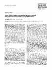
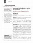
Fig. 1. Western blot analysis of protein lysates from postnatal and adult
rat sciatic nerve. Both the GABAB1a (130 kDa) and the GABAB2 subunits (100 kDa) were expressed during postnatal development but
decreased with age and the progression of myelination. The overall
expression of GAD65 and GAD67 increased somewhat during development. To confirm the loading of equal amounts of protein, the
membrane was also labeled with b-actin (n 5 5).
as GAD65/67 were expressed in the postnatal sciatic
nerve. The GABAB receptors clearly decreased with age,
whereas the GAD increased with a maximum at P15.
Both GAD65 and GAD67 were present in the adult nerve
but at a lower level than at P15 (Fig. 1).
GABA and GABAB Receptors Are Present
Primarily in Nonmyelinating Schwann Cells
The GABAB receptor expression was investigated in
sections of adult rat DRG and in teased preparations of
adult rat sciatic nerve. In DRGs, both neurons and satellite glia expressed GABAB receptors, and the expression
was particularly high in the satellite glia (Fig. 2A,B;
arrows). In the sciatic nerve, GABAB receptors were
found primarily in nonmyelinating Schwann cells (Fig.
2C,C0 ,D), and this cell type was also found to be GABA
and GAD65/67 positive (Fig. 2E,F). These data suggest
that nonmyelinating Schwann cells both generate and
store GABA.
GABAB Receptors GABA and GAD65/67 Are
Present in a Small Population of Myelinating
Schwann Cells
Most myelinating Schwann cells did not express
GABAB receptors, GAD65/67, or GABA. A small population of axons with thick myelin sheaths expressed the
GABAB receptor, GABA, and GAD65/67 in the nodal
region (Fig. 2G–L). Whereas GAD65/67 was on the axonal side of the node (Fig. 2J), the GABAB receptor was
present on the glial side (Fig. 2G,H,K,L). The distribution
of the GABAB receptor covers not only the nodal area
but also the paranodal and juxtaparanodal segment of
these Schwann cells (Fig. 2G,K). The labeling partially
overlapped with Caspr I labeling, an axonal paranodal
marker. Furthermore, the axon connected with the node
Journal of Neuroscience Research
289
was labeled with peripherin, indicating that the axon is
sensory. The exact location of GABA itself was somewhat
difficult to determine since it was unclear whether it was
distributed on the glial or axonal side (Fig. 2I).
We next investigated where the fibers containing
GABAB positive nodes originated. Cryosections were
made from the DRG with intact dorsal and ventral roots
that were labeled with antibodies to the GABAB receptor
subunits. The glia of the ventral root completely lacked
GABAB receptors. GABAB-positive nodes as well as nonmyelinating Schwann cells were localized to the dorsal
root (Fig. 2M). The fibers surrounded by these Schwann
cells were labeled with NF200 (Fig. 2K) but also contained a population that was labeled with peripherin (Fig.
2L), which specifically labels sensory fibers. The fact that
the GABAB-positive nodes are present only in the dorsal
root further demonstrates that these nodes have a sensory
origin.
The GABAB Receptor Is Upregulated in
Degenerated Nerves and in the Distal Segment of
Crushed Rat Sciatic Nerves
Because the GABAB receptor is downregulated with
age and rate of myelination, we also wanted to investigate
possible changes in the expression induced by an injury,
especially in the distal segment, where the Schwann cells
lose axonal contact after injury. To this end, adult sciatic
nerve was dissected, cut into 3–5-mm segments, and
cultured for 1 or 2 weeks. The GABAB receptor
was upregulated in the segments (Figs. 2O,P and 3).
Immunochemistry also indicated that the GABAB receptor partially colocalized with the MBP labeling
(Fig. 2O,P). The distal segment of the in vivo crushed and
degenerated sciatic nerve was investigated next. The segment was crushed 6 days prior to sacrifice of the animal.
Immunolabeled cryosections made from these nerves
showed an increase in GABAB receptor expression similar
to that seen in the in vitro degenerated segments
(Fig. 2N).
Expression of GABAB Receptor, GABA, and
GAD65/67 in Schwann Cell Development
To further study the distribution of the GABAB
receptor, GABA, and GAD65/67 in Schwann cell development, dissociated E17 rat DRG cultures were used.
Shortly after plating, both DRG neurons and Schwann
cells expressed GABAB1 and GABAB2 receptor subunits
in cultures, as is the case in vivo (Fig. 4A,A0 ,B,B0 ,I,J). The
receptors were localized evenly in the Schwann cell
membrane (Fig. 4A,A0 ,B,B0 ). After 4 DIV, ascorbic acid
was added to the medium to induce myelination. The
cultures were kept for 7–28 DIV to obtain thick myelin.
The nonmyelinating Schwann cells of the cultures continued to express the GABAB receptor during the entire
time in culture (data not shown). Cells that started their
myelination program downregulated the GABAB receptor
expression, and fully myelinated segments had no or low
expression (Fig. 4E,E0 ,F,F0 ).
290
Corell et al.
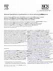
Fig. 2. In sections of the adult dorsal root ganglia many neurons
express the GABAB1 (red) and GABAB2 subunits (red, A,B). The
labeling was even more intense in Satellite cells (arrows, A,B). DRG
neurons are double-labeled with neuronal marker neurofilament 200
kDa (NF). In preparations of teased adult sciatic nerve (C-F) both
GABAB1 (red) and GABAB2 receptor (red) were expressed uniformly
in nonmyelinating Schwann cells (C,D). GABA (red, E) and GAD65/
67 (red, F) were also expressed in these cells. The nonmyelianting
Schwann cells were double-labeled with GFAP (green, C0 ,F). DAPI
was used as nuclei staining (blue). In preparations of teased adult sciatic
nerve, the expression of GABAB1 (red) and GABAB2 (red) were
localized to the myelin sheath at the node of Ranvier (G,H) in a small
population of larger caliber axons; double-labeled with the myelin
marker Rip (green, G,H). In these myelinated fibers, GABA was localized at the node of Ranvier (I) and GAD65/67 was expressed in the
axon and node of Ranvier (J). All axons positive for GABA receptors
at the nodes expressed neurofilament 200 kDa (NF, green, K0 ) and
peripherin (green, L0 ). The paranodes in these axons were labeled with
Caspr (blue, K,K0 ,L,L0 ). To determine the origin of this population,
the dorsal root (DR) and the ventral root (VR) were labeled with
anti-GABAB receptor (M). Interestingly, GABAB receptors exist only
in Schwann cells of sensory axons. Myelin sheaths are double-labeled
with myelin basic protein (MBP, I,J,M). (N) The distal segment of an
in vivo crushed nerve that was removed from the rat after 6 days. The
section show a clear up-regulation of GABAB1 (red) in the myelinated
fibers labeled with MBP (green). In segments of the sciatic nerve, cultured in vitro for on week, to mimic the distal segment after injury, the
same upregulation of both GABAB receptor subunits could be seen
(O, P). This experiment show that the upregulation of the GABAB
receptor was not due to external factors such as macrophage infiltration
of externally produced growth factors. The GABAB receptor subunits
in red and MBP in green. Scale bars 5 50 mm.
Journal of Neuroscience Research
GABA and Its B Receptor in Schwann Cells
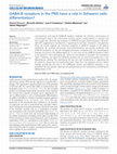
Fig. 3. Adult sciatic nerve was dissected cut in 3-5 mm segments and
cultured for 1 and 2 weeks. This procedure was made to mimic Wallerian degenerate. Both receptor subunits were upregulated in segments cultured for 1 and 2 weeks compared to control. This western
confirms the data shown in figure 2N-P, indicating the increased
GABAB receptor expression may be an attempt by the Schwann cells
to be more responsive to excreted GABA from growing axons. The
stimulation of the GABAB receptor could potentially lead to a production of growth factors and other chemoattractants necessary for
axonal guidance and sprouting.
Both GAD65/67 and GABA were found primarily
in the neuronal cell bodies of the cell cultures (Fig. 4K,L),
but they were also present in the axon itself (data not
shown). Similarly to the nonmyelinating Schwann cells
observed in vivo, GABA and GAD65/67 were present in
the premyelinating young Schwann cells (Fig. 4C,D), and
the expression decreased as the cells started to myelinate
axons (data not shown). Just as in vivo, fully myelinated
segments expressed very little GABA or GAD65/67 (Fig.
4G,H).
Functional Studies of the GABAB Receptor in
DRG Cell Cultures
It has previously been shown that baclofen, a potent
GABAB receptor agonist, decreases proliferation and
reduces the synthesis of myelin proteins in pure cultures
of Schwann cells from neonatal rats (Magnaghi et al.,
2004; Faroni et al., 2011). However, the proliferation and
myelination in these cultures was induced by using forskolin, a potent adenylate cyclase agonist. Because our
cultures proliferated spontaneously and also contained
neurons that actively produced GABA in the culture, we
wanted to investigate the response to GABA agonists and
antagonists in this more complex system. In DRG cultures, Schwann cell proliferation is spontaneous and visibly high the first days after plating but decreases with
time. We first determined that the highest rate of proliferation was at 48 hr (2 DIV) after plating (data not shown).
DRG primary cultures were then exposed to 100 mM of
the GABAB receptor agonist baclofen or 10 mM of the
antagonist CGP55845 for 2 DIV, and cell proliferation
was then evaluated by using bromodeoxyuridine (BrdU)
incorporation. The number of BrdU-incorporating cells
compared with the total number of cells did not decrease
with treatment of either baclofen or CGP55845 compared with DMSO-treated controls (Fig. 5A).
To test whether baclofen or CGP55845 would
interfere with the myelination process, other DRG cultures were treated with these compounds for 28 DIV.
Journal of Neuroscience Research
291
To sustain the levels of the agonist or the antagonist, fresh
compounds were applied twice per week during normal
medium change. With real-time PCR, the relative
mRNA expression levels of myelin markers were normalized with the housekeeping gene GAPDH and compared
among the treatment groups. The mRNA expression levels for MAG and MBP decreased in the baclofen-treated
cultures and increased in cultures treated with GCP55485
(Fig. 5B–D). The mRNA levels for PMP22 showed a
similar change, but it did not reach statistical significance
(Fig. 5C). The mRNA levels of GFAP did not change
with addition of baclofen or CGP55485 (Fig. 5E).
DISCUSSION
GABA and Its B Receptor in Nonmyelinating
Schwann Cells of the Sciatic Nerve
The GABA, GAD65/67, and GABAB receptors
are all present in premyelinating and nonmyelinating
Schwann cells, indicating that these cells both generate
and store GABA and that the cells can respond to GABA
through their B receptors in vivo and in vitro. It is conceivable that GABA and its receptors have a function similar
to that in astrocytes, in which focused GABA release activates GABAB receptors on neighboring neurons and
modulates neuronal activity (Beenhakker and Huguenard,
2010). Premyelinating and nonmyelinating Schwann cells
might use GABA for similar glia–neuron communication,
for example, to regulate nociceptive fibers and sensory
functions. Mice that lack the GABAB1 subunit, which
makes this receptor nonfunctional, show altered pain
reception as well as hyperalgesia to thermal stimuli, corroborating this theory (Magnaghi et al., 2008; Faroni
et al., 2014).
During PNS development, the number of premyelinating Schwann cells decreases with age and the rate of
myelination. This was also true for the GABAB receptor
expression in vivo as well as in vitro. GABA and its B
receptor were absent in most of the myelinating Schwann
cells, whereas the GAD activity was sustained. This indicates that the GABA synthesized in the adult sciatic nerve
acts via the ionotropic GABAA, which is also present in
Schwann cells (Magnaghi et al., 2006).
GABAB Receptors at the Node of Ranvier in a
Small Population of Axons in the Sciatic Nerve
A population of myelinated fibers displayed
GABAB receptors as well as GABA and GAD65/67 at
the node of Ranvier. The GABAB receptor was present on the glial side of the node, whereas GAD65/67
was localized to the axon. The localization of GABA
was somewhat difficult to interpret; it might, in part,
be restricted to the microvilli covering the node and
would thus be in the glial compartment. This indicates
that GABA might be generated in the axon, then
transported and stored in the surrounding myelinating
Schwann cells. These fibers had thick myelin sheaths
and expressed both NF200 and peripherin, a label for
292
Corell et al.
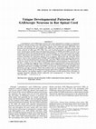
Fig. 4. In primary DRG cultures, all glial cells expressed both
GABAB1 (red) and GABAB2 (red) subunits shortly after plating
(A,B). These cells also expressed GABA and GAD65/67 (C,D;
red). The premyelinating Schwann cells were double labeled with
GFAP (A0 ,B0 ,C,D; green). At 28 DIV, many Schwann cells had
myelinated axons. These myelinating Schwann cells had lost their
GABAB receptor expression (E,F; red). GABA and GAD65/67
expression also decreased in the cells that started to myelinate
(G,H; red). GAD65/67 expression was localized to the cell membrane of the Schwann cells (D). Myelin was double labeled with
the myelin markers CNP (E0 ; green) and MBP (F0 ,G,H; green).
DRG neurons express GABAB receptors, GABA, and GAD65/67
in culture (I–L; red) and are double labeled with NF200. Scale
bars 5 50 mm.
Journal of Neuroscience Research
GABA and Its B Receptor in Schwann Cells
sensory neurons. This was corroborated by the finding
that nodal GABA, GAD65/67, and GABAB receptors
all originated from the dorsal root. It has previously
been shown that mice lacking the GABAB1 receptor
subunit have irregularities in the myelin sheaths and an
increased expression of several myelin proteins in large
myelinated fibers expressing NF200 (Magnaghi et al.,
2008). This group of neurons might be the same population found in this study. The glial cells myelinating
these axons could be more dependent on GABA’s
direct actions via B receptors and, thus, more vulnerable in a mouse lacking the GABAB1 receptor subunit.
293
This finding is especially interesting in light of the new
phenotypes of Schwann cells that are being described
(Hoke et al., 2006; Jesuraj et al., 2012). To date, phenotypic differences in Schwann cells, have been related
to motor and sensory neurons, but a phenotypic difference between sensory neurons of different calibers is
plausible.
Distribution of GABA and Its B Receptor in DRG
Cell Culture
Schwann cells in cultures devoid of neurons possess
functional GABAB receptors and generate GABA (Magnaghi et al., 2004). To mimic the environment of the
developing PNS better, we chose to investigate the
GABAB receptor-mediated actions of GABA in primary
fetal DRG cultures containing a mixture of sensory neurons and glia. The Schwann cells in these cultures proliferate, mature, and myelinate axons without the addition of
growth-promoting agents (Fex Svenningsen et al., 2003).
The sensory neurons of the cultures expressed the
GABAB receptor, GAD65/67, and GABA. This is in
accordance with what is known about the distribution of
these proteins in vivo (Towers et al., 2000; Charles et al.,
2001). The distribution of the GABAB receptor in
Schwann cells also correlated with Western blots and the
immunochemical results from in vivo material. Pre- and
nonmyelinating Schwann cells both expressed GABAB
receptors that were downregulated as the myelination
process commenced. The same was true for GABA and
GAD65/67. These data show that the Schwann cells in
primary DRG cultures correlate well with the in vivo
findings. Schwann cells, both in vivo and in vitro, have the
ability to generate GABA and respond to this neurotransmitter. Such autocrine/paracrine signaling has previously
been suggested. For example, GABA, released by
Schwann cells, mediated by protein kinase C, regulates
the synthesis and transport of the glutamate transporter
EAAC1, activating an autocrine loop that controls the
uptake of glutamate, a precursor of GABA (Perego et al.,
2012). The developmental downregulation of GABA and
Fig. 5. The function of the GABAB receptor was studied in primary
dissociated DRG cultures. Cultures were treated with either 100 mM
baclofen or 10 mM CGP55485 for 2 DIV. The BrdU-incorporating
cells were manually counted and compared with the total number of
cells. No change in proliferation rate was detected with either treatment (A; t-test, P 5 0.2868, n 5 8; C: P 5 0.4260, n 5 8; error bars
represent SEM). To investigate the effect of these compounds on
myelination, the DRG cultures were treated with either 100 mM
baclofen or 10 mM GCP55485 for 28 DIV. The cultures were then
used for mRNA expression analysis with quantitative real-time PCR.
The relative expression levels of the markers MAG (B), PMP22 (C),
MBP (D), and GFAP (E) were normalized with the housekeeping
gene GAPDH (one-way ANOVA, with LSD post hoc test; B: control
vs. baclofen *P 5 0.049, control vs. CGP55485 *P 5 0.025, n 5 4; C:
control vs. baclofen P 5 0.47, control vs. CGP55485 P 5 0.156,
n 5 4; D: control vs. baclofen *P 5 0.032, control vs. CGP55485
**P 5 0.01, n 5 5; E: control vs. baclofen P 5 0.807, control vs.
CGP55485 P 5 0.838, n 5 4; error bars represent SEM).
Journal of Neuroscience Research
294
Corell et al.
its B receptor, seen both in vivo and in vitro, also suggests
that GABA might play a role in the maturation of the sciatic nerve.
GABAB Receptor Is Not Involved in Spontaneous
Schwann Cell Proliferation
During in vivo developmental Schwann cell proliferation and subsequent myelination, axons are always present. In pure cultured Schwann cells in which the
proliferation and myelination are induced by forskolin, a
potent activator of the cAMP/protein kinase A pathway,
both the proliferation and the expression of myelin proteins are affected by the activation of the GABAB receptor
with the GABA agonist baclofen (Magnaghi et al., 2004).
Although these data suggest that GABA might be implicated in both proliferation and myelination in pure
Schwann cells, it is not clear whether GABA has this effect
in a culture system containing neurons or whether this is
the case in vivo. It fact, it has recently been shown that
Schwann cells devoid of neurons and Schwann cells
grown with neurons present respond differently and that
neuronal contact results in a more in-vivo-like behavior
(Stassart et al. 2013). It is thus far from clear whether the
same signal transduction pathways are used by forskolininduced proliferating Schwann cells and by those grown
in the presence of axons. In an attempt to mimic nerve
development, we repeated the experiments performed by
Magnaghi et al. (2004) but instead used dissociated developing DRG primary cultures, in which the Schwann cells
proliferate spontaneously in vitro. Neither baclofen nor the
GABAB receptor antagonist CGP55485 had any effect on
this Schwann cell proliferation. These results suggest that
neither GABA nor the GABAB receptor is involved in
developmental Schwann cell proliferation. The difference
between the earlier data and our data likely depends on
the presence of the neurons.
The Inhibition of GABAB Receptor Activity
Increases Myelination
We also investigated the effect of the GABAB receptor agonists and antagonists on long-term myelination in
the DRG cultures. In cultures treated with baclofen
MAG and MBP mRNA decreased, whereas GCP55485
had the reverse effect. These data indicate that GABA
might be involved in the myelination process through its
B receptor, controlling myelin protein expression and the
myelination process.
GABAB Receptors Increase After Injury
GABAB receptor expression is high in undifferentiated Schwann cells and nonmyelinating Schwann cells.
We therefore hypothesized that Schwann cells in damaged nerves would upregulate their GABAB receptor
expression. To investigate this, we studied the expression
in adult sciatic nerve segments that were allowed to
degenerate in vitro for 1 and 2 weeks, mimicking the
Wallerian degeneration of the distal segment after nerve
transection. In the predegenerated neurons, no axonal
sprouting occurs, but Schwann cell proliferation decreases
after 72 hr in culture (Fex Svenningsen and Kanje, 1998).
A Western blot of these segments shows that the expression of the GABAB receptor increases. We also investigated segments of the rat sciatic nerve crushed. In vivo, 6
days after nerve injury, Schwann cell proliferation had
decreased, and the bands of B€
ugner had formed (in
rodents). There is also an increase in NRG1 type III at
this time point, secreted from outgrowing neurons. This
NRG1 type III is likely involved in the initiation of myelination (Fex Svenningsen and Dahlin, 2013; Stassart
et al., 2013). Teased preparations of the in vitro segments
and cryosections of the in vivo crushed nerve show a clear
upregulation of the GABAB receptor in MBP-positive
Schwann cells. The increase is somewhat greater in the
in vivo segments, which might indicate an involvement in
the remyelination process. It is not likely that the GABAB
receptors are involved in the injury-induced Schwann cell
proliferation since this happens relatively early during the
regeneration process (Pellegrino et al., 1986; Fex Svenningsen and Kanje, 1998). Because the GABAB receptor
expression increases over time for at least 2 weeks, it
might instead be involved in efforts to communicate with
the nerve sprouts that should appear if there was a proximal segment. The Schwann cells of the bands of B€
ungner,
in the distal segment, normally secrete growth factors and
chemoattractants that stimulate and guide axonal growth
(Mudo et al., 1993). The increased GABAB receptor
expression might be an attempt by the Schwann cells to
be more responsive to excreted GABA from the growing
axons. The stimulation of these GABAB receptors could
potentially lead to a production of growth factors and
other chemoattractants necessary for axonal guidance and
sprouting. It is possible that GABA production by the
Schwann cells might be directly involved in axonal
sprouting. This is the case in neural development, when
the growth cone must respond accurately to stimuli that
direct its growth. This axonal navigation depends on
extracellular concentration gradients of numerous
guidance cues, including GABA (Bouzigues et al., 2007).
Xenopus spinal growth cone behavior as well as olfactory
neuron axonal guidance and target recognition are both
modulated by a gradient of GABAB receptors (Xiang
et al., 2002). It is conceivable that the increase in GABAB
receptors seen in Schwann cells in the injured nerve is a
natural response to stimulate arriving axons to grow.
ACKNOWLEDGMENTS
We are grateful for the expert technical assistance of
Karen Rich. The MBP and Caspr antibodies were a kind
gift from the laboratory of Dr. David R. Colman. The
authors have no conflicts of interest.
REFERENCES
Beenhakker MP, Huguenard JR. 2010. Astrocytes as gatekeepers of
GABAB receptor function. J Neurosci 30:15262–15276.
Bettler B, Tiao JY. 2006. Molecular diversity, trafficking and subcellular
localization of GABAB receptors. Pharmacol Ther 110:533–543.
Journal of Neuroscience Research
GABA and Its B Receptor in Schwann Cells
Bettler B, Kaupmann K, Mosbacher J, Gassmann M. 2004. Molecular
structure and physiological functions of GABAB receptors. Physiol Rev
84:835–867.
Bouzigues C, Morel M, Triller A, Dahan M. 2007. Asymmetric redistribution of GABA receptors during GABA gradient sensing by nerve
growth cones analyzed by single quantum dot imaging. Proc Natl Acad
Sci U S A 104:11251–11256.
Charles KJ, Evans ML, Robbins MJ, Calver AR, Leslie RA, Pangalos
MN. 2001. Comparative immunohistochemical localisation of
GABAB1a, GABAB1b, and GABAB2 subunits in rat brain, spinal cord,
and dorsal root ganglion. Neuroscience 106:447–467.
Charles KJ, Deuchars J, Davies CH, Pangalos MN. 2003. GABA B
receptor subunit expression in glia. Mol Cell Neurosci 24:214–223.
Desarmenien M, Feltz P, Occhipinti G, Santangelo F, Schlichter R.
1984. Coexistence of GABAA and GABAB receptors on A delta and C
primary afferents. Br J Pharmacol 81:327–333.
Faroni A, Castelnovo LF, Procacci P, Caffino L, Fumagalli F, Melfi S,
Gambarotta G, Bettler B, Wrabetz L, Magnaghi V. 2014. Deletion of
GABA-B receptor in Schwann cells regulates remak bundles and small
nociceptive C-fibers. Glia 62:548–565.
Fex Svenningsen Å, Kanje M. 1998. Regulation of Schwann cell proliferation in cultured segments of the adult rat sciatic nerve. J Neurosci
Res 52:530–537.
Fex Svenningsen A, Dahlin LB. 2013. Repair of the peripheral nerve—
remyelination that works. Brain Sci 3:1182–1197.
Fex Svenningsen A, Shan W-S, Colman D, Pedraza L. 2003. Rapid
method for culturing embryonic neuron–glial cell cultures. J Neurosci
Res 72:565–573.
Fields RD, Stevens-Graham B. 2002. New insights into neuron–glia
communication. Science 298:556–562.
Fraser DD, Mudrick-Donnon LA, MacVicar BA. 1994. Astrocytic
GABA receptors. Glia 11:83–93.
Grubbs FE. 1969. Procedures for detecting outlying observations in samples. Technometrics 11:1–21.
Hanani M. 2010. Satellite glial cells in sympathetic and parasympathetic
ganglia: in search of function. Brain Res Rev 64:304–327.
Hoke A, Redett R, Hameed H, Jari R, Zhou C, Li ZB, Griffin JW,
Brushart TM. 2006. Schwann cells express motor and sensory phenotypes that regulate axon regeneration. J Neurosci 26:9646–9655.
Jessen KR, Hills JM, Saffrey MJ. 1986. Immunohistochemical demonstration of GABAergic neurons in the enteric nervous system. J Neurosci
6:1628–1634.
Jesuraj NJ, Nguyen PK, Wood MD, Moore AM, Borschel GH,
Mackinnon SE, Sakiyama-Elbert SE. 2012. Differential gene expression
in motor and sensory Schwann cells in the rat femoral nerve.
J Neurosci Res 90:96–104.
Kang J, Jiang L, Goldman SA, Nedergaard M. 1998. Astrocyte-mediated
potentiation of inhibitory synaptic transmission. Nat Neurosci 1:683–
692.
Kuner R, Kohr G, Grunewald S, Eisenhardt G, Bach A, Kornau HC.
1999. Role of heteromer formation in GABAB receptor function. Science 283:74–77.
Lujan R, Shigemoto R, Lopez-Bendito G. 2005. Glutamate and
GABA receptor signalling in the developing brain. Neuroscience 130:
567–580.
Luyt K, Slade TP, Dorward JJ, Durant CF, Wu Y, Shigemoto R,
Mundell SJ, Varadi A, Molnar E. 2007. Developing oligodendrocytes
express functional GABAB receptors that stimulate cell proliferation and
migration. J Neurochem 100:822–840.
Magnaghi V. 2007. GABA and neuroactive steroid interactions in glia:
new roles for old players? Curr Neuropharmacol 5:47–64.
Journal of Neuroscience Research
295
Magnaghi V, Ballabio M, Cavarretta IT, Froestl W, Lambert JJ, Zucchi
I, Melcangi RC. 2004. GABAB receptors in Schwann cells influence
proliferation and myelin protein expression. Eur J Neurosci 19:2641–
2649.
Magnaghi V, Ballabio M, Consoli A, Lambert JJ, Roglio I, Melcangi
RC. 2006. GABA receptor-mediated effects in the peripheral nervous
system: a cross-interaction with neuroactive steroids. J Mol Neurosci
28:89–102.
Magnaghi V, Ballabio M, Camozzi F, Colleoni M, Consoli A, Gassmann
M, Lauria G, Motta M, Procacci P, Trovato AE, Bettler B. 2008.
Altered peripheral myelination in mice lacking GABAB receptors. Mol
Cell Neurosci 37:599–609.
Magnaghi V, Parducz A, Frasca A, Ballabio M, Procacci P, Racagni G,
Bonanno G, Fumagalli F. 2010. GABA synthesis in Schwann cells is
induced by the neuroactive steroid allopregnanolone. J Neurochem
112:980–990.
Mudo G, Persson H, Timmusk T, Funakoshi H, Bindoni M, Belluardo
N. 1993. Increased expression of trkB and trkC messenger RNAs in the
rat forebrain after focal mechanical injury. Neuroscience 57:901–912.
Nicoll RA. 1988. The coupling of neurotransmitter receptors to ion
channels in the brain. Science 241:545–551.
Oka M, Wada M, Wu Q, Yamamoto A, Fujita T. 2006. Functional
expression of metabotropic GABAB receptors in primary cultures of
astrocytes from rat cerebral cortex. Biochem Biophys Res Commun
341:874–881.
Owens DF, Kriegstein AR. 2002. Is there more to GABA than synaptic
inhibition? Nat Rev Neurosci 3:715–727.
Pellegrino RG, Politis MJ, Ritchie JM, Spencer PS. 1986. Events in
degenerating cat peripheral nerve: induction of Schwann cell S phase
and its relation to nerve fibre degeneration. J Neurocytol 15:17–28.
Perego C, Di Cairano ES, Ballabio M, Magnaghi V. 2012. Neurosteroid
allopregnanolone regulates EAAC1-mediated glutamate uptake and triggers actin changes in Schwann cells. J Cell Physiol 227:1740–1751.
Pfaffl MW. 2001. A new mathematical model for relative quantification
in real-time RT-PCR. Nucleic Acids Res 29:e45.
Pfaffl MW, Tichopad A, Prgomet C, Neuvians TP. 2004. Determination
of stable housekeeping genes, differentially regulated target genes, and
sample integrity: BestKeeper—Excel-based tool using pair-wise correlations. Biotechnol Lett 26:509–515.
Ramakers C, Ruijter JM, Deprez RH, Moorman AF. 2003. Assumption-free analysis of quantitative real-time polymerase chain reaction
(PCR) data. Neurosci Lett 339:62–66.
Schoenen J, Delree P, Leprince P, Moonen G. 1989. Neurotransmitter
phenotype plasticity in cultured dissociated adult rat dorsal root ganglia:
an immunocytochemical study. J Neurosci Res 22:473–487.
Stassart RM, Fledrich R, Velanac V, Brinkmann BG, Schwab MH,
Meijer D, Sereda MW, Nave KA. 2013. A role for Schwann cellderived neuregulin-1 in remyelination. Nat Neurosci 16:48–54.
Stefansk W. 1972. Rejecting outliers in factorial designs. Technometrics
14:469–479.
Towers S, Princivalle A, Billinton A, Edmunds M, Bettler B, Urban L,
Castro-Lopes J, Bowery NG. 2000. GABAB receptor protein and
mRNA distribution in rat spinal cord and dorsal root ganglia. Eur J
Neurosci 12:3201–3210.
Urazaev AK, Grossfeld RM, Fletcher PL, Speno H, Gafurov BS,
Buttram JG, Lieberman EM. 2001. Synthesis and release of Nacetylaspartylglutamate (NAAG) by crayfish nerve fibers: implications
for axon–glia signaling. Neuroscience 106:237–247.
Xiang Y, Li Y, Zhang Z, Cui K, Wang S, Yuan XB, Wu CP, Poo
MM, Duan S. 2002. Nerve growth cone guidance mediated by G
protein-coupled receptors. Nat Neurosci 5:843–848.
Keep reading this paper — and 50 million others — with a free Academia account
Used by leading Academics

Maro G Machizawa
Hiroshima University

Ludwig Kappos
University of Basel, University Hospital
John Slevin
University of Kentucky

Carlo Semenza
Università degli Studi di Padova