IJMS
Vol 34, No 3, September 2009
Case Report
Gastrointestinal Tuberculosis with Cecum
Involvement in a 33-Year-Old Woman
1
1
Davood Yadegarynia , Farhad Abbasi ,
1
Maryam Keshtkar-Jahromi ,
1
Sharareh Gholamin
Abstract
Abdominal tuberculosis is one of the most prevalent forms of
extra-pulmonary tuberculosis. Various regions of gastrointestinal tract including cecum, terminal ileum, peritoneum, lymphatic system, and solid viscera can be affected by tuberculosis. Here we report a 33-year-old woman presented with fever,
chills, and a history of abdominal discomfort. Lymphadenopathy was detected on physical examination. Contrast computed
tomography of chest and abdomen showed patchy densities
and thickening of the ileocecal wall respectively. Histological
studies of the biopsy samples documented the existence of
tuberculosis.
Iran J Med Sci 2009; 34(3): 213-216.
Keywords ● Tuberculosis ● extra-pulmonary ● abdominal
Introduction
uberculosis (TB) has plagued the human beings since
ancient times. Despite advances in medicine, TB is
still a major problem in developing and some developed countries. Difficulty in controlling the disease is attributed
to factors such as transglobal migration, increasing population
age, socioeconomic deprivations, and the acquired immunodeficiency syndrome (AIDS).
As long as pulmonary TB is a problem, extra-pulmonary TB
1
is also a health problem. In order of frequency, the extrapulmonary sites most commonly involved by TB are the lymph
nodes, pleura, genitourinary tract, bones and joints, meninges,
peritoneum, and pericardium. However, virtually all organ sys2
tems may be affected. Extra-pulmonary TB can be classified
based on the pathogenesis into three groups. The first group
comprises superficial mucosal foci resulting from the spread of
infectious pulmonary secretions via the respiratory and gastrointestinal tracts. Such lesions were once almost inevitable complications of extensive cavitary pulmonary disease but are now rare.
The second group comprises foci established by contiguous
spread, such as from a subpleural focus into the pleural space.
The third group comprises foci established by lymphohematogenous dissemination, either at the time of primary infection or,
less commonly, from established chronic pulmonary or ex3
trapulmonary foci. Failure of pulmonary tubercular disease
control resulted in increasing number of extra-pulmonary tuberculosis, such as abdominal tuberculosis.1
Abdominal TB can affect the gastrointestinal tract, the peritoneum, lymph nodes of the small bowel mesentery, or the
solid viscera including the liver, spleen, and pancreas. The
gastrointestinal tract is involved in 66-75% of patients with
T
1
Infectious Diseases and Tropical
Medicine Research Center,
Shaheed Beheshti University of
Medical Sciences,
Tehran, Iran.
Correspondence:
Davood Yadegarynia MD.
Infectious Diseases and Tropical
Medicine Research Center,
Shaheed Beheshti University of Medical
Sciences,
Tehran, Iran.
Tel: +98 21 22439963
Fax: +98 21 22439964
Email: yadegarinia@yahoo.com
Received: 12 November 2008
Revised: 3 January 2009
Accepted: 19 April 2009
Iran J Med Sci September 2009; Vol 34 No 3 213
D. Yadegarynia, F. Abbasi, M. Keshtkar-Jahromi, Sh. Gholamin
abdominal TB. The terminal ileum and the ileocecal region are the most common sites, fol4
lowed by the jejunum and colon.
Case Presentation
A 33-year-old woman presented with a history of
4-month intermittent fever and chills. There was a
history of abdominal discomfort and pain of 3
weeks duration since a month ago. She also
complained of weight loss during the last month.
She had not been exposed to any ill people.
Physical examination of head and neck,
heart, lungs, extremities, and nervous system
were normal. Apart from axillary lymphadenopathy, nothing was remarkable on physical examination. No hepatosplenomegaly was found.
In initial evaluation, erythrocyte sedimentation rate of 72 mm/hr and tuberculin skin test
with induration exceeding 20 mm were detected. The chest radiograph was normal but
spiral computed tomography (CT) of thorax
with and without contrast showed bilateral peripheral patchy densities of upper lobes with focal
pleural thickening of the right side (figure 1).
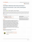
Figure 2: Marked thickening of cecal wall with irregular
lumen surface
history of abdominal discomfort, lymphoma
with abdominal source was suspected.
Colonoscopic biopsy of cecum demonstrated
eight soft pale tan tissue fragments measuring
1.2 × 0.2 × 0.2 cm in gross view whereas microscopic view of the sections revealed ulcerated colonic mucosa with polymorphous leukocytic infiltration and epithelioid granulomas,
consistent with tuberculosis. Acid fast staining
and PCR for mycobacterium tuberculosis were
positive on the biopsy specimen. There was an
ulcerovegetan mass in cecum extended to
terminal ileum (figure 3).
Figure 1: Bilateral peripheral patchy densities of upper
lobes
Bronchoalveolar lavage was done for detection of malignancy. Also the specimen taken
on the lavage was sent for detection of mycobacterium tuberculosis through acid fast bacilli
staining and PCR technique. The report was
negative in both studies. Abdominal CT revealed marked thickening of cecal wall and
terminal ileum with irregular lumen surface.
Other parts of colon, liver, spleen, kidneys and
other organs were normal (figure 2).
Since inflammatory process was detected in
abdominal evaluation and the patient had a
214 Iran J Med Sci September 2009; Vol 34 No 3
Figure 3: Ulcerovegetan mass is seen in cecum extended
to terminal ileum
Axillary lymph node biopsy showed chronic
necrotizing granulomatous lymphadenitis, consistent with TB and large areas of granular eosinophilic necrosis with surrounding epithelioid histiocytes and giant cells. Anti-TB drugs isoniazid
(300 mg daily), rifampin (600 mg daily),
Tuberculosis of cecum
ethambutol (800 mg daily), and pyrazinamide
(1200 mg daily) were administered. After several weeks, abdominal pain and discomfort
gradually decreased and finally disappeared.
After 6 months, the patient's general condition
was good and there was no abdominal pain.
Discussion
Gastrointestinal TB, once considered common,
and then a relatively rare disease is now reemerging in association with AIDS and multidrug resistant Mycobacterium tuberculosis.
Intestinal involvement with TB may be either
primary from ingesting of the organism, or secondary usually from a pulmonary source. 5
Gastrointestinal TB can affect any part of
the tract, from the mouth to the anus. The most
common site is the ileocecal area. This is
probably caused by several factors; I. A massive amount of lymphoid tissue, II. Physiologic
stasis causing increased contact time between
the bacteria and the intestinal lumen, III. Increased rate of fluid and electrolyte absorption,
and IV. Minimal digestive activity, permitting
1
greater contact time.
Other commonly involved sites are the colon and the jejunum. Uncommon involvements
of the esophagus, duodenum and small bowel
6
in isolation have also been reported.
Clinical manifestations of gastrointestinal
TB are non-specific. The most common complaint is abdominal pain, occurring in approxi1
mately 80% of cases. Patients with primary
TB may present with abdominal pain, fever,
and a tender, fixed palpable mass in the ileocecal area. Weight loss is more common in
secondary intestinal tuberculosis. Only one
third of the patients with gastrointestinal TB
may present with diarrhea. 5 The exact mechanism for diarrhea is unknown but it may be
caused by generalized inflammatory response
of the intestine and the subsequent effect of
the cytokines, leukotrienes, and prostaglandins
1
on fluid and electrolyte transport. Hemorrhage
and the presence of gross blood in the stool
are distinctly uncommon.5
Diagnosis of intestinal TB may be difficult
5
radiologically and even histologically. Radiological and histological manifestations of intestinal TB may resemble other diseases such as
1
Crohn’s, lymphoma, or malignancies. It must
also be distinguished from regional enteritis,
sarcoidosis, actinomycosis, amebiasis, carcinoma, and periappendiceal abscess. 5 In imaging studies, chest radiology is usually normal,
but evidence of pulmonary TB in chest radiography or high resolution CT supports the diagnosis. Radiographs of the abdomen are useful
in patients with intestinal obstruction and perfo4
ration. Lymphadenopathy is a common manifestation of abdominal TB. Mesenteric, omental, and peripancreatic lymph nodes are most
commonly involved. Contrast enhanced CT
shows peripherally enhancing lymph nodes
with low density centers explained by a peripheral inflammatory reaction and central caseous
necrosis. This appearance is highly suggestive
7
but not pathognomonic of abdominal TB. In
histological study, epithelioid cell granulomata
that resembles Crohn’s disease makes the
diagnosis difficult. However, the epithelioid cell
granulomata with the peripheral rim and
plasma cells, giant cells and central caseating
necrosis, fibrosis, and calcification in healing
lesions can be used as histological criteria for
4,8
making differentiation.
The first line of treatment for abdominal TB
9
is medical treatment. More recent reports
show that 6 months of treatment is adequate
for abdominal tuberculosis. Diagnosis in the
early phase results in a good response to
medical treatment. Patients usually show signs
of improvements as early as 2 weeks after
starting the treatment. Even patients with signs
of incomplete gut obstruction have shown improvement and cessation of symptoms with
1
medical treatment alone. Surgery for abdominal TB is reserved for patients who develop
complications, such as obstruction, perforation,
and stricture formation.1,4 Stomach and duodenum are involved in just 0.3-2.3% of patients
10
with TB of the gut. Abdominal TB is a rare
manifestation of extrapulmonary TB. In a study
by Chen and co-workers 21 patients with abdominal TB were identified during a 20-year
period. Tuberculous peritonitis was noted in 11
patients. The remaining patients were diagnosed
as having TB of gastrointestinal tract (n = 6),
11
urinary tract (n = 2), and pelvis (n = 2).
In another study by Akinkuolie and colleagues, the clinical records of 47 patients who
diagnosed as having abdominal TB between
January 1986 and December 2005 in Nigeria,
were reviewed. Common presenting symptoms
and signs were abdominal pain 76.6%, ascites
59.6%, weight loss 53.2%, and fever 29.8%.
Average duration of symptoms before presentation was 3 months. Thirteen percent of the
patients had earlier been treated for pulmonary
tuberculosis in the hospital. Mantoux test was
positive in 33% and ascitic fluid evaluation was
diagnostic for TB in 29%. Chest radiography
showed abnormal findings in 25% of the patients and laboratory evaluation of sputum
12
samples showed acid fast bacilli in 14.3%.
PCR might be a rapid alternative for identification of mycobacterium tuberculosis in culture
Iran J Med Sci September 2009; Vol 34 No 3 215
D. Yadegarynia, F. Abbasi, M. Keshtkar-Jahromi, Sh. Gholamin
and allow for earlier setup of susceptibility test13
ing. Overall sensitivity and specificity of PCR
are 100% and 99.7%, respectively. In the study
by Smith and co workers, the sensitivity and
specificity of PCR were 93% and 100%, re14
spectively. In another study by Webster and
colleagues the Roche Amplicor Mycobacterium
tuberculosis PCR test (RMtb-PCR) was compared with mycobacterial culture, with the
BACTEC 460 (Becton Dickinson, USA) system
and inoculation on Lowenstein-Jensen media.
The results were interpreted with an adjusted
"gold standard" incorporating clinical diagnosis.
The sensitivity, specificity, and positive and
negative predictive values of RMtb-PCR compared with the adjusted gold standard for clinical specimens were 79%, 99%, 93%, and
98%, respectively. This study demonstrates
the value of a commercial nucleic acid amplification kit for rapid diagnosis of Mycobacterium tuberculosis, particularly in smear-positive speci15
mens or BACTEC culture-positive specimens.
6
7
8
9
10
11
Conflict of Interest: None declared
References
12
1
2
3
4
5
Faylona JMV, Chung SCS. Abdominal tuberculosis revisited. Ann Coll Surg 1999;
3: 65-70.
Raviglione MC, O’Brien RJ. Tuberculosis. In:
Kasper DL, Braunwald E, Fauci AS et al, editors. Harrison’s principles of internal medith
cine. 16 edition. McGraw-Hill, 2005. p. 957.
Mandlle, Bennett, & Dolin. Principles and
th
practice of infectious diseases, 6 ed.
2005 Chuerchill Livingstone. An Imprint of
Elsevier. Extrapulmonary Tuberculosis.
MD Consult www.mdconsult.com
Kapoor VK. Abdominal tuberculosis. Medicine 2007; 35: 257-60.
Mandlle, Bennett, & Dolin. Principles and
th
practice of infectious diseases, 6 ed.
216 Iran J Med Sci September 2009; Vol 34 No 3
13
14
15
2005 Chuerchill Livingstone. An Imprint of
Elsevier. Chronic inflammatory processes.
MD Consult www.mdconsult.com
Ong WC, Cheemalakonda R. Colonic tuberculosis mimicking a diminutive sessile
polyp. Dig Endosc 2005; 17: 257-8.
Yilmaz T, Sever A, Gür S. CT findings of
abdominal tuberculosis in 12 patients. Comput Med Imaging Graph 2002; 26: 321-5.
Pulimood AB, Peter S, Ramakrishna B.
Segmental colonoscopic biopsies in the
differentiation of ileocolic tuberculosis from
Crohn’s disease. J Gastroenterol Hepatol
2005; 20: 688-96.
Mert A; Bilir M; Tabak F. Miliary tuberculosis. Clinical manifestations, diagnosis and
outcome in 38 adults. Respirology 2001; 6:
217-24.
Padussis J, Loffredo B, McAneny D. Minimally invasive management of obstructive
gastroduodenal tuberculosis. Am Surg
2005; 71: 698-700.
Chen HL, Wu MS, Chang WH, et al. Abdominal tuberculosis in southeastern Taiwan: 20 years of experience. J Formos
Med Assoc 2009; 108: 195-201.
Akinkuolie AA, Adisa AO, Agbakwuru EA,
et al. Abdominal tuberculosis in a Nigerian
teaching hospital. Afr J Med Med Sci 2008;
37: 225-9.
Forbes BA, Hicks KE. Ability of PCR assay
to identify Mycobacterium tuberculosis in
BACTEC 12B vials. J Clin Microbiol 1994;
32: 1725-8.
Smith MB, Bergmann JS, Woods GL. Detection of Mycobacterium tuberculosis in
BACTEC 12B broth cultures by the Roche
Amplicor PCR assay. J Clin Microbiol
1997; 35: 900-2.
Wobeser WL, Krajden M, Conly J, et al.
Evaluation of Roche Amplicor PCR assay
for Mycobacterium tuberculosis. J Clin Microbiol 1996; 34: 134-9.
Gastrointestinal Tuberculosis with Cecum Involvement in a 33-Year-Old Woman
Abdominal tuberculosis is one of the most prevalent forms ofextra-pulmonary tuberculosis. Various regions of gastrointestinaltract including cecum, terminal ileum, peritoneum, lymphaticsystem, and solid viscera can be affected by tuberculosis.Here we report a 33-year-old woman presented with fever,chills, and a history of abdominal discomfort. Lymphadenopathywas detected on physical examination. Contrast computedtomography of chest and abdomen showed patchy densitiesand thickening of the ileocecal wall respectively. Histologicalstudies of the biopsy samples documented the existence oftuberculosis....Read more