ACTA CHROMATOGRAPHICA, NO. 15, 2005
APPLICATION OF RP-HPLC–ED ASSAY
TO ANALYSIS OF THE BLOOD
OF PARKINSON’S DISEASE PATIENTS
B. K. Głód1,* and K. I. Stańczak2
1
Meat and Fat Research Institute, Jubilerska 4, 04-190 Warsaw, Poland
Medical Research Centre, Polish Academy of Sciences, Pawińskiego 5, 03-187 Warsaw,
Poland
2
SUMMARY
Results from application of reversed-phase HPLC with electrochemical detection to analysis of catecholamines and total antioxidant potential are presented. The assay was used to estimate their daily profiles in
blood serum of patients treated with anti-Parkinson drugs. It was found that
increases in the concentration of hydroxy radicals in blood serum, and of
the total antioxidant potential related to the hydroxy radical, was inversely
correlated with the concentration of catecholamines in the serum. This suggests that catecholamines are pro-oxidants of hydroxy radicals. The method
also enabled us to study changes in the concentrations of catecholamines
during their storage at room temperature. It was found that change of drug
color was caused by the oxidation of benseraside.
INTRODUCTION
Free radicals are implicated in the pathogenesis of many diseases
including, e.g., cancer, Alzheimer’s disease, rheumatoid arthritis, and Parkinson’s disease [1]. For this reason the literature contains descriptions of
many methods of analysis of free radicals [2–6]. Because of the interaction
between free radicals and antioxidants, in pathology changes of antioxidant
concentration are observed. Antioxidant concentrations can also be measured [7–10]. It has been found that an estimate of total antioxidant potential (TAP) is frequently much more useful then separate analysis of all the
antioxidants [11–17]. Cooperation between different antioxidants sometime
results in better protection than separate compounds. Examples of this synergism are glutathione, which regenerates ascorbate [18], and ascorbic acid,
which regenerates α-tocopherol [19]. Both these quantities, i.e. oxidative
- 276 -
�stress and total antioxidant potential, are correlated with each other and even
at times identified [20,21].
TAP has been introduced for evaluating, mainly, the antioxidant
capacity of complex biological samples, such as serum. These tests exploit
different free-radical generators (usually thermolabile diazo compounds
generating peroxy radical) and oxidation of the samples analyzed. Because
of competition, the sample inhibits interaction between the detector and
radical. Results are calculated from the delay time during which antioxidants are consumed. The basic requirement of analyzed sample is that its
oxidation rate constant is much higher then that of the compound used as
a ‘detector’. One literature method is based on generation of peroxy radicals and then fluorimetric or chemiluminescence kinetic measurement of
the product of their reaction with the so-called detector, i.e. a compound
which is easy to detect. A derivative of fluorescein is usually used as detector for fluorimetric [15] or photometric [12,21] measurement; luminol is
used for the chemiluminescence method [22].
Parkinson’s disease (PD) usually results from a reduction in the
efficiency of the extrapyramidal movement system. Dopamine (DA) plays
a crucial role in the pathomechanism of PD [23]. Injury of the substantia
nigra by free radicals reduces the concentration of dopamine, its main neurotransmitter [23,24]. Patients are usually treated substitutionally with Ldopa (and inhibitors of L-dopa decarboxylase), a precursor of dopamine
readily absorbed in the brain. Analysis of L-dopa in the serum enables diagnosis of PD and enables individualization of dose selection, and thus avoidance of side-effects (fluctuations and dyskinesis).
Catecholamines (among them L-dopa) can be analyzed by many
methods, for example capillary electrophoresis [25], gas chromatography
with mass spectrometric detection (GC–MS) [26,27], radioimmunoenzymatic methods [25,28] and thin-layer chromatography [26,27]. Most frequently, however, they are analyzed by HPLC, usually in the reversedphase mode, with amperometric detection [29,30]. In this technique sample
retention is directly proportional to its hydrophobicity, which means that
ionized (dissociated) compounds are usually not retained on the column
and are eluted in the dead column volume. Retention of acidic catecholamines therefore increases with decreasing pH whereas that of basic catecholamines decreases.
In the blood serum of PD patients changes of free radical concentrations and, indirectly, total antioxidant potential (TAP) are expected for
two main reasons. Patients are treated with huge dose of L-dopa and other
- 277 -
�catecholamines, which are strong antioxidants [31,32]. Also, hypermetabolism caused by the characteristic PD tremor increases free radical concentrations.
In this paper we present results from application of RP-HPLC to
the estimation of TAP. TAP was measured by generation of hydroxy radicals in the Fenton reaction and their spin trapping with hydroxybenzoate
in the presence of the sample. The daily profile of L-dopa in PD patients
will be correlated with TAP values.
EXPERIMENTAL
Instrumentation
Measurements were performed by means of a chromatograph comprising interface box, K-5004 four-channel degasser, K-1500 solvent organizer, dynamic mixing chamber, K-1001 HPLC pump, K-2600 fast scanning UV detector, Eurochrom 2000 chromatographic data acquisition and
analysis software (all from Knauer, Berlin, Germany), Basic+ Marathon
autosampler (Spark Holland, Emmen, The Netherlands), Jet-Stream Plus
column thermostat (Industrial Electronics, Langenzersdorf, Austria), and
LaChrom L-3500A amperometric detector (E. Merck, Darmstadt, Germany). Samples were separated on a 250 mm × 4 mm i.d., 5 µm particle, Hibar
RP-18 column (Merck).
Reagents
p-Hydroxybenzoic acid (pHBA), 3,4-dihydroxybenzoic acid (3,4DHBA), and phosphate buffered-saline (PBS) tablets were obtained from
Sigma (St Louis, MO, USA). All other reagents (Sigma; Fluka, Buchs,
Switzerland; and POCh, Gliwice, Poland) were of analytical-reagent grade
and were used without further purification. Water was passed through Millipore (Bedford, USA) Milli-RO4 and Milli-Q water-purification systems.
Mobile phases were filtered through a 0.22-µm membrane filter (Millipore).
Procedures
Chromatographic experiments were performed at a flow rate of 1
mL min−1. The column was stabilized at 30°C by passage of mobile phase
for 1 h before chromatographic measurements. Acetate–citrate buffer (pH
4.3) containing 0.125 mmol L−1 EDTA and 5% methanol was used as mobile phase.
- 278 -
�Stock solutions (10 mmol L−1) of the analyzed compounds were
prepared in Milli-Q water and diluted to the required concentration before
use. Samples (20 µL) were injected by means of an autosampler. Output
signals from the photometric detector working simultaneously at 210, 254,
and 280 nm and from the amperometric detector working at +0.8 V relative to Ag/AgCl were continuously displayed by the computer. Each sample
was injected six times and the average was taken for further processing.
Hydroxy radicals were generated by means of the Fenton reaction
[33], by incubation, at 37 ºC for 1 min, of 0.5 mmol L−1 Fe2+, 2 mmol L−1
ADP, and 2 mmol L−1 H2O2 in 50 mmol L−1 phosphate buffer (pH 7.4) in
the presence of 1 mmol L−1 p-hydroxybenzoic acid and the analyzed sample. The product of reaction of pHBA with hydroxy radicals, i.e. 3,4-DHBA
was detected amperometrically. Addition of sample (serum or dopamine)
to the reaction mixture reduced its peak, because of competitive reaction
with the radicals. The reaction was stopped by addition of 2 mmol L−1
DMSO and 0.1 mg mL−1 Desferal, and the reaction mixture was immediately analyzed by HPLC.
Subjects
Blood plasma was obtained from sixteen patients with idiopathic
PD (12 men and 4 women, age from 53 to 80 years, average 64 ± 7 years,
duration of disease 5–15 years). Informed written consent was obtained
from every patient. Permission for the study was obtained from the BioEthnic Commission of the Military Medical Chamber. Patients were hospitalized for two weeks in the Rehabilitation Clinic of the Military Medical
Institute in Warsaw. They received chronically an oral dose of 250 mg of
L-dopa four times daily (Madopar; F. Hoffmann–La Roche, Bazylea, Switzerland). Each session started at 7 a.m. Blood samples were taken immediately and 1, 2, 3, and 5 h after L-dopa administration. Blood plasma was
deproteinized by use of saturated uranyl acetate, excess of which was removed by use of pH 4 phosphate buffer.
Data Analysis
Intrasubject comparisons were performed by using Student’s t-test
for dependent variables. Significance was set at P < 0.05.
- 279 -
�RESULTS AND DISCUSSION
Separation of Catecholamines
The aim of our measurements was to separate L-dopa from the other
amines and from other reducing agents present in the serum. We also wanted to use the same conditions for estimation of TAP. This seemed possible because TAP measurements are based on analysis of a catechol groupcontaining compound, 3,4-dihydroxybenzoic acid.
For mobile phases we tested buffers of different pH and containing
different concentrations of methanol. It was found that reducing the pH
improved the separation of L-dopa. Addition of methanol, at concentrations
as low as 5%, to the mobile phase improved peak symmetry and reduced
retention of the amines up to 20 min. Further increasing the methanol concentration reduced retention so much that the amines were not separated
from compounds used for estimation of TAP.
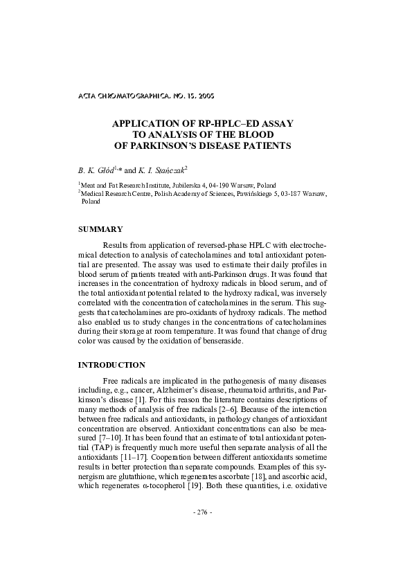
Fig. 1
Effect of potential ∆Φ [mV] (relative to Ag/AgCl) on the peak height of catecholamines:
1. L-dopa, 2. dopamine, 3. benserazide, 4. carbidopa. Chromatographic conditions: 250
mm × 4 mm i.d., 5 µm particle, Hibar RP-18 column with precolumn (Knauer); temperature 30°C; mobile phase acetate–citrate buffer, pH 3.5–methanol, 95:5 (v/v), flow rate
1 mL min−1
- 280 -
�Detection Conditions
Catecholamines, because they are aromatic compounds, can be monitored by use of a photometric detector. Amperometric detection is, however, much more sensitive (approximately three orders of magnitude)
[30]. Some of the catecholamine peaks were above the range of the amperometric detector and were, therefore, analyzed by use of serially connected photometric detector.
Hydrodynamic voltammograms obtained from different catecholamines are presented in Fig. 1. In subsequent experiments a potential of
+0.8 V relative to the AgCl/Ag electrode was selected as the optimum
compromise between the peak height and noise level. It was found that for
the catecholamines investigated linear calibration plots were obtained (detection limit approximately 1 nM, R2 = 0.97; linear dynamic range four orders of magnitude and RSD = 5%).
It was found that, in contrast with photometric measurements [12,
30], chromatographic measurements of TAP are not characterized by a
linear calibration plot (Fig. 2). This is because we measure the decrease of
CPA [rel. units]
1.0
0.8
0.6
0.4
0.2
0.0
0
1
2
3
4
cl-dopa [µg/ml]
Fig. 2
Effect of L-dopa concentration on TAP of the hydroxy radical (measured as relative
changes of 3,4-DHBA peak height). Chromatographic conditions: 250 mm × 4 mm i.d.,
5 µm particle, Hibar RP-18 column with precolumn (Knauer); temperature 30°C; mobile
phase acetate–citrate buffer, pH 3.5–methanol, 95:5 (v/v), flow rate 1 mL min−1; volume
injected 20 µL; amperometric detection at +0.8 V relative to Ag/AgCl
- 281 -
�the chromatographic peak. When it completely disappears further increasing the sample concentration has no effect. It is, therefore, convenient to
present results on a scale between 0 (lack of the interaction between radical and sample) and 1 (complete scavenging of free radicals by the sample),
as is presented in Fig. 2.
Pharmacokinetics of the Catecholamines
Estimation of the daily profile of the L-dopa concentration in the
serum of PD patients is very important because of possibility to individualizing therapy for each patient [34]. It was found that the largest concentration of L-dopa in serum was observed 1 h after uptake by the patient (Fig.
3) [35]. Although the concentration of carbidopa also increased during the
first hour, it subsequently remained stable (Fig. 4). Changes of concentrations (data not presented) of other amines and other antioxidants present
in serum at high concentrations (observation by UV detection) were not
statistically significant [36].
C [µM]
0.8
*
0.4
#
0
0
1
2
3
5
time [h]
Fig. 3
Changes of L-dopa concentration in the blood serum of Parkinson’s disease patients before medicine uptake and after one, two, three, and five hours. *P < 0.05 relative to time 0;
#
P < 0.05 relative to time 1. Chromatographic conditions: 250 mm × 4 mm i.d., 5 µm
particle, Hibar RP-18 column with precolumn (Knauer); temperature 30°C; mobile phase
acetate–citrate buffer, pH 3.5–methanol, 95:5 (v/v), flow rate 1 mL min−1; volume injected 20 µL; amperometric detection at +0.8 V relative to Ag/AgCl
- 282 -
�9
*
8
c [µM]
*
*
*
7
6
5
0
1
2
t [h]
3
5
Fig. 4
Changes of carbidopa concentration in the blood serum of Parkinson’s disease patients
before medicine uptake and after one, two, three, and five hours. *P < 0.05 relative to
time 0. Chromatographic conditions: 250 mm × 4 mm i.d., 5 µm particle, Hibar RP-18
column with precolumn (Knauer); temperature 30°C; mobile phase acetate–citrate buffer,
pH 3.5–methanol, 95:5 (v/v), flow rate 1 mL min−1; volume injected 20 µL; amperometric detection at +0.8 V relative to Ag/AgCl
Pro- and Antioxidant Properties of Dopamine
In this paper we would like to examine the oxidative properties of
DA (related to hydroxy radical) using results obtained by use of this RPHPLC–ED assay. It was found that addition of DA to the reaction mixture
(as described in ‘Procedures’) reduced the intensity of the 3,4-DHBA peak
(compare Figs 5A and 5B). This means that in the presence of the strong
antioxidant (ADP) DA scavenges hydroxy radicals. When ADP is absent
from the reaction mixture, however, addition of dopamine increases the
intensity of the 3,4-DHBA peak (compare Figs 5C and 5D). This means
that under these conditions DA is a pro-oxidant, probably because it reduces iron ions, which catalyze the Fenton reaction. Similar results were obtained for L-dopa. This result means that, depending on its concentration
and the presence in the solutions of other antioxidants, DA can be a proor antioxidant.
The literature contains contradictory information about oxidative
properties of dopamine [37–39]. On the one hand dopamine is involved in
oxidative stress (because of its catalysis of the Fenton reaction by reduc- 283 -
�Fig. 5
Pro- and antioxidant properties of dopamine in relation to the hydroxy radical. Description in the text. Chromatographic conditions: 250 mm × 4 mm i.d., 5 µm particle, Hibar
RP-18 column with precolumn (Knauer); temperature 30°C; mobile phase acetate–citrate
buffer, pH 3.5–methanol, 95:5 (v/v), flow rate 1 mL min−1; volume injected 20 µL;
amperometric detection at +0.8 V relative to Ag/AgCl
tion of transition metal ions and generation of semiquinone and superoxide
anion radicals during their reduction); on the other hand it acts as an antioxidant (because of its ability to scavenge free radicals, because of its catechol and aromatic groups, and by complexation with transition metal ions).
Enzymatic oxidation of dopamine by monoaminooxidase type B
(MAO B) and its auto-oxidation produce hydrogen peroxide (H2O2) [37].
In the Fenton reaction (which DA promotes because of its ability to reduce
Fe(III) to Fe(II), which catalyzes the reaction) H2O2 generates extremely
reactive hydroxy radicals [24]. Also, metabolism of DA produces 6-hydroxydopamine and semiquinone radical, which are oxidants. It can, therefore, damage neurons, even causing their death by apoptosis [38].
On the other hand, the catechol group in the DA molecule is characterized by strong antioxidant properties. DA can also complex Fe(II), therefore, preventing the Fenton reaction. We can see that the effect of DA on
free radicals is rather complicated and its pro- or antioxidant properties
depend on its concentration [39].
- 284 -
�Changes of TAP in Parkinson’s Disease
As was described above, catecholamines can be either strong antioxidants or pro-oxidants. PD patients consume them in quite large quantities (up to 1500 mg per day). This reduces the tremor of patients, which
increases hypermetabolism and production of free radicals. We therefore
wished to check the correlation between their concentration and the TAP
value of blood serum. It was found that increasing the concentration of the
amine increased the pro-oxidant properties of serum (compare Figs 4–6).
It is worth noting that blood serum scavenges peroxy radicals (i.e. it has
antioxidant properties) [36]. It is probable that L-dopa metabolism, controlled by the enzyme MAO B, increases the production of superoxide anion
radical [37].
Fig. 6
Changes of TAP in the blood serum of the Parkinson’s disease patients before medicine
uptake and after one, two, three, and five hours. *P < 0.05 relative to time 0. Chromatographic conditions as for Fig. 1. The results are means ± SEM from five independent
experiments with triplicate analysis (*P < 0.05 compared with control value)
Oxidation of Catecholamines During Storage
We have observed that catecholamine solutions change color during
storage. Because they are strong reducing agents, we supposed that they
could be oxidized by atmospheric oxygen. Using this method of catechol- 285 -
�amine analysis we decided to analyze their concentration during storage at
25°C. As the compounds tested we selected catecholamines used as antiparkinsonian drugs (L-dopa, carbidopa, and benserazide). It was found that
the concentration of benseraside deceased the most quickly (Fig. 7) and
that this was responsible for the change of color of anti-parkinsonian drugs.
Fig. 7
Changes of concentrations of catecholamines (L-dopa, carbidopa, benseraside) during
storage at 25°C. Chromatographic conditions: 250 mm × 4 mm i.d., 5 µm particle, Hibar
RP-18 column with precolumn (Knauer); temperature 30°C; mobile phase acetate–citrate
buffer, pH 3.5–methanol, 95:5 (v/v), flow rate 1 mL min−1; volume injected 20 µL; amperometric detection at +0.8 V relative to Ag/AgCl
ACKNOWLEDGEMENTS
The authors are grateful to Dr Waldemar Pakszys from the Central
Clinical Hospital of Military Medical Institute for supplying us with the
blood serum of PD patients.
- 286 -
�REFERENCES
[1] B.K. Głód, G.A. Czapski, and P.R. Haddad, Trends Anal. Chem.,
19, 492 (2000)
[2] P.G. Osborne and K. Yamamoto, J. Chromatogr. B, 707, 3 (1998)
[3] B. Halliwell, H. Kaur, and M. Ingelman-Sundeberg, Free Radical
Biol. Med., 10, 439 (1991)
[4] B.K. Głód and P. Grieb, Chem. Anal. (Warsaw), 47, 399 (2002)
[5] A. Rehman, M. Whiteman, and B. Halliwell, Br. J. Pharmacol.,
122, 1702 (1997)
[6] G. Ellis, I. Adatia, M. Yazdanpanah, and S.K. Makela,
Clin. Biochem., 31, 195 (1998)
[7] D.G. Watson, C. Atsriku, and E.J. Oliveira, Anal. Chim. Acta,
492, 17 (2003)
[8] K. Kostner, S. Bayai, M. Jansen, G. Khoschsorur, W.H. Horl,
G. Maurer, B. Winklhofer-Roob, and K. Derfler, Clin. Chim. Acta,
288, 21 (1999)
[9] A.H. Waterfall, G. Singh, and J.R. Fry, C.A. Marsden,
Neurosci. Lett., 200, 69 (1995)
[10] I.N. Acworth and B. Bailey, The Handbook of Oxidative
Metabolism, ESA, 1995
[11] C.A. Rice-Evans, Free Radical Res., 33, 59 (2000)
[12] M. Valkonen and T. Kuusi, J. Lipid Res., 38, 823 (1997)
[13] A. Ghiselli, M. Serafini, F. Natella, and C. Saccini, Free Radical
Biol. Med., 29, 1106 (2000)
[14] F. Tubaro, A. Ghiselli, P. Rapuzzi, M. Maiorino, and F. Ursini,
Free Radical Biol. Med., 24, 1228 (1998)
[15] A. Krasowska, D. Rosiak, K. Szkapiak, and M. Łukasiewicz,
Curr. Topics Biophys., 24, 89 (2000)
[16] H. Wang and J.A. Joseph, Free Radical Biol. Med., 27, 612 (1999)
[17] D. Genser, M-H. Kang, H. Vogelsang, and I. Elmadta, Eur. J. Clin.
Nutrit., 53, 675 (1999)
[18] J.E. Packer, T.F. Slater, and R.L. Willson, Nature, 278, 737 (1979)
[19] R. Stocker, M.J. Weidemann, and N.H. Hunt, Biochim. Biophys. Acta,
881, 391 (1986)
[20] H. Wang and J.A. Joseph, Free Radical Biol. Med., 27, 612 (1999)
[21] W. Malinka, M. Kaczmarz, B. Filipek, J. Sapa, and B. Głód,
Il Farmaco, 57, 737 (2002)
- 287 -
�[22] E. Lissi, M. Salim-Hanna, C. Pascual, and M.D. del Castillo,
Free Radical Biol. Med., 18, 153 (1995)
[23] O. Hornykiewicz and S.J. Kish, Adv. Neurol., 45, 19 (1986)
[24] S.G. Diamond, C.H. Markham, and M.M. Hoehn, Ann. Neurol.,
22, 8 (1987)
[25] L. L. Iverson and B. Jarrett, Brit. J. Pharmacol., 384, 816 (1970)
[26] H.G. Lovelandy, Biochem. Med., 15, 130 (1976)
[27] T.C. Lhuguenot and B.F. Maume, J .Chromatogr., 139, 218 (1977)
[28] D.C. Siggers, C. Calter, and P.A. Toseland, Clin. Chim. Acta,
30, 373 (1970)
[29] D.A. Richards, J. Chromatogr., 175, 293 (1979)
[30] B.K. Głód, K.I. Stańczak, A. Woźniak, and W. Pakszys,
Acta Chromatogr., 14, 142 (2004)
[31] B.K. Głód, K.I. Stańczak, A. Woźniak, and W. Pakszys,
J. Chromatogr. Sci., submitted for publication
[32] C.A. Rice-Evans, N.J. Miller, and G. Paganga, Free Radical Biol.
Med., 7, 933 (1996)
[33] R.A. Floyd, J.J. Watson, and P.K. Wong, J. Biochem. Biophys.
Methods, 10, 221 (1984)
[34] T. Zieliński, A. Drabik, W. Pakszys, and B.K. Głód, in preparation
[35] N. Simon, R. Gantcheva, B. Bruguerolle, and F. Viallet,
Parkinsonism Relat Disord., 10, 137 (2004)
[36] K.I. Stańczak, M.Sc. Thesis, CMDiK PAN, Warszawa 2004
[37] L. Antkiewicz-Michalczuk, A. Krygowska-Wajs, J. Michaluk,
I. Romańska, A. Szczudlik, and J. Vetulani, Neurosci. Res.
Commun., 25, 97 (1999)
[38] G. Cohen, J. Neural. Transm., 19(Suppl.), 89 (1983)
[39] J.W. Miller, J. Selhub, and J.A. Joseph, Free Radical Biol. Med.,
21, 241 (1996)
- 288 -
View publication stats
�
 Bronislaw Glod