Academia.edu no longer supports Internet Explorer.
To browse Academia.edu and the wider internet faster and more securely, please take a few seconds to upgrade your browser.
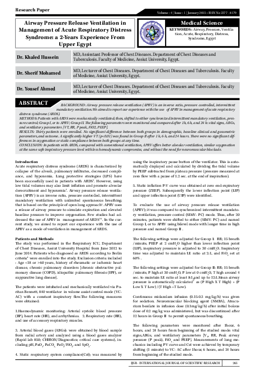
Airway Pressure Release Ventilation in Management of Acute Respiratory Distress Syndrome: a 2-Years Experience From Upper Egypt
Airway Pressure Release Ventilation in Management of Acute Respiratory Distress Syndrome: a 2-Years Experience From Upper Egypt
BACKGROUND: Airway pressure release ventilation (APRV) is an inverse ratio, pressure controlled, intermittent mandatory ventilation.We aimed to report our experience with the use of APRV in management of acute respiratory distress syndrome (ARDS). METHODS: Patients with ARDS were mechanically ventilated; then, shifted to either synchronized intermittent mandatory ventilation, pres-sure control; Group I, or to APRV; Group II. The following parameters were monitored and compared after 1h, 6 h, and 24 h: vital signs, ABGs, and ventilatory parameters [VT, RR, P peak, FiO2, PEEP]. RESULTS: Thirty patients were enrolled. No significant difference between both groups in demographic, baseline clinical and gasometric parameters,and outcome. A significantly higher VT (p<0.01) was found in Group II after 1 h, 6 h, and 24 hours. There were no significant dif-ferences in oxygenation or static compliance between both groups at any time. CONCLUSION: In patients with ARDS, compared with conventi...
Related Papers
Hiroshima journal of medical sciences
Practical use of airway pressure release ventilation for severe ARDS--a preliminary report in comparison with a conventional ventilatory support2009 •
Airway pressure release ventilation (APRV) is a ventilatory mode that allows unsupported spontaneous breathing at any phase of the ventilatory cycle with high mean airway pressures. We hypothesized that use of APRV might produce potential beneficial effects on oxygenation, reducing mortality in patients with severe acute respiratory distress syndrome (ARDS) in comparison with synchronized intermittent mandatory ventilation (SIMV) as a conventional mode of ventilation. We retrospectively reviewed data of 58 patients with severe ARDS (the ratios of partial arterial oxygen tension to fraction of inspired oxygen, PaO2/F(I)O2 ratio <150). The patients' data were divided into two groups: SIMV-group and APRV-group. Patients' backgrounds, oxygenation on day 0, 1, 3, 5 and 7 following initiation of each mode, vasopressor dependence, duration of ventilation, duration of ICU stay, and mortality in ICU were analyzed. PaO2/F(I)O2 ratios were statistically higher in the APRV-group (APR...
Cleveland Clinic Journal …
Airway pressure release ventilation: An alternative mode of mechanical ventilation in acute respiratory distress syndrome2011 •
Acute respiratory distress syndrome (ARDS) results in collapse of alveoli and therefore poor oxygenation. In this article, we review airway pressure release ventilation (APRV), a mode of mechanical ventilation that may be useful when, owing to ARDS, areas of the lungs are ...
The Egyptian Journal of Intensive Care and Emergency Medicine
Early transition to airway pressure release ventilation may facilitate weaning and improve the outcome of acute respiratory distress syndrome patients2002 •
Appropriate ventilatory intervention is life saving in acute respiratory distress syndrome (ARDS). Pressure controlled inverse ratio ventilation (PC-IRV) is the likely mode of ventilation benefiting in extreme conditions of ARDS. However, guidelines when to start PC-IRV is not yet well defined. The ventilation-related dilemma, which we faced in two illustrative cases of ARDS are presented. The first patient presenting clinically with ARDS but with high peak airway pressure (PIP) and low dynamic lung compliance, PC-IRV helped in reducing PIP, improved haemodynamics and the oxygenation of blood. In second patient with similar clinical presentation of ARDS, where although PIP was high but the dynamic compliance was better, the PC-IRV caused deterioration in PaO2. Here, patient rather did better with high PEEP (15 cm H2O) and usual I: E ratio (1:2). It is probable that the dynamic lung compliance (< 20ml/cmH2O), PIP (> 50 cm H2O) at conventional I: E ratio (1:2) ventilation (10 ml/kg) with hypotension might form the basis to develop a scoring system for guidance to switch over to PC-IRV ventilation. Further randomised prospective controlled clinical trials will then be required to establish indication to start PC-IRV in ARDS.
Critical Care Medicine
Ventilatory management of acute respiratory distress syndrome: A consensus of two2004 •
Purpose: Low tidal volume ventilation improves the outcomes of acute respiratory distress syndrome (ARDS). However , no studies have investigated the use of a rescue therapy involving mechanical ventilation when low tidal volume ventilation cannot maintain homeostasis. Inverse ratio ventilation (IRV) is one candidate for such rescue therapy, but the roles and effects of IRV as a rescue therapy remain unknown. Methods: We undertook a retrospective review of the medical records of patients with ARDS who received IRV in our hospital from January 2007 to May 2014. Gas exchange, ventilation, and outcome data were collected and analyzed. Results: Pressure-controlled IRV was used for 13 patients during the study period. Volume-controlled IRV was not used. IRV was initiated on 4.4 ventilation days when gas exchange could not be maintained. IRV significantly improved the PaO 2 /FiO 2 from 76 ± 27 to 208 ± 91 mmHg without circulatory impairment. The mean duration of IRV was 10.5 days, and all survivors were weaned from mechanical ventilation and discharged. The 90-day mortality rate was 38.5 %. Univariate analysis showed that the duration of IRV was associated with the 90-day mortality rate. No patients were diagnosed with pneumothorax. Conclusions: Pressure-controlled IRV provided acceptable gas exchange without apparent complications and served as a successful bridge to conventional treatment when used as a rescue therapy for moderate to severe ARDS.
Tuberculosis and respiratory diseases
Clinical Practice Guideline of Acute Respiratory Distress Syndrome2016 •
There is no well-stated practical guideline for mechanically ventilated patients with or without acute respiratory distress syndrome (ARDS). We generate strong (1) and weak (2) grade of recommendations based on high (A), moderate (B) and low (C) grade in the quality of evidence. In patients with ARDS, we recommend low tidal volume ventilation (1A) and prone position if it is not contraindicated (1B) to reduce their mortality. However, we did not support high-frequency oscillatory ventilation (1B) and inhaled nitric oxide (1A) as a standard treatment. We also suggest high positive end-expiratory pressure (2B), extracorporeal membrane oxygenation as a rescue therapy (2C), and neuromuscular blockage for 48 hours after starting mechanical ventilation (2B). The application of recruitment maneuver may reduce mortality (2B), however, the use of systemic steroids cannot reduce mortality (2B). In mechanically ventilated patients, we recommend light sedation (1B) and low tidal volume even wit...
Alveolaroverdistention and cyclicreopening of collapsed alveoli have been implicated in the lung damage found in animals submitted to artificial ventilation. To test whether these phenomena are impairing the recovery of patients with acute respiratory distress syndrome (ARDS) submitted to conventional 'mechanical ventilation (MV), we evaluated the impact of a new ventilatory strategy directed at minimizing "cyclic parenchymal stretch." After receiving pre-established levels of hemodynamic, infectious, and general care, 28 patients with early ARDS were randomly assigned to receive either MV based on a new approach (NA, consisting of maintenance of end-expiratory pressures above the lower inflection point of the P x V curve, VT < 6 ml/kg, peak pressures < 40 cm H20, permissive hypercapnia, and stepwise utilization of pressure-limited modes) or a conventional approach (C =conventional volume-cycled ventilation, VT = 12 ml/kg, minimum PEEP guided by F102 and hemodynamics and normal PaC02 levels). Fifteen patients were selected to receive NA, exhibiting a better evolution of the Pao2/Fl02 ratio (p < 0.0001) and of compliance (p = 0.0018), requiring shorter periods under Flo2 > 50% (p = 0.001) and a lower F102 at the day of death (p =0.0002). After correcting for baseline imbalances in APACHE II, we observed a higher weaning rate in NA (p =0.014) but not a significantly improved survival (overall mortality: 5/15 in NAversus 7113 in C, p =0.45). We concluded that the NAventila-tory strategy can markedly improve the lung function in patients with ARDS, increasing the chances of early weaning and lung recovery during mechanical ventilation. Amato MBP, Barbas CSV, Medeiros DM, Schettino GdPP, Filho GL, Kairalla RA, Deheinzelin D, Morais C, Fernandes EdO, Takagaki TY, de Carvalho CRR. Beneficial effects of the "open lung approach" with low distending pressures in acute respiratory distress syndrome: a prospective randomized study on mechanical ventilation.
New England Journal of Medicine
Effect of a Protective-Ventilation Strategy on Mortality in the Acute Respiratory Distress Syndrome1998 •
RELATED PAPERS
Unpublished
A Note About Evidence and the Coronado Expedition in the Río Sonora and San Pedro River Valleys2024 •
Periodic Research
Assessment of Grit and Decision Making Styles among Males and Females of different Age Groups2018 •
2023 •
Al-Ghazali and the Rationalization of Sufism
Al Ghazali and the Rationalization of Sufism2019 •
2011 •
Journal of the Mechanical Behavior of Materials
An atomistic study on the strain rate and temperature dependences of the plastic deformation Cu–Au core–shell nanowires: On the role of dislocationsAnalytical and Bioanalytical Chemistry
Passive sampling and/or extraction techniques in environmental analysis: a review2004 •
Journal of Kathmandu BernHardt College
Public Spending and Economic Growth: A Study of Nepal During Fiscal Year 1990/91-2020/212022 •
2010 •
Tobacco Prevention & Cessation
Patterns and related factors of bidi smoking in India2020 •
Audiology and Neurotology
Progressive Deterioration of Central Components of Auditory Brainstem Responses during Postnatal Development of the Myelin Mutant taiep Rat2000 •
RELATED TOPICS
- Find new research papers in:
- Physics
- Chemistry
- Biology
- Health Sciences
- Ecology
- Earth Sciences
- Cognitive Science
- Mathematics
- Computer Science

 yousef ahmed