Labelled Diagram of Neuron with Detailed Explanations
Last Updated :
12 Jan, 2024
A diagram of a neuron also known as the nerve cell is useful as a visual tool to illustrate the various components of the neuron. It also helps us to understand the functions of the neuron. This article incorporates a well-labeled diagram and a brief description of the components of a neuron highlighting their functions.
Definition of Neuron
The building blocks of the system responsible for transmitting and processing information are called neurons.
What are Neurons?
Neurons are specialised cells that plays an important role in facilitating communication among different parts of our body. Neurons are made up of three components-the cell body, dendrites and an axon. The complex and extensive network formed by neurons within our bodies allows us to think, perceive and carry out functions. They are crucial to the central nervous system which includes the brain and spinal cord. And also to the peripheral nervous system that governs our sensory experiences and motor abilities.
The study of neurons holds importance in unravelling the complexities of the brain and its impact, on our cognition and behaviour. 3 different types of neurons can be found in humans, based on their respective roles in the human nervous system, these are- sensory, motor and interneurons.
Labeled Diagram of a Neuron
The different components of the neuron are illustrated below, with each area specified and labeled.
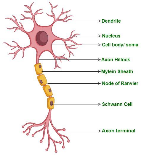
Structure of Neuron
A neuron is a complex and specialized cell with several key components, such as
- Dendrites: The reception of signals, from neighbouring neurons is primarily carried out by dendrites, which are crucial for this process. These branch like structures extend outward from the cell body providing an expanded surface area to receive information. Dendrites receive impulses known as neurotransmitters, from neurons. By receiving and processing these signals dendrites facilitate the integration of information allowing the neuron to comprehend the received signals and decide on a response.
- Soma or Cell body: The neuron’s central region, which is known as the soma or cell body, contains the nucleus, along with several important organelles. Paramount to the neuron’s overall performance and metabolic processes, the soma’s role is invaluable. It encompasses the cell’s nucleus in which DNA, genetic coding essential for protein synthesis and various cellular functions, resides. In addition, it is a crucial function of the soma to absorb and process the information received from dendrites. It is then that the decision whether to trigger or inhibit an electrical signal, known as the action potential, is made.
- Axon: The axon is like a long and thin projection extending out from the soma. It is responsible for carrying electrical impulses which are known as action potentials away from the soma. It acts as the primary source of communication between the neurons. These electrical messages are important for sending information over long distances. Around the axon, there is a layer of fatty substance which is known as the myelin sheath. This myelin sheath acts as the insulator which allows for more efficient and faster processing of the electrical message along the length of the Axon.
- Nodes of Ranvier: Nodes of Ranvier are small gaps along the axon where the myelin sheath is absent. These nodes play a crucial role in enhancing the speed of signal transmission. As the action potential jumps from one node to another, a process known as saltatory conduction, it drastically speeds up the signal propagation compared to non-myelinated axons. This phenomenon is vital for efficient communication within the nervous system and ensuring swift responses to stimuli.
- Synapses: There are little ends present at the ends of the axon known as axon terminals. Sometimes these axon terminals are also called as boutons. These terminals contain synaptic vesicles, which are tiny sacs that store and release neurotransmitters. These neurotransmitters play a very important role in carrying the electrical message from one neuron to the next neuron. It even carries the electrical message from one neuron to the muscle or a gland. The release of these neurotransmitters takes place at specialised junctions called as synapses. This process allows a smooth transfer of information from one neuron to the next, or from one neuron to the muscle or the gland.
- Schwann cells: Schwann sells provide support to the axons in our bodies peripheral nervous system. Schwann cells also maintain the structural component of the axon and manages the environment around the axon. It also aids in the regeneration of the damaged nerve fibres. These cells also produce the myelin sheath for the axon. This myelin sheath insulates the axon.
Please Login to comment...