VECTOR-BORNE DISEASES, SURVEILLANCE, PREVENTION
Longitudinal Studies of Japanese Encephalitis Virus Infection in Vector
Mosquitoes in Kurnool District, Andhra Pradesh, South India
N. ARUNACHALAM,1 U.S.N. MURTY,2 D. NARAHARI,3 A. BALASUBRAMANIAN, P. PHILIP SAMUEL,
V. THENMOZHI, R. PARAMASIVAN, R. RAJENDRAN, AND B. K. TYAGI
Centre for Research in Medical Entomology (Indian Council of Medical Research), 4, Sarojini Street,
Chinna Chokkikulam, Madurai, Tamil Nadu, India
KEY WORDS Japanese encephalitis, Culex tritaeniorhynchus, Culex gelidus, Kurnool, south India
Japanese encephalitis virus (family Flaviviridae, genus
Flavivirus, JEV) is currently one of the most important
arboviral childhood viral encephalitis in Asia, causing at
least 50,000 clinical cases and 10,000 deaths every year
(WHO 2005). Japanese encephalitis (JE) is a zoonotic
disease, with a complex life cycle involving pigs and
ardeid birds and vector mosquitoes. Humans are only
occasionally infected and are a “dead end” host, because
viremia in human blood is too low and transient to infect
mosquitoes. JE is predominantly a rural disease, and it
occurs scattered over extensive areas and seldom in peripheral localities of cities (Reuben and Gajanana 1997).
In India, JE was Þrst reported in 1955 (Work and
Shah 1956); subsequently, many epidemics have occurred in different parts of the country. JE outbreaks
have been reported as many as 25 states/union territories of India (Kabilan et al. 2004). In some states,
such as Uttar Pradesh, Bihar, and Andhra Pradesh, JE
has emerged as a perennial public health problem
during the rainy season. In 2005, a major encephalitis
outbreak was reported in Gorakhpur, Uttar Pradesh,
which was the most severe JE epidemic so far reported, affecting ⬎5,700 persons, mainly children,
with ⬎1,300 deaths (WHO 2006).
JE virus has been recovered from several mosquito
species (19 species) in different parts of India, and the
most important vectors are Culex tritaeniorhynchus Giles
and Culex vishnui Theobald from which the largest number of isolations have been made (Geevarghese et al.
2004). In Asia, development of irrigation systems and the
expansion of rice (Oryza sativa L.)-growing areas have
facilitated the increase of JEV vectors (Lacey and Lacey
1990). Although epidemics invariably are preceded by
increase in vector abundance, several other factors, including mosquito infection rate are involved (Gajanana
et al. 1997). Our knowledge of the epidemiology of JE
virus is still incomplete considering the diverse ecogeographical regions in India. A detailed understanding of JE
epidemiology will lead to the achievement of prediction
of outbreaks and sound mosquito control methods. The
present studies were undertaken to investigate the dynamics JEV transmission between June 2002 and July
2006 in a JE-endemic district, Kurnool, Andhra Pradesh
(AP) state, south India. The main objective was to determine the seasonal abundance and JE infection rates of
mosquito vector population to understand their role in
the transmission of JE.
Materials and Methods
Corresponding author, e-mail: crmeicmr@icmr.org.in.
2 Indian Institute of Chemical Technology, Hyderabad, Andhra
Pradesh, India 500007.
3 Department of Health, Andhra Pradesh, India 500001.
1
Study Area. The study was carried out in Kurnool
district, AP (Fig. 1), and its total population is
1,724,795. Most of the people are working in the ag-
0022-2585/09/0633Ð0639$04.00/0 䉷 2009 Entomological Society of America
Downloaded from https://academic.oup.com/jme/article/46/3/633/861274 by guest on 13 February 2022
J. Med. Entomol. 46(3): 633Ð639 (2009)
ABSTRACT A 4-yr (2002Ð2006) entomological study was carried out in Kurnool district, Andhra
Pradesh state, south India, to identify the mosquito vectors of Japanese encephalitis virus (family Flaviviridae, genus Flavivirus, JEV). In total, 37,139 female mosquitoes belonging Þve genera and 18 species
resting on vegetation were collected in villages and periurban areas at dusk. Mosquito species composition
and pattern of JEV infection in mosquitoes varied in periurban and rural areas. In periurban area, Culex
gelidus Theobald was abundant, msking up 49.7% of total catch followed by Culex tritaeniorhynchus Giles
(44.5%). In rural area, Cx. tritaeniorhynchus was predominant, making up 78.9% of total catch followed by
Culex quinquefasciatus Say (10.8%), Anopheles subpictus Grassi (7.1%), and Cx. gelidus (1.1%). In light trap
collections, Cx. gelidus and Cx. tritaeniorhynchus predominated in periurban and rural areas, respectively.
Of 50,145 mosquitoes screened JEV isolations were made only from Cx. gelidus and Cx. tritaeniorhynchus.
Based on high abundance and frequent JEV isolation, Cx. tritaeniorhynchus was found to be the principal
vector in both areas, whereas Cx. gelidus plays a secondary vector role in periurban areas only.
�634
JOURNAL OF MEDICAL ENTOMOLOGY
Vol. 46, no. 3
ricultural sector, are of low-income, and live in substandard housing. Domestic animals such as cattle,
pigs, and poultry are commonly sharing the living
space with the human population. A single crop of rice
was raised in irrigated Þelds of study villages from
August to January.
The climate of Kurnool is topical, and it can be divided
into three seasons: dry (FebruaryÐJune), wet or monsoon (JulyÐOctober), and winter (NovemberÐJanuary).
Southwest monsoon gives more dependable rains in the
area, which begins in June and persists through October.
During the study period (2002Ð2006), the maximum
temperature ranged from 30.3⬚C (December 2005) to
42.8⬚C (May 2003) (Fig. 2). Two suburban localities and
six villages where at least one JE case occurred during
1997Ð2002 were selected as index areas and sampled
once every 2 mo. The suburban localities located on the
margin of Kurnool are deÞned as periurban areas, which
are outside the municipal boundaries and thus without
basic civic amenities.
Mosquitoes resting on vegetation and bushes
around cattle sheds were collected by three insect
collectors for 1 h after dusk by mouth aspirator. Two
cattle sheds were sampled in each area, and the same
sheds were sampled on each occasion by three insect
collectors. Mosquito (only females) abundance was
calculated as number collected per person per hour.
Adult mosquitoes were also collected by using light
traps outdoors for 1 h simultaneously, and the samples
were also used for virus isolation. Mosquitoes were
transported to the laboratory for identiÞcation and
enumeration.
Downloaded from https://academic.oup.com/jme/article/46/3/633/861274 by guest on 13 February 2022
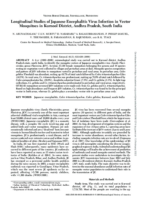
Fig. 1. (A) Map of India showing location of the Kurnool district, AP, India. (B) Map of Kurrnool district showing location
of study areas.
�May 2009
ARUNACHALAM ET AL.: JE VIRUS INFECTION IN VECTOR MOSQUITOES
635
350
80
60
250
Rainfall (mm)
50
200
40
150
100
20
50
10
0
JUN
JULY
AUG
SEP
OCT
NOV
DEC
JAN
FEB
MAR
APR
MAY
JUN
JULY
AUG
SEP
OCT
NOV
DEC
JAN
FEB
MAR
APR
MAY
JUN
JULY
AUG
SEP
OCT
NOV
DEC
JAN
FEB
MAR
APR
MAY
JUNE
JULY
AUG
SEPT
OCT
NOV
DEC
JAN
FEB
MAR
APR
MAY
JUNE
JULY
0
2002
2003
TOTAL Rainfall (mm)
2004
Max. Temp.
2006
2005
Min. Temp.
Relative Humidity 17.30hrs
Fig. 2. Meteorological data recording during the study.
Female mosquitoes were tested in single-species pools
of 3Ð50 for JE virus by using an antigen-capture enzymelinked immunosorbent assay (ELISA) for the initial
screening for ßavivirus and inoculation of Toxorhynchites
splendens Wiedemann combined with an indirect immunoßuorescence assay (Toxo-IFA) to conÞrm infection with JEV (Gajanana et al. 1997): 1) antigen-capture
ELISA by using monoclonal antibody 6B4A-10 (reactive
against all the viruses in the JE-WN-SLE-MVE complex)
as capture antibody and monoclonal antibody peroxiTable 1.
dase conjugate SLE MAB 6B6C-1 (reactive against all
ßaviviruses) as detector antibody, and 2) insect bioassay
by using mosquito pools positive in ELISA inoculated
intracerebrally into Tx. splendens larvae (early stage of
third instar). In total, eight larvae from a laboratory cyclic
colony were used for each positive pool, and ⬇0.17 l of
ELISA-positive mosquito pools was inoculated. Inoculated larvae were incubated at 29⬚C for 7 d, and head
squashes of inoculated larvae were tested by IFA by
using a JEV-speciÞc monoclonal antibody, MAB 112
Species composition of mosquitoes in Kurnool
Species
Aedes aegypti (L.)
Ae. albopictus (Skuse)
Ae. vexans (Meigen)
Ae. vittatus
Anopheles barbirostris
An. hyrcanus
An. peditaeniatus
An. stephensi Liston
An. subpictus Grassi
An. vagus
Armigeres subalbatus
C.x gelidus Theobald
Cx. infula Theobald
Cx. pseudovishnui
Cx. quinquefasciatus Say
Cx. tritaeniorhynchus Giles
Cx. vishnui Theobald
Lutzia fuscanus Wiedemann
Mansonia indiana Edwards
Ma. uniformis Theobald
Total
Resting collection at dusk
Light trap
Rural
Periurban
Rural
Periurban
Total
collected
31
0
0
0
77
42
18
26
1,531
4
99
237
0
0
2315
16,948
124
17
0
1
21,470
29
3
4
0
3
6
5
79
295
1
48
7,793
0
9
363
6,979
25
6
1
20
15,669
3
0
0
1
98
54
18
0
1,134
1
78
194
1
0
1,673
4,405
0
0
0
0
7,660
16
0
1
1
6
8
0
14
405
0
66
11,602
0
3
491
4,388
20
0
1
27
17,049
79
3
5
2
184
110
41
119
3,365
6
291
19,826
1
12
4,842
32,720
169
23
2
48
61,848
%
0.13
0.005
0.008
0.002
0.3
0.18
0.07
0.19
5.44
0.01
0.47
32.06
0.002
0.02
7.83
52.9
0.27
0.04
0.002
0.08
Downloaded from https://academic.oup.com/jme/article/46/3/633/861274 by guest on 13 February 2022
30
Temperature 0C / Relative Humidity (17.30hrs)
70
300
�636
JOURNAL OF MEDICAL ENTOMOLOGY
Vol. 46, no. 3
500
4
3.5
3
350
2.5
300
2
250
200
1.5
150
1
Minimum infection rate / 1000
400
100
0.5
50
0
Ju
l.
Se
p.
N
o
Ja v.
n.
05
M
ar
.
M
ay
.
Ju
l.
Au
g.
Se
p.
N
ov
Ja .
n.
06
M
ar
.
M
ay
.
Ju
l.
ar
.
ay
.
M
M
Ju
n
e
02
Se
p.
D
ec
Fe .
b.
03
Ap
r.
Ju
l.
Au
g.
Se
p.
O
ct
.
N
ov
.
D
e
Ja c.
n.
04
0
Month
Urban MIR
Fig. 3.
Urban PMH
Vector abundance and JEV infection of Cx. tritaeniorhynchus in rural areas of Kurnool district.
(Kimura-Kuroda and Yasuri 1983), and virus was detected by anti-mouse immunoglobulin conjugated with
ßuorescein isothiocyanate (Dakoppats, Glostrup, Denmark).
Virus infection rate in mosquitoes was expressed as
minimum infection rate (MIR) per 1,000 females tested
(Chiang and Reeves 1962). The pool size of adult females
for virus isolations varied from 3 to 50 specimens per
pool. MIR is not an appropriate parameter for expressing
infection rates and for comparison between vector species when pool sizes are unequal. To overcome this
problem maximum likelihood estimate method was also
used to calculate the infection rate (http://www.
cdc.gov/ncidod/dvbid/westnile/software.htm). Virus
infection rate was expressed as maximum likelihood estimate with 95% conÞdence intervals.
Results
Mosquito abundance varies in periurban and rural
ecosystems. In total, 37,139 female mosquitoes representing six anopheline and 12 culicine species were
collected in dusk collections (Table 1). In periurban
areas, Cx. gelidus was the most abundant species, making up 49.7% of the total mosquitoes collected, followed by Cx. tritaeniorhynchus (44.5%), whereas in
rural areas Cx. tritaeniorhynchus was the most abundant species, making up 78.9% of the total collected,
and Cx. gelidus consisted only 1.1% of the total catch.
In light trap collection, 24,709 female mosquitoes
representing six anopheline and 12 culicine species
were collected. Cx. gelidus was the most abundant
species in periurban areas, making up 68.1% of the
total mosquitoes collected, followed by Cx. tritaeniorhynchus (25.7%), whereas in rural areas Cx. tritaeniorhynchus was the most abundant species, making up
57.5% of the total mosquitoes collected, and Cx. gelidus
consisted only 2.5% of the total.
Cx. tritaeniorhynchus population ßuctuates in a similar pattern in both rural and periurban areas (Figs. 3
and 4). Abundance of Cx. tritaeniorhynchus was lowest
in summer, and it increased from September onward
coinciding with monsoon and rice cultivation. Cx.
tritaeniorhynchus abundance did not show signiÞcant
correlation with meteorological factors except for
temperature in rural and periurban areas. Correlation
analysis of abundance of Cx. tritaeniorhynchus with
temperature showed a signiÞcant negative relationship in rural areas (r ⫽ ⫺0.50, P ⬍ 0.01) and in periurban areas (r ⫽ ⫺0.49, P ⬍ 0.01). Abundance of Cx.
gelidus did not show any major seasonal ßuctuations in
periurban areas (Fig. 5).
Altogether, 951 pools (30,264 specimens) of Cx.
tritaeniorhynchus, 594 pools (19,245 specimens) of Cx.
gelidus, seven pools (142 specimens) of Cx. vishnui,
two pools (11 specimens) of Cx. pseudovishnui, 14
pools (483 specimens) of Anopheles subpictus were
tested for ßavivirus infection. JEV was isolated only
from Cx. tritaeniorhynchus and Cx. gelidus. Cx. tritaeniorhynchus yielded more isolates (19), with an overall
MIR of 0.63, followed by Cx. gelidus (11) with an MIR
of 0.57. Minimum infection rate of Cx. tritaeniorhynchus ranged from 1.03 to 3.18 in rural areas (Fig. 3) and
from 0.72 to 3.70 in periurban areas (Fig. 4). Infection
in Cx. gelidus was observed only from periurban areas,
and the MIR ranged from 0.17 to 23.26 (Fig. 5).
Maximum likelihood estimates of JEV infections
for Cx. tritaeniorhynchus and Cx. gelidus were calculated. Maximum likelihood estimates of JEV infections of Cx. tritaeniorhynchus were almost similar
in periurban 0.64 and rural areas (0.63). Cx. tritaeniorhynchus showed a higher rate of infection (0.64)
Downloaded from https://academic.oup.com/jme/article/46/3/633/861274 by guest on 13 February 2022
PMH (female Cx. tritaeniorhynchus collected
per man-hour)
450
�637
140
3.5
120
3
100
2.5
80
2
60
1.5
40
1
20
0.5
Minimum infection rate / 1000
ARUNACHALAM ET AL.: JE VIRUS INFECTION IN VECTOR MOSQUITOES
0
Ju
l.
Se
p.
N
o
Ja v.
n.
05
M
ar
.
M
ay
.
Ju
l.
Au
g.
Se
p.
N
ov
Ja .
n.
06
M
ar
.
M
ay
.
Ju
l.
ar
.
ay
.
M
M
Ju
n
e
02
Se
p.
D
ec
Fe .
b.
03
Ap
r.
Ju
l.
Au
g.
Se
p.
O
ct
.
N
ov
.
D
e
Ja c.
n.
04
0
Month
MIR
Fig. 4.
PMH
Vector abundance and JEV infection of Cx. tritaeniorhynchus in periurban areas of Kurnool district.
than Cx. gelidus (0.56). However, 95% conÞdence
intervals of the two species overlap; hence, the
difference is not signiÞcant (Table 2).
Discussion
The seasonal distribution of vector mosquitoes varies in time and space depending upon environmental
conditions and availability of breeding habitats. The
study sites (rural/periurban) with different ecological
conditions show striking differences in relative abun-
dance of JE vectors. Cx. tritaeniorhynchus populations
were more abundant during the monsoon and immediate postmonsoon seasons due to the availability of
paddy Þelds (only rural) and rainwater pools. Because
these breeding habitats were absent during summer,
Cx. tritaeniorhynchus showed a negative relationship
with temperature. It shows Cx. tritaeniorhynchus population is strongly affected by the availability of suitable breeding habitats. Cx. gelidus was the predominant species in periurban areas, and no seasonal
pattern on population density was observed, unlike for
25
500.0
350.0
15
300.0
250.0
200.0
10
150.0
5
100.0
50.0
e
Se
p.
D
ec
Fe .
b.
03
Ap
r.
Ju
l.
Au
g.
Se
p.
O
ct
.
N
ov
.
D
ec
Ja .
n.
04
M
ar
.
M
ay
.
Ju
l.
Se
p.
N
ov
Ja .
n.
05
M
ar
.
M
ay
.
Ju
l.
Au
g.
Se
p.
N
ov
Ja .
n.
06
M
ar
.
M
ay
.
Ju
l.
0
02
0.0
Month
MIR
Fig. 5.
PMH
Vector abundance and JEV infection of Cx. gelidus in periurban areas of Kurnool district.
Minimum infection rate /1000
20
400.0
Ju
n
PMH (female Cx. gelidus collected per manhour)
450.0
Downloaded from https://academic.oup.com/jme/article/46/3/633/861274 by guest on 13 February 2022
PMH (female Cx. tritaeniorhynchus collected
per man-hour)
May 2009
�638
JOURNAL OF MEDICAL ENTOMOLOGY
Table 2.
Vol. 46, no. 3
Maximum likelihood estimate of JE virus infection in Cx. tritaeniorhynchus and Cx. gelidus
Mosquito pools positive by IFA/examined (no. female specimens)
Parameter
JE virus infection (IFA)
Maximum likelihood estimate
95% conÞdence intervals
Cx. tritaeniorhynchus
Cx. gelidus
Rural
Periurban
Rural
Periurban
12/617 (19,179)
0.63
0.34, 1.07
7/334 (11,085)
0.64
0.28, 1.26
0/20 (389)
0
11/574 (18,856)
0.56
0.29, 0.97
the 2003 animal census, which is quite high compared
with other states of India. Pigs were maintained in
small groups in backyard pens. Consequently, JE vectors and the amplifying hosts were in proximity, favoring the transmission of JEV to humans.
Acknowledgments
We thank the Director General, Indian Council of Medical
Research, New Delhi, for encouragement and facilities. The
technical assistance rendered by Shriyuts C. Sundararaju, A.
Veerapathiran, V. K. Alagan, and K. Moorthy is acknowledged. The assistance of V. Rajamannar in bibliographical
search is appreciated. Shri A. Venkatesh rendered help in the
preparation of manuscript. We thank three anonymous reviewers for valuable comments and suggestions for improving the manuscript.
References Cited
Banerjee, K., P.V.M. Mahadev, M. A. Ilkal, A. C. Mishra, V.
Dhanda, G. B. Modi, G. Geevarghese, H. N. Kaul, P. S.
Shetty, and P. J. George. 1979. Isolation of Japanese encephalitis virus from mosquitoes collected in Bankura
District (West-Bengal) during October 1974 to December 1975. Indian J. Med. Res. 69: 201Ð205.
Chiang, C. L., and W. C. Reeves. 1962. Statistical estimation
of virus infection rates in mosquito vector populations.
Am. J. Hyg. 75: 377Ð391.
Dhanda, V., and H. N. Kaul. 1980. Mosquito vectors of Japanese encephalitis virus and their bionomics in India.
Proc. Indian Nat. Sci. Acad. India 46B: 759Ð768.
Dhanda, V., V. Thenmozhi, N. P. Kumar, J. Hiriyan, N.
Arunachalam, A. Balasubramanian, A. Ilango, and A.
Gajanana. 1997. Virus isolation from wild-caught mosquitoes during a Japanese encephalitis outbreak in Kerala
in 1996. Indian J. Med. Res. 106: 4 Ð 6.
Gajanana, A., R. Rajendran, P. Philip Samuel, V. Thenmozhi,
T. F. Sai, J. Kimura-Kuroda, and R. Reuben. 1997. Japanese encephalitis in South Arcot district, Tamil Nadu,
India: a three-year longitudinal study of vector abundance and infection frequency. J. Med. Entomol. 34: 651Ð
659.
Geevarghese, G., A. C. Mishra, and P. George Jacob, H. R.
Bhat. 1994. Studies on the mosquito vectors of Japanese
encephalitis virus in Mandya district, Karnataka, India.
Southeast Asian J. Trop. Med. Public Health 25: 378Ð382.
Geevarghese, G., P. C. Kanojia, and A. C. Mishra. 2004. Japanese
encephalitis: vector ecology. In A. C. Mishra [ed.], National
Institute of Virology Commemorative Compendium, Pune,
India.
Kabilan, L., R. Rajendran, and N. Arunachalam, S. Ramesh,
S. Srinivasan, P. Philip Samuel, and A. P. Dash. 2004.
Japanese encephalitis in India: an overview. Indian J. Pediatr. 71: 609 Ð 615.
Downloaded from https://academic.oup.com/jme/article/46/3/633/861274 by guest on 13 February 2022
Cx. tritaeniorhynchus due to availability of perennial
larval habitats such as semipermanent pools, riverbed
pools created by rain, which persist throughout the
year because of urban efßuents.
Our entomological assessment indicated that Cx.
tritaeniorhynchus was the primary vector based on
relative abundance, and more number of virus isolations. Vector competence of Cx. tritaeniorhynchus was
well demonstrated in laboratory studies (Mourya et al.
1991). Cx. tritaeniorhynchus has been incriminated as
the principal vector of JE in many parts of India
(Dhanda et al. 1997; Gajanana et al. 1997). The MIR
of Cx. tritaeniorhynchus (0.63) was higher in Kurnool
compared with MIR of Cx. tritaeniorhynchus (0.28) in
Cuddalore, India (Gajanana et al. 1997).
JEV infection in Cx. gelidus was observed only from
periurban areas. JEV isolations also have been made
from Cx. gelidus in India (Banerjee et al. 1979, Dhanda
and Kaul 1980, Gajanana et al. 1997). MIR of Cx.
gelidus (0.57) was higher in Kurnool compared with
the MIR of Cx. gelidus (0.52) in Cuddalore, India
(Gajanana et al. 1997). MIR and maximum likelihood
estimates of JEV infections of Cx. gelidus were lower
compared with the values obtained for Cx. tritaeniorhynchus, which indicate the primary role played by
Cx. tritaeniorhynchus. Cx. gelidus is highly zoophagic
and poorly anthrophagic; therefore, it may have an
important role in amplifying JEV transmission (Reuben et al. 1992, Geevarghese et al. 1994).
The seasonality of JEV transmission depends on
various factors, among which the relative abundance
of the vector species is important (Pant 1979). Our
entomological investigations have improved our understanding of the seasonal patterns of JEV transmission which is highly seasonal in AP. In Kurnool, JE
epidemic season begins in August, peaks in November,
and then it declines dramatically in December each
year as per state health records. The results indicated
that Cx. tritaeniorhynchus abundance was highest during JE epidemic months, and JEV infections in vector
mosquitoes also were found only during these months.
JE is basically a rural disease because of the major
JE vectors breed in rice Þelds. It is estimated that ⬇1.9
billion people currently live in rural JE-prone areas of
the world. Among them, 220 million people live in
proximity to rice irrigation schemes (Keiser et al.
2005). Pigs can become infected and act as amplifying
hosts, bringing the virus closer to human habitats,
especially in parts of Asia where pigs are kept near
homes (Solomon 2006). In AP, pig rearing is very
common among the weaker section of the communities, and there were 570 million pigs available as per
�May 2009
ARUNACHALAM ET AL.: JE VIRUS INFECTION IN VECTOR MOSQUITOES
Reuben, R. Thenmozhi, V. Samuel, P. P. Gajanana, A., and
T. R. Mani. 1992. Mosquito blood feeding patterns as a
factor in the epidemiology of Japanese encephalitis in
southern India. Am. J. Trop. Med. Hyg. 46: 654 Ð 663.
Solomon, T. 2006. Control of Japanese encephalitisÑwithin
our grasp? N. Engl. J. Med. 355: 869 Ð 871.
Work, T. H., and K. V. Shah. 1956. Serological diagnosis of
Japanese B type encephalitis in North Arcot District of
Madras State, India, with epidemiological notes. Indian
J. Med. Res. 10: 582Ð592.
[WHO] World Health Organization. 2005. Global Advisory
Committee on vaccine safety, 9Ð10. Wkly. Epidemiol. Rec.
80: 242Ð243.
[WHO] World Health Organization. 2006. Out break encephalitis 2005: cases of Japanese encephalitis: Gorakhpur, Uttar Pradesh, India. 2005. Core Programme Clusters. Communicable Disease Surveillance. World Health
Organization, Geneva, Switzerland.
Received 7 May 2007; accepted 20 October 2007.
Downloaded from https://academic.oup.com/jme/article/46/3/633/861274 by guest on 13 February 2022
Keiser, J., M. F. Maltese, T. E. Erlanger, R. Bos, M. Tanner,
B. H. Singer, and J. Utzinger. 2005. Effect of irrigated
rice agriculture on Japanese encephalitis, including challenges and opportunities for integrated vector management. Acta Trop. 95: 40 Ð57.
Kimura-Kuroda, J., and K. Yasuri. 1983. Topographical analysis of antigenic determinants on envelope glycoprotein
V3 (E) of Japanese encephalitis virus, using monoclonal
antibodies. J. Virol. 45: 124 Ð132.
Lacey, L. A., and C. M. Lacey. 1990. The medical importance of riceland mosquitoes and their control using alternatives to chemical insecticides. J. Am. Mosq. Control
Assoc. 6 (Suppl.): 1Ð93.
Mourya, D. T., A. C. Mishra, and R. S. Soman. 1991. Transmission of Japanese encephalitis virus in Culex pseudovishnui and Culex tritaeniorhynchus mosquitoes India. Indian J. Med. Res. 93: 250 Ð252.
Pant, C. P. 1979. Vectors of Japanese encephalitis and their
bionomics. WHO/VBC/79.732. World Health Organization, Geneva, Switzerland.
Reuben, R., and A. Gajanana. 1997. Japanese encephalitis in
India. Indian J. Pediatr. 64: 243Ð251.
639
�
 Philip Samuel