VECTOR/PATHOGEN/HOST INTERACTION, TRANSMISSION
Japanese Encephalitis in Kerala, South India: Can Mansonia
(Diptera: Culicidae) Play a Supplemental Role in Transmission?
N. ARUNACHALAM,1, 2 P. PHILIP SAMUEL,1 J. HIRIYAN,1 V. THENMOZHI,1
AND
A. GAJANANA3
J. Med. Entomol. 41(3): 456Ð461 (2004)
KEY WORDS Japanese encephalitis, secondary vector, Mansonia, Culex, India
JAPANESE ENCEPHALITIS (JE) is a mosquito-borne ßavivirus responsible for thousands of clinical cases,
mostly children, in Eastern and Southeastern Asia
each year (Vaughn and Hoke 1992). JE was clinically
diagnosed for the Þrst time in India in 1955 at Vellore
in North Arcot District in Tamil Nadu (Webb and
Pereira 1956). Several JE outbreaks of varying intensity were reported from different parts of India (Rodrigues 1984). Currently, JE remains a major public
health problem in India, where major outbreaks occur
(Arunachalam et al. 2002).
JE is a zoonosis, affecting many species of animals
and birds of which pigs and ardeid birds are known to
be important maintenance and amplifying hosts for
the virus. Humans are a “dead end” host, playing no
role in the maintenance cycle. The mosquito vectors
breed in paddy Þelds, irrigation channels, rainwater
pools, and seepages (Reuben 1987). The Þrst JE outbreak was reported in Kerala in 1996, when JE virus
was isolated from Culex tritaeniorhynchus Giles, and
Mansonia indiana and Ma. uniformis (Theobald) were
found naturally infected (Dhanda et al. 1997). Previously, JE virus was isolated from Ma. uniformis in
Malaysia (Macdonald et al. 1967) and Sri Lanka (Peiris
et al.1992), and members of the subgenera Mansonioides and Coquillettidia have been implicated in the
epidemiology of several arboviruses in Africa (Theiler
and Downs 1973). Isolation of any arbovirus from
naturally caught mosquitoes is not sufÞcient evidence
to implicate a species as a biological vector, because
infected mosquitoes might be ecologically insigniÞcant or dead-end hosts (Scherer et al. 1971). To in1 Centre for Research in Medical Entomology (Indian Council of
Medical Research), 4, Sarojini Street, Chinna Chokkikulam, Madurai
625002, Tamil Nadu, India.
2 E-mail: crmeicmr@satyam.net.in.
3 146 11th Main Road, Hanumantha Nagar, Bangalore 560 019, India.
criminate a mosquito species as a vector, it is necessary
to demonstrate that the species acquire the infection
in nature, that it is capable of transmitting the infection by bite, that it feeds on humans, and that it is
abundant (Scherer et al. 1971). Some vectors like Cx.
gelidus Giles and Cx. fuscocephala Theobald from
which isolations of JE virus have been reported from
India are highly zoophagic and poorly anthropophagic
and therefore may have an important role in amplifying JE virus but not in transmitting virus to humans
(Reuben et al.1992). Entomological studies were carried out in 1999 and 2000 in Kuttanadu, Kerala, to
determine the seasonal abundance and JE virus infection rates in Cx. tritaeniorhynchus. Suspected Mansonia
vectors were studied coincidentally to understand
their role in the transmission of JE virus.
Materials and Methods
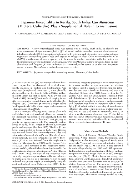
The villages of Kavalam, Molagan Thurthy, Neelamperoor, Nehru Trophy Ward, Pulimcunnu, and
Veliyanadu in the Kuttanadu region of Kerala state
near Lake Vembanad were selected as index villages
for this study (Fig. 1). At least one JE case occurred
in each village during the 1996 and 1997 epidemic.
Kuttanadu is a warm humid region with fairly uniform
temperature, which ranges from 21 to 35⬚C throughout the year (Fig. 2). Most villagers work as agricultural laborers. Cattle, goat, pigs, dogs, fowl, and ducks
are the most common domestic animals (Alappuzha
district Livestock Census). The area receives most of
its rainfall (83%) from JuneÐAugust under the inßuence of southwest monsoons and less rainfall from
OctoberÐDecember under the inßuence of the northeast monsoons. The annual average rainfall is ⬃300
cm.
0022-2585/04/0456Ð0461$04.00/0 䉷 2004 Entomological Society of America
Downloaded from https://academic.oup.com/jme/article/41/3/456/917545 by guest on 13 February 2022
ABSTRACT A 2-yr entomological study was carried out in Kerala, south India, to identify the
mosquito vectors of Japanese encephalitis (JE) virus and to determine their seasonal abundance and
infection. In total, 150,454 mosquitoes belonging to Þve genera and 18 species were collected from
vegetation surrounding cattle sheds and pigsties in villages at dusk. Culex tritaeniorhynchus Giles
(66.7%) was the most abundant species, with increases in numbers associated with rice cultivation.
JE virus isolations were made from Cx. tritaeniorhynchus and Mansonia indiana Edwards. Based on high
abundance and frequent JE virus infection, Cx. tritaeniorhynchus seems to be the most important
vector, whereas Ma. indiana is probably a secondary vector.
�May 2004
ARUNACHALAM ET AL.: JE IN KERALA, SOUTH INDIA
457
Each study village was sampled at monthly intervals
during 1999 and 2000. Mosquitoes resting on vegetation and bushes around cattle sheds and pigsties were
collected for 1 h after dusk by oral aspirator and
transported to the laboratory for identiÞcation and
enumeration. Mosquito (only females) abundance
was calculated as number collected per man-hour.
Male mosquitoes also were collected resting in and
around cattle sheds and pigsties.
Wild-caught mosquitoes were counted into pools of
25Ð50 and were stored at ⫺80⬚C until processed for JE
virus detection and isolation. Two systems were used
(Gajanana et al. 1995). (1) Antigen capture ELISA:
monoclonal antibody 6B4A-10 (reactive against all
viruses in JE/WN/SLE/MVE complex) was used as
capture antibody and monoclonal antibody peroxidase conjugate SLE MAB 6B6C-1 (reactive against all
ßaviviruses) as detector antibodies (Supplied by Dr.
T. F. Sai, Centers for Disease Control and Prevention,
Fort Collins, CO). (2) Insect bioassay: Toxorhynchites
splendens Wiedemann larvae were inoculated intracerebrally, incubated for 7Ð10 d at 29⬚C, and tested by
indirect immunoßuorescence assay (IFA) on head
squash preparations (Toxo-IFA). Smears were Þrst
screened using an anti-JE virus immune serum raised
in rabbits that was broadly reactive against ßaviviruses
and detected by ßuorescein isothiocyanate (FITC)
conjugated anti-rabbit immunoglobulin (Dakoppats,
Glostrup, Denmark). For conÞrmation, duplicate
smears were tested with JE virusÐspeciÞc monoclonal
antibody, MAB 112 (supplied by Dr. Kimura Kuroda,
Tokyo Metropolitan Institute of Neurosciences, Tokyo, Japan), and detected by FITC-conjugated antimouse immunoglobulin (Dakoppats).
Results
In total, 150,454 female mosquitoes representing 6
anopheline and 12 culicine species were collected. Cx.
tritaeniorhynchus was the most abundant species, comprising 66.9% of the total collected. This was followed
in decreasing order by Cx. gelidus (11.1%), Ma. uniformis (9.6%), Ma. indiana (8.2%), and Ma. annulifera
(Theobald) (3.0%); the remaining species comprised
⬍2% of the mosquitoes collected (Table 1).
The abundance of Cx. tritaeniorhynchus was lowest
in JuneÐAugust, increased in September, and reached
a maximum in DecemberÐMarch (Fig. 3). The increase corresponded with the period of rice cultivation. Monthly abundance of Cx. tritaeniorhynchus was
Downloaded from https://academic.oup.com/jme/article/41/3/456/917545 by guest on 13 February 2022
Fig. 1. Map of Alappuzha district showing the study villages in Kuttanadu region.
�458
JOURNAL OF MEDICAL ENTOMOLOGY
Vol. 41, no. 3
negatively correlated with rainfall (r ⫽ ⫺0.61, df ⫽ 23,
P ⬍ 0.05) and was not correlated with temperature or
humidity (P ⬎ 0.05).
Culex gelidus was least abundant during the monsoon months, but unlike Cx. tritaeniorhynchus, it did
not show major seasonal ßuctuations (Fig. 3). Mansonia species (Ma. annulifera, Ma. indiana, and Ma.
uniformis) were collected throughout the year. Ma.
uniformis was more abundant than the other two species (Fig. 4). Although the abundance of the Mansonioides was not correlated signiÞcantly with any meTable 1. Species composition of mosquitoes collected in Kuttanadu, Kerala
Species
No.
collected
Ae. aegypti
An. barbirostris
An. jamesii
An. pallidus
An. peditaeniatus
An. subpictus
An. tesellatus
Ar. subalbatus
Cx. fuscanus
Cx. fuscocephala
Cx. gelidus
Cx. infula
Cx. quinquefasciatus
Cx. tritaeniorhynchus
Cx. vishnui
Ma. annulifera
Ma. indiana
Ma. uniformis
Total
2
602
157
85
133
106
2
362
2
1
16,658
2
333
100,611
3
4,530
12,362
14,503
150,454
%
⬍1
⬍1
⬍1
⬍1
⬍1
⬍1
⬍1
⬍1
⬍1
⬍1
11.1
⬍1
⬍1
66.7
⬍1
3.0
8.2
9.6
teorological parameters, Ma. uniformis was most
abundant during the monsoon months (Fig. 4).
JE virus was isolated from Cx. tritaeniorhynchus and
Ma. indiana. Overall, 146,560 mosquitoes were tested
for JE virus in 3,374 pools, of which 64 pools of Cx.
tritaeniorhynchus, 10 pools of Cx. gelidus, 3 pools of Ma.
annulifera, 12 pools of Ma. indiana, and 5 pools of Ma.
uniformis tested positive by ELISA. JE virus infection
was conÞrmed by Toxo-IFA only from Cx. tritaeniorhynchus and Ma. indiana. Virus infection in Mansonia
was observed in May, July, and August in 1999 and
May, August, and October in 2000. Infection in Cx.
tritaeniorhynchus was observed during JanuaryÐApril
and November in 1999 and JanuaryÐMarch and SeptemberÐDecember in 2000.
JE virus also was isolated from male mosquitoes
collected resting in and around cattle sheds and pigsties. Of the 4 pools of male Cx. gelidus, 33 of Cx.
tritaeniorhynchus, 25 of Ma. indiana, and 35 of Ma.
uniformis tested, 3 male poolsÑ2 Ma. indiana and 1
Ma. uniformisÑwere found positive for ßavivirus antigen. Of the three ßavivirus-positive pools, one (of
Ma. indiana) was conÞrmed to be positive for JE virus
by Toxo-IFA, indicating vertical transmission by Ma.
indiana.
Discussion
The seasonality of JE virus transmission depends on
various factors, among which the relative abundance
of the vector species is most important (Pant 1979).
Our entomologic assessment indicated that Cx. tritaeniorhynchus was the primary vector based on relative
Downloaded from https://academic.oup.com/jme/article/41/3/456/917545 by guest on 13 February 2022
Fig. 2. Meteorological data recorded during the study period.
�May 2004
ARUNACHALAM ET AL.: JE IN KERALA, SOUTH INDIA
459
abundance, widespread distribution, and frequent virus infection. Vector competence of Cx. tritaeniorhynchus has been demonstrated in laboratory studies
(Carey et al.1969, Mourya et al.1991). Cx. tritaeniorhynchus accounted for 67% of mosquitoes collected,
was the most abundant Culex during the transmission
season, and was the most frequently infected species
during our study. If vector abundance and JE virus
infection rates were indicators of potential spillover of
JE virus to humans, then January and April would be
the months of greatest risk. Abundance of Cx. tritaeniorhynchus was very high (up to 397 females/manhour) during the rice cultivation season (JanuaryÐ
April), which also was the main transmission season
Fig. 4. Monthly rainfall and abundance of Mansonia species in Kuttanadu (1999Ð2000).
Downloaded from https://academic.oup.com/jme/article/41/3/456/917545 by guest on 13 February 2022
Fig. 3. Abundance of Cx. tritaeniorhynchus and Cx. gelidus and total rainfall per month in Kuttanadu (1999Ð2000).
�460
JOURNAL OF MEDICAL ENTOMOLOGY
transmission in Japan and Taiwan (Wu et al.1999). The
removal of the pigs from within the Badu Island community also has reduced the potential contact between viremic pigs and vectors within the community
(Van den Hurk et al. 2001). The relocation of domestic
pigs could be adopted as a control strategy in Kerala
to prevent/reduce JE transmission to humans.
Acknowledgments
The authors thank SEARO/WHO New Delhi for Þnancial
support. This publication is an outcome of WHO project SN
1094. We thank A. Veerapathiran, V. Kodangi Alagan, and V.
Rajamannar of Vector Biology and training division of Centre
for Research in Medical Entomology for excellent technical
assistance. We appreciate the excellent help rendered by A.
Venkatesh (CRME) in preparation of this manuscript, particularly in DTP work.
References Cited
Arunachalam, N., P. Philip Samuel, J. Hiriyan, V. Thenmozhi, A. Balasubramanian, A. Gajanana, and K. Satyanarayana. 2002. Vertical transmission of Japanese encephalitis virus in Mansonia species, in an epidemicprone area of southern India. Ann. Trop. Med. Parasitol.
96: 419 Ð 420.
Burton, G. J. 1959. Studies on the bionomics of mosquito
vectors, which transmit Þlariasis in India. I. Attachment of
Mansonia annulifera and Mansonia uniformis larvae to
host plants occurring in Pistia tanks in Kerala, south India.
Ind. J. Malariol. 13: 75.
Carey, D. E., R. Reuben, and R. M. Myers. 1969. Japanese
encephalitis studies in Vellore, South India. Part V. Experimental infection and transmission, Ind. J. Med. Res.
37: 282.
Dhanda, V., D. T. Mourya, A. C. Mishra, M. A. Ilkal, U. Pant,
P. George Jacob, and H. R. Bhat. 1989. Japanese encephalitis virus infection in mosquitoes reared from Þeldcollected immatures and in wild-caught males. Am. J.
Trop. Med. Hyg. 41: 732Ð736.
Dhanda, V., V. Thenmozhi, N. P. Kumar, J. Hiriyan, N.
Arunachalam, A. Balasubramanian, A. Ilango, and A. Gajanana. 1997. Virus isolation from wild-caught mosquitoes during a Japanese encephalitis outbreak in Kerala in
1996. Ind. J. Med. Res. 106: 4 Ð 6.
Gajanana, A., R. Rajendran, V. Thenmozhi, P. Philip Samuel,
T. F. Tsai, and R. Reuben. 1995. Comparative evaluation
of bioassay and ELISA for detection of Japanese encephalitis virus in Þeld collected mosquitoes. Southeast Asian
J. Trop. Med. Publ. Hlth. 26: 91Ð97.
Macdonald, W. W., C.E.G. Smith, P. S. Dawson, A. Ganapathipillai, and S. Mahadevan. 1967. Arbovirus infections in Sarawak: further observations on mosquitoes.
J. Med. Entomol. 4: 146 Ð157.
Mourya, D. T., A. C. Mishra, and R. S. Soman. 1991. 1991.
Transmission of Japanese Encephalitis virus in Culex
pseudovishnui and Culex tritaeniorhynchus mosquitoes India. Ind. J. Med. Res. 93: 250 Ð252.
Pant, C. P. 1979. Vectors of Japanese encephalitis and their
bionomics. WHO/VBC/79.732: 1Ð18.
Peiris, J.S.M., F. P. Amarasinghe, P. H. Amarasinghe, C. B.
Ratnayaka, S.H.P. Karunatne, and T. F. Tsai. 1992. Japanese encephalitis in Sri Lanka. A study of an epidemic:
vector incrimination, porcine infection and human disease. Am. J. Trop. Med. Hyg. 86: 307Ð313.
Downloaded from https://academic.oup.com/jme/article/41/3/456/917545 by guest on 13 February 2022
for JE in this area. Ma. indiana and Ma. uniformis
abundance (maximum 62 females/man-hour) peaked
during the monsoon months (JulyÐNovember), when
the paddy Þelds were ßooded, and few Cx. tritaeniorhynchus could be found (collections falling as low as
2.4 females/man-hour). The monsoon rains permit
large stands of hydrophytes (such as Pistia, Salvinia,
and Eichhornia) to develop, and these plants are essential for the larval development of Mansonia (Burton 1959). Most of our study area was water-logged at
an elevation of 0.5Ð2 m below mean sea level; this
created swampy areas supporting dense stands of
aquatic vegetation, which permited the breeding of
Mansonia.
Vertical transmission of JE virus was documented
by virus isolation from male Ma. indiana (females as
well as males were infected). Vertical transmission in
which the virus is transmitted from an infected female
mosquito to her eggs as they pass through the genital
tract is known to support the persistence of some
arboviruses in nature (Rosen 1988). There is Þeld
evidence of this vertical transmission of JE virus in
south India. Dhanda et al. (1989) detected JE virus in
wild-caught males of Cx. tritaeniorhynchus, indicating
the occurrence of vertical transmission in Culex. Thenmozhi et al. (2001) detected JE virus infection in
Þeld-collected immature stages of Cx. tritaeniorhynchus. JE virus could use mosquitoes as reservoir hosts
for local survival of the virus during adverse conditions
of hot seasons, when vector abundance is low and
nonimmune pigs are few in number (Thenmozhi et
al.2001). The infected Mansonia mosquitoes were collected toward the end of the monsoon, at a time when
few adult Cx. tritaeniorhynchus can be caught. If the
larval stages of local Mansonia are sometimes infected
with JE virus, the relatively slow development of Mansonia (Srinivasan and Viswam 1986) may help the virus
to survive a period when its main vector is scarce.
Mansonia is probably a secondary vector, with Cx.
tritaeniorhynchus as the primary vector.
Blood meals of Cx. tritaeniorhynchus and Mansonia
were analyzed against antisera of six hosts, cattle, pigs,
ducks, goats, fowl, and humans, to Þnd the preferred
hosts of vectors in this region. Cx. tritaeniorhynchus fed
predominantly on cattle (76%), with pigs accounting
for 5% of blood meals identiÞed. Pigs constituted 6%
of Ma. indiana blood meals tested (Centre for Research in Medical Entomology [CRME], unpublished
data). A serological survey revealed a high prevalence
of (69%) of JE antibodies in pigs (CRME, unpublished
data). Pigs were maintained in small groups in backyard pens surrounded by paddy Þelds. Consequently,
the primary vector Cx. tritaeniorhynchus and the potential bridge vector Ma. indiana were in close proximity to the amplifying pig hosts and humans, favoring
the spillover transmission of JE virus to humans. Similarly, intense transmission of JE on Badu Island, Australia, was attributed to a large domestic pig population housed in small backyard pens throughout the
community (Van den Hurk et al. 2001). The relocation
of pigs to specialized farms separate from human habitation was useful in reducing pig-mosquito-human JE
Vol. 41, no. 3
�May 2004
ARUNACHALAM ET AL.: JE IN KERALA, SOUTH INDIA
Thenmozhi, V., R. Rajendran, P. Philip Samuel, J. Hiriyan,
K. Ayanar, A. Balasubramanian, and A. Gajanana. 2001.
Natural vertical transmission of Japanese encephalitis virus in south Indian mosquitoes. Trop. Biomed. 18: 19 Ð27.
Van den Hurk, A. F., D. J. Nisbet, C. A. Johansen, P. N. Foley,
S. A. Ritchie, and J. S. Mackenzie. 2001. Japanese encephalitis on Badu Island, Australia: the Þrst isolation of
Japanese encephalitis virus from Culex gelidus in the
Australasian region and the role of mosquito host-feeding
patterns in virus transmission cycles. Trans. Roy. Soc.
Trop. Med. Hyg. 95: 595Ð 600.
Vaughn, D. W., and C. H. Hoke. 1992. The epidemiology of
Japanese encephalitis: prospects of prevention. Epidemiol. Rev. 14: 197Ð221.
Webb, J.K.G., and S. M. Pereira. 1956. Clinical diagnosis of
an arthropod-borne type of encephalitis in North Arcot
district, Madras state, India. Ind. J. Med. Res. 10: 583Ð588.
Wu, Y. C., Y. S. Huang, L. J. Chien, T. L. Lin, Y. Y. Yueh, W. L.
Tseng, K. J. Chang, and G. R. Wang. 1999. The epidemiology of Japanese encephalitis on Taiwan during 1966 Ð
1997. Am. J. Trop. Med. Hyg. 61: 78 Ð 84.
Received 23 May 2003; accepted 7 November 2003.
Downloaded from https://academic.oup.com/jme/article/41/3/456/917545 by guest on 13 February 2022
Reuben, R. 1987. The epidemiology of Japanese encephalitis in Tamil Nadu, pp. 135Ð142. In Proceedings of the
Symposium of Alternatives to Synthetic Insecticides, Madurai, India.
Reuben, R., V. Thenmozhi, P. P. Samuel, A. Gajanana, and
T. R. Mani. 1992. Mosquito blood feeding patterns as a
factor in the epidemiology of Japanese encephalitis in
southern India. Am. J. Trop. Med. Hyg. 46: 654 Ð 663.
Rodrigues, F. M. 1984. Epidemiology of Japanese encephalitis in India: a brief overview, pp. 1Ð9. In Proceedings of
the National Conference of Japanese Encephalitis, 1982,
New Delhi, India.
Rosen, L. 1988. Further observations on the mechanism of
vertical transmission of ßaviviruses by Aedes mosquitoes.
Am. J. Trop. Med. Hyg. 39: 123Ð126.
Scherer, W. F., R. W. Dickerman, A. Diaz-Najera, B. A. Ward,
M. H. Miller, and P. A. Schaffer. 1971. Ecological studies
of Venezuelan encephalitis virus in South Eastern Mexico
III Infection of mosquitoes. Am. J. Trop. Med. Hyg. 20:
969 Ð979.
Srinivasan, R., and K. Viswam. 1986. Laboratory studies on
the biology of Mansonia annulifera Theobald 1901.
(Diptera:Culicidae) Ind. J. Med. Res. 83: 384 Ð386.
Theiler, M., and W. G. Downs. 1973. The arthropod-borne
viruses of vertebrates. Yale University Press, New Haven,
CT.
461
�
 Philip Samuel