Obesity Surgery, 10, 409-412
Open vs Laparoscopic Vertical Banded Gastroplasty:
A case control study with 1-year follow-up
Andrea Dávila-Cervantes, MD1; Gabriella Ganci-Cerrud, MD1; Rosa
Gamino, SW1; José Gallegos-Martínez, MD2; Jorge González-Barranco,
MD2; Miguel F. Herrera, MD1
1 Department
of Surgery. and 2The Obesity Clinic, Instituto Nacional de la Nutrición Salvador
Zubirán, Mexico City, Mexico
Background: Vertical Banded Gastroplasty (VBG) is
one of the most common bariatric operations. It can
be performed by open or laparoscopic methods. The
purpose of this study was to analyze and compare
the 1-year results of 40 patients who underwent
laparoscopic (20) and open (20).
Methods: The initial 20 patients undergoing
Laparoscopic VBG and the initial 20 patients in whom
an Open VBG were performed in our Institution were
comparatively evaluated. Demography, surgical
details, complications, and 1-year weight loss were
analyzed.
Results: Both groups were highly comparable in
terms of age, sex and body mass index. Laparoscopic
VBG was a more prolonged procedure (median 4 hr)
than the open VBG (median 3 hr). On the other hand,
hospital stay was significantly shorter in the laparoscopic procedure (median 10 days for the open and 6
days for the laparoscopic). One year weight loss and
complications were similar in both groups.
Conclusions: Laparoscopic VBG is a safe procedure for the treatment of morbid obesity. This initial
series shows comparable results.
Key words: Morbid obesity, bariatric surgery, gastroplasty, laparoscopy
defined as a body mass index (BMI) >40 kg/m2 or
a weight of 100% or more above the ideal body
weight. Morbid Obesity is associated with a high
frequency of complications and early mortality.
Bariatric surgery is currently the most effective
treatment for this group of patients.1
Many bariatric operations have been described
since Kremen, Linner and Nelson reported their
first jejunoileal bypass in 1954.2,3 Among the operations most frequently used is the vertical banded
gastroplasty (VBG): in a series of 14,641 bariatric
operations from the International Bariatric Surgery
Registry, VBG had been performed in 36.3% of
this population with a long-term mean loss of
excess body weight of 58%.4
Important advances in endoscopic surgery have
made it possible to perform bariatric operations
laparoscopically. Laparoscopic VBG was first
described by Chua and Mendiola in 1995 and some
subsequent reports have been published.5,6
The aim of the present study is to analyze our 1year results in the first 20 patients undergoing
laparoscopic VBG and to compare those results
with a non-concurrent group of 20 patients who
underwent open VBG.
Introduction
Morbid obesity is a major health problem. It is
Patients and Methods
Reprint requests to: Miguel F. Herrera, MD, Department of
Surgery, Instituto Nacional de la Nutrición Salvador Zubirán,
Vasco de Quiroga 15, Tlalpan 14000 México, D.F., MEXICO.
Tel: (52-5) 573-1200, x2144; fax: (52-5) 573-9321;
E-mail: herreram@quetzal.innsz.mx
Twenty morbidly obese patients underwent laparoscopic VBG between July of 1997 and December of
1998. Demography, surgical details, surgical com-
© FD-Communications Inc.
Obesity Surgery, 10, 2000
409
�Dávila-Cervantes et al
plications, and 1-year weight loss were analyzed and
compared to the initial 20 patients who underwent
open VBG in our Institution from 1992 to 1998.
Patient selection and preoperative evaluation: A
multidisciplinary evaluation was performed on all
patients including a complete physical examination, laboratory work-up and a cardiopulmonary
evaluation. All were non sweet-eater patients with
a BMI between 40 and 50 kg/m2.
Surgical technique: VBG was performed through a
midline supraumbilical incision following the
technique described by Mason in 1982 without
calibration of the pouch.7 A 25-mm circular stapler
was used to create the window, a TA 90-B linear
stapler was selected for the partition, and a 5-cm
Marlex mesh collar was placed to restrict the
reservoir outlet.
Laparoscopic VBG was performed using 5 trocars in the standard position for gastric surgery. A
25-mm circular stapler was used to create the window, a non-cutting 60-mm linear stapler was
employed for the partition in 10 patients, and division of the stomach with a 60-mm cutting linear
stapler was selected in the last 10 patients. A 5-cm
Marlex mesh collar was also placed and fixed
around the outlet to restrict the pouch.
All patients underwent a postoperative
Gastrografin ® upper gastrointestinal (GI) x-ray
study. Liquid diet was administered once leaks
were ruled out and adequate emptying was confirmed. Minced food was administered from the
3rd postoperative day and maintained for a 6-week
period. Patients were followed monthly for 3
months and every 3 months for a year. In all visits,
a diet history was taken, patients were weighed
and a careful interrogatory looking for complications was carried out. An upper GI contrast series
was performed at 1 year after surgery to evaluate
the status of the operation.
the laparoscopic group, and hospital stay was more
prolonged in the open group (Table 2). One patient
in the open group developed superficial vein
thrombophlebitis. There was one non-operative
mortality at 3 months follow-up in the open group
due to an unexpected coagulopathy. The remaining
patients completed 1-year follow-up.
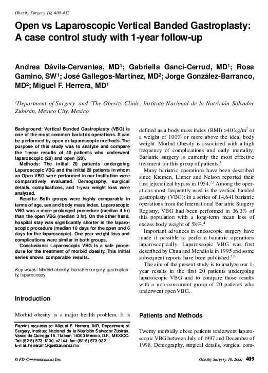
Weight loss was faster during the initial 6 months
regardless of the surgical approach. Mean weight
loss at this period was 28 kg in the open and 20 kg in
the laparoscopic group. Body weight modifications
during the 1st year are shown in Figure 1. Excess
weight loss at 6 months follow-up was 42% and 44%
for the open and the laparoscopic groups respectively. At one-year follow-up, these figures increased up
to 57% and 52% respectively (Figure 2). Changes in
Table 1. Demography
Open
Laparoscopic
n=20
n=20
Age, years
38 ± 10
32 ± 10
Sex, M:F
3:17
2:18
Preoperative excess weight, % 94 ± 15
92 ± 14
Preoperative BMI kg/m2
45 ± 6
44 ± 6
Table 2. Surgical details and complications
Operative time, hours
Hospital stay, days
Transfusions, n
Complications, n
Additional procedures
Open
3±1
10 ± 6
0
1
5*
Laparoscopic
4±1
6±1
0
0
0
*cholecystectomies
120
D
110
Open
Laparoscopic
Kg 100
90
Results
Both groups were highly comparable in terms of
age, sex distribution and preoperative weight
(Table 1). All patients underwent a satisfactory
VBG with no conversions in the laparoscopic
group. Mean operative time was slightly longer in
410
Obesity Surgery, 10, 2000
80
70
Pre-op
6 months
1 year
Figure 1. Mean Weight. Changes during the first year
after surgery. All values are mean ± standard error.
�Open vs Laparoscopic VBG
the BMI are shown in Table 3. The number of
Discussion
patients who achieved different excess weight
loss was also similar in both groups (Table 4).
Postoperative Gastrografin® study was normal in
all patients. The upper GI contrast study 1 year
later showed stomal stenosis in one patient of the
open group and partial staple-line disruption in one
patient of the laparoscopic group. The stomal
stenosis required endoscopic dilatation, and the
patient with partial staple-line disruption achieved
a 20-kg weight loss at 1-year follow-up with no
additional treatment.
11
D Open
Laparosocpic
0
% 90
70
50
Pre-op
6 months
1 year
a) Excess body weight
b) Excess body weight (%)
%
6 months
Open
42 ± 7
Laparoscopic
57 ± 5
1 year
44 ± 6
52 ± 2
Figure 2. Excess body weight and excess body weight
loss during the first year after surgery. All values are
mean ± standard error.
Table 3. Body mass index
BMI kg/m2
Preop
6 months
1 year
Open n=19
45 ± 6
34 ± 5
32 ± 5
Laparoscopic n= 20
44 ± 6
38 ± 3
33 ± 5
Table 4. Number of patients at different % of excess
weight loss at 1-year follow-up
EWL (%)
<25
26-50
51-75
>75
Total
Open
n
3
10
6
0
19
%
16
53
31
0
100
Laparoscopic
n
%
1
5
11
55
8
40
0
0
20
100
VBG is one of the most frequently performed
bariatric operations. It provides satisfactory results
in terms of weight loss and has a very low incidence of complications.
The introduction and rapid development of
laparoscopic techniques has permitted performance of a wide variety of procedures in a safe
way. Laparoscopic techniques in general have been
demonstrated to produce less trauma, to reduce
hospital stay, to diminish postoperative pain, and to
improve the early return to full normal activities. 6-9
Reports of laparoscopic bariatric surgery have been
consistent with these findings and have shown an
additional improvement in postoperative respiratory function.6
Our initial experience has shown that laparoscopic VBG is a relatively simple operation that
can be carried out almost identically to the original
open technique.7 Considering that the laparoscopic
technique does not differ from the open procedure,
the same long-term results should be anticipated.
However, some technical details need special consideration. The original operation was described
with intraoperative calibration of the gastric pouch.
Several authors have performed the technique
without calibration, obtaining similar results. 10,11
Calibration in the laparoscopic procedures is generally omitted.
Although the aim is always to create a very small
reservoir, the lack of tridimensional view and palpation might influence the ultimate size of the gastric pouch. A comparative analysis of pouch size
using both techniques needs to be evaluated.
Several surgical techniques and stapling devices
have been used for the construction of the gastric
pouch. In the original technique, gastric partition
without division was recommended. This can be
performed in open surgery using one or two firings
of a standard non-cutting lineal stapler or one or
two firings of a special bariatric stapler (TA-90B)
which gives 4 rows of staples in each application.
An endoscopic non-cutting linear stapler was available for a period of time and was used in our initial
10 laparoscopic operations. However, current
instrumentation only permits us to perform the
operation by dividing the stomach.
Obesity Surgery, 10, 2000
411
�Dávila-Cervantes et al
MacLean and colleagues11 reported in 1993 a
modification of VBG consisting of division of the
stomach. With this technique the authors reported
a higher incidence of gastric fistula. 11-13 This
complication was considered as a primary factor
contributing to an unsatisfactory outcome and
need for reoperation. In the 10 laparoscopic
patients of this series in whom the stomach was
divided, no gastric fistula was seen. Comparative
resistance of the laparoscopic instruments and the
frequency of gastric fistula in laparoscopic operations need to be evaluated in a larger number of
procedures.
At 1-year follow-up, no differences were found
in the amount of weight loss with either technique.
Partial staple-line disruption was found in only one
laparoscopic case and corresponded to the group of
patients in whom the non-cutting stapler was used.
Results of bariatric operations have two different
components: the maximum weight loss achieved
and the overall time that patients maintain a
reduced weight. This initial series only shows that
the amount of weight lost during the 1st year is
comparable in both techniques. Weight behavior in
the long-term needs further evaluation.
Surgical time in our study was longer in the
laparoscopic approach. As in most initial reports,
laparoscopic techniques take longer operative time.
We believe that this only represents our learning
curve and should decrease with time. The hospital
stay was significantly shorter after laparoscopic
VBG, and we believe this is more related to
changes in our surgical practice than to the technique itself. Since we take care of patients from
several parts of our country and we send them
home when they are totally recovered, long postoperative periods are common in our hospital. In
our initial practice a Gastrografin® GI x-ray study
was performed on the 5th postoperative day and
diet was administered afterwards. Currently, we
perform our Gastrografin® studies on the day after
surgery and send the patients home as soon as they
are able to walk and eat minced food. This explains
the dramatic changes in postoperative hospital stay.
This initial experience shows that laparoscopic
VBG compares with the open procedure in terms
412
Obesity Surgery, 10, 2000
of short-term results and complications. Further
studies need to be carried out to analyze specific
details of both techniques.
References
1. National Institutes of Health Consensus Development
Conference Draft Statement on Gastrointestinal
Surgery for Severe Obesity. Obes Surg 1991;1:25765.
2. Hall JC, Watts JM, O’Brien PE. Gastric Surgery for
morbid obesity. The Adelaide Study. Ann Surgery
1990;211:419-27.
3. Linner JH, Drew RL. Why the operation we prefer is
the Roux-Y gastric bypass. Obes Surg 1991;1:305-6.
4. Mason E, Shenghui T, Renquist K et al. A decade of
change in obesity surgery. Obes Surg 1997;7:189-97.
5. Chua T, Mendiola R. Laparoscopic vertical banded
gastroplasty: The Milwaukee Experience. Obes Surg
1995;5:77-80.
6. Lönroth H, Dalenback J, Haglind E et al. Vertical
banded gastroplasty by laparoscopic technique in the
treatment of morbid obesity. Surg Laparosc Endosc
1996; 6:102-7.
7. Mason EE. Vertical banded gastroplasty for obesity.
Arch Surg 1982;117:701-6.
8. Majeed AW, Troy G, Nicholl J et al. Randomized,
prospective, single blind comparison of laparoscopic
versus small-incision cholecystectomy. Lancet
1996;347:989-94.
9. Hinder R, Filipi C. The technique of laparoscopic
Nissen fundoplication. Surg Laparosc Endosc
1992;2:265-73.
10.Deitel M. Update general surgery: morbid obesity.
Annals RCPSC 1990;23:241-6.
11.MacLean I, Rhode BM Forse A. A gastroplasty that
avoids stapling in continuity. Surgery 1993;113:380-8.
12.Toppino M, Nigra I, Olivieri F et al. Staple-line disruptions in vertical banded gastroplasty related to different stapling techniques. Obes Surg 1994;4:256-61.
13.Melissas J, Christodoulakis M, Schoeretsanitis G et
al. Staple-line disruption following vertical banded
gastroplasty. Obes Surg 1998;8:15-20.
(Received February 28, 2000; accepted August 1, 2000)
�
 miguel herrera