International Journal of Pediatric Otorhinolaryngology (2004) 68, 1417—1421
www.elsevier.com/locate/ijporl
Management of nasal septal abscess in childhood:
our experience
C. Dispenza*, C. Saraniti, F. Dispenza, C. Caramanna, F.A. Salzano
Clinica Otorinolaringoiatrica, Università di Palermo, Azienda Ospedaliera Policlinico ‘‘P. Giaccone’’,
Via P. E. Giudici n.37, Palermo 90127, Italy
Received 18 March 2004; received in revised form 24 May 2004; accepted 26 May 2004
KEYWORDS
Nasal trauma;
Septum abscess;
Surgical treatment
Summary
A nasal septal abscess is usually the result of an infected hematoma of the septum. A
secondary septal abscess may be the result of infections extending from any of the
neighbouring tissues.
The necrosis of septal cartilage may lead to nasal deformities and severe impairment
of nasal patency and growth.
Objectives: Assess if the drainage of the abscess and the immediate reconstruction
of the destroyed nasal septum in the acute phase is the best treatment to prevent
short- and long-term effect on nasal and midface growth.
Methods: Three pediatric patients treated with drainage and immediate implantation of homologous bank cartilage prior to 1990 and four treated with mosaic plastic
using small pieces of residual septal cartilage assembled with fibrin glue.
Result: No complication were observed in the follow-up and any deformities in the
long-term controls.
Conclusions: The drainage and immediate reconstruction of the nesal septum are the
golden standard in the treatment of the septum infected haematoma.
# 2004 Elsevier Ireland Ltd. All rights reserved.
1. Introduction
A nasal septal abscess is defined as a collection of
pus between the cartilage or bony septum and the
mucoperichondrium or mucoperiostium [1] (Fig. 1).
The recognition of the nasal septal abscess is traced
* Corresponding author. Tel.: +39 091 6554243.
E-mail address: francesco.dispenza@poste.it (C. Dispenza).
back to 1810 when Cloquet healed an abscess by
drainage [2].
This pathology is often the result of an infected
septal hematoma which can be a serious complication of trauma or surgery. The rupture of the small
vessels that supply the nasal septum forms a hematoma that separates the mucoperichondrium from
the septal cartilage. Cartilage destruction follows
as a result of ischemic and pressure necrosis. The
0165-5876/$ — see front matter # 2004 Elsevier Ireland Ltd. All rights reserved.
doi:10.1016/j.ijporl.2004.05.014
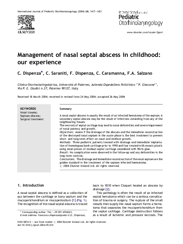
�1418
Fig. 1
C. Dispenza et al.
A nasal septal abscess in a 2-year-old child.
static blood and the necrotic cartilage form an
adequate medium for the growth of the bacteria
which normally colonize the nasal mucosa [3].
Beck defines this process as a post-trauma primary abscess and groups all the other pathologies
as secondary complications to the infections
extending from the neighbouring tissues (dental
infections, septic embolism, furuncle of the nasal
vestibule); in such cases, the infection focus
should be identified and treated at the same time
as the nasal septal abscess (secondary abscesses)
[15]. The most common organisms cultured from
nasal abscesses are Staphylococcus aureus, Streptococcus pneumoniae, Haemophilus influenzae,
[3—5] and group A beta-haemolytic streptococcus
[1]; Brook reports two cases in which cultures for
anaerobic bacteria revealed Prevotella intermedia
and Peptostreptococcus magnus in one patient,
and Peptostreptococcus anaerobius, Streptococcus intermedius and Prevotella melaninogenica in
the second patient. Brooks believes these organisms are fairly unremarkable as they are usually
cultured from oropharynx and retropharynx infections, cervical ademolymphitis and parotidean
abscesses [13].
Pirsig believes the cartilage lysis is not just a
consequence of the avascular necrosis but occurs
for the digestion process caused by leukocytes and
Cathepsin D enzyme. The latter is present in the
septal cartilage and belongs to a group of enzymes
which re-shape the quadrangular cartilage. Its
acid pH accelerates the cartilage digestion [2]
(Fig. 2).
Inspection with palpation is initially required to
detect the destroyed cartilage. The nose undergoes
a slow collapse in the weeks following the injury
because of a retraction in the connective tissue,
skin, mucous covering, dorsum and valve. The
short-term effects of the septal abscess are the
Fig. 2 Activity of the enzyme cathepsin D in five areas of
the human septal cartilage. The enzymatic activity is
expressed in mg tyrosine generated per 60 min per
0.1 mg septal cartilage (adopted from ref. [2]).
sagging and widening of the nasal pyramid as well
as impaired breathing (Fig. 3a and b). The long-term
effect of the septum destruction is the disturbance
of growth, form and function of the nose [6].
Fig. 3 (a) and (b) Patient with typical nasal deformities
due to absence of septal cartilage (saddle nose).
�Management of nasal septal abscess in childhood
1419
Fig. 4 Homologous L-shaped cartilage from the cartilage bank used for septal graft.
2. Materials and methods
Seven patients, aged 3—12 years, were treated for
nasal abscess in the period from 1987 to 2000 (ratio
male:female, 6:1). All patients had a history of
nasal trauma. The diagnosis was made 1—15 days
(mean, 8 days) after the episode of trauma. Nasal
obstruction, fever and epistaxis were the common
presenting symptoms, which, in three cases, were
associated with fractures of the nasal bones and
deformity of the nasal pyramid.
All patients underwent drainage of the abscess
and immediate implantation of the cartilage; a
reduction was performed in the cases of nasal bone
fractures.
Homologous cartilage from the cartilage bank
was used on three patients prior to 1990 (Fig. 4);
the mosaic technique, consisting in the reimplantation of multiple small septal pieces in to the septal
space fixed together by fibrin glue, was performed
on four patient using residual septal cartilage [7].
All patients received packing with Merocel1 or
gauze which was removed in 3 days. Only two
patients required additional packing.
Fig. 5 (a) and (b) The same patient as Fig. 1 (follow-up
at the age of 5): regular development of the nasal pyramid
(the treatment consisted of drainage of the abscess and
mosaicplasty of the nasal septum).
4. Discussion
3. Results
No complications were observed in the immediate
follow-up period. No saddle deformity nor retraction of the columella were observed at six month
follow-up and the functional results were satisfactory in all patients. The long-term results observed
at 5 and 10 years follow-up periods consisted in a
normal development of the face and nasal pyramid
(Fig. 5a and b); a sub-stenosis deviation of the nasal
septum was observed in four patients (Fig. 6a
and b).
The management of nasal septal abscess should
consider short-term effects, nasal obstruction, saddle nose, retraction of the columella, without
neglecting long-term ones such as the disturbance
of growth and form of the nose and midface region
[6]. The two main treatments published in literature
are the surgical drainage [1,3—5,8,9,12] and the
drainage and immediate reconstruction of the
destroyed septal cartilage [2,6,7,10,11] Cottle
has proposed the implantation of nasal septum
since 1953 recommending the treatment within
8—12 weeks from the abscess when the infection
�1420
C. Dispenza et al.
Fig. 7 A boomerang-shaped septal graft (adopted from
ref. [7]).
The first and third treatment are unsuitable for
children. The former, in fact could destruct the
posterior septum and impedes septal growth, while
the latter would be too invasive for young patients.
Whenever the cartilage fragment is sufficiently
wide, Hellmich suggests a ‘‘boomerang-shaped’’
implantation which provides two supports (K area
and anterior nasal spina) and allows a good development of the midface region [7] (Fig. 7).
Fig. 6 (a) and (b) A patient aged 15 (follow-up at 10
years): harmonic development of the nasal pyramid and of
the midface region (the treatment consisted of drainage
of the abscess, reconstruction of the cartilaginous septum
by grafting of homologous cartilage and reduction of the
fracture of the nasal bones). Dislocation of antero-inferior septal cartilage in right vestibule (arrow).
resolution could assure a successful implantation
[14]. Afterwards, Huizing e Masing proposed a septal reconstruction in the acute phase of a septal
abscess, at the same time as drainage [2]. Various
authors [2,7,10] have adopted the latter treatment
reporting satisfactory results. Hellmich suggested
three possible options for the reconstruction of the
destroyed septal infrastructure:
(1) the use of posterior cartilage or bony septum
residues to adjust deformities in the anterior
septum (‘‘exchange technique ’’);
(2) the mosaicplasty with small fragments of residual cartilage;
(3) the use of preserved rib cartilage allograft
whenever the septal material is not available.
5. Conclusions
Long-term results have led us to consider the drainage and immediate reconstruction of the nasal
septum as a very effective treatment of the septum
infected hematoma. It allows a good development
of the nasal pyramid and midface region while
preventing the short-term effects of this pathology
(saddle nose and nasal obstruction) A symptomatic
deviation of the septum, as reported in literature,
was observed in four cases.
References
[1] P.S. Ambrus, R.D. Eavey, A.S. Baker, W.R. Wilson, J.H.
Kelly, Management of nasal septal abscess, Laryngoscope
91 (1981) 575—582.
[2] W. Pirsig, Historical notes and actual observations on the
nasal septal abscess especially in children, Int. J. Ped.
Otorhinolaryngol. 8 (1984) 43—54.
[3] C.M. Ginsburg, J.L. Leach, Infected nasal septal hematoma,
Ped. Infect. Dis. J. (1995) 14.
[4] M. da Silva, J. Helman, I. Eliachar, H.Z. Joachims, Nasal
septal abscess of dental origin, Arch. Otolaryngol. 108
(1982) 380—381.
�Management of nasal septal abscess in childhood
[5] J. Viscasillas, S. Viscasillas, P. Claros, Abscesos y hematomas del tabique nasal en la edad pediatrica, Anales ORL
Iber-Am. 11 (1984) 419—429.
[6] E.H. Huizing, Long-term results of reconstruction of the
septum in the acute phase of a septal abscess in children,
Rhinology 22 (1984) 55—63.
[7] S. Hellmich, Reconstruction of the destroyed septal infrastructure, Otolaryngol. Head Neck Surg. 100 (1989) 92—94.
[8] A. Ehrlich, Nasal septal abscess: an unusual complication of
nasal trauma, Am. J. Emerg. Med. 11 (1993) 149—150.
[9] P.A. Canty, R.G. Berkowitz, Haematoma and abscess of
nasal septum in children, Arch. Otolaryngol. Head Neck
Surg. 122 (1996) 1373—1376.
[10] P. Vase, J. Johannessen, Homograft cartilage in the treatment of an abscess in the nasal septum, J. Laryngol. Otol. 95
(1981) 357—359.
1421
[11] L.F. Grymer, C. Bosch, The nasal septum and the development of the midface. A longitudinal study of a pair of
monozygotic twins, Rhinology 35 (1997) 6—10.
[12] H. Alvarez, J. Osorio, J.I. De Diego, M.P. Prim, C.
De La Torre, J. Gavilan, Sequelae after nasal septum
injuries in children, Auris Nasus Larynx 27 (2000) 339—
342.
[13] I. Brook, Recovery of anaerobic bacteria from a post-traumatic nasal septal abscess. A report of two cases, Ann. Otol.
Rhinol. Laryngol. 107 (1998) 959—960.
[14] M.H. Cottle, T.J. Quilty, R.A. Buckingham, Nasal implants in
children and in adults; with preliminary note on the use of ox
cartilage, Ann. Otol. 62 (1953) 169—175.
[15] A.L. Beck, Abscess of the nasal septum complicating
acute ethmoiditis, Arch. Otolaryngol. 42 (1945) 275—
279.
�
 F. Dispenza