International Journal of COPD
Dovepress
open access to scientific and medical research
OrIgInal researCh
Open access Full Text article
Do frequent moderate exacerbations contribute
to progression of chronic obstructive pulmonary
disease in patients who are ex-smokers?
Jorge Dreyse 1
Orlando Díaz 1
Paula B repetto 2
arturo Morales 1
Fernando saldías 1
Carmen lisboa 1
1
Department of Pulmonary Diseases,
school of Medicine, 2school of
Psychology, Pontificia Universidad
Católica de Chile, santiago, Chile
Background: In addition to smoking, acute exacerbations are considered to be a contributing
factor to progression of chronic obstructive pulmonary disease (COPD). However, these findings come from studies including active smokers, while results in ex-smokers are scarce and
contradictory. The purpose of this study was to evaluate if frequent acute moderate exacerbations
are associated with an accelerated decline in forced expiratory volume in one second (FEV1)
and impairment of functional and clinical outcomes in ex-smoking COPD patients.
Methods: A cohort of 100 ex-smoking patients recruited for a 2-year follow-up study was evaluated at inclusion and at 6-monthly scheduled visits while in a stable condition. Evaluation included
anthropometry, spirometry, inspiratory capacity, peripheral capillary oxygen saturation, severity of
dyspnea, a 6-minute walking test, BODE (Body mass index, airflow Obstruction, Dyspnea, Exercise
performance) index, and quality of life (St George’s Respiratory Questionnaire and Chronic Respiratory Disease Questionnaire). Severity of exacerbation was graded as moderate or severe according
to health care utilization. Patients were classified as infrequent exacerbators if they had no or one
acute exacerbation/year and frequent exacerbators if they had two or more acute exacerbations/
year. Random effects modeling, within hierarchical linear modeling, was used for analysis.
Results: During follow-up, 419 (96% moderate) acute exacerbations were registered. At baseline, frequent exacerbators had more severe disease than infrequent exacerbators according to
their FEV1 and BODE index, and also showed greater impairment in inspiratory capacity, forced
vital capacity, peripheral capillary oxygen saturation, 6-minute walking test, and quality of
life. However, no significant difference in FEV1 decline over time was found between the two
groups (54.7±13 mL/year versus 85.4±15.9 mL/year in frequent exacerbators and infrequent
exacerbators, respectively). This was also the case for all other measurements.
Conclusion: Our results suggest that frequent moderate exacerbations do not contribute to
accelerated clinical and functional decline in COPD patients who are ex-smokers.
Keywords: chronic obstructive pulmonary disease, acute exacerbations, disease progression,
FEV1, BODE index, health status
Introduction
Correspondence: Carmen lisboa
Department of Pulmonary Diseases,
School of Medicine, Pontiicia Universidad
Católica de Chile, Marcoleta 350,
santiago 8330033, Chile
Tel +56 2 2633 1541
email clisboa@med.puc.cl
Progression of chronic obstructive pulmonary disease (COPD) assessed by decline
in forced expiratory volume in one second (FEV1) is closely related to active tobacco
smoking. It has been shown that smoking cessation decelerates the FEV1 decline in
patients with mild-to-moderate COPD.1,2 It is also widely assumed that acute exacerbations contribute to disease progression.3 Exacerbations increase respiratory symptoms,
impair exercise capacity, and cause a deterioration in quality of life.4,5 However, most
studies evaluating the functional and clinical changes associated with an acute exacerbation and its recovery have included current smokers, making it difficult to separate the
525
submit your manuscript | www.dovepress.com
International Journal of COPD 2015:10 525–533
Dovepress
© 2015 Dreyse et al. This work is published by Dove Medical Press Limited, and licensed under Creative Commons Attribution – Non Commercial (unported, v3.0)
License. The full terms of the License are available at http://creativecommons.org/licenses/by-nc/3.0/. Non-commercial uses of the work are permitted without any further
permission from Dove Medical Press Limited, provided the work is properly attributed. Permissions beyond the scope of the License are administered by Dove Medical Press Limited. Information on
how to request permission may be found at: http://www.dovepress.com/permissions.php
http://dx.doi.org/10.2147/COPD.S76475
�Dovepress
Dreyse et al
effects of exacerbations from those of smoking.3–6 Studies in
ex-smokers are scarce and have yielded conflicting results.7–9
Analysis of the Lung Health Study data by Kanner et al
indicate that acute exacerbations evaluated by self-reported
episodes of lower respiratory infections resulting in physician visits during the previous year in ex-smoking patients
do not contribute to FEV1 decline, whereas in a follow-up
of 102 patients for 3 years, Makris et al reported that acute
exacerbations produced a decline in FEV1 in ex-smokers,
albeit of a lesser magnitude that in active smokers.
On the other hand, there is no information about the effect
of acute exacerbations on patient-centered outcomes in exsmoking COPD patients. The aim of the present study was to
evaluate if frequent acute exacerbations in ex-smoking COPD
patients are associated with disease progression by assessing
functional and clinical indices over 2 years of follow-up.
Patients and methods
Patients
A cohort of 105 consecutive ex-smoking patients with a long
history of COPD according to Global Initiative for Chronic
Obstructive Lung Disease (GOLD) criteria10 was enrolled
in a 2-year follow-up study, based on scheduled visits every
6 months. Recruitment criteria included: age older than
40 years; smoking history greater than ten packs/year; cessation of smoking for at least 6 months before recruitment,
confirmed by urine cotinine levels; absence of an acute
exacerbation in the previous month at least; absence of
any physical condition precluding the ability to perform a
6-minute walking test (6MWT); or a short life expectancy.
Patients with asthma, bronchiectasis, sequelae of tuberculosis, or known malignancy were excluded. The institutional
review board at our institution approved the study, and written informed consent was obtained from all patients.
exacerbations
For diagnosis of acute exacerbation, the following definition
was applied:
A sustained worsening of the patient’s condition from the stable state and beyond normal day-to-day variation that is acute
in onset and necessitates a change in regular medication.11
Patients were instructed to contact one of the investigators
if they had an acute increase in symptoms (dyspnea, cough,
sputum, and/or purulent sputum) for 2 consecutive days with
or without symptoms of upper respiratory tract infection or
fever and to attend our clinic to confirm the presence of an
acute exacerbation.12
526
submit your manuscript | www.dovepress.com
Dovepress
Severity of exacerbation was graded according to the
health care utilization classification. Thus, exacerbation was
considered: mild if it required increases in regular inhaled
medication; moderate if courses of antibiotics and/or systemic corticosteroids were needed; and severe if the patient
required hospital admission.13
Exacerbations were treated at our facility or at other
health institutions. Those exacerbations treated at other
institutions were registered during the scheduled visits, and
their severity was categorized according to the treatment
received using the above classification.
Measurements
Evaluations were performed at recruitment and at every
scheduled visit while patients were in a stable condition. At
each visit, assessments included anthropometry, dyspnea,
health status, peripheral capillary oxygen saturation (SpO2),
lung function, 6MWT, and comorbidities.14
Magnitude of dyspnea was assessed using the modified
Medical Research Council scale (mMRC).15 Lung function included spirometric testing and inspiratory capacity
before and after 400 µg of albuterol following international
guidelines,16 and was standardized as percentages of predicted values by using prediction equations.17,18 Measurements were carried out by the same laboratory personnel
and with the same equipment over the 2 years of follow-up.
Health status was assessed with two questionnaires, ie, the
Spanish version of the St George’s Respiratory Questionnaire (SGRQ)19 and the Spanish version of the Chronic
Respiratory Disease Questionnaire (CRQ).20 The 6MWT
was measured according to current guidelines21 and its values were expressed as a predicted percentage using reference
values from Troosters et al.22 The BODE (body mass index,
airflow obstruction, dyspnea, exercise performance) index23
was calculated according to Celli et al. Inhalation therapies
were registered at baseline and at each scheduled visit. We
considered a drug as being the main therapy if the patients
reported its use for at least more than 50% of the follow-up
period. Disease progression was assessed by lung function
decline and progressive impairment of symptoms, functional
capacity (6MWD), health status, and BODE index.
statistical analysis
The results are expressed as the mean ± one standard deviation
or median and interquartile range in tables and text, and as the
mean ± one standard error in figures. The Kolmogorov-Smirnov
and Shapiro-Wilk tests were used for checking distribution
normality. Baseline variables were compared by the Student’s
International Journal of COPD 2015:10
�Dovepress
t-test for independent samples, the Mann–Whitney U-test,
or chi-square test according to the type of variable and its
distribution.
For analyses, patients were grouped into two categories
according to the annual rate of total number of exacerbations
experienced. Since the median exacerbation frequency for
the whole group was two per year, those patients experiencing more than the median annual exacerbation rate (two or
more acute exacerbations per year) were termed frequent
exacerbators whereas those with less than the median were
considered infrequent exacerbators.
Differences in changes across time in functional, clinical,
and health status indices between infrequent exacerbators and
frequent exacerbators were assessed by linear mixed effects
models using hierarchical linear modeling (HLM). Using
HLM, we examined patterns of change for all measurements
(ie, fixed effects) and if there were variations among individuals in these patterns of change (ie, random effects) based on
the frequency of exacerbations (ie, covariate). Baseline values for all measurements were used as intercept, allowing us
to examine change from these while controlling for the initial
values. For these analyses, we used data for all subjects who
had at least two measurements. Although linear and quadratic
changes were examined (based on number of measures over
time), the simplest pattern of change was selected, ie, the
most parsimonious model, for further analyses.
All analyses were conducted with the HLM version 6.08
statistical package (Scientific Software International, Inc.,
Lincolnwood, IL, USA).
Results
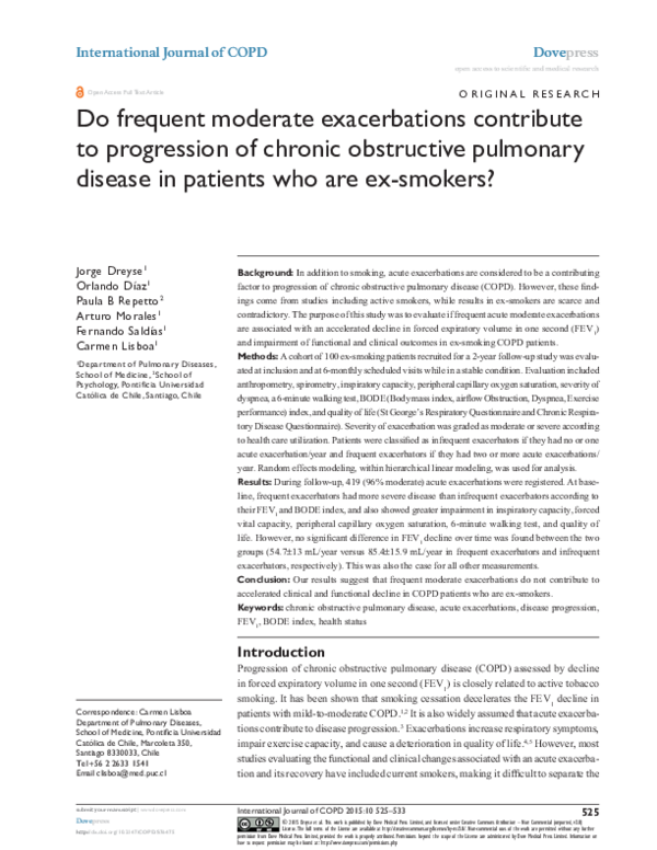
The recruited cohort included 105 patients. Two patients died
and another three were lost to follow-up before the first scheduled visit and were consequently excluded from the analyses.
During follow-up, seven patients died and four declined to
continue in the protocol, mainly due to transport difficulties (Figure 1). Compared with patients who completed the
study, these eleven patients were older, had more severe
disease according to GOLD criteria and BODE index, and
nine reported frequent exacerbations during the year before
recruitment. The baseline characteristics of the 100 patients
included in the analyses are shown in Table 1.
number, distribution, and severity
of exacerbations
During follow-up, 83 patients had 419 exacerbations. Two
hundred and fifty-six patients (61%) were seen and treated
at our institution, and the remaining 163 (39%) at other
International Journal of COPD 2015:10
Frequent exacerbations in ex-smokers with COPD
Q ���
��GHDG
��ORVW
��PRQWKV
Q ���
��GHDG
��ORVW
���PRQWKV
Q ��
��GHDG
��ORVW
���PRQWKV
Q ��
��GHDG
��ORVW
���PRQWKV
Q ��
Figure 1 Follow-up diagram for cohort of patients with chronic obstructive pulmonary disease.
Table 1 Characteristics of COPD patients at study entry
sex, male/female
age, years
smoking history pack/year
Years since quitting smoking
gOlD stage, n (%)
Mild
Moderate
severe
Very severe
BMI, kg/m2
mMrC
0–1 points
2 points
3 points
4 points
IC, % predicted
FVC, % predicted
FeV1, % predicted
FeV1/FVC, %
6MWD, % predicted
BODe index
0–2 points
3–4 points
5–6 points
7–10 points
Charlson comorbidity index, points
Urine cotinine levels, ng/ml
CrQ global, points
sgrQ total, points
58/42
68.8±7.7
42.8±23.5
11.5±8.7
8 (8)
48 (48)
28 (28)
16 (16)
26.6±3.7
15 (15)
60 (60)
21 (21)
4 (4)
77.9±22.9
90.9±22.1
52.6±20.6
42.7±12.7
84.5±19.9
50 (50)
33 (33)
11 (11)
6 (6)
4.46±1.84
12.9±28.5
92.4±24.4
49.0±19.8
Notes: Values are expressed as the mean ± standard deviation or number (percent).
Abbreviations: BMI, body mass index; COPD, chronic obstructive pulmonary
disease; mMRC, modiied Medical Research Council; IC, inspiratory capacity; FVC,
forced vital capacity; FeV1, forced expiratory volume in 1 second; gOlD, global
Initiative for Chronic Obstructive lung Disease; 6MWD, 6-minute walking distance;
BODe, body mass index, airlow obstruction, dyspnea, exercise performance; SGRQ,
st george’s respiratory Questionnaire; CrQ, Chronic respiratory Questionnaire.
submit your manuscript | www.dovepress.com
Dovepress
527
�Dovepress
Dreyse et al
��
The number of exacerbations per patient over the 2 years of
follow-up was variable (Figure 2). Seventeen patients did not
experience an exacerbation, whereas five experienced 14 or
more exacerbations during the observation period. Median
exacerbation frequency was two and one per year in patients
classified as frequent exacerbators and infrequent exacerbators, respectively (P,0.001).
��
3DWLHQWV� Q
��
��
��
Characteristics of patients according
to frequency of exacerbations
��
�
�
�
�
�
�
�
�
�
([DFHUEDWLRQV�SHU�\HDU� Q
�
Figure 2 number of exacerbations per patient.
Note: Patient numbers include patients treated at our institution and in other health
institutions.
institutions. According to the classification employed,
exacerbations were moderate (96%) or severe (4%). Thirteen
subjects had one hospitalization, one subject had two hospitalizations, and a third subject had three hospitalizations.
At baseline, patients with frequent exacerbations showed
significant differences in the number of acute exacerbations
during the previous year, FEV1 % predicted, forced vital
capacity (FVC) % predicted, inspiratory capacity % predicted, and SpO2, as well as a greater impairment in mMRC,
health status (SGRQ and CRQ) and the BODE index, as
compared with infrequent exacerbators (see Table 2).
The slopes for all these variables, representing changes
over the 2-year follow-up period, were not significantly different between the two groups (Table 3). Thus, FEV1 decreased
Table 2 Baseline characteristics according to frequency of exacerbations in 100 patients with COPD
Male/female, n
age, years
BMI, kg/m2
smoking history, pack-years
ae previous year
mMrC
0–1 points
2 points
3 points
4 points
IC, % predicted
FeV1, % predicted
FVC, % predicted
FeV1/FVC, %
6MWD, % predicted
gOlD
stage I
stage II
stage III
stage IV
spO2, %
BODe index
0–2 points
3–4 points
5–6 points
7–10 points
sgrQ total, points
CrQ global, points
Infrequent exacerbators
n=51
Frequent exacerbators
n=49
P-value
31/20
68.8±6.5
26.7±3.5
42.5±21.1
1 (0–2)
27/22
68.8±8.8
26.5±4.0
43.1±26.0
2 (1–3)
ns
ns
ns
ns
,0.001
0.043
8 (16)
36 (71)
6 (12)
1 (2)
83.8±20.7
57.6±19.3
96.4±21.6
44.4±12.6
87.8±16.6
7 (14)
24 (49)
15 (31)
3 (6)
71.6±23.7
47.6±20.8
85.1±21.4
40.9±12.8
80.9±22.5
5 (10)
27 (53)
17 (33)
2 (4)
93.6±2.71
3 (6)
21 (43)
11 (22)
14 (29)
92.5±2.29
29 (57)
19 (37)
2 (4)
1 (2)
44.0±19.5
97.3±26.1
21 (43)
14 (29)
9 (18)
5 (10)
54.2±19.0
87.3±21.7
0.008
0.013
0.011
ns
0.087
0.030
0.026
0.027
0.010
0.044
Notes: Values are expressed as the mean ± standard deviation, number (percent), or median (interquartile range).
Abbreviations: BMI, body mass index; COPD, chronic obstructive pulmonary disease; mMRC, modiied Medical Research Council; IC, inspiratory capacity; FVC, forced
vital capacity; FeV1, forced expiratory volume in 1 second; gOlD, global Initiative for Chronic Obstructive lung Disease; 6MWD, 6-minute walking distance; BODe, body
mass index, airlow obstruction, dyspnea, exercise performance; NS, not statistically signiicant; SGRQ, St George’s Respiratory Questionnaire; CRQ, Chronic Respiratory
Questionnaire; spO2, peripheral capillary oxygen saturation.
528
submit your manuscript | www.dovepress.com
Dovepress
International Journal of COPD 2015:10
�Dovepress
Frequent exacerbations in ex-smokers with COPD
Table 3 slopes of all indices according to the random effect
modeling
IC, ml
FVC%
FeV1, ml
FeV1, % predicted
FeV1/FVC
6MWD, % predicted
mMrC, points
BODe index, points
sgrQ, points
Slope infrequent
Slope frequent
P-value
38
-2.20
-85.57
-2.84
-1.46
-2.04
-0.04
0.136
-0.74
105
-1.73
-54.72
-1.7
-0.84
-2.88
-0.04
0.276
0.73
0.420
0.762
0.129
0.212
0.357
0.672
0.259
0.387
0.331
Abbreviations: IC, inspiratory capacity; FVC, forced vital capacity; FeV1, forced
expiratory volume in 1 second; 6MWD, 6-minute walking distance; mMRC, modiied
Medical research Council; BODe, body mass index, airlow obstruction, dyspnea,
exercise performance; sgrQ, st george’s respiratory Questionnaire; CrQ,
Chronic respiratory Questionnaire.
57±13 mL/year (1.7% predicted/year) and 85±15.8 mL/year
(2.8% predicted/year) in frequent exacerbators and infrequent
exacerbators, respectively.
Figure 3 shows lung function indices at baseline and at
each scheduled visit in both groups of patients. Over the
study period, frequent exacerbators had consistently lower
values for inspiratory capacity, FVC, FEV1, and FEV1/FVC
than infrequent exacerbators, but changes across time did not
differ between the two groups. Similar behavior was observed
for functional exercise capacity (6MWT) and health-related
quality of life (Figure 4).
Patients with frequent exacerbations were treated more
frequently with inhaled corticosteroids, whereas short-acting
bronchodilators were used as the main therapy more often
by infrequent exacerbators (Table 4).
Discussion
The main findings of this prospective observational study in
patients with COPD who had quit smoking and were followed
up for 2 years are: frequent moderate exacerbations did not
accelerate the decline in FEV1 nor worsen other functional
indices as compared with infrequent exacerbations, and there
were no significant differences in patient-centered outcomes
over time between the two groups. Our results also confirm
that exacerbations are more frequent in patients with more
severe disease and that previous history of exacerbations is
a predictor of frequent exacerbations.24
To our knowledge, this is the first study performed
solely in ex-smoking COPD patients that included several
clinical indices in addition to lung function to evaluate the
effect of exacerbations on disease progression. Our findings
are consistent with results reported in other studies, where
no significant differences were found in the FEV1 decline
��
���
)9&� ��SUHG
���
,&� ��SUHG
���
��
��
��
�
�
�
�
��
��
��
�
�
�
�
��
��
��
��
��
��
�����
��
�����
��
)(9�� P/
)(9�� ��SUHG
��
��
�
�
��
��
��
��
�
�
��
�
�
��
0RQWKV
��
��
�����
�����
���
���
�
0RQWKV
Figure 3 lung function indices progression.
Notes: Baseline (0) and scheduled visits (every 6 months) during 2 years of follow-up; values are expressed as the mean ± one standard error. (◊) indicates infrequent
exacerbators; () indicates frequent exacerbators.
Abbreviations: IC, inspiratory capacity; FVC, forced vital capacity; FeV1, forced expiratory volume in 1 second; pred, predicted.
International Journal of COPD 2015:10
submit your manuscript | www.dovepress.com
Dovepress
529
�Dovepress
Dreyse et al
��
���
P05&� SRLQWV
%0,� .J P±�
��
��
��
��
�
�
�
��
��
���
���
�
��
��
%2'(� SRLQWV
�0:'� ��SUHG
��
��
��
�
�
�
��
��
��
��
�
�
��
��
��
�
�
��
��
��
�
�
�
�
��
���
&54�JOREDO� SRLQWV
��
6*54�WRWDO� SRLQWV
��
�
��
��
��
��
�
�
�
�
��
�
�
�
�
��
��
��
���
��
��
�
�
0RQWKV
0RQWKV
Figure 4 Clinical indices and health status progression.
Notes: Baseline (0) and scheduled visits (every 6 months) during 2 years of follow-up; values are expressed as the mean ± one standard error. (◊) indicates infrequent
exacerbators, () indicates frequent exacerbators.
Abbreviations: BMI, body mass index; mMRC, modiied Medical Research Council dyspnea scale; 6WD, 6-minute walking distance; BODE, body mass index, airlow
obstruction, dyspnea, exercise performance; sgrQ, st george’s respiratory Questionnaire; CrQ, Chronic respiratory Questionnaire.
Table 4 Inhaled treatment during follow-up
Therapy
Infrequent
exacerbators
n=51
Frequent
exacerbators
n=49
P-value
laBa alone
laMa alone
saBa and/or saMa alone
laBa + ICs
laBa + laMa + ICs
ICsa
2
5
20
14
10
24
5
2
9
16
17
33
ns
ns
0.021
ns
0.07
0.032
Note: aPlus any other inhaled therapy.
Abbreviations: laBa, long-acting β agonist; laMa, long-acting muscarinic
antagonist; NS, not statistically signiicant; SABA, short-acting β agonist; saMa,
short-acting muscarinic antagonist; ICs, inhaled corticosteroids.
530
submit your manuscript | www.dovepress.com
Dovepress
between frequent and infrequent exacerbators.4,7,9,25,26 Some
of these studies included current smokers.4,25,26 Two studies
have previously assessed the effects of exacerbations on FEV1
decline in ex-smokers, with conflicting results.7,9 Kanner et al
performed a secondary analysis of the Lung Health Study and
found that, in intermittent and continuous smokers, frequent
lower respiratory infections were associated with a greater
5-year averaged annual rate of FEV1 loss (-52 mL/year
and -69 mL/year, respectively) than in sustained quitters
(-12 mL/year). Makris et al evaluated the effect of acute
exacerbations in current and ex-smokers during a 3-year
International Journal of COPD 2015:10
�Dovepress
prospective study including 58 ex-smokers and 44 current
smokers. The FEV1 decline was greater in current smokers
than in ex-smokers.
Our results are in agreement with those of the ex-smoker
group reported by Kanner et al but differed from those
reported by Makris et al. This may be explained by differences
between the present study and the study by Makris et al with
regard to the number of ex-smoking patients (100 versus 58,
respectively) and length of follow-up (2 versus 3 years,
respectively), but mainly in the annual rate of hospitalizations
(0.10 versus 0.35 per year, respectively), indicating more
severe exacerbations in their study.
The effect of exacerbations on decline in lung function
has not been clearly established. The role of exacerbation
frequency in FEV1 decline is largely based on findings of
studies that have included active smokers.3–6 Even so, differences in FEV1 decline between frequent exacerbators and
infrequent exacerbators have been small: 8 mL/year was
reported by Donaldson et al in a group of 32 patients followed
for 4 years and, according to Makris et al frequent exacerbations added -1.4% predicted/year to FEV1 decline.
In the present study, which included only ex-smoking
patients, the decline in FEV1, although not significant, was
smaller in frequent exacerbators (54.7 mL/year) than in
infrequent exacerbators (85.4 mL/year). This unexpected
nonsignificant faster decline in infrequent exacerbators
could be explained by their higher baseline FEV1 value, in
agreement with previous data from Casanova et al.27 It is
also consistent with the findings from the analysis of data
obtained from the placebo arms of several recent clinical
series by Tantucci and Modena,28 which showed that “the loss
of lung function, assessed as expiratory airflow reduction,
seems more accelerated and therefore more relevant in the
initial phases of COPD”. Other possible explanations for the
lack of a more rapid FEV1 decline in frequent exacerbators
are frequent treatment with systemic corticosteroids due to
acute exacerbations as well as more regular use of inhaled
corticosteroids than in infrequent exacerbators. Moreover,
the effects of acute exacerbations on FEV1 decline are
difficult to assess due to the heterogeneity of the patient
behavior over time. Several recent studies, independent of
the frequency of exacerbations, show a heterogeneous FEV1
decline in COPD.29–31 According to these studies, FEV1
may remain stable in some patients, increase in others, or
experience a slow or accelerated decline. Accelerated FEV1
decline was associated with a high baseline FEV1, low body
mass index, active smoking, bronchodilator reversibility,
and greater magnitude of emphysema. Our results confirm
International Journal of COPD 2015:10
Frequent exacerbations in ex-smokers with COPD
the heterogeneous FEV1 decline reported by these authors,
independently of the frequency of exacerbations.29–31
In addition to the lack of effect of acute exacerbations on
FEV1 decline, the present results also demonstrate no effects
on other physiological or clinical outcomes in ex-smoking
COPD patients. To our knowledge, the effects of exacerbations on patient-centered outcomes in ex-smoking COPD
patients have not been previously assessed. The few data
available derive from the study by Cote et al4 in a cohort
of 205 patients (95% men), including active smokers, who
were followed for 2 years after their first acute exacerbation. Although no significant changes in FEV1 decline were
observed, they found a significant deterioration of mMRC,
6MWT, and BODE index in their frequent exacerbators. In
addition to differences in cohort characteristics, our results
differ from theirs in the percentage of patients who required
hospital admissions. Fifty of their patients with frequent acute
exacerbations were hospitalized, of whom 39% required
one hospitalization, 35% required two hospitalizations, and
26% required three or more hospitalizations. In our cohort,
13 patients required hospitalization and only four of them
required a second hospitalization. As it is known that severe
acute exacerbations contribute to deterioration and mortality
in COPD patients,32 the greater number of patients and
hospital admissions reported by Cote et al4 may explain the
clinical deterioration of their patients.
The most advanced therapy (long-acting muscarinic antagonist + long-acting beta-2 agonists + inhaled
corticosteroids), mostly received by the frequent exacerbators
given their greater COPD severity, could partly explain the
lack of significant differences in the measured indices.
Our study has some limitations: the sample size was
relatively small; most exacerbations were moderate and
treated in an outpatient setting; severe exacerbations, which
are known to contribute to mortality and hospitalization, were
infrequent; no mild acute exacerbations, ie, those needing
only an increase in bronchodilator therapy, were registered,
probably because they resolved without medical assistance;
and follow-up was relatively short. However, we believe that
these limitations do not affect our main findings, indicating
no effect of moderate acute exacerbations on FEV1 decline
or clinical progression of COPD in ex-smokers.
Conclusion
In summary, our findings suggest that acute moderate exacerbations in ex-smoking patients do not contribute to COPD
progression assessed by FEV1 decline and by the BODE
index during a follow-up of 2 years. They also confirm that
submit your manuscript | www.dovepress.com
Dovepress
531
�Dreyse et al
changes in FEV1 over time are heterogeneous independent
of exacerbation frequency. Although patients with frequent
exacerbations had greater baseline impairment of functional
and clinical indices than those with infrequent acute exacerbations, their parameters behaved similarly over time. Replication of these findings may help us to better understand how
the effects of frequency of exacerbations among ex-smoking
COPD patients impacts their disease behavior.
Acknowledgment
This work was funded by a grant from FONDECYT
(1085268).
Disclosure
The authors report no conflicts of interest in this work.
References
1. Fletcher C, Peto R. The natural history of chronic airflow obstruction.
BMJ. 1977;1(6077):1645–1648.
2. Anthonisen NR, Connett JE, Murray RP. Smoking and lung function of
Lung Health Study participants after 11 years. Am J Respir Crit Care
Med. 2002;166(5):675–679.
3. Donaldson GC, Seemungal TA, Bhowmik A, Wedzicha JA. Relationship
between exacerbation frequency and lung function decline in chronic
obstructive pulmonary disease. Thorax. 2002;57(10):847–852.
4. Cote CG, Doderlly LJ, Celli BR. Impact of COPD exacerbations on
patient-centered outcomes. Chest. 2007;131(3):696–704.
5. Seemungal TA, Donaldson GC, Paul EA, Bestall JC, Jeffries DJ,
Wedzicha JA. Effect of exacerbations on quality of life in patients with
chronic obstructive pulmonary disease. Am J Respir Crit Care Med.
1998;157(5 Pt 1):1418–1422.
6. Halpin DMG, Decramer M, Celli BR, Kesten S, Liu D, Tashkin DP.
Exacerbation frequency and course of COPD. Int J Chron Obstruct
Pulmon Dis. 2012;7:653–661.
7. Kanner RE, Anthonisen NR, Connet JE; for the Lung Health Study
Research Group. Lower respiratory illnesses promote FEV1 decline
in current smokers but not ex-smokers with mild chronic obstructive
pulmonary disease. Results from the Lung Health Study. Am J Respir
Crit Care Med. 2001;164(3):358–364.
8. Silverman EK. Exacerbations in chronic obstructive pulmonary disease
Do they contribute to disease progression? Proc Am Thorac Soc. 2007;
4(8):586–590.
9. Makris D, Moschandreas J, Damaniaki A, et al. Exacerbations and lung
function decline in COPD: new insights in current and ex-smokers.
Respir Med. 2007;101(6):1305–1312.
10. Rabe KF, Hurd S, Anzueto A, et al. Global strategy for the diagnosis,
management and prevention of chronic obstructive lung disease:
GOLD executive summary. Am J Respir Crit Care Med. 2007;176(6):
532–555.
11. Rodríguez-Roisin R. Towards a consensus definition for COPD exacerbations. Chest. 2000;117(5 Suppl 2):398S–401S.
12. Anthonisen NR, Manfreda J, Warren CPW, Hershfield ES, Harding GKM,
Nelson NA. Antibiotic therapy in exacerbations of chronic obstructive
pulmonary disease. Ann Intern Med. 1987;106(2):196–204.
532
submit your manuscript | www.dovepress.com
Dovepress
Dovepress
13. Wedzicha JA, Seemungal T. COPD exacerbations: defining their cause
and prevention. Lancet. 2007;370(9589):786–796.
14. Charlson M, Szatrowski TP, Peterson J, Gold J. Validation of a combined comorbidity index. J Clin Epidemiol. 1994;47(11):1245–1251.
15. Mahler D, Wells C. Evaluation of clinical methods for rating dyspnea.
Chest. 1988;93(3):580–586.
16. Miller MR, Hankinson J, Brusasco V, et al. Standardization of spirometry. Eur Respir J. 2005;26(2):319–338.
17. Hankinson JL, Odencrantz JR, Fedan KB. Spirometric reference values
from a sample of the general U.S population. Am J Respir Crit Care
Med. 1999;159(1):179–187.
18. Lisboa C, Leiva A, Pinochet R, Repetto P, Borzone G, Diaz O. [Valores
de referencia de la capacidad inspiratoria en sujetos sanos no fumadores mayores de 50 años]. Arch Bronconeumol. 2007;43(9):485–489.
Spanish.
19. Quirk FH, Jones PW, Baveystock CM, Littlejohns P. A self complete
measure for chronic airflow limitation: the St George’s Respiratory
Questionnaire. Am Rev Respir Dis. 1992;145(6):1321–1327.
20. Williams JE, Singh SJ, Sewell L, Guyatt GH, Morgan MD. Development of a self-reported chronic respiratory questionnaire (CRQ-SR).
Thorax. 2001;56(12):954–959.
21. ATS Committee on Proficiency Standards for Clinical Pulmonary Function Laboratories. American Thoracic Society Statement. Guidelines
for the six-minute walk test. Am J Respir Crit Care Med. 2002;166(1):
111–117.
22. Troosters T, Gosselink S, Decramer M. Six minute walking distance
in healthy elderly subjects. Eur Respir J. 1999;14(2):270–274.
23. Celli BR, Cote CG, Marin JM, et al. The body-mass index, airflow
obstruction, dyspnea and exercise capacity index in chronic obstructive
pulmonary disease. N Engl J Med. 2004;350(10):1005–1012.
24. Hurst JR, Vestbo J, Anzueto A, et al; for the Evaluation of COPD
Longitudinally to Identify Predictive Surrogate Endpoints (ECLIPSE)
Investigators. Susceptibility to exacerbation in chronic obstructive
pulmonary disease. N Engl J Med. 2010;363(12):1128–1138.
25. Miratvilles M. Ferrer M, Pont A, et al. Effect of exacerbations on quality
of life in patients with chronic obstructive pulmonary disease: a 2 years
follow up study. Thorax. 2004;59(5):387–395.
26. Spencer S, Calverley PMA, Burge PS, Jones PW. Impact of preventing
exacerbations on deterioration of health status in COPD. Eur Respir J.
2004;23(5):698–702.
27. Casanova C, Cote CG, Marin JM, et al. The six-minute walk distance:
long term follow up in patients with COPD. Eur Respir J. 2007;29(3):
535–540.
28. Tantucci C, Modena D. Lung function decline in COPD. Int J Chron
Obstruct Pulmon Dis. 2012;7:95–99.
29. Casanova C, de Torres JP, Aguirre-Jaime A, et al. The progression of
chronic obstructive pulmonary disease is heterogeneous. The experience of the BODE cohort. Am J Respir Crit Care Med. 2011;184(9):
1015–1021.
30. Vestbo J, Edwards LD, Scanlon PD, et al; for the ECLIPSE Investigators. Changes in forced expiratory volume in 1 second over time in
COPD. N Engl J Med. 2011;365(13):1184–1192.
31. Nishimura M, Makita H, Nagai K, et al; for the Hokkaido COPD Cohort
Study Investigators. Annual change in pulmonary function and clinical
phenotype in chronic obstructive pulmonary disease. Am J Respir Crit
Care Med. 2012;185(1):44–52.
32. Soler-Cataluña JJ, Martínez-García MA, Román Sanchez P, Salcedo E,
Navarro M, Ochando R. Severe acute exacerbation and mortality in
patients with chronic obstructive pulmonary disease. Thorax. 2005;
60(11):925–931.
International Journal of COPD 2015:10
�Dovepress
Frequent exacerbations in ex-smokers with COPD
International Journal of COPD
Dovepress
Publish your work in this journal
The International Journal of COPD is an international, peer-reviewed
journal of therapeutics and pharmacology focusing on concise rapid
reporting of clinical studies and reviews in COPD. Special focus is given
to the pathophysiological processes underlying the disease, intervention
programs, patient focused education, and self management protocols.
This journal is indexed on PubMed Central, MedLine and CAS. The
manuscript management system is completely online and includes a
very quick and fair peer-review system, which is all easy to use. Visit
http://www.dovepress.com/testimonials.php to read real quotes from
published authors.
Submit your manuscript here: http://www.dovepress.com/international-journal-of-chronic-obstructive-pulmonary-disease-journal
International Journal of COPD 2015:10
submit your manuscript | www.dovepress.com
Dovepress
533
�
 Fernando Saldías