BR A IN RE S EA RCH 1 3 08 ( 20 1 0 ) 6 8 –78
available at www.sciencedirect.com
www.elsevier.com/locate/brainres
Research Report
Motor area localization using fMRI-constrained cortical current
density reconstruction of movement-related cortical
potentials, a comparison with fMRI and TMS mapping
Alberto Inuggi a , Massimo Filippi b , Raffaella Chieffo a , Federica Agosta b , Maria A. Rocca b ,
Javier J. González-Rosa a , Marco Cursi a , Giancarlo Comi a , Letizia Leocani a,⁎
a
Department of Neurology, Neurophysiology and Neurorehabilitation, Experimental Neurology Institute, IRCCS San Raffaele, Milan, Italy
Neuroimaging Research Unit, IRCCS San Raffaele, Milan, Italy
b
A R T I C LE I N FO
AB S T R A C T
Article history:
The localization of human hand primary motor area (M1) has been the object of several
Accepted 16 October 2009
studies during the last decades. EEG source analysis, functional magnetic resonance
Available online 22 October 2009
imaging (fMRI) and focal transcranial magnetic stimulation (TMS) are non-invasive methods
for localizing M1 with good accuracy compared to direct electrocorticography (ECoG) results.
Keywords:
EEG sources were reconstructed with Cortical Current Density (CCD) method, allowing to
EEG source analysis
evaluate simultaneous and distributed patterns of activation and to increase accuracy by
Multimodal integration
constraining on information derived from fMRI (fMRI-CCD). The aim of this study was to
fMRI constrained CCD
compare the M1 contribution of movement-related cortical potentials (MRCP) with TMS and
fMRI results and to test the effect of constraints strength, algorithm norm and localization
methods over CCD reconstruction. Seven right-handed healthy subjects underwent 64channel EEG recording of MRCP to right thumb movement, focal TMS mapping of the right
abductor pollicis brevis muscle and fMRI during right hand movement. We found fMRI
activations, EEG sources and TMS mapping corresponding to the anatomical landmark of
the hand area in all subjects with fMRI and TMS center-of-gravity and in almost all subjects
using fMRI-CCD with moderate constraint. A significant improvement was found using
fMRI-CCD compared to CCD alone. This study confirms the usefulness of multimodal
integration of fMRI, EEG and TMS in localizing M1 and the possibility to increase EEG spatial
resolution using fMRI information.
© 2009 Elsevier B.V. All rights reserved.
⁎ Corresponding author. Department of Clinical Neurophysiology, IRCCS San Raffaele, Via Olgettina 60, 20132 Milan, Italy. Fax: +39 2 2643
3085.
E-mail address: leocani.letizia@hsr.it (L. Leocani).
Abbreviations: BOLD, blood oxygenation level dependent; CCD, cortical current density; CCD-POSnorm,K,method, Anatomical position of
CCD sources calculated with a specific norm, M1 reconstruction method and fMRI over-weighting factor strength K; CCD-fMRInorm,K,method,
Distance between CCD source and fMRI maxima calculated with a specific norm, M1 reconstruction method and K fMRI over-weighting
factor strength; COG, center of gravity in one single latency; COGT, average center of gravity in one period; CSF, cerebrospinal fluid; EEG,
electroencephalography; fMRI, functional magnetic resonance imaging; fMRI-CCD, fMRI constrained CCD; HS, hot spot; K, strength of fMRI
over-weighting factor over fMRI-CCD; M1, primary motor area; MEP, motor evoked potential; METHOD, method to calculate M1 position
(HS, COG, COGT); MRCP, movement related cortical potentials; NORM, type of CCD algorithm (L1, L2); PMd, dorso-lateral premotor cortex;
SMA, supplementary motor area; SNR, signal-to-noise ratio; TMS, transcranial magnetic stimulation
0006-8993/$ – see front matter © 2009 Elsevier B.V. All rights reserved.
doi:10.1016/j.brainres.2009.10.042
�BR A IN RE S E A RCH 1 3 08 ( 20 1 0 ) 6 8 –7 8
1.
Introduction
The localization of human hand primary sensory and motor
areas (SM1) has been object of several studies during the last
decades. Preoperative direct cortical electrical stimulation
69
(Yousry et al., 1995) and electrocorticography with recording
of movement-related cortical potentials (MRCP) (Ikeda et al.,
1996) localized the primary human hand motor area (M1) in
BA4 on the precentral gyrus. Its position was later associated to
the precentral knob region (Yousry et al., 1997). Several non-
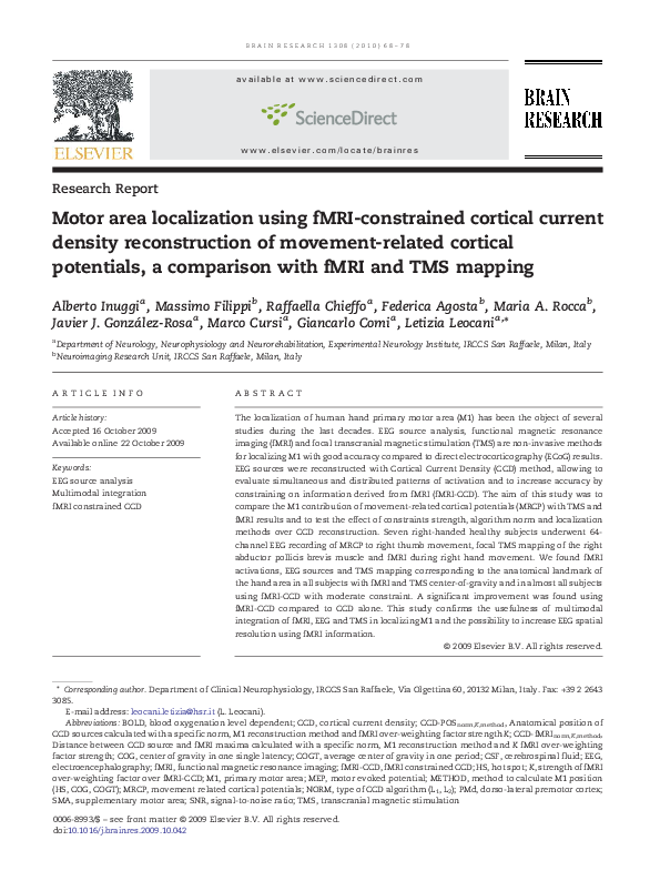
Fig. 1 – Multimodal localization of M1. Each column represents a different subject, each row a different method. (fMRI) axial
slices of fMRI activation to fingers movement at fMRI maximum depth superimposed over anatomical MRI. In all subsequent
rows, fMRI contours are overlaid over cortex surface (black line), intensities, either of current density and MEP values, are
expressed in a color scale ranging from red (lowest values) to white (highest values). (TMS) results from MEP mapping over CSF
surface. TMS COG and HS are represented with blue square and triangle respectively, fMRI maxima with a black circle. When
the TMS COG and HS distance is below 3 mm only COG is shown. TMS points are projected according to the procedure described
in Fig. 4. (remaining rows) CCD with both norms (L1 and L2) and two representative K: 1.0 shows unconstrained CCD and 1.4
shows sources with the K producing the lowest distance from fMRI. In L2 CCD, sources carrying currents below 50% of the
largest current (full width at half maximum, FWHM) are clipped (Fuchs et al., 1999).
�70
BR A IN RE S EA RCH 1 3 08 ( 20 1 0 ) 6 8 –78
invasive methods have been developed with increasing
accuracy. Among these, functional magnetic resonance imaging (fMRI) measures the blood oxygenation level dependent
(BOLD) contrast related to the increased blood demand in the
vicinity of neuronal activity (Logothetis, 2003). fMRI of voluntary movement localized M1 area with differences ranging
from 3 to 10 mm compared with intra-operative electrical
stimulation (Yousry et al., 1995). The analysis of motor evoked
potentials (MEPs) to focal transcranial magnetic stimulation
(TMS) applied to different scalp positions over the motor cortex
allows to map the scalp representation of a given muscle
(Wassermann et al., 1992; Rossini et al., 1994). Cortical
projection of these maps produced average localization
discrepancies from 2 to 14 mm compared with fMRI activation
maps (Terao et al., 1998; Herwig et al., 2002; Lotze et al., 2003;
Neggers et al., 2004). High-resolution electroencephalography
(EEG) has been used to investigate the spatio-temporal pattern
of cortical activity by reconstructing the generators of scalp
recorded potentials using dipole and distributed source
models such as cortical current density (CCD) reconstruction.
Several approaches have been developed to increase the
spatial resolution of EEG (Graves de Peralta Menendez and
Gonzalez Andino, 1998; Fuchs et al., 1999; Wagner et al., 2001a,
b) that, however, remains quite poor in comparison to fMRI.
EEG source reconstruction accuracy can be improved with a
spatial constraints approach (Kiebel and Friston, 2004), using
fMRI activated regions of interest (ROI) as priors in the solution
of the inverse problem (Babiloni et al., 2000; Wagner et al.,
2001b; Dale and Halgren, 2001; Babiloni et al., 2003; Liu and He,
2008). fMRI-CCD methods, to solve the inverse problem, utilize
anatomo-functional correlates of cortical activity instead of
arbitrary mathematical hypotheses. The analysis of MRCP
(Deecke and Lang, 1996; Wildgruber et al., 1997; Ball et al., 1999;
Cui et al., 1999; Toma et al., 2002, Inuggi et al., 2009) is the most
investigated electrophysiological protocol for investigating M1
activity particularly close to movement onset. In this paper, for
the first time, the three non-invasive methodologies (fMRI,
TMS and EEG) will be applied to the same subjects in order to
investigate the reciprocal relationships among these methods
and the precentral knob position. A particular focusing will be
given to the issue of MRCP generators reconstruction through
the CCD model, providing the effects of fMRI constraints over
its accuracy in real rather than simulated data as previously
performed.
2.
Results
2.1.
fMRI
All subjects showed activations in several areas, which are part
of the “classical” sensorimotor network, including the primary
sensorimotor cortex (SM1), bilaterally, the supplementary
motor area (SMA) and the ipsilateral cerebellum. One subject
also showed an additional activation in the contralateral
secondary somatosensory cortex. The fMRI-maximum (ROI
showing the highest T-value) was always precentral inside the
‘precentral knob’ region where the hand area is located (Yousry
et al., 1997), 8 ± 5 mm deep into the precentral gyrus. In Fig. 1,
fMRI maxima (row L1 1.0) and contours have been overlaid over
rendered cortex and compared with TMSHS, TMSCOG and MRCP
sources. No mirror movements could be observed.
Table 1 – Anatomical localization of EEG sources and TMS measures.
Subject
Method
Norm
TMS
L2
L1
A
B
C
D
E
F
G
HS
COG
COGT
HS
COG
COGT
HS
COG
COGT
HS
COG
COGT
HS
COG
COGT
HS
COG
COGT
HS
COG
COGT
K=1
K = 1.1
K = 1.4
K=3
K=1
K = 1.1
K = 1.4
K=3
BA6⁎
BA6⁎
BA6⁎
BA6⁎
BA6⁎
BA6⁎
BA4⁎
BA4
BA4
BA1⁎
BA1
BA1
PK
BA4
BA4
BA3b
BA3b
BA3b
BA3b
BA3b
BA3b
BA4⁎
BA4
BA4
BA3b
BA6⁎
BA6⁎
BA4⁎
BA4
BA4
BA1⁎
BA1
BA1
BA3b
BA4
BA4
BA3b
BA3b
BA3b
BA3b
BA3b
BA3b
BA4
BA4
PK
PK
BA4
BA4
PK
PK
PK
PK
BA1
BA1
PK
BA4
BA4
PK
PK
PK
BA4
BA4
BA4
BA4
BA4
PK
PK
BA4
BA4
PK
PK
PK
PK
BA1
BA1
PK
BA4
BA4
PK
BA3b
PK
PK
PK
PK
BA4⁎
BA3b
BA3b
BA6⁎
BA4
BA4
BA4⁎
BA4
PK
BA4⁎
BA4
BA4
PK
PK
PK
BA6⁎
BA6⁎
BA6⁎
BA3b
BA4
BA4
BA4
BA3b
BA3b
PK
BA4
BA4
PK
BA4
PK
BA4
BA4
BA4
PK
PK
PK
BA6⁎
BA6⁎
BA6⁎
BA4
BA3b
BA4
PK
BA4
PK
PK
BA4
BA4
PK
PK
PK
PK
PK
PK
PK
PK
PK
PK
PK
PK
BA4
BA4
PK
BA3b
BA4
PK
PK
BA3b
BA4
BA3b
PK
PK
PK
PK
PK
PK
PK
PK
PK
PK
PK
BA4
BA4
BA3b
PK
PK
–
BA6
PK
–
BA1
PK
–
PK
PK
–
BA3b
PK
–
BA6
PK
–
PK
PK
–
Anatomical positions of EEG sources inside the confidence region. PK indicates that source was located in the precentral knob. EEG sources not
included inside fMRI bounds are represented with the symbol ‘⁎’.
�BR A IN RE S E A RCH 1 3 08 ( 20 1 0 ) 6 8 –7 8
2.2.
EEG
Left EPB EMG analysis showed that no mirror movements
were performed by any subject. The independent component
analysis revealed three components with signal-to-noise ratio
(SNR) values of 3.2 ± 0.9 , 1.2 ± 0.4, 0.6 ± 0.3. The first was located
over mesial areas, the second located over primary sensorymotor cortex (SM1), the third pattern varied and was not used
in all subjects as its SNR resulted below noise threshold
(SNR < 1) during the whole interval. CCD analysis of the EEG
signal reconstructed a distributed activation pattern mainly
localized in contralateral primary sensorimotor cortex (M1
and S1) and SMA, but also embracing ipsilateral M1 and
bilateral dorso-lateral premotor cortex. Applying fMRI constraints, activations mostly concentrated in contralateral M1
and SMA only. In Fig. 1 MRCP-CCD reconstructions with two
norms and two overweighting factors are shown. CCD sources
anatomical positions are summarized in Table 1 and plotted in
Fig. 2. The best reconstruction methods resulted the HS with
K = 3 for L1 and COGT and HS with K = 1.4 for L2 norm
respectively (one M1 reconstruction in BA4 and six into the
71
precentral knob). Good performance was also achieved for L1
using the HS with K = 1.4 (two M1 reconstructions in BA4, five
into PK). Significant effects of the three factors (norm, K,
method) and their interactions over anatomical localization of
CCD sources respect to precentral knob (CCD-POSnorm,K,method)
are shown in Table 2. Concerning the interaction between
norm and K strength, cortical activity resulted closer to
precentral knob using K = 1.4 and K = 3 compared to K = 1.0
with both norms and using L2 compared to L1 norm with all K
but K = 3. Concerning the method for M1 localization, HS
performed significantly better than COG and COGT using L1
norm. Although no interaction between method and L2 norm
emerged, HS resulted the only method for which L2 did not
perform better than L1 in unconstrained CCD.
2.3.
TMS
On average, APB responses over 50 μV have been obtained in
22 ± 5.5 stimulated points. In Fig. 1 (row TMS), TMS-derived CSF
maps and their corresponding measures positions (TMSCOG
and TMSHS), projected orthogonally to scalp surface until CSF
Fig. 2 – Distances of CCD sources from fMRI and precentral knob. Effect of norm (L1 and L2), fMRI constraints strength (K) and
method (HS, COG, COGT) on CCD Euclidean distance, in mm, from fMRI maxima (left) and anatomical position respect to
precentral knob (right). L1 and L2 norms are shown in left and right columns, respectively, and the different localization method
(COGT, COG and HS) in upper, middle and lower row, respectively. Results are plotted with different K strengths (on the
abscissas) and each subject is represented by a different symbol.
�72
BR A IN RE S EA RCH 1 3 08 ( 20 1 0 ) 6 8 –78
Table 2 – Statistical analysis.
Effect
Distance from fMRI maxima
K
Norm
Method
norm⁎K
K = 1.4, 3 < K = 1
L2 < L 1
norm⁎ method
method⁎ k
norm⁎K⁎ method
Anatomical position
K = 1.4, 3 < K = 1
L2 < L1
HS, COGT < COG
all norms : K = 1.4, 3 < K = 1
K = 1, 1.1, 1.4 : L2 < L1
L1 : HS < COG, COGT
all norms : K = 1.4, 3 < K = 1
all K : L2 < L1
L1 : HS, COGT << COG
L2 : COG, COGT << HS
all methods : L2 < L1
all methods : K = 1.4, 3 < K = 1
COG , COGT : L2 < L1
HS : K = 1.4 < K = 1,
COG , COGT : K = 1.4, 3 < K = 1
K = 1.4 : HS < COG
all methods (L1 ): K = 1.4, 3 < K = 1,
all methods (L2 ): K = 1.4 < K = 1,
COG, COGT (K = 1) : L2 < L1,
K = 3 & L1: HS < COG
all methods (L1) : K = 1.4, 3 < K = 1,
HS, COGT (L2) : K = 1.4, 3 < K = 1,
all methods (K = 1, 1.1) : L2 < L1,
COG (K = 1.4, 3) : L2 < L1
Significant effect of norm, K and method on CCD distance from fMRI maxima (left) and CCD anatomical position respect to precentral knob
(right), investigated with non-parametric related samples Wilcoxon signed-rank (for norm) and Friedman's several-related (for K and method)
tests. Post-hoc analysis, performed with Wilcoxon signed-rank test (two related samples test, p < 0.05), is displayed. Symbol “A, B < C” indicates
that measure in both conditions A and B are significantly lower than C one.
surface, were overlaid over rendered cortex. The presence of
both TMSCOG and TMSHS inside the following areas, precentral
gyrus, precentral knob region and fMRI bounds, is summarized
in Table 1 and shows that TMSCOG resulted significantly closer
to precentral knob than TMS HS (TMS-POS COG = 0, TMSPOSHS = 1.14, P = 0.046).
CCDL1,1.4,HS (Z = −2.366, p = 0.018), as shown in Fig. 3. Ventraldorsal differences among fMRI and CCD results showed that
CCDL1,1.4,HS activation resulted more ventral than fMRI
maxima.
3.
2.4.
Euclidean distance of CCD sources from fMRI maxima, plotted
in Fig. 2, resulted significantly affected by K and norm factors
(Table 1). The post-hoc analysis showed that it resulted
significantly lower in both K = 1.4 and K = 3 compared to
K = 1.0 and significantly lower in L2 than L1 norm. Their
interactions (norm⁎K) showed that K factor highly influenced
both norms and that CCD reconstruction with L2 were
significantly closer to fMRI than L1 for all values of K.
Interactions between method and norm showed that COG
and HS represent the worst M1 estimator for L1 and L2 norm
respectively. Norm⁎K⁎method analyses confirmed the higher
accuracy of L2 compared to L1, regardless of the method used,
with K = 1 and K = 1.1. With K = 1.4 and K = 3 instead, such
improvement occurred considering COG only. No significant
differences emerged instead between TMS measures distance
from fMRI maxima (TMSCOG = 8.5 ± 3 mm, TMSHS = 10.7 ±
11 mm). Comparing TMS measures with EEG ones, choosing
within the latter those closer to fMRI and precentral knob with
each both norm (CCD-fMRIL1,1.4,HS, CCD-fMRIL2,3,COG, CCDPOSL1,3,HS and CCD-POSL2,1.4,COGT), no significant differences
emerged.
2.5.
Discussion
EEG and TMS distances from fMRI
Coordinate differences
The analysis of antero-posterior and medial-lateral position
differences among TMS, fMRI and EEG results showed that
fMRI maxima was significantly posterior than TMSHS (Z =
−2.201, p = 0.028) and nearly significant posterior than TMSCOG
(Z = − 1.859, p = 0.061) and that CCDL2,1.4,COG was posterior than
The present study investigated two different aspects of hand
M1 localization. First, the comparison of different noninvasive techniques that already proved to localize M1 with
good precision, but that were never applied to the same
subjects. Second, EEG source analysis was performed with
fMRI constraints of different strength, with two different
algorithm norms and with three source definition methods
in order to investigate their effect, particularly those
concerning fMRI constraints, over real rather than simulated
data. The three non-invasive techniques used in the present
study provided comparable results in localizing M1, confirming previous studies performing pairwise comparisons
(Yousry et al., 1995; Terao et al., 1998; Ball et al., 1999; Hlustík
et al., 2001; Toma et al., 2002; Herwig et al., 2002; Lotze et al.,
2003; Neggers et al., 2004). Since we focused on anatomical
M1 activation only, neither considering its temporal evolution nor the activity within secondary motor areas, differences between our metronome-paced block-design fMRI and
self-initiated movement related EEG tasks should have been
minor and not involving the position of the M1 cortical
region generating each movement. While homologous mirror
movements could be definitely excluded from EEG analysis,
the fMRI paradigms ones could have instead escaped our
visual monitoring and be inserted in the analysis, as in all
previous studies comparing fMRI with EEG and TMS.
Actually, mirror movements presence is considered a rare
event in healthy adults (Cincotta and Ziemann, 2008),
particularly when using their dominant hand (Leocani et
al., 2000). Concerning the localization of the involved cortical
areas, the assessment of different muscles (four fingers for
�BR A IN RE S E A RCH 1 3 08 ( 20 1 0 ) 6 8 –7 8
Fig. 3 – Position differences. Antero-posterior positions (in
millimeters) of M1 localization reconstructed with, starting
from the left, fMRI, TMSCOG, TMSHS, CCDL1,1.4,HS and
CCDL2,1.4,COG .The asterisk (*) represents a significant
difference between two measures, the plus sign (+) a trend
(p = 0.061). Each subject is represented by a different symbol.
fMRI and thumb extension/abduction for EEG/TMS respectively) to locate hand M1 needs some comment. Although M1
is known to be somatotopically organized (Penfield and
Rasmussen, 1950), intracortical microstimulation in nonhuman primates confirmed such spatial segregation at the
level of major-subdivision (arm, leg, trunk and head) but not
for the single muscles areas, where a diffused overlapping
could be observed (Asanuma, 1989; Humphrey, 1986; Lemon,
1990). The degree of overlapping further increases when
analysed with fMRI (Sanes and Donoghue, 1997; Kleinschmidt et al., 1997) where COG distance between thumb and
little finger were 2.5 ± 2 mm and the extent activation for
each muscle was 46 ± 10 mm (Hlustik et al., 2001). These
differences are below both EEG and TMS spatial resolution
that cannot hence affordably discriminate between a finger
and the thumb movement. EEG signal is in fact smeared by
the non-homogeneous differences in electrical conductivities
between skull and spinal fluid (Shibasaki, 2008) while the
magnetic field distribution of eight-shaped TMS coil is no
less than 1 cm wide (Bohning et al., 2001) and the
corresponding induced electric field is even larger.
Concerning fMRI and EEG comparability, previous studies
investigating their respective physiologic origin (Logothetis,
2003; Kim et al., 2004) stressed that, along with a good
correlation between BOLD loci and neuronal activity, the
locations of neurons and involved vessels are expected to be
different. Therefore, a perfect overlapping between fMRI
maxima and EEG sources could not represent a CCD
algorithm success because the actual active neuronal population generating the BOLD signal might not reside in its
maxima and, at worst, locate around fMRI activations
borders or even outside them. In order to also overweight
such areas, fMRI regions were enlarged by 5 mm, before
inserting them into the sources space, reducing de facto the
possibility of having erroneously biased CCD to primary
motor areas not specific for thumb activation. According to
73
all these considerations, differences among involved muscles
and paradigms did not invalidate the correct localization of
hand motor area into the precentral knob.
The effect of fMRI constraints on CCD accuracy is one of
the main topic of interest of the present paper. In fMRI-CCD,
by rising the over-weighting factor strength (K), sources
proportionally concentrate in fMRI constraints locations but
a general agreement does not exist about the correct value of
K that increases accuracy in over weighted ROIs without
preventing to detect activations in non-over-weighted areas.
Two values were considered to represent a good compromise:
K = 1.4 (Wagner et al., 2001b) and K = 3 (Babiloni et al., 2003).
Considering CCD distance from both fMRI maximum and
precentral knob, a significant reduction was observed with
both these two values compared to unconstrained CCD with
both norms. The resulting CCD-fMRI distance (around 911 mm) corresponded to those obtained in a previous study
comparing self paced movement CCD with fMRI (Ball et al.,
1999). Notably, the fMRI constrained CCD did not simply shift
the M1 localization inside the fMRI contours but also in their
precentral part and, six out of seven times (considering L1 HS
and L2 COGT) into the precentral knob region. While the
activations shift inside fMRI bounds was expected, both the
precentral and into-the-knob localization were not. In fact,
since fMRI activations extended in both pre and postcentral
regions and their respective loci were equally over-weighted,
the algorithm did not have any further a priori information to
localize the precentral knob. Such improvement was observed with K = 1.4, but did not further increase at K = 3. In
addition, although not statistically, the anatomical score
worsened using K = 3 with L2 norm, hence suggesting that too
powerful over-weightings should be applied with caution. In
a previous simulation study (Babiloni et al., 2003) in fact, the
lowest reconstruction error was found with the lowest K
value tested. Concerning the effect of algorithm norm, L2
norm showed a higher accuracy compared to L1 presumably
for the latter high sensitivity to SNR, that in the present study
was low (5.34) and only partly (36%, corresponding to a 1.92
SNR) generated by the lateral component associated to M1
activity. Most of SNR was associated instead to the mesial
component, consistently with the higher SMA intensity, at
the latencies investigated, resulted in previous self-paced
movement studies (Ball et al., 1999; Toma et al., 2002). The
low SNR of the lateral component could be also the origin of
either the presence of unconstrained reconstructions outside
fMRI bounds and the anatomical score high variability among
subjects observed within each reconstruction. Particular for
the latter phenomena, where BA4/BA6 classifications differed
by only 1–2 mm from precentral knob one, a much lower
distance than EEG spatial resolution. Noteworthy, the poor
accuracy of unconstrained CCD could be highly increased
using fMRI constraints. The M1 definition method had minor
effect over CCD accuracy but consistent with the different
nature of the two norms. With the focal and sparse L1 norm,
HS resulted the best method to locate M1 while, using the
smoothly changing distributed source activity of L2, COGT
and COG performed better than HS. Coherently, source
position reconstructed by L2 COG was significantly posterior
to L1 HS presumably for some postcentral activity that was
also averaged into L2 COG calculation.
�74
BR A IN RE S EA RCH 1 3 08 ( 20 1 0 ) 6 8 –78
In the present paper, the lack of a frameless stereotactic
device able to assess the real coil orientation could have
limited our results, as we could not confirm that TMS pulses
were actually delivered orthogonally to scalp surface, as
hypothesized while projecting COG/HS according to that
direction. Nevertheless, our COG distance from fMRI maxima
(8.5 ± 3 mm) was consistent with 13 and 4.6 mm found in two
previous TMS-PET studies (Wassermann et al., 1996; Classen
et al., 1998) and with distances ranging from 2 to 14 mm in
previous TMS-fMRI studies using conventional (Terao et al.,
1998; Lotze et al., 2003) and neuro-navigated TMS (Herwig et
al., 2002; Neggers et al., 2004). TMS measures resulted anterior
than fMRI one consistently with previous study (Herwig et al.,
2002; Lotze et al., 2003). Moreover, COG resulted located over
the precentral knob and fMRI activation bounds in all cases
and confirmed to be a better M1 estimator than HS as a result
of evaluating their anatomical position differences. The
variability of each single MEP, influenced by both physiological
(Kiers et al., 1993; Darling et al., 2006; Rosler et al., 2008) and
methodological factors, should mostly affect HS. COG calculation process instead, through its weighted averaging across
several locations, might partly reduce such variability (Classen
et al., 1998; Herwig et al., 2002; Neggers et al., 2004; Sparing et
al., 2008) due to its good stability versus non-systematic MEP
amplitude variations (Thickbroom et al., 1999). Considering
that MEP could be evoked from postcentral and premotor
regions also, the 1- to 2-cm resolution of the 8-shaped coil
could be confirmed (Bohning et al., 2001) and may indicate
that an M1 localization consistent with such resolution could
be achieved with our method. On the contrary, a neuronavigator device proved to be very useful working with either more
focal coils and lower stimulation intensities, in longitudinal
studies that need to minimize errors in coil repositioning and
to speed up stimulation procedure allowing to localize the
area of interest without searching for the “hot spot.”
In conclusion, the multimodal approach used in the
present study demonstrated a strong relationship among
fMRI, TMS and EEG results in locating human hand M1 as the
three non-invasive techniques correctly located activation
into the precentral knob hand region in all subjects, considering fMRI and TMS COG, and in most of them using fMR-CCD
with moderate over-weighting (K = 1.4 and K = 3) and HS for L1
and COG and COGT for L2. EEG source analysis significantly
improved when fMRI constraints were applied and, even if the
over-weighted regions extended in nearby precentral and
postcentral regions, most of the solutions concentrated into
the precentral knob area confirming that the spatial resolution
of CCD algorithm can be increased by fMRI information, also in
case of low SNR data. In the specific case of M1 localization,
the fMRI constraints could have been substituted by a manual
segmentation of the precentral knob, but the main aim of this
study was to test their effect over CCD and compare it with
TMS and anatomy, in order to validate this methodology and
support its usefulness in other paradigms. In clinical settings
the non-invasive EEG localization of M1 may be useful not only
for the study of brain reorganization and plasticity but also to
investigate those patient that are not eligible for tests
involving high magnetic fields like those produced by TMS
and MRI devices. Therefore, a validated method for noninvasive M1 localization could reveal to be very useful in the
functional study of motor cortex in physiological and pathological conditions.
4.
Experimental procedures
Seven right-handed healthy subjects (5 F, 2 M; age 26–58 years,
score 10/10 at Edinburgh scale, Oldfield, 1971) first underwent
EEG to self-paced movement of the right thumb, then fMRI to
right hand movement and anatomical MRI and finally TMS
mapping of the right abductor pollicis brevis (APB). Subjects
gave their informed consent to participate in the study, which
was approved by the local ethics committee.
4.1.
EEG recording
EEG was recorded with 64 Ag-AgCl electrodes, positioned
according to the 10-20 international system, with two 32channel EEG amplifier (Synamps Amplifiers, Neuroscan Inc.,
Herndon, VA), using the right earlobe as reference. Electrode
impedance was kept below 5 K Ohm. The 3D coordinates of the
scalp electrodes and of anatomical landmarks (NAS, PAR, PAL)
were obtained using the Polhemus FastTrak. Continuous EEG
was recorded while the subject performed a series of 60 brisk
extensions of the right thumb, at 7- to 10-s intervals (250 Hz
sampling frequency, filtering DC-70 Hz). MRCPs were obtained
by averaging epochs from −2.7 s to 1.5 s from EMG onset. The
interval −2.7 to −2.5 s was used for baseline correction and
noise estimate. Bipolar EMG from the extensor pollicis brevis
(EPB) of the two sides was recorded to detect EMG onset and to
monitor for relaxation; eye movements were monitored using
bipolar electro-oculogram. Trials with artifacts, incomplete
muscle relaxation between movements or mirror movements
(MM), were excluded from further analysis. MM were counted
in each subject.
4.2.
MRI
Using a block design (ABAB), where five periods of activation
were alternated with six periods of rest (each period of
activation and rest consisting of five measurements), the
subjects were scanned while performing a simple motor task
consisting in repetitive flexion-extension of the last four fingers
of the right hand simultaneously. The movements were paced
by a metronome at a 1-Hz frequency and was visually inspected
by an MRI operator in order to exclude blocks with mirror
movements. Using a 1.5 Tesla MR scanner (Vision, Siemens,
Erlangen, Germany), fMRI scans were acquired using a T2⁎weighted single shot echo-planar imaging (EPI) sequence (TE/
TR= 66 ms/3.0 s, flip angle [FA]= 90°, matrix size= 128 × 128, field
of view [FOV] = 256×256 mm, 24 axial contiguous slices,
thickness= 5 mm). On the same occasion, a whole brain sagittal
3D T1-weighted magnetization-prepared rapid acquisition
gradient echo (MP-RAGE) sequence (TR/TE= 11.4/4.4, FA= 15°,
FOV= 256× 256, matrix size= 256 × 256, slab thickness = 160 mm,
voxel size = 1 × 1 × 1 mm), including the three anatomical landmarks (nasion, NAS, right and left pre-auricular points, PAR and
PAL) required for the MRI-TMS-EEG co-registration process, was
also acquired. fMRI was analyzed using the statistical parametric mapping (SPM99) software (Friston et al., 1995). Prior to
�BR A IN RE S E A RCH 1 3 08 ( 20 1 0 ) 6 8 –7 8
statistical analysis, all images were realigned to the first one to
correct for subject motion, spatially normalized into the
standard SPM space, and smoothed with a 10-mm, 3D-Gaussian
filter. Changes in BOLD contrast associated with the performance of the motor tasks were assessed on a pixel-by-pixel
basis, using the general linear model (Friston et al., 1995) and the
theory of Gaussian fields (Worsley and Friston, 1995). Specific
effects were tested by applying appropriate linear contrasts.
Significant haemodynamic changes for each contrast were
assessed using t statistical parametric maps (SPMt). Activations
below a threshold of p < 0.05 corrected for multiple comparisons
were reported. MP-RAGE images were reorientered in neurological convention. Then, using the graphical interface of the vtkCISG tool (Hartkens et al., 2002), EPI mean images were coregistered to the transformed MP-RAGE images, and the
resulting transformation was applied to SPMt maps of activations, to transform them from EPI coordinate system into the
MP-RAGE one.
4.3.
TMS
Focal TMS was applied using a 70 mm eight-shaped coil
(Magstim 200 stimulator, Magstim Co., Whitland, Dyfed, UK),
orientated perpendicularly to scalp surface, on a grid with
75
1 cm steps drawn on a cap tightly adherent to the scalp and
centered over the C3–C3A line. Bipolar EMG from the right
abductor pollicis brevis (APB) muscle was recorded, TMS
pulses were delivered at intensity 15% above motor threshold
on its hot spot. Starting from such position, in order to
improve mapping accuracy (Classen et al., 1998), adjoining
grid points were stimulated until no response could be
obtained. The 3D positions of the stimulation sites and the
three anatomical landmarks (NAS, PAR, PAL) have been
digitized using the Polhemus FastTrak digitizer (Polhemus,
Kaiser Aerospace & Electronics, Colchester, VT). The average
peak-to-peak amplitude values from 3 subsequent MEPs, with
amplitudes over 50 μV (set as threshold value), were used for
mapping. To visualize TMS results, the stimulated scalp points
were projected over the cerebrospinal fluid (CSF) outer surface.
This process, performed using Curry v4.6 software (Neuroscan, Inc., Herndon, VA), collapses each grid point in lateral to
medial and antero-posterior direction (Fig. 4), using the three
landmark points to calculate the center of such projection.
Their corresponding MEP intensities were mapped by interpolation creating hundreds of CSF projected MEP values. CSF
was selected for mapping being a regular surface only few mm
from the cortex, allowing to clearly visualize results. In order
to compare TMS results with those from fMRI and EEG, the
Fig. 4 – MEPs projection process. Left: TMS montage over scalp and CSF surface. Stimulation positions over the scalp (TS) were
projected to CSF according to TSC segment, connecting TS with Curry coordinate system origin, individuated from the
intersection of PAL, PAR and Nasion (not shown). t and n are respectively the tangential and orthogonal planes to scalp surface
in TS position. Right upper: TMS montage projection over CSF (left) and scalp (right) surfaces. Right lower: 2D representation of
the 3D procedure to calculate distances among TMS scalp measures (HS and COG) and cortical EEG sources and fMRI maxima
(both represented by S). COG and HS, calculated over scalp surface, were projected along the segment n, perpendicular to scalp
surface (t), until its distance from the TS (TSTP) coincided with that of TS from S (TSS). The Euclidean distance TPS was then
calculated. Segment TcLTnL represents the differences, at CSF level, between Curry projection (along TSC) and the one
orthogonal to scalp surface (n).
�76
BR A IN RE S EA RCH 1 3 08 ( 20 1 0 ) 6 8 –78
coordinates of hotspot (HS), as the stimulation site with the
highest MEP, and center of gravity (COG), as the average of
stimulated position coordinates weighted by MEP intensity
(Lotze et al., 2003; Neggers et al., 2004), calculated as following:
COGX = (ΣX MEPX × X) / ΣX MEPx, where MEPX is the intensity
of the x-th MEP value at X-th grid position
COGY = (ΣY MEPY × Y/ΣY MEPY , where MEPX is the intensity
of the x-th MEP value at X-th grid position
Considering that projection using Curry was performed
according to an angle different from the one used to stimulate
the scalp (perpendicular to it) and that fMRI maxima, CCD
sources and neurons activated by magnetic stimulation reside
into the cortex, their comparisons with COG and HS projections to CSF would have been affected by two errors. The first,
acting in an antero-posterior and medial–lateral direction, was
corrected by projecting them perpendicularly to scalp surface.
To accomplish that, the spheres interpolating the stimulation
points and the direction cosines of the radius passing from HS
and COG (segment TSTP of Fig. 4) were calculated. The second
bias, acting in a dorsal-ventral direction, was corrected by
projecting COGs and hotspots until their distance from the
original scalp point coincided with that of the compared fMRI
or CCD source (Fig. 4, lower right). To allow their graphical
representation, they were projected to CSF surface and
overlaid on the cortical surface.
4.4.
MRCP sources reconstruction
A highly detailed anatomical MP-RAGE (voxel dimension
1 × 1 × 1 mm) was used to create the volume conductor
consisting in a 3-compartment (scalp, skull, and dura mater
triangulated with about 1000 triangles for each surface, with
conductivity values of 0.33–0.0042–0.33 1/Ωm) boundary element method (BEM) and the cortex surface (3 mm discretization, ∼ 16000 investigated sources) (Fuchs et al., 2001). A CCD
distributed sources model was used, with sources constrained
to cortex surface and orientations fixed normal to the cortical
area the elementary dipole arise from (Dale and Sereno, 1993),
in order to prevent unrealistic activations. Lead-field matrix
normalization was performed with an optimal componentwise depth weighting method (Fuchs et al., 1999). To improve
the signal-to-noise ratio (SNR), an independent component
analysis (ICA) algorithm (Hyvarinen et al., 1999) was applied to
MRCP and the resulting noise-normalized components, whose
SNR was below 1 across all interval of interest, were excluded
by reconstruction algorithm (Inuggi et al., 2009). For fMRI
constrained CCD, different lead field matrix over-weighting
factor (K) were tested: K = 1.0 (no overweight), K = 1.1, K = 1.4
and K = 3 (Wagner et al., 2001b; Babiloni et al., 2003). fMRI
locations were integrated into the sources space after a 5 mm
enlargement. MRCP generators have been calculated using
CCD with a focal L1 norm and a more distributed L2 norm
(Fuchs et al., 1999; Wagner et al., 2001a,b; Babiloni et al., 2000).
CCD analysis was performed from -100 to 52 ms respect to
EMG onset where motor potential (MP) could be evaluated
(Toma et al., 2002; Shibasaki and Hallett, 2006). The volume
conductor and the sources space were co-registered to EEG
montage and TMS stimulation grid by identifying the three
anatomical landmarks (NAS, PAR, PAL) from the MPRAGEderived skin surface. All these operations and source reconstructions were performed using Curry v4.6 software (Neuroscan, Inc., Herndon, VA).
4.5.
MRCP M1 source selection
M1 position in each CCD reconstruction was calculated using
different methods. A confidence region composed by pre- and
postcentral gyri (BA4 and BA1), pre and postcentral banks of
central sulcus (BA4 and BA3b) and dorso-lateral premotor
cortex (PMd, BA6) was defined for each individual anatomical
MRI. Only sources included in such region were used to
calculate M1 position. Two different spatial criteria were used.
In the first, M1 was associated to the source with the highest
amplitude (CCDHS), in the second, to the center-of-gravity of
sources activity (CCDCOG). Concerning the temporal aspects
and according to the characteristic of the two norms, two
different methods were selected to define the proper latency.
Using L2 norm, providing more distributed and continuous
activations, the latency used to locate M1 was the one showing
the smallest deviations between measured and computed
potentials, a measure calculated by Curry to evaluate the
reconstruction accuracy (Fuchs et al., 1999). With L1 instead
this approach could not be followed, as such norm produces
very few and non-continuous activations and the latency with
the best fit could just show the SMA source, the other cortical
area intensively active during this interval (Shibasaki et al.,
2006). In the latter case, the latency used to calculate M1 was
the one showing the source with the highest amplitude in the
region of confidence. The center-of-mass, averaged across all
timepoints, of sources activity within the confidence region
was also calculated (CCDCOGT) for both norms according to the
following formula:
COGT =
�X
s 4Aij
ij ij
� X
=
s ;
ij ij
where sij is the intensity of the i-th source at timepoint j.
Aij are the x, y, z position of the i-th source at timepoint j.
In L2 reconstructions, only those sources carrying currents
over 50% of the largest current (full width at half maximum,
FWHM) were considered. fMRI constrained CCDs were then
analyzed in the same latency as unconstrained one. Each
reconstructed M1 position was classified, according to its
anatomical localization, in three categories: inside the precentral knob, in BA4 and in the remaining areas (BA1, BA3b, BA6).
4.6.
Statistical analysis
Considering the fMRI maxima as the reference positions, their
distances from CCD localizations and TMS measures were
calculated in each subject. They will be referred as CCDfMRInorm,K,method, TMSCOG and TMSHS. In addition, a score was
attributed to each CCD localization and TMS measure
according to its anatomical classification: ‘0’ to sources
inside the precentral knob, ‘1’ to sources inside BA4 and ‘2’
to the remaining sources. They will be referred as CCDPOSnorm,K,method, TMS-POSCOG and TMS-POSHS. To investigate
the effect of norm (L1 and L2), K value (1, 1.1, 1.4, 3) and method
(HS, COG, COGT) and their interactions (norm⁎method, norm⁎k,
k⁎method and norm⁎k⁎method) over CCD-fMRInorm,K,method
�BR A IN RE S E A RCH 1 3 08 ( 20 1 0 ) 6 8 –7 8
and CCD-POSnorm,K,method, non-parametric related samples
tests (Wilcoxon signed-rank and Friedman's several-related
for norm and K/method respectively) were performed. Posthoc analysis for the latter factors was performed with a nonparametric pairwise comparison (two-related samples Wilcoxon signed-rank test). The same test was used to compare
TMSCOG with TMSHS and TMS-POSCOG with TMS-POSHS.
Anterior-posterior and medial-lateral position differences
among TMS COG, TMS HS, fMRI maximum and CCD sources
and ventral–dorsal position differences for fMRI and CCD
measures were separately analysed using two related
samples Wilcoxon signed-rank test.
Acknowledgments
This study was supported by the Ministry of Health, project
“Tecniche robotizzate per la valutazione ed il trattamento
riabilitativo delle disabilità motorie dell’arto superiore” no. ICS
030.8/RF01.175. Authors would like to thank Eng. E. Pagani and
Eng. M. Cursi for their helpful advices and Dr. Anna Cercignani
for her help in the language editing of the manuscript.
REFERENCES
Asanuma, H, 1989. The Motor Cortex. Raven Press, New York.
Babiloni, F., Carducci, F., Cincotti, F., Del Gratta, C., Roberti, G.M.,
Romani, G.L., Rossini, P.M., Babiloni, C., 2000. Integration of
high resolution EEG and functional magnetic resonance in the
study of human movement-related potentials. Methods Inf.
Med. 39, 179–182.
Babiloni, F., Babiloni, C., Carducci, F., Romani, G.L., Rossini, P.M.,
Angelone, L.M., Cincotti, F., 2003. Multimodal integration of
high-resolution EEG and functional magnetic resonance
imaging data: a simulation study. Adv. Neurol. 19, 1–15.
Ball, T., Schreiber, A., Feige, B., Wagner, M., Lucking, C.H.,
Kristeva-Feige, R., 1999. The role of higher-order motor areas in
voluntary movement as revealed by high-resolution EEG and
fMRI. Neuroimage 10, 682–694.
Bohning, D.E., He, L., George, M.S., Epstein, C.M., 2001.
Deconvolution of transcranial magnetic stimulation (TMS)
maps. J. Neural Transm. 108, 35–52.
Cincotta, M., Ziemann, U., 2008. Neurophysiology of unimanual
motor control and mirror movements. Clin. Neurophysiol. 119,
744–762.
Classen, J., Knorr, U., Werhahn, K.J., Schlaug, G., Kunesch, E.,
Cohen, L.G., Seitz, R.J., Benecke, R., 1998. Multimodal output
mapping of human central motor representation on different
spatial scales. J. Physiol. 255, 163–179.
Cui, R.Q., Huter, D., Lang, W., Deecke, L., 1999. Neuroimage
of voluntary movement: topography of the
Bereitschaftspotential, a 64-channel DC current source density
study. Neuroimage 9, 124–134.
Dale, A.M., Halgren, E., 2001. Spatiotemporal mapping of brain
activity by integration of multiple imaging modalities. Curr.
Opin. Neurobiol. 11, 202–208.
Dale, A.M., Sereno, M., 1993. Improved localization of cortical
activity by combining EEG and MEG with MRI cortical
surface reconstruction: a linear approach. J. Cogn. Neurosci. 5,
162–176.
Darling, W.G., Wolf, S.L., Butler, A.J., 2006. Variability of motor
potentials evoked by transcranial magnetic stimulation
depends on muscle activation. Exp. Brain Res. 174, 376–385.
77
Deecke, L., Lang, W., 1996. Generation of movement-related
potentials and fields in the supplementary sensorimotor area
and the primary motor area. Adv. Neurol. 70, 127–146.
Friston, K.J., Holmes, A.P., Poline, J.B., Grasby, P.J., Williams, S.C.,
Frackowiak, R.S., Turner, R., 1995. Analysis of fMRI time-series
revisited. Neuroimage 2, 45–53.
Fuchs, M., Wagner, M., Kohler, T., Wischmann, H.A., 1999. Linear
and nonlinear current density reconstructions. Adv. Neurol. 16,
267–295.
Fuchs, M., Wagner, M., Kastner, J., 2001. Boundary element method
volume conductor models for EEG source reconstruction. Clin.
Neurophysiol. 112, 1400–1407.
Graves de Peralta Menendez, R., Gonzalez Andino, S.L., 1998.
Distributed source models: standard solutions and new
developments. In: Uhl, C (Ed.), Analysis of Neurophysiological
Brain Functioning. Springer Verlag, New York, pp. 176–201.
Hartkens, T., Rueckert, D., Schnabel, JA., Hawkes, DJ., 2002. VTK
CISG Registration Toolkit: an open source software package for
affine and non-rigid registration of single- and multimodal 3D
images. BVM2002. Springer-Verlag, Leipzig.
Herwig, U., Kolbel, K., Wunderlich, A.P., Thielscher, A., von
Tiesenhausen, C., Spitzer, M., Sch?nfeldt-Lecuona, C., 2002.
Spatial congruence of neuronavigated transcranial magnetic
stimulation and functional neuroimaging. Clin. Neurophysiol.
113, 462–468.
Hlustík, P., Solodkin, A., Gullapalli, R.P., Noll, D.C., Small, S.L., 2001.
Somatotopy in human primary motor and somatosensory
hand representations revisited. Cereb. Cortex 11, 312–321.
Humphrey, D.R., 1986. Representation of movements and muscles
within the primate precentral motor cortex: historical and
current perspectives. Adv. Neurol. 45, 2687–2699.
Hyvarinen, A., 1999. Fast and robust fixed-point algorithms for
independent component analysis. IEEE Trans. Neural Netw. 10,
626–634.
Ikeda, A., Luders, H.O., Collura, T.F., Burgess, R.C., Morris, H.H.,
Hamano, T., Shibasaki, H., 1996. Subdural potentials at
orbitofrontal and mesial prefrontal areas accompanying
anticipation and decision making in humans: a comparison
with Bereitschaftspotential. Adv. Neurol. 98, 206–212.
Inuggi, A., Amato, N., Magnani, G., González-Rosa, J.J., Chieffo, R.,
Comi, G., Leocani, L., 2009. Cortical control of unilateral simple
movement in healthy aging. Neurobiol Aging.
Kiebel, S.J., Friston, K.J., 2004. Statistical parametric mapping
for event-related potentials: I. Generic considerations.
Neuroimage 22, 492–502.
Kiers, L., Cros, D., Chiappa, K.H., Fang, J., 1993. Variability of motor
potentials evoked by transcranial magnetic stimulation
Electroencephalogr. Clin. Neurophysiol. 89, 415–423.
Kim, D.S., Ronen, I., Olman, C., Kim, S.G., Ugurbil, K., Toth, L.J.,
2004. Spatial relationship between neuronal activity and BOLD
functional MRI. Neuroimage 21, 876–885.
Kleinschmidt, A., Nitschke, M.F., Frahm, J., 1997. Somatotopy in
the human motor cortex hand area. A high-resolution
functional MRI study. Adv. Neurol. 9, 2178–2186.
Lemon, R., 1990. Mapping the output functions of the motor
cortex. In: Edelman, G., Gall, W., Cowan, W. (Eds.), Signal and
Sense. Wiley-Liss, New York, pp. 315–355.
Leocani, L., Cohen, L.G., Wassermann, E.M., Ikoma, K., Hallett, M.,
2000. Human corticospinal excitability evaluated with
transcranial magnetic stimulation during different reaction
time paradigms. Brain 123, 1161–1173.
Liu, Z., He, B., 2008. fMRI-EEG integrated cortical source imaging by
use of time-variant spatial constraints. Neuroimage 39,
1198–1214.
Logothetis, N.K., 2003. The underpinnings of the BOLD
functional magnetic resonance imaging signal. J. Neurosci. 23,
3963–3971.
Lotze, M., Kaethner, R.J., Erb, M., Cohen, L.G., Grodd, W., Topka, H.,
2003. Comparison of representational maps using functional
�78
BR A IN RE S EA RCH 1 3 08 ( 20 1 0 ) 6 8 –78
magnetic resonance imaging and transcranial magnetic
stimulation. Clin. Neurophysiol. 114, 306–312.
Neggers, S.F., Langerak, T.R., Schutter, D.J., Mandl, R.C., Ramsey,
N.F., Lemmens, P.J., Postma, A., 2004. A stereotactic method for
image-guided transcranial magnetic stimulation validated
with fMRI and motor-evoked potentials. Neuroimage 21,
1805–1817.
Oldfield, R.C., 1971. The assessment and analysis of handedness:
the Edinburgh inventory. Neuropsychologia 9, 97–9113.
Penfield, W., Rasmussen, T., 1950. The Cerebral Cortex of Man.
A Clinical Study of Localization of Function. Macmillan,
New York.
Rosler, K.M., Roth, D.M., Magistris, M.R., 2008. Trial-to-trial size
variability of motor-evoked potentials. A study using the triple
stimulation technique. Exp. Brain Res. 187, 51–59.
Rossini, P.M., Barker, A.T., Berardelli, A., Caramia, M.D., Caruso, G.,
Cracco, R.Q., Dimitrijević, M.R., Hallett, M., Katayama, Y.,
Lücking, C.H., 1994. Non-invasive electrical and magnetic
stimulation of the brain, spinal cord and roots: basic principles
and procedures for routine clinical application. Report of an
IFCN committee. Electroencephalogr. Clin. Neurophysiol. 91,
79–92.
Sanes, J.N., Donoghue, J.P., 1997. Static and dynamic organization
of motor cortex. Adv. Neurol. 73, 277–296.
Shibasaki, H., 2008. Human brain mapping: hemodynamic response
and electrophysiology. Clin. Neurophysiol. 119, 731–743.
Shibasaki, H., Hallett, M., 2006. What is the Bereitschaftspotential?
Clin. Neurophysiol. 117, 2341–2356.
Sparing, R., Buelte, D., Meister, I.G., Paus, T., Fink, G.R., 2008.
Transcranial magnetic stimulation and the challenge of coil
placement: a comparison of conventional and stereotaxic
neuronavigational strategies. Hum. Brain Mapp. 29, 82–96.
Terao, Y., Ugawa, Y., Sakai, K., Miyauchi, S., Fukuda, H., Sasaki, Y.,
Takino, R., Hanajima, R., Furubayashi, T., Pütz, B., Kanazawa, I.,
1998. Localizing the site of magnetic brain stimulation by
functional MRI. Adv. Neurol. 121, 145–152.
Thickbroom, G.W., Byrnes, M.L., Mastaglia, F.L., 1999. A model of
the effect of MEP amplitude variation on the accuracy of TMS
mapping. Adv. Neurol. 110, 941–943.
Toma, K., Matsuoka, T., Immisch, I., Mima, T., Waldvogel, D.,
Koshy, B., Hanakawa, T., Shill, H., Hallett, M., 2002. Generators
of movement-related cortical potentials: fMRI-constrained EEG
dipole source analysis. Neuroimage 17, 161–173.
Wagner, M., Fuchs, M., Kastner, J., 2001a. fMRI-constrained dipole
fits and current density reconstruction. BIOMAG 2000. Helsinki
university of Technology, Espoo, pp. 785–788.
Wagner, M., Kohler, T., Fuchs, M., Kastner, J., 2001b. An extended
source model for current density reconstructions. BIO-MAG
2000. Helsinky University of technology, Espoo, pp. 749–752.
2001.
Wassermann, E.M., McShane, L.M., Hallett, M., Cohen, L.G., 1992.
Noninvasive mapping of muscle representations in human
motor cortex. Electroencephalogr. Clin. Neurophysiol. 85, 1–8.
Wassermann, E.M., Wang, B., Zeffiro, T.A., Sadato, N.,
Pascual-Leone, A., Toro, C., Hallett, M., 1996. Locating the motor
cortex on the MRI with transcranial magnetic stimulation and
PET. Adv. Neurol. 3, 1–9.
Wildgruber, D., Erb, M., Klose, U., Grodd, W., 1997. Sequential
activation of supplementary motor area and primary motor
cortex during self-paced finger movement in human evaluated
by functional MRI. Neurosci. Lett. 227, 161–164.
Worsley, K.J., Friston, K.J., 1995. Analysis of fMRI time-series
revisited—again. Neuroimage 2, 173–181.
Yousry, T.A., Schmid, U.D., Jassoy, A.G., Schmidt, D., Eisner, W.E.,
Reulen, H.J., Reiser, M.F., Lissner, J., 1995. Topography of the
cortical motor hand area: prospective study with functional MR
imaging and direct motor mapping at surgery. Radiology 195,
23–29.
Yousry, T.A., Schmid, U.D., Alkadhi, H., Schmidt, D., Peraud, A.,
Buettner, A., Winkler, P., 1997. Localization of the motor hand
area to a knob on the precentral gyrus. A new landmark. Brain
120, 141–157.
�
 Marco Cursi