Reproductive Medicine and Biology 2005; 4: 7–30
Review Article
Blackwell Publishing, Ltd.
Sperm functions
Sperm function and assisted reproduction technology
RALF HENKEL,1,2* GESA MAAß,2 ROLF-HASSO BÖDEKER,3 CHRISTINE SCHEIBELHUT,3
THOMAS STALF,4 CLAAS MEHNERT,4 HANS-CHRISTIAN SCHUPPE,2 ANDREAS JUNG2
and WOLF-BERNHARD SCHILL2
1
Department of Urology, Friedrich Schiller University, Jena, 2Center for Dermatology and Andrology, 3Institute for Medical
Informatics, Working Group for Medical Statistics, and 4Institute for Reproductive Medicine, Justus Liebig University,
Giessen, Germany
The evaluation of different functional sperm parameters
has become a tool in andrological diagnosis. These assays
determine the sperm’s capability to fertilize an oocyte. It also
appears that sperm functions and semen parameters are
interrelated and interdependent. Therefore, the question arose
whether a given laboratory test or a battery of tests can predict
the outcome in in vitro fertilization (IVF).
One-hundred and sixty-one patients who underwent an IVF
treatment were selected from a database of 4178 patients who
had been examined for male infertility 3 months before or after
IVF. Sperm concentration, motility, acrosin activity, acrosome
reaction, sperm morphology, maternal age, number of transferred embryos, embryo score, fertilization rate and pregnancy
rate were determined. In addition, logistic regression models
to describe fertilization rate and pregnancy were developed.
All the parameters in the models were dichotomized and
intra- and interindividual variability of the parameters were
assessed. Although the sperm parameters showed good correlations with IVF when correlated separately, the only essential
parameter in the multivariate model was morphology. The
enormous intra- and interindividual variability of the values
was striking. In conclusion, our data indicate that the andrological status at the end of the respective treatment does not
necessarily represent the status at the time of IVF. Despite a
relatively low correlation coefficient in the logistic regression
model, it appears that among the parameters tested, the most
reliable parameter to predict fertilization is normal sperm
morphology. (Reprod Med Biol 2005; 4: 7–30)
Key words: assisted reproduction, high intra- and
interindividual variability, multivariate approach, prediction
of outcome of IVF, sperm functions.
important for sperm function. Most of them, however,
determine biological functions of spermatozoa, and consequently the capability to fertilize an oocyte (i.e. motility,
membrane integrity, morphology, zona binding, acrosome
reaction, acrosin activity, oolemma binding, chromatin
condensation or DNA integrity) (Fig. 1). All these parameters repeatedly showed a moderate or strong relationship to both fertilization in vitro and pregnancy when they
were examined in spermatozoa from the ejaculate, which
was used for IVF treatment, at the same time. In this
present review, the impact of these functional sperm
parameters shall first be discussed separately and then
in a multivariate approach.
In addition, the occurrence of leukocytes in ejaculates, which physiologically produce large amounts of
highly detrimental substances, reactive oxygen species
(ROS), is common, even in healthy men not regarded
as leukocytospermic (leukocyte count < 1 × 10⁶/mL)3
needs to be considered in order to assess the male fertility potential. There is: (i) still no common agreement
on the accurate determination of active leukocytes in
INTRODUCTION
M
ALE SUBFERTILITY IS the reason for an unfulfilled
wish for children in approximately 50% of childless
couples. In Germany alone, the number of andrologically caused childless partnerships amounts to more
than 1 500 000. This high incidence of male factor infertility mandates a complete andrological consultation
in all male partners of couples consulting for infertility.
During recent years, apart from the light microscopical
determination of sperm count and morphological malformations, evaluation of functional sperm parameters has
become a powerful tool in andrological laboratories.
Some of these assays determine biochemical parameters,
such as α-glucosidase1,2 or the polymorphonuclear granulocyte (PMN)-elastase3,4 which have been found to be
*Correspondence: Dr Ralf Henkel, Department of Urology, Friedrich
Schiller University of Jena, Lessingstrasse 1, D-07740 Jena, Germany.
Email: ralf.henkel@med.uni-jena.de
Received 17 August 2004; accepted 13 September 2004.
7
�8 R. Henkel et al.
Figure 1 Schematic depiction of functional parameters of
spermatozoa. Note that the function of capacitation goes
together with acrosome reaction and sperm hyperactivation.
In addition, chromatin decondensation goes together with the
condensation of the sperm DNA material during spermatogenesis and subsequent sperm maturation in the epididymis.
ejaculates, the effect of (ii) ROS; and (iii) leukocytes on
human sperm function and male fertility. Currently, no
simple solution for these problems is available, especially in view of the high variability of these biological
parameters.
Motility
Motility, the most obvious sperm function, is an essential prerequisite for fertilization and conventional
methods of assisted reproduction. Under in vivo conditions, potentially fertile spermatozoa separate from
immotile spermatozoa, debris and seminal plasma in
the female genital tract by active migration through the
cervical mucus.5 During this process, not only progressively motile spermatozoa are selected, but male germ
cells also undergo physiological changes called ‘capacitation’, which are fundamental prerequisites for the
sperm’s functional competence.6 With regard to in vitro
fertilization (IVF), Acosta et al.7 reported that even low
percentages of motile spermatozoa in the ejaculate did
not have a significant negative influence on fertilization
in vitro and pregnancy rates. However, it may be possible that motility values less than 10% may represent
a problem in IVF. Sukcharoen and Keith8 concluded
that even detailed motility grading and sperm motility
after 24 h does not have a practical value in predicting
the fertilization outcome in an IVF program. However,
Shulman et al.9 emphasized that none of the standard
semen characteristics, such as volume, sperm count or
motility, has prognostic value for the outcome after
Reproductive Medicine and Biology 2005; 4: 7–30
intrauterine insemination. The only parameter that
could predict treatment outcome was the percentage
of motile spermatozoa after appropriate sperm separation. On principle, Kasai et al.10 recently confirmed these
results. These authors also concluded that there is a
close positive relationship between mitochondrial membrane potential and sperm motility. Therefore, these parameters are indicative of the male fertility potential.
For assisted reproduction, motile spermatozoa are
normally selected by different methods of sperm
separation (i.e. swim-up, glass wool filtration, glass bead
column separation, migration-sedimentation, density
gradient centrifugation) (for review see Henkel &
Schill11). Some of these methods can also be employed
in cases in which epididymal or testicular spermatozoa
were aspirated to be used in IVF or intracytoplasmic
sperm injection (ICSI). Since the spermatozoon’s ability to self-propelled movement is closely correlated
with other parameters, such as morphology, this results
in an increased percentage of morphologically normal
sperm after sperm separation.12–14 Therefore, motility
is an important sperm parameter that is essential for
successful fertilization in an assisted reproduction
program. In addition, it is a sign of vitality, and scientists
in the IVF laboratory make use of this feature to identify viable spermatozoa for ICSI. However, one must
approach each male patient as an individual and
assisted reproduction laboratories must have different
separation techniques available in order to obtain the
best result.
Morphology
Sperm morphology, as evaluated by strict criteria,15 is
one of the most important parameters of the standard
semen analysis and has repeatedly been proven a good
predictor for fertilization in vivo16 and assisted reproduction.17–19 In contrast to the evaluation of the other
functional parameters of spermatozoa, morphology is
a simple and cost-effective method that can be performed in every andrological and IVF laboratory after
thorough training.20 In this context, it is also important
to mention that sperm morphology also correlates
significantly with sperm motility21 and its ability to
bind to the zona pellucida (ZP).22,23 In addition, Liu
and Baker,24 and Menkveld et al.25 demonstrated that
normal sperm acrosomal morphology correlated significantly with sperm binding to the ZP, while Franken
et al.26 and Menkveld et al.27 showed a strong relationship between normal sperm morphology and the
inducibility of the acrosome reaction.
�Sperm functions 9
It also appears that there is a correlation between
poor sperm morphology, especially the presence of a
residual cytoplasmic droplet, and the sperm cell’s own
excessive production of reactive oxygen species,28,29 which
significantly affects sperm fertilizing potential.30 Spermatozoa that have cytoplasmic residues, have a higher
content of cytoplasmic enzymes, such as creatine kinase
or glucose-6-phosphate dehydrogenase,31,32 which are
thought to stimulate the generation of ROS in the spermatozoa themselves.32,33 The clinical importance of this
connection, between sperm morphology and the sperm
cell’s own ROS production, is underlined by considerably stronger correlations of the percentage of ROSproducing spermatozoa with the different parameters.34
The fact that morphological disturbances affect
the sperm cell’s functional competence to fertilize an
oocyte, in many respects, is most probably the reason
why this parameter has consistently been reported
to have a high predictive power for the outcome of
assisted reproduction (for review see Coetzee et al.19).
Consequently, these manifold correlations, between a
specific biological sperm function and its morphologically related structures, also reflect the importance of
normal sperm morphology and its central role, which
the evaluation of sperm morphology currently plays
in many IVF centres. This is also an indication of the
interdependent and interrelated nature of mammalian
sperm functions and normal morphology. Nevertheless, the knowledge of specific disturbances of biological functions of spermatozoa is not less important, as
this gives an insight in the pathophysiology of spermatozoa and their functions. These parameters are, therefore, discussed separately below.
Reactive oxygen species, membrane integrity
and DNA integrity
Closely correlated with motility and sperm function is
membrane integrity,35,36 which is reportedly affected
by ROS, such as hydrogen peroxide (H2O2), superoxide
−
anion ( O2) and/or hydroxyl radical (·OH).37–40 These
highly reactive substances, which exhibit half-life times
in the nano-second (·OH) to the milli-second range
−
( O2), are very strong oxidants and are physiologically
produced in any living cell during respiration. Compared to somatic cells, sperm contain an unusually
high percentage of polyunsaturated fatty acids in their
membranes.29 However, this feature is an essential prerequisite for normal sperm membrane function, but
makes sperm in particular susceptible for oxidation by
ROS, which causes lipid peroxidation. 28
Since the first report by McLeod,41 on the influence
of ROS on human spermatozoa, it is now believed that
oxidative stress is associated with male infertility.29,37 In
extreme cases this might result in a dramatic loss of
normal sperm function (e.g. markedly reduced motility 36
and penetration in the zona-free hamster ovum
penetration test,42 or impaired membrane integrity 43),
therefore indicating decreased fertilizing capability of
spermatozoa. In addition, oxidative damage to spermatozoa is closely correlated with inflammatory processes
in the genital tract and occurrence of leukocytes, particularly granulocytes, that generate about 1000-times
more ROS than spermatozoa themselves.44 In addition,
a highly positive correlation between ROS, PMNelastase − a specific parameter of inflammation, sperm
concentration and motility has been found.45
Several authors have revealed that 30–40% of ejaculates
from infertile men generate excessive levels of ROS.46,47
Oligozoospermic patients tend to have high ROS production of spermatozoa.45 From a clinical view, it is
therefore important to determine semen samples that
produce excessive amounts of ROS, and to separate
leukocytes and damaged spermatozoa from those sperm
cells which still do not show signs of lipid peroxidation. Because of the sensitivity of spermatozoa to oxidative damage, sperm separation should be performed
very carefully, preferably by means of density gradient
centrifugation or glass wool filtration (for review see
Henkel & Schill11). Although it is difficult to remove
leukocytes completely from semen,42 even after Percoll
gradient centrifugation, leukocytes play a major role
in the production of ROS.48 Using the glass wool filtration technique, Sánchez et al.49 were able to reduce leukocyte contamination in human ejaculates to an extent
higher than 90%. Moreover, with this technique, it
was possible to distinguish between ejaculates showing
ROS production by spermatozoa or by leukocytes.47 In
addition, both density gradient centrifugation and glass
wool filtration have been shown to maintain normal
sperm function with regard to motility and penetration
into zona-free hamster oocytes.36,37
At present, research is focused on scavenging free
oxygen radicals, produced by either active leukocytes or
the sperm cells themselves. This includes approaches
to separate excessively high ROS-producing cells from
those producing only very little amounts of free radicals
by means of Percoll-centrifugation or glass wool filtration,47 or by adding scavengers for ROS to the semen
or sperm separation medium. For in vitro treatment of
spermatozoa with glutathione during sperm separation,
contradictory results have been published. Following
Reproductive Medicine and Biology 2005; 4: 7–30
�10 R. Henkel et al.
swim-up preparation of human spermatozoa in the
presence of glutathione, Griveau and Le Lannou50
found an improved acrosome reaction and 24 hmotility on the same level as for Percoll gradient centrifugation, and suggest that glutathione has a therapeutic
potential. In contrast, Donnelly et al.51 provided data
indicating that this drug has no significant effect on
progressive motility, neither by itself, nor in combination with hypotaurine. However, the treatment still
afforded a significant protection against ROS-induced
DNA damage. Another approach to treat oxidative stressrelated male infertility was performed by Oeda et al.52
These authors used N-acetyl-L-cysteine (ACC) and
succeeded in a dose- and time-dependent significant
reduction of the ROS production, and significantly
improved motility. However, Hughes et al.53 demonstrated that the addition of ACC to a sperm separation
medium, induced sperm DNA damage. Moreover, in vivo
attempts of scavenging ROS by antioxidants, such as
vitamin E and glutathion, have been performed.54–56
However, ROS do not only oxidize the sperm plasma
membrane, but also the DNA causing DNA fragmentation,57 which is also closely related to fertilization.34,58–60
It seems that patients treated with assisted reproductive
technologies, especially with ICSI, have a significantly
higher risk that sperm with fragmented DNA fertilize
oocytes, which may lead to embryo death.61 Since the
ROS produced by leukocytes and pre-damaged spermatozoa affect sperm functions at a late stage, there is also
evidence that ROS may be a cause of testicular damage.62
This might be due to a production of ROS because of their
regulatory role in programmed cell death, apoptosis.63
In this regard, one can speculate whether increased levels
of ROS in the testis are the reason for sperm damage or
its consequence. The former is supported by the observation by Erkkilä et al.64 that the antioxidant ACC significantly inhibits apoptosis in human male germ cells
in vitro. This would not be possible if ROS production
was a consequence of apoptotic events.
Causes of DNA fragmentation could be internal influences, such as apoptosis or ROS production of the
spermatozoa or external inducers, such as leukocytes.
ROS production in the ejaculate by leukocytes seems
to have a low level of influence on sperm DNA fragmentation. However, as even low amounts of ROS are
harmful to sperm DNA integrity,65 a causality between
leukocytes in the ejaculate and DNA fragmentation
should not be neglected.34 These cells play an important
role in the immunosurveillance in the ejaculate and
produce high amounts of oxidants, including hydrogen
peroxide.66 In addition, it has been shown that this oxygen
Reproductive Medicine and Biology 2005; 4: 7–30
metabolite accounts for most human sperm damage.46
Also, because it is not charged, hydrogen peroxide can
easily penetrate plasma membranes, enter the spermatozoa
and damage DNA integrity. Henkel et al.67 could corroborate this concept, and it even appeared that the cutoff value for leukocytospermia (1 × 10⁶ leukocytes/mL
ejaculate) set by the World Health Organization (WHO)3
may be too high.
Finally, it is important to mention the consequences
of fertilization of oocytes with sperm derived from an
ejaculate containing a high incidence of DNA fragmentation in IVF and especially ICSI patients. According
to present knowledge, sperm DNA fragmentation may
not only cause impaired embryonic development and
early embryonic death,68–70 but also an increased risk of
childhood cancer in the offspring.71,72 The latter is due
to the vulnerability of human sperm DNA during late
stages of spermatogenesis and epididymal maturation.
At this stage, DNA repair mechanisms have been switched
off, resulting in a genetic instability of the male germ
cells,73 especially on the Y-chromosome, resulting in malespecific cancers.74 Therefore, the pathophysiology of ROS
and the impact of leukocytes on spermatozoa and DNA
integrity should be better understood.
Zona pellucida binding
Direct interaction between mammalian spermatozoa
and the oocyte is an essential step of fertilization taking
place at two different physiological barriers. The first
barrier for sperm entry into the oocyte is the ZP, and
the second is the oolemma. The ZP is a non-cellular
coat of the female gamete, which is synthesized by the
oocyte and the surrounding follicle cells.75 At its peak,
the messenger ribonucleic acid (mRNA) content for zona
proteins in oocytes amounts to approximately 1.5% of
the total.76 In the human, the ZP has an average thickness of about 22 µm. Early studies have shown the ZP
to be composed of different layers with varying thickness among species and three to four glycoproteins.77
Structural studies indicate that the ZP appears like a
sponge,78,79 and consists of interconnected microfilaments,
each filament being formed by alternating molecules
of ZP2 and ZP3. These filaments are long (2–3 µm) and of
uniform width (7–18 nm), with the structure repeated
every 14–15 nm, reflecting the periodic arrangement of
several heterodimers ZP2–ZP3. They are bridged by the
glycoprotein ZP1, which itself is composed of two peptide chains connected by disulfide bridges.80
The ZP has several important features. Apart from
mediation of sperm binding to the oocyte,81 species-
�Sperm functions 11
specific recognition of spermatozoa,82 and prevention
of polyspermy,83 the ZP is a physiological inducer of
the acrosome reaction.84,85 Sperm binding is mediated
by means of O-linked carbohydrate side chains of the
glycoproteins ZP1/ZPB, ZP2 /ZPA and ZP3/ZPC, composing
the zona of many species.81,86 In the pig, an additional
low molecular weight (21 kDa) glycoprotein (ZP4) has
been identified.80 Results obtained by Hasegawa et al.87
have provided evidence that porcine ZP4 and ZP2 are
derived from a common parent polypeptide by proteolytic
cleavage. Porcine ZPC seems to be the primary receptor
and ZPB the secondary receptor. Interestingly, only the
ZPB-ZPC heterocomplex possesses zona-binding abilities in the pig, but not the free subunits.80 The carbohydrate structures that are responsible for sperm binding
in the pig have been clarified.88,89 In acrosome-intact
porcine spermatozoa, the binding site for zona proteins is located on the anterior portion of the sperm
head, forming a band over the acrosomal ridge.90
ZP3 is particularly involved in sperm-zona binding
and induction of the acrosome reaction.91 This protein
serves as a primary receptor for spermatozoa and
induces the acrosome reaction.92 While ZP2 is the secondary sperm receptor,93 ZP1 forms the matrix of the
ZP.94 In mice, Rankin et al.95 showed that zonae without
ZP1 are structurally defective, resulting in decreased
fecundity due to early embryonic loss. However, if
ZP3 is missing, no 2-cell embryos are formed and the
respective females are infertile.96 Meanwhile, full-length
ZP cDNA from a series of species have been cloned,
implying that most mammalian species express the
ZPA, ZPB and ZPC proteins.87 Recently, recombinant
human ZP proteins were coexpressed in the human
embryonic kidney cell line, 293T.97 However, despite
the presence of all three zona proteins, the biological
activity to induce acrosome reaction was not observed.
Zona maturity78,98 and proper sperm-zona binding
ability have repeatedly been shown to be predictive
of successful fertilization in vitro.99,100 In order to test
sperm-zona binding prior to IVF treatment, few zona
binding assays have been developed in the past. In a
competitive zona-binding assay, described by Liu et al.,
spermatozoa from patients and donors were marked
with different fluorescent dyes and the ratio of the differently marked spermatozoa bound to the zona was
calculated.101 In this assay, at least 20 oocytes are necessary to obtain valid results. The hemizona assay (HZA)
gained practical importance in the diagnosis of male
factor infertility and has been evaluated in an IVF
program.102 In this assay, only 2–4 devitalized human
oocytes were microbisected into two hemispheres and
incubated with the patient’s or donor’s sperm. A
threshold of 30% for the hemizona assay index (HZI)
was established, with better prognosis in IVF for those
sperm samples with an index of >30%.100 It is noteworthy that most of the spermatozoa bound to the
hemizonae were morphologically normal22 and 80%
acrosome-reacted.103 However, due to species specificity,
human spermatozoa will bind firmly only to human ZP.
In addition, availability of human ZP material is limited.
Therefore, zona-binding assays using human material can
only be performed in a selected group of patients. However, the test is complicated, time-consuming, requires
highly skilled staff and an inverted microscope, including
micromanipulation equipment.
Acrosome reaction
The acrosome reaction (AR) is another essential prerequisite for successful mammalian fertilization. The
AR is a modified exocytotic event in which the outer
acrosomal membrane fuses with the plasma membrane
of the spermatozoon at discrete points,104 resulting in
hybrid membrane vesicles. These vesicles then detach
from the spermatozoa and finally lead to the complete
loss of the acrosome with the release of the acrosomal
enzymes, which are thought to play a role in the penetration of spermatozoa through the outer oocyte vestments.105 The AR can be induced after the spermatozoa
have spent a period of time in the female genital tract
or in vitro by incubating the spermatozoa in specific
culture media. During this time, a series of poorly
understood cellular and molecular changes, collectively
known as capacitation, takes place.104,106 The loss of
cholesterol is an essential step in capacitation of human
sperm, which is thought to increase membrane fluidity.107
However, preventing the loss of sterols inhibited capacitation.108 While capacitation is a reversible process, the
execution of AR is irreversible. In addition, with the
execution of the AR, spermatozoa not only render morphological changes, but also a functional change in
terms of the loss of the ability to bind to the zona, and
the acquisition of the ability to bind to the oolemma
takes place.
Components of the natural environment of the spermatozoa along their way to the oocyte are of particular
interest. In addition to the ZP,85 the cumulus oophorus,109 secretion products of the fallopian tube epithelium,110 as well as follicular fluid have been discussed as
possible inducers of the AR in vitro.111 Recent studies
with human follicular fluid have concentrated primarily on a 50 kDa protein112–114 or progesterone115 as the
Reproductive Medicine and Biology 2005; 4: 7–30
�12 R. Henkel et al.
inducer of which the corticosteroid binding globuline
(CBG)-like protein-progesterone complex is thought
to modulate AR in vivo.110 Blackmore and Lattanzio,116
Tesarik et al.117 and Baldi et al.118 found a novel nongenomic progesterone receptor on the plasma membrane, which, in contrast to the classical mechanism of
steroid action, explains the velocity of the progesterone
effects.
Apart from the physiological inducers, such as ZP,
follicular fluid, progesterone or the cortico-steroid
binding globulin progesterone complex, which have
been shown to be predictive for fertilization in vitro,
non-physiological inducers, such as calcium ionophore
A 23187 or low temperature119 can be used. Whereas a
close correlation between the induction by means of
low temperature and follicular fluid was observed,120 no
significant correlation between the ionophore induction
and a physiological inducer could be found.121 However, both methods are frequently used in andrological
diagnosis and were shown to be predictive for fertilization in vitro.122,123 In cases where the spermatozoa do not
respond to the stimulus of the ZP to AR (disordered
ZP-induced AR), the men are also infertile.124,125
Since only acrosome-reacted spermatozoa can penetrate the ZP, patients showing aberrations of the acrosome or an impaired AR are subfertile or infertile. Data
obtained by Henkel et al.,123 supports the hypothesis by
Tesarik,126 that higher levels of acrosome-reacted spermatozoa are required for fertilization, which will occur
under physiologic induction of the AR. This means that
the spontaneous AR of capacitated spermatozoa is not
sufficient for fertilization of oocytes. Apart from a certain minimum of acrosome-reacted sperm in a sample,
the inducibility of AR, that is, the difference between
spontaneous AR and the percentage of acrosome-reacted
sperm after induction of AR, is the most important
parameter.123 By means of receiver operating curve (ROC)
analysis, Henkel et al.123 calculated cut-off values for the
induced AR and the inducibility of 13% and 7.5%,
respectively. In patients whose sperm AR is above these
cut-off values but showed poor fertilization, the cause
for IVF failure can most obviously attributed to another
sperm parameter, such as decreased acrosin activity.
Acrosin activity
Determination of acrosin, which is one of the best
characterized sperm-specific enzymes, is a suitable
approach to evaluate the fertilizing capacity of human
spermatozoa. Acrosin is a trypsin-like serine proteinase
that is exclusively located within the mammalian
Reproductive Medicine and Biology 2005; 4: 7–30
sperm acrosome.127,128 It is considered the major penetration enzyme required for zona penetration through
limited proteolysis of zona proteins. Another important function is its ability to bind to the ZP.129 Acrosin
is apparently also involved in capacitation and AR.130,131
Although contradictory results on the contribution of
acrosin to the fertilization process have been published,132–135 its importance for fertilization and its determination for diagnostic purposes has repeatedly been
emphasized.136,138 In addition, it may act as a spermstimulating agent during intrauterine sperm migration
when it is released from the acrosome of dead spermatozoa, since it is able to liberate kinins from kininogen.
Kinins were demonstrated to enhance sperm metabolism and sperm motility in vitro.139
Several methods have been described to assess
the acrosin activity in human spermatozoa.139 A very
simple method is the determination of the proteolytic
potential of spermatozoa on gelatine plates.138 Acrosin
is released by hyperosmolaric rupture of the acrosome,
and leads to halo formation during incubation in a
humid chamber at 37°C. Halo formation is predominantly brought about by living spermatozoa, which is
supported by correlation with the eosin test (r = 0.619).
The more dead spermatozoa are identified, the lower is
the halo formation rate. Normal acrosin activity indices
are observed in men with high fertilization rates,
whereas the halo diameters and halo formation rates
are smaller in most cases of poor fertilization (<50%).138
Therefore, the method may give information about the
fertilizing potential of a sperm population. Patients
showing normal acrosin activity index but low fertilization, probably have defects other than impaired acrosin
activity (e.g. impaired AR, impaired sperm–oolemma
interaction, or disturbance of chromatin decondensation). This is also a reason why statistical calculations
show a low sensitivity (26%), whereas high specificity
(98%), and a high predictive value (positive predictive
value 90%, negative predictive value 74%) exist for
human IVF outcome,138 thus supporting the concept
that acrosin determination is a useful parameter to
predict the fertilizing potential of spermatozoa.139 The
rate of false negative results of this assay is 3.5%. No
acrosin is available in case of globozoospermia.141 The
method of gelatinolysis is advantageous in that its equipment is simple and acrosin activity can be determined
in individual spermatozoa. It shows good correlation
with the biochemical assay.142
Compared to patients with normozoospermia, significantly lower acrosin activity is observed in patients
with severe teratozoospermia and polyzoospermia, the
�Sperm functions 13
latter with an average of <60%.139 By immunological
methods, it was shown that the acrosomal membrane
integrity is severely disturbed in most spermatozoa
from polyzoospermic men. Therefore, polyzoospermic
patients equal men with severe oligozoospermia, showing reduced fertility compared to normozoospermic
controls.
Oolemma binding
Successful fertilization is the result of a variety of different interactive functional parameters of both the oocyte
and the spermatozoon. Spermatozoa have to surmount
two biological barriers before entry into the oocyte,
the ZP and the oolemma. Therefore, direct interactions
of spermatozoa with the oocyte can be divided into
two phases, early and late. Following binding to the
ZP, spermatozoa undergo AR, penetrate the zona
and acquire their ability to bind to the oolemma.
Therefore, only acrosome-reacted spermatozoa can
penetrate the zona143 and then get in contact with the
oolemma.144
The morphological and functional changes in
spermatozoa taking place during AR also reflect in the
kind of interaction. While the interaction at the ZP is
mediated by carbohydrate binding, adhesion molecules
(integrins β1, β3, β4) and matrix proteins (fibronectin,
laminin) mediate sperm–oolemma binding.145,146 The
molecular mechanism is thought to be analogous to
the cell–cell interactions between somatic cells. Although
the arginine-glycine-aspartic acid (RGD) sequence147 is
known to inhibit sperm–oolemma binding148 and indicates the involvement of integrins, the actual role of
integrins in sperm–egg interaction remains to be clarified. As the sperm–oolemma interaction also plays a
critical role in the process of fertilization and can be
regarded as an independent sperm function,149 the determination of the sperm–oolemma binding has been
suggested for andrological diagnosis by the WHO.3
A measure for the sperm–oolemma binding, is the
sperm penetration assay (SPA) using zona-free hamster
oocytes. This heterologous bioassay evaluates the ability of acrosome-reacted sperm to bind to the oolemma,
to fuse with the oocyte, and to decondense within
hamster eggs. Several authors demonstrated higher
penetration rates in the sperm penetration assay after
induction of AR.150,151 Despite SPA being often used as a
prognostic test to assess male fertility in many centres,
no consensus of a correlation between SPA and conventional semen parameters has been attained. This is
because of the varied experimental conditions and
assessment criteria used by different laboratories. Moreover, the percentage of acrosome-reacted sperm in a
certain sample has not been taken into account. Therefore, one does not know whether low binding and/or
penetration results from a poorly induced AR or from
an impaired binding of sperm to the oolemma. However, Henkel et al.149 revealed that sperm binding to the
oolemma has to be considered as a late interaction
between spermatozoa and oocyte representing a discrete
parameter of sperm function. Therefore, it seems obvious
that spermatozoa showing insufficient penetration,
express significantly less fibronectin, which might be
one reason for poor results in the sperm penetration
assay and failed fertilization in IVF. Miranda and Tezon152
observed that human spermatozoa express fibronectin
during epididymal maturation. Therefore, expression
of fibronectin might be of particular importance for
sperm–oolemma interaction, that is, binding of spermatozoa. Furthermore, expression of β1 integrins and
fibronectin could be demonstrated on spermatogenic
cells in human testis.153 Recently, Ford et al. and
Freeman et al. confirmed the diagnostic advantage of
the SPA.154,155
Chromatin condensation
Another parameter of spermatozoal function that has
been shown to be predictive of fertilization in vitro is
chromatin condensation. During spermiogenesis, lysinerich histones are normally replaced by protamines. This
process is a prerequisite for the decondensation of the
sperm head in the oocyte to form a male pronucleus.
Recently, Steger et al. showed that the protamine 1mRNA to protamine 2-mRNA ratio in round spermatids may serve as a predictive factor for the outcome of
ICSI.156 In case of disturbed chromatin condensation,
histones persist and can be identified by staining with
acidic aniline blue.157 Therefore, the ratio of replacement is a measure to determine quality of chromatin
condensation. Since nuclear proteins play a significant
role in chromatin condensation, this method is an
attempt to discriminate between fertile men and those
suspected of being infertile,158,159 using nuclear maturity
as a parameter; disturbed chromatin condensation is
often observed in combination with an increased number
of acrosomal defects.160
According to studies by Dadoune et al.161 and Hofmann
et al.,160 a normal ejaculate should contain at least 75%
aniline blue-negative spermatozoa, which indicates
normal chromatin condensation. These data were confirmed by Haidl and Schill162 and Hammadeh et al.,159 and
Reproductive Medicine and Biology 2005; 4: 7–30
�14 R. Henkel et al.
therefore show that normal chromatin condensation
is mandatory to induce fertilization. The aniline blue
stain is highly predictive and may be used as an easy
performable laboratory test that should precede all
methods of assisted reproduction. However, its value
is apparently restricted to conventional IVF procedures,
since recent studies assessing chromatin condensation
in spermatozoa, used for intracytoplasmic sperm injection, failed to predict the outcome of fertilization by
ICSI.163,164 In this connection, it should be mentioned
that Henkel et al. demonstrated that glass wool filtration has a selective capacity to enrich the number of
normal chromatin condensed spermatozoa,165 suggesting its beneficial effect for the various procedures of
assisted reproduction. In addition, the same working group revealed that the chromatin condensation
of human spermatozoa is clearly subject to seasonal
changes which show a shift of 6 months on the southern hemisphere.166 This might have a clinical impact on
the results in IVF. Should a patient be examined in winter when the quality of sperm chromatin condensation
is high, and referred to IVF in summer when the percentage of normally chromatin-condensed spermatozoa is significantly lower, IVF for this patient might fail.
Thus, for these patients, a sperm separation by means
of glass wool filtration (PureSperm; Hunter Scientific,
Saffron Walden, UK) or migration-sedimentation might
be beneficial.
Fertilization as a multifactorial process
It is postulated that if an abnormality in a specific step
of the binding-fertilization chain could be identified,
the information gained could then be used in selecting
optimal therapy for a specific patient. Therefore, the
fertilizing ability should be evaluated in a sequential
analysis. Oehninger et al.99 proposed that by combining
the two bioassays, the HZA, a zona penetration assay,
and the heterologous SPA, using zona-free hamster
oocytes, it might be possible to evaluate tight spermoocyte binding, zona penetration, sperm oocyte fusion
and sperm head decondensation in sequence. This is an
interesting concept that may refine our ability to diagnose male factor infertility.
Since Amann already pointed out that fertilization is
a multifactorial process,167 prediction of the outcome of
assisted reproduction technology (ART) will be more
accurate the more parameters are tested.168 This multifactorial nature of fertilization has to be considered
even more as the female gamete, the oocyte, plays
an essential role in the process as a whole as well.
Reproductive Medicine and Biology 2005; 4: 7–30
The female organism is coordinating and modulating
events of the male gametes, such as capacitation, AR,
adherence to the epithelial surface of the female reproductive tract or in sustaining normal sperm function.169
All these complex, dynamic interactions eventually lead
to the selection of functionally competent spermatozoa
in vivo. Since the oocyte cannot be tested for diagnostic
purposes in human ART, the question raises which of
the functions of the male gamete, the spermatozoon, is
most predictive.
However, clinicians and scientists are still confronted
with the question of whether a given laboratory test or
a battery of tests can predict the outcome in assisted
reproduction. In addition, it is also not clear whether
these tests should be performed in advance of an ART
treatment, as is presently the rule in the clinical procedure of counseling patients and which actually makes
sense, if the testing should be carried out shortly before
IVF/ICSI, or if such tests are not useful because semen
samples differ from each other. In 1987, Aitken et al.
remarked that the prediction of the IVF outcome
with sperm functional parameters was excellent when
the same semen sample was used on which the IVF
was performed, but markedly worse if the functional
testing was performed on a previous sample.170 Later,
Sukcharoen et al. concluded in a study using two semen
samples from IVF and from the same patient some
weeks before IVF, that this time delay between testing of
sperm fertilizing capacity and performing IVF did not
affect predictive accuracy.171 Considering that these questions are of paramount importance for clinical routine
in andrology and assisted reproduction, we aimed at
investigating the relationship of different functional parameters of spermatozoa on the outcome of IVF, if tested
significantly, that is, up to 3 months before or after the
IVF treatment.
MULTIVARIATE APPROACH
Materials and methods
F
ROM A DATABASE of 4178 patients who attended
the Andrological Outpatient Clinic at the Center
for Dermatology and Andrology, Justus Liebig University (JLU), Giessen, Germany, a total number of 161
patients who underwent an IVF treatment at the Institute of Reproductive Medicine, JLU, and had been
examined for male infertility 3 months before or after
the IVF treatment were selected. Andrological diagnosis
and IVF techniques were performed according to standard procedures. On the andrological side, sperm
�Sperm functions 15
concentration, vitality, motility, acrosin activity, AR and
normal sperm morphology were taken into consideration. On the female/IVF side, age of the female, number of transferred embryos, embryo score, fertilization
rate and pregnancy rate were taken into consideration.
Patients who received an ICSI treatment were not included
in the study.
Considering that fertilization and pregnancy are
processes that are influenced by a multitude of sequential parameters of both the male and the female partner, we tried to develop a logistic regression model to
describe fertilization rate and pregnancy, and investigated the multivariate relationship of andrological
parameters and the IVF success. At first, we examined
the relationship of these parameters with embryo score,
fertilization rate and pregnancy rate by means of the
Spearman rank correlation and, due to the statistical
characteristics of these parameters, the Median or
Wilcoxon test. In addition, the correlation between the
andrological parameters was investigated. From the
clinical point of view and under consideration of
the results of the above mentioned analyses, three sets
of variables were selected as possible predictors for IVF
success. Thus, the coefficients for the following models
were calculated:
(i) Fertilization rate = f(motility, sperm concentration, morphology, AR)
(ii) Embryo score = f(motility, sperm concentration, morphology, female age)
(iii) Pregnancy = f(motility, morphology, female age, number of embryos)
To assess the inter- and intraindividual variability
of sperm count, total motility, progressive motility and
normal sperm morphology, 26 patients with six successive examinations were randomly chosen from the
initial group of patients (n = 4178) who showed at least
six repetitive measurements. To exclude the effects of
therapy, seven patients with four successive measurements who had no therapy were selected from this
subgroup. In order to obtain an unbiased estimation
for the inter- and intraindividual variability, a variance
component estimation was performed. This mathematical model is hierarchical and includes the factors ‘the
patient’ and repeated measurements as random factors.
Since normal distribution of the values was not given, a
logarithmic transformation of the data was performed.
Therefore, the variability between patients and within a
patient can only be described by an interval around the
geometric mean of all observed values.
Furthermore, to investigate the influence of leukocytes and ROS, we aimed at investigating the impact of
extrinsic ROS produced by leukocytes, and intrinsic ROS
produced by the spermatozoa themselves on motility
and DNA fragmentation in 63 non-leukocytospermic
(leukocyte count less than 1 × 10⁶/mL semen) patients in
a separate set of experiments. Ejaculates were randomly
selected and analyzed for morphology, DNA fragmentation (TdT-mediated dUTP nick-end-labelling TUNEL
assay), sperm count, and motility before and after sperm
separation by swim-up. In addition, ROS production by leukocytes (extrinsic) and by the spermatozoa
themselves (intrinsic) was evaluated by chemiluminescence and a fluorescence technique, respectively. The
techniques applied for the TUNEL assay, the chemiluminescent and fluorescent detection of ROS are described
elsewhere.34
RESULTS
I
N THE FIRST data set, a relevant relation of the different
andrological parameters with fertilization and pregnancy could be detected for progressive motility, sperm
concentration and normal morphology (Table 1). For
the other parameters (sperm vitality, AR and acrosin
activity), no relevant association could be found.
Table 1 Results of the analysis of relation between different sperm parameters, fertilization and pregnancy. Only the sperm
count and normal sperm morphology appeared to be significantly correlated with fertilization
Fertilization rate
Pregnancy
Sperm parameters
n
Statistics†
P-value
n
Statistics‡
P-value
Total motility
Progressive motility
Sperm concentration
Normal sperm morphology
Maximal induced acrosome reaction
Inducibility of acrosome reaction
161
161
161
156
69
69
0.13
0.14
0.21
0.16
−0.17
−0.07
0.10
0.08
0.01
0.04
0.16
0.58
133
133
133
128
55
55
1.77
1.49
0.34
1.04
0.11
−0.62
0.08
0.14
0.73
0.30
0.91
0.53
†Spearman rank correlation coefficient; ‡Wilcoxon test, z-approximation.
Reproductive Medicine and Biology 2005; 4: 7–30
�16 R. Henkel et al.
In order to assess the influence of andrological and
gynecologic parameters on fertilization rate, embryo
score and pregnancy, logistic regression model with the
dependent variable dichotomised fertilization rate and
the independent parameters sperm motility, sperm
concentration, normal sperm morphology and AR were
determined. Although the different sperm parameters
showed moderate to good correlations/relations with
fertilization in vitro in the univariate analysis and the
model was successful (Pmodel = 0.049), the only essential
parameter in this model was morphology (P = 0.028)
(Table 2a). The attempt to apply a model for the
embryo score with the explained independent parameters motility, sperm count, normal sperm morphology
and female age was also successful (Pmodel = 0.031).
However, the only essential parameter was female age
(P = 0.026) (Table 2b). Finally, the attempt to develop
a model for pregnancy with the independent parameters
motility, normal sperm morphology, maternal age and
number of transferred embryos failed (Pmodel = 0.173)
(Table 2c). Not even one of the parameters included in
the model showed a significant relationship to pregnancy. Due to weak (not relevant) correlations/relations with fertilization rate, pregnancy or embryo score,
other parameters, such as acrosin activity, were not
included in the models.
In all three models, only one or none the four dependent
parameters were essential. Looking for explanations for
this result, the intra- and interindividual variability were
examined. The variability between the 26 patients selected
for this analysis was determined for the first observation
of each patient. As these parameters were not normally
distributed, the geometric mean and the corresponding
2 s-intervals were computed: sperm concentration 26,
21.07 million/mL, (2.12 million/mL; 209.13 million/mL);
motility 26, 32.20%, (7.36%; 140.78%); progressive
motility: 25, 29.23%, (9.57%; 89.29%); normal sperm
morphology: 24, 14.70%, (1.86%, 115.99%) (sample size,
geometric mean, 2 s interval).
To obtain estimators for the intra- and interindividual variability for sperm count, total motility, progressive motility and normal sperm morphology, all
patients (n = 7) who had no therapy during four successive measurements were selected. Using the method
of variance component analysis under the assumption
of log-normal distribution, estimations for the intraand interindividual variability were computed. As the
parameters were log-normal distributed, the results are
described by means of geometric mean and the corresponding ‘2 s-interval’. For total motility and normal
sperm morphology, we determined the following
Reproductive Medicine and Biology 2005; 4: 7–30
Table 2a Fertilization rate described as function of sperm
motility, sperm concentration, normal sperm morphology
and acrosome reaction, that is, model: fertilization rate =
f(motility, sperm concentration, morphology, scrosome teaction), n = 67
Testing global null hypothesis (likelihood ratio)
Chi-square
Degrees of freedom
9.550
4
P-value
0.049
Analysis of maximum likelihood estimates
Parameter
Odds ratio and 95% CI
Motility
0.719 (0.250; 2.065)
Sperm concentration
2.238 (0.740; 6.767)
Morphology
3.348 (1.139; 9.841)
Acrosome reaction
0.625 (0.206; 1.898)
P-value
0.540
0.153
0.028
0.407
Table 2b Embryo score described as function of sperm motility,
sperm concentration, normal sperm morphology and female
age, that is, Embryo score = f(motility, sperm concentration, morphology, female age),
n = 156
Testing global null hypothesis (likelihood ratio)
Chi-squared
Degrees of freedom
10.657
4
P-value
0.031
Analysis of maximum likelihood estimates
Parameter
Odds ratio and 95% CI
Motility
1.549 (0.793; 3.027)
Sperm concentration
0.887 (0.460; 1.711)
Morphology
1.602 (0.817; 3.140)
Female age
0.476 (0.247; 0.917)
P-value
0.200
0.720
0.170
0.026
Table 2c Pregnancy described as function of sperm motility,
normal sperm morphology, female age and number of embryos
transferred, that is, Pregnancy = f(motility, morphology, female age, no. embryos),
n = 128
Testing global null hypothesis (likelihood ratio)
Chi-squared
Degrees of freedom
6.380
4
P-value
0.173
Since fertilization and pregnancy are multifactorial processes
influenced by sequential parameters of both the male and
the female partner, a logistic regression model was applied to
describe fertilization rate and pregnancy (no. patients: n = 161).
Different sets of parameters were used as independent
variables. All the parameters in the models were dichotomized.
intervals: intra-individual variability (26.4%; 63.8%),
(5.4%; 33.9%), and interindividual variability (18.3%;
92.1%), (2.1%; 86.5%). For the other parameters, these
values are shown in Table 3. Very high values for
intra- and interindividual variability are obvious and
are depicted in Figure 2.
�Sperm functions 17
Table 3 Variance component estimation for sperm parameters sperm count, total motility, progressive motility and morphology
of seven patients who did not receive a treatment with four repeated measurements in order to obtain unbiased estimates. The
high variability is obvious
Parameter
n
Geometric
mean
Sperm concentration (million/mL)
Total motility (%)
Progressive motility (%)
Morphology (%)
7
7
7
7
20.44
41.06
33.35
13.57
Inter-individual variability†
s-interval
Intra-individual variability†
2 s-interval
Lower border
Upper border
Lower border
Upper border
5.33
26.42
20.34
5.42
78.46
63.81
54.67
33.98
4.91
18.30
11.20
2.13
85.13
92.11
99.27
86.53
†Computation of the variance components is based on the assumption of log-normal distribution of the parameters of interest.
The variability is characterized by the 2 s-interval calculated around the total geometric mean computed for all 28 observed values.
Figure 2 Intra- and interindividual
variation of (a) sperm concentration,
(b) morphology, (c) total motility, and
(d) progressive motility of four successive examinations of seven different
patients who had no therapy. The high
variation of these sperm functions is
obvious.
Seminal leukocytes correlated significantly with
extrinsic ROS production (r = 0.576; P < 0.0001), but
markedly less with intrinsic ROS production (r = 0.296;
P = 0.0218). Sperm count, morphology and motility in
the ejaculate were markedly more affected by extrinsic
than by intrinsic ROS. However, DNA fragmentation
was strongly positively correlated with intrinsic ROS
production (r = 0.504; P = 0.0001), while this correlation
was weaker for extrinsic ROS production (r = 0.400;
P = 0.0019). No correlation was found between DNA
fragmentation and the number of leukocytes (r = 0.237;
P = 0.0716), while the correlations with motility in the
ejaculate (r = −0.523; P < 0.0001) and the motile sperm
count were highly significant (r = −0.598; P < 0.0001).
Moreover, significant differences were observed for
extrinsic (P = 0.0003) and intrinsic ROS production
(P = 0.0047), sperm DNA fragmentation (P = 0.0125) and
sperm motility in the ejaculate (P = 0.0013) between
Reproductive Medicine and Biology 2005; 4: 7–30
�18 R. Henkel et al.
groups of patients having a high (≥0.1 million/mL) and
a low number (<0.1 million/mL) of leukocytes in the
ejaculate.
DISCUSSION
Influence of different sperm parameters on
the fertility status
T
HE QUESTIONS WHETHER a male is fertile and how
fertility can be predicted have repeatedly been
approached in domestic animals and human.168,172–175
In stockbreeding, the aspects for a correct fertility evaluation and prediction are clearly oriented towards the
selection of the most fertile animals at the lowest possible
cost. In human reproduction, scientists and clinicians
are dealing with men who are sub- or infertile due
to various reasons and who want to father their own
child. The high financial cost of ART can be restrictive
for some patients. In human reproduction, health and
ethical aspects weigh higher than in animals. However,
the basic questions whether or not the outcome of ART
can be predicted, and how accurate this prediction
would be are the same. In addition, questions are
raised as to which fertility tests should be applied and
can fertility be predicted from a given semen sample.176
As fertilization is a multifactorial process that is
not only influenced by the distinct sperm functions,
as pointed out in Figure 1, but also by other sperm
parameters, such as the ability of the spermatozoa to
undergo capacitation or the mitochondrial membrane
potential, an answer to the above questions is not easy.
Also, the weight of each parameter contributing to the
process as a whole is still unknown. However, in a
meta-analysis, using data from studies that investigated
sperm parameters in the same ejaculate that was also
used for insemination, Oehninger et al.175 reported a
high predictive power of sperm-ZP binding and the
inducibility of AR. Moreover, as the female organism
is selecting functionally competent spermatozoa, modulating sperm functions and coordinating sperm and
female genital tract functions, thus sustaining normal sperm
functions, female parameters influencing the fertilization
process must not be neglected. Therefore, factors that have
an effect on the fertilization process and the onset of
pregnancy are, to the present knowledge, summarized
in Table 4, however, this Table may be still incomplete.
When focusing on andrology and the contribution of
spermatozoa to the fertilization process in the past, the
vast majority of papers published on the prediction of
the outcome of ART investigated sperm functions when
Reproductive Medicine and Biology 2005; 4: 7–30
Table 4 Factors influencing the (correlating with) fertilization
process and the onset of pregnancy
Male/sperm factors
Female/oocyte factors
Sperm count
Motility
Progressive motility
Morphology
Maturity of oocyte
Maturity of spindle
Maturity of zona pellucida
Ability of the zona to induce
acrosome reaction
Intact cumulus-oocyte
complex
Ability to modulate sperm
functions
DNA fragmentation
Ability of spermatozoa to
undergo capacitation
Ability to induce
hyperactivation
Ability to bind to the zona
pellucida
Ability to undergo acrosome
reaction
Energy metabolism
Mitochondrial membrane
potential
Membrane integrity
Normal chromatin
condensation
Ability to: bind to and fuse
with the oolemma
DNA fragmentation
Influence of leukocytes
Sufficient contribution of
accessory sex glands
Autoantibodies against
spermatozoa
Sperm maturity as tested by
creatine kinase or HspA2
chaperone levels
Sufficient elimination of the
element zinc from the
spermatozoa during
epididymal maturation
Sufficient selection of
functionally competent
spermatozoa
Sufficient transport of
functionally competent
spermatozoa
Sufficient supply of energy
resources for spermatozoa
Ability to decondense the
sperm head
Ability to restore limited
DNA damages
Endometriosis
NA
NA
NA
NA
NA
NA
the tests were performed with spermatozoa from the
same ejaculate that was also used for insemination.
In these studies, significant correlations between the
respective sperm functions and fertilization in vitro
were observed repeatedly.15,34,60,100,123,138 They reflect the
importance of sperm functionality for the fertilization
process, and significantly contributed to our understanding of the fertilization process itself. In addition,
in most of the studies, only a single parameter was
tested and correlated with the fertilization result and
�Sperm functions 19
pregnancy outcome. Most of these sperm function tests
showed a more or less high predictive power as well as
sensitivities and specificities of the test system. However, by all qualities of these test systems, they are used
in andrological diagnosis under the premise that the
conditions in an ejaculate obtained months before the
actual fertility treatment takes place are the same in
the ejaculate that will also be used for the insemination.
In addition, as to the multifactorial nature of fertilization,
most fertility centres perform only a selection of the
apparently most relevant and most cost-effective tests,
with the consequence that not all relevant male and
sperm parameters are tested. This can lead to a lack of
information and ‘surprise’ if the expected result, fertilization and pregnancy does not materialize. In turn,
patients, especially the women, will be disappointed
and may suffer from depression.
For this reason and in order to improve andrological
diagnosis, Oehninger et al.99 suggested investigating
sperm-zona binding and sperm-oolemma binding as
key sperm functions in a sequential manner. While
Parinaud et al.172,177 proposed a scoring method that
included sperm functions, such as normal morphology,
sperm viability, rapid motility, linearity of motility,
spontaneous AR and the acrosomal response, Duran
et al.178 suggested a logistic regression model composed
of the evaluation of sperm morphology and DNA
strand breakages by means of the acridine orange stain.
However, consideration was even given to describe the
success of ART by means of complex mathematical formulas based on the ultrastructural evaluation of sperm
morphology.179,180 Since the attempt to predict male fertility is very difficult, in search of the most practical and
most predictive, even different mathematical approaches
have been discussed.181 However, as mammalian reproduction is not simply an event of one single individual,
but a result of the interaction of the gametes of both
sexes, the female contribution has not yet been taken
into consideration.
Mainly gynecological-oriented working groups have
addressed this problem. In a multivariate analysis of
factors predicting the success of live births after IVF,
Minaretzis et al.182 reported that previous pregnancies
and the number of embryos transferred, correlated
positively, while maternal age was negatively correlated
with live birth. These conclusions could basically be
confirmed by Hunault et al.183 and Chuang et al.184 In
addition, there is evidence for a predictive value of the
number of retrieved oocytes and an embryo score of
the transferred embryos for ongoing pregnancy.183,185
Our observation that maternal age is most important
for the prediction of the embryo quality corresponds
with these ideas. As a possible cause for the lower quality of oocytes from older women, Catt and Henman186
discussed inherent age-related defects in oocytes and
embryos, because oocytes of older women may suffer
oxidative stress due to the higher production of reactive
oxygen species by their mitochondria.187
However, in an attempt to address parameters of
both sexes, Ashkenazi et al.188 applied logistic regression analysis to sperm concentration, sperm motility
index, hypoosmotic swelling and woman’s age, and
found that these parameters would be sufficient to
predict sperm fertilizing capacity in IVF. In a study
performed on 522 intrauterine insemination (IUI) cycles,
the Tygerberg group189 revealed that on the gynecologic
side, the number of follicles and the woman’s age were
significantly correlated with pregnancy. On the andrological side, sperm motility and normal morphology
have been identified as male factors that could significantly and independently predict the outcome of the
fertility treatment. However, all these approaches to
predict IVF outcome were still based on the same ejaculate, which was also used for the insemination in IUI
and IVF.
In the present study, we analyzed data from IVF
patients, where the male partner underwent andrological diagnostics 3 months (cycle of spermatogenesis)
before or after the IVF treatment. The results of this
study, and in particular the high variability of the
sperm parameters analyzed, point out the problems of
the prediction of fertilization and pregnancy in IVF.
Since experienced and quality-controlled technologists
performed all andrological laboratory diagnostics,
these results indicate that the andrological status at the
end of a respective andrological treatment seems not
necessarily represent the status at the time of IVF. This
assumption is supported by the variance component
estimation. Despite a relatively low correlation coefficient in the logistic regression model, it appears that
among the parameters tested, the most reliable parameter to predict fertilization in vitro is normal sperm
morphology, even from ejaculates analyzed up to
3 months before or after the IVF treatment, which
is the case in normal clinical routine. Therefore, our
data confirmed the paramount contribution of normal
sperm morphology to successful fertilization and the
importance of an accurate evaluation of the percentage
of morphologically normal spermatozoa, as well as its
value for andrological diagnosis.14,19,190–192
Obviously, this good predictive power of normal
sperm morphology is based on the fact that sperm
Reproductive Medicine and Biology 2005; 4: 7–30
�20 R. Henkel et al.
morphology appears to be closely correlated with
other sperm functions, as it could be shown for sperm
zona binding ability,22,193 acrosomal functionality,26,27,194
motility21,58 or the sperm cell’s own ROS production.34
However, it is a good reason to believe that sperm morphology is a stable or even the most stable parameter,
as the morphological phenotype of spermatozoa is
genetically determined and stays relatively constant
with respect to the type of abnormalities and the percentage of normal forms.195–198 However, sperm morphology is also a very sensitive parameter with regard
to physical conditions or environmental factors.199,200
Nevertheless, the concept of the relative stability of
sperm morphology could not be confirmed in this
study, as there is a high intra-individual variability,
which in turn would be in accordance with findings
of Tielemans et al.201 However, compared with the other
functional parameters investigated, intra-individual
variability of sperm morphology seems to be less, thus
resulting in a parameter that is reliable and useful for
the prediction of the outcome of assisted reproduction.14,202 The only prerequisites for reproducible and
reliable results are thorough training, continuous quality control of the laboratory personnel and a standardized methodology.20,203
According to Tielemans et al.,201 sperm concentration
had the largest reliability coefficient for conception,
followed by motility and morphology. Although sperm
concentration showed the highest level of significance
with regard to fertilization in our correlation model,
a significant relationship between these two parameters
could no longer be observed after logistic regression,
including motility, morphology and AR. Instead, normal sperm morphology was the only parameter that
could predict fertilization. The fact that seminal sperm
concentration does not count for successful fertilization or pregnancy, may be associated with the
adjustment of sperm concentration for the in vitro
insemination, together with the selection of functionally competent spermatozoa by means of sperm separation methods, even in patients with low seminal
sperm concentration. In addition, Tielemans et al.201
performed the study at a time where ICSI had just started
and was therefore not available in many centers.
However, since by ICSI, all physiological barriers are
bypassed and therefore the sperm–oocyte interactions
are reduced to a minimum, causing an anomalous
fertilization process; ICSI was expressly excluded in our
study. However, in a randomized controlled trial and
a meta-analysis, Tournaye et al.204 showed that sperm
concentration has an influence on fertilization if
Reproductive Medicine and Biology 2005; 4: 7–30
patients were treated with normal IVF, although they
should have been treated with ICSI because of their
poor semen analysis. In these cases, the overall fertilization rate was significantly lower after IVF. Nevertheless,
if the IVF insemination was performed with a five times
higher sperm concentration, results in terms of fertilization were comparable with ICSI.
Our attempt to predict pregnancy by including those
parameters from both sexes (sperm motility, normal
sperm morphology, maternal age, number of transferred embryos) in our logistic regression model, that
are reportedly most predictive for IVF success, failed,
and not even one of the parameters included showed
a significant relationship to pregnancy. The reasons
for this failure are currently only speculative, but the accumulation of other parameters, such as oocyte or sperm
DNA integrity, which has been shown to have a significant influence on the occurrence of pregnancy,60,205 that
were not included in the model, might be responsible.
However, this impossibility to predict the outcome of
ART also clearly reflects the enormous problems clinicians and scientists have in their endeavour to do so.
The more parameters will be involved in the occurrence
of a certain event, such as pregnancy, the more difficult
is the prediction.
When focusing only on male fertility and on the relevant laboratory parameters, the first question that has
to be addressed is about which of the different known
sperm parameters should be tested. Obviously, this will
also include a financial aspect, as some of the laboratory techniques are rather expensive, large-scaled and
require highly trained personnel, and not every laboratory can afford these costs. Any seminal sample contains a heterogeneous population of spermatozoa of
different functional competence, and fertilization is a
multifunctional process that is also determined by the
probability of the presence of a sufficient number of
functionally competent spermatozoa in the close vicinity of a matured and functionally competent oocyte.
Amann and Hammerstedt168 described two prerequisites that must be met in order to predict the fertility
status of a given male individual. Fertilizing competence must be given for: (i) each spermatozoon where
all functional parameters of the spermatozoon must be
normal at the right site and right time; and (ii) for the
population of spermatozoa as a whole, where this population contains a certain percentage of functionally
competent spermatozoa. Considering these two prerequisites, an individual spermatozoon would have to be
considered infertile if only one of its functional parameter was low-ranged. However, the fertility status would
�Sperm functions 21
be fertile, if all of its functional parameters were normal. For a whole population of spermatozoa, either in
the native semen or in a separated sperm fraction, this
sperm population would have to be considered infertile
if only one of the parameters tested was abnormal.
However, if the tested parameters are normal, one cannot regard this sperm population as fertile yet, because
another functional parameter that was not tested can
be abnormal, therefore the fertility status of this sperm
population has to be regarded as unknown.
This concept is depicted in Figure 3 for the percentage
of acrosome-reacted spermatozoa (Fig. 3a) and for the
acrosin activity index (Fig. 3b). The ‘blank’ bars represent patients who fertilized oocytes in IVF, the ‘black’
bars such patients who showed poor IVF results (fertilization rate < 50%). The cut-off value for the percentage
of acrosome-reacted spermatozoa and the acrosin activity
index are indicated at 13% and 6, respectively. A patient
whose AR or acrosin activity was low-ranged, that is,
had less than 13% acrosome-reacted spermatozoa after
induction of AR in the ejaculate or had and acrosin
activity index less than 6, showed poor fertilization
results. However, there are also patients who had poor
fertilization results in IVF, but whose test parameters
were normal. Apparently, in these patients, a parameter
other than the one tested was low-ranged, causing the
poor fertilization. In this regard, functional parameters
of the oocyte should of course not be forgotten. Therefore,
if one does not count a diagnostic IVF, which is actually
prohibited in countries such as Germany, the wish of
some clinicians for the ultimate fertility assay is and
will be in future utopia. Moreover, this is even more the
case as other external parameters, such as leukocytes,
do influence the fertilizing potential of individual spermatozoa as well as of whole sperm population.206,207
Influence of the oxidative status and
leukocytes on sperm function
Previous findings revealed that leukocyte-derived
extrinsic ROS production is positively correlated with
the sperm cell’s own intrinsic ROS production,34,208 and
could clearly be confirmed in this study. The noticeably
lower correlation coefficient and level of significance
for the correlation between the number of leukocytes
in the ejaculate and intrinsic ROS production, as compared to the correlation between the number of leukocytes in the ejaculate and extrinsic ROS production, is
indicative for a different action of ROS from a different
origin. Therefore, the site of ROS production, either
inside the spermatozoa themselves or outside by leuko-
Figure 3 Cumulative number of patients for the percentage
of acrosome-reacted spermatozoa after induction by means of
the low-temperature method of 76 patients (a), and of the
acrosin activity index measured by means of the gelatinolysis
technique (b) of 110 patients. Patients with a fertilization rate
higher than 50% show a normal distribution while those
who show poor fertilization (fertilization rate < 50%) are leftshifted. Arrows indicate the cut-off points of the tests. Patients
with a normal acrosome reaction and normal acrosin activity
index, but low fertilization probably have fertilization
disorders other than decreased acrosome reaction or acrosin
activity. Therefore, only in those cases where the specific test,
acrosome reaction or acrosin activity, is abnormally low, male
infertility can be attributed to that particular sperm function.
For patients where these functional tests were normal, the
fertility status has therefore to be regarded as unknown. (䊏)
Fertilization rate < 50%, (䊐) fertilization rate > 50%.
cytes, appears to play a role for sperm function. This
effect can be explained by the fact that among the oxidants produced by leukocytes, H2O2 is persistent and
can even penetrate plasma membranes, while other ROSlike superoxide or the ·OH are non-membrane permeable. Consequently, externally produced non-membrane
penetrating ROS will oxidize the phospholipids in
Reproductive Medicine and Biology 2005; 4: 7–30
�22 R. Henkel et al.
the sperm plasma membrane and cause lipid peroxidation, which would affect sperm functions, such as
motility.36 H2O2, however, can penetrate the plasma
membrane and therefore damage DNA integrity.209
Considering the membrane permeability of H2O2, it is
obvious that the external production of ROS must have
a negative effect on sperm motility and DNA integrity.
Here, and in a previous report,34 we showed that extrinsic ROS significantly affects motility and DNA integrity,
while intrinsic ROS affects DNA fragmentation.
This finding is consistent with those of Alvarez
et al.210 and Erenpreiss et al.,211 who showed that leukocytospermia negatively affects sperm DNA integrity.
Even in non-leukocytospermic patients, seminal leukocytes significantly impair sperm DNA integrity and
motility. Therefore, we can confirm the observation
of Sharma et al.207 that oxidative stress, which reduces
sperm fertilizing capacity, can even occur in patients
with seminal leukocyte counts much less than 1 × 10⁶/
mL. Since Aitken et al.212 demonstrated that leukocyte
contaminations of more than 2 × 10⁴ leukocytes/mL
have detrimental effects on separated spermatozoa, the
question raises whether the WHO threshold for leukocytospermia (1 × 10⁶/mL) is still justified, because even
low amounts of ROS are harmful to sperm DNA integrity.65 Our results on ROS show that the origin of ROS
deriving from both leukocytes and the sperm cells
seems to have an influence on the site of the damage.
In addition, since leukocyte counts much less than the
normal value, as defined by the WHO, contributed to
a significant decrease of motility and DNA integrity, this
definition given by the WHO should be re-evaluated.
Moreover, a significant effect of leukocyte action in the
semen on fertilization and pregnancy might be possible
in cases with elevated leukocyte concentrations and/or
elevated levels of ROS accompanied by low levels of
antioxidant capacity. Pasqualotto et al.213 demonstrated
that infertile patients did not only have elevated ROS
levels, but also reduced levels of antioxidant capacity.
This observation supports the concept that the balance
between ROS generation and antioxidant capacity in
the semen plays a critical role in the pathophysiology
of genital tract inflammations and their impact on
sperm functions and fertilization/pregnancy.214
Do sperm function tests contribute to fertility
diagnosis?
From the above data and facts, it is obvious that fertilization is a multifactorial process and answering this
question is not easy. If this question should be answered
Reproductive Medicine and Biology 2005; 4: 7–30
from a clinical point of view, the prediction of the male
fertility status is certainly not accurate and the factor
time must be taken into consideration, as the intra- and
interindividual variation of the parameters is enormous.
In addition, other parameters, such as the presence of
leukocytes in the ejaculate, motility, chromatin condensation and seasonal variations of sperm concentration,
are frequently found playing a paramount role and can
therefore affect the outcome of assisted reproduction.
Therefore, this study points out that the andrological
status of a patient at the time of examination does not
necessarily represent the status at the time of IVF.
Should the above question refer to the importance of
functional sperm parameters influencing the fertilization process, it needs to be answered with a clear yes, if
the correlation between these parameters and fertilization is investigated in the same ejaculate, which is also
used for insemination. However, in a routine clinical
setup, where patients are examined months before the
treatment takes place, the high intra- and interindividual variability clearly reduces the clinical value of these
parameters. Therefore, scientists and clinicians are urged
to search for parameters with lower variability to improve
and to standardize the laboratory methods.
Finally, as only a selection of diagnostic methods
will be employed in the clinical routine, the examination of the male fertility status will always be fragmentary. However, in order to give the best counseling and
most efficient treatment to a couple, an andrological
examination should include as many functional parameters as possible, because only by doing so, the risk
of a failed treatment in assisted reproduction with all
its complications can be minimized. An example for
this necessity of determining functional sperm parameters is the detrimental influence of smoking and varicocele on human sperm acrosin activity and AR.214 It can
be noted that not all these patients are prone to this
negative influence and smoking appears not to affect
sperm motility or sperm viability; a potential risk of
fertilization failure in IVF would not be identified if
these functional sperm parameters had been tested
during andrological examination. In addition, the detrimental effect of leukocytes present in the ejaculate and/
or an imbalanced antioxidative protection system in
the seminal fluid on functional parameters of spermatozoa should not be neglected.
ACKNOWLEDGMENTS
T
HE AUTHORS ARE grateful to Dr R. Menkveld, Department of Obstetrics and Gynecology, University of
�Sperm functions 23
Stellenbosch, Tygerberg Hospital, Tygerberg, South Africa,
for his helpful discussion on sperm morphology and
Ms S. Henkel for her expert linguistic review. In addition, the assistance of Ms B. Agel and Ms M. Blasevic
in retrieving the andrological data from the database
is acknowledged.
REFERENCES
1
2
3
4
5
6
7
8
9
10
11
12
13
14
Jochum M, Pabst W, Schill WB. Granulocyte elastase as a
sensitive diagnostic parameter of silent male genital tract
inflammation. Andrologia 1986; 18: 413–419.
Viljoen MH, Bornman MS, van der Merve MP, du Plessis DJ.
Alpha-glucosidase activity and sperm motility. Andrologia
1990; 22: 205–208.
World Health Organization. WHO laboratory manual
for the examination of human semen and sperm–cervical
mucus interaction, 4th edn. Cambridge: Cambridge University Press, 1999.
Zorn B, Sesek-Briski A, Osredkar J, Meden-Vrtovec H.
Semen polymorphonuclear neutrophil leukocyte elastase
as a diagnostic and prognostic marker of genital tract
inflammation − A review. Clin Chem Laboratory Med 2003;
41: 2–12.
Mortimer D. Sperm transport in the human female reproductive tract. In: Finn CA, ed. Oxford Reviews of Reproductive
Biology. Vol. 5. Oxford: Oxford University Press, 1989; 30.
Bedford JM. Significance of the need for sperm capacitation before fertilization in eutherian mammals. Biol Reprod
1983; 28: 108–120.
Acosta AA, Kruger TF, Swanson RJ, Ackerman S. In vitro
fertilization in male infertility. In: Garcia CR, Mastroianni
L Jr, Dubin L, eds. Current Therapy of Infertility 3. Philadelphia: Decker B.C. Inc., 1988; 233–239.
Sukcharoen N, Keith J. Evaluation of the percentage of
sperm motility at 24 h and sperm survival ratio for prediction of in vitro fertilization. Andrologia 1996; 28: 203–210.
Shulman A, Hauser R, Lipitz S et al. Sperm motility is a
major determinant of pregnancy outcome following intrauterine insemination. J Assist Reprod Genet 1998; 15: 381–385.
Kasai T, Ogawa K, Mizuno K et al. Relationship between
sperm mitochondrial potential, sperm motility and fertility potential. Asian J Androl 2002; 4: 97–103.
Henkel R, Schill WB. Sperm preparation for ART. Reprod
Biol Endocrinol 2003; 1: 108.
Zavos PM, Centola GM. Methods of semen preparation for
intrauterine insemination and subsequent pregnancy rates.
Tohoku J Exp Med 1992; 168: 583–590.
Sánchez R, Stalf T, Khanaga O, Turley H, Gips H, Schill
WB. Sperm selection methods for intracytoplasmic sperm
injection (ICSI) in andrological patients. J Assist Reprod
Genet 1996; 13: 110–115.
Kruger TF, Coetzee K. The role of sperm morphology in
assisted reproduction. Hum Reprod Update 1999; 5: 172–
178.
15
16
17
18
19
20
21
22
23
24
25
26
27
28
29
Menkveld R, Stander FSH, Kotze TJVW, Kruger TF, van Zyl JA.
The evaluation of morphological characteristics of human
spermatozoa according to stricter criteria. Hum Reprod 1990;
5: 586–592.
Van Zyl JA, Kotze TJVW, Menkveld R. Predictive value
of spermatozoa morphology in natural fertilization. In:
Acosta AA, Swanson RJ, Ackerman SB, Kruger TF, Van Zyl JA,
Menkveld R, eds. Human Spermatozoa in Assisted Reproduction.
Baltimore: Williams & Wilkins, 1990; 319–324.
Kruger TF, Menkveld R, Stander FSH et al. Sperm morphology as a prognostic factor in in vitro fertilization. Fertil
Steril 1986; 46: 1118–1126.
Toner JP, Massad H, Grow DR, Moreshdy M, Swanson RJ,
Oehninger S. Value of sperm morphology assessed by strict
criteria for prediction of the outcome of artificial (intrauterine) insemination. Andrologia 1995; 27: 143–148.
Coetzee K, Kruger TF, Lombard CJ. Predictive value of
normal sperm morphology: a structured literature review.
Hum Reprod Update 1998; 4: 73–82.
Franken DR, Smith M, Menkveld R et al. The development
of a continuous quality control programme for strict
sperm morphology among sub-Saharan African laboratories. Hum Reprod 2000; 15: 667–671.
Katz DF, Diel L, Overstreet JW. Differences in the movement of morphologically normal and abnormal human
seminal spermatozoa. Biol Reprod 1982; 26: 566–570.
Menkveld R, Franken DR, Kruger TF, Oehninger S,
Hodgen GD. Sperm selection capacity of the human zona
pellucida. Mol Reprod Dev 1991; 30: 346–352.
Liu DY, Baker HWG. Morphology of spermatozoa bound
to the zona pellucida of human oocytes that failed to fertilize in vitro. J Reprod Fert 1992; 94: 71–84.
Liu DY, Baker HWG. The proportion of human sperm
with poor morphology but normal intact acrosomes
detected with pisum sativum agglutinin correlates with
fertilization in vitro. Fertil Steril 1988; 50: 288–293.
Menkveld R, Rhemrev JPT, Franken DR, Vermeiden JPW,
Kruger TF. Acrosomal morphology as a novel criterion
for male fertility diagnosis: relation with acrosin activity,
morphology (strict criteria), and fertilization in vitro. Fertil
Steril 1996; 65: 637–644.
Franken DR, Bastiaan HS, Kidson A, Wranz P, Habenicht UF.
Zona pellucida mediated acrosome reaction and sperm
morphology. Andrologia 1997; 29: 311–317.
Menkveld R, El-Garem Y, Schill WB, Henkel R. Relationship between acrosomal function and sperm morphology.
J Assisted Reprod Genet 2003; 20: 432–438.
Alvarez JG, Touchstone JC, Blasco L, Storey BT. Spontaneous lipid peroxidation and production of hydrogen peroxide
and superoxide in human spermatozoa. Superoxide dismutase as major enzyme protectant against oxygen toxicity.
J Androl 1987; 8: 338–348.
Aitken RJ, Clarkson JS. Cellular basis of defective sperm
function and its association with the genesis of reactive
oxygen species by human spermatozoa. J Reprod Fertil
1987; 81: 459–469.
Reproductive Medicine and Biology 2005; 4: 7–30
�24 R. Henkel et al.
30
31
32
33
34
35
36
37
38
39
40
41
42
43
44
Keating J, Grundy CE, Fivey PS, Elliot M, Robinson J.
Investigation of the association between the presence of
cytoplasmic residues on the human sperm midpiece and
defective sperm function. J Reprod Fertil 1997; 110: 71–77.
Huszar G, Vigue L, Corrales M. Sperm creatine kinase
activity in fertile and infertile oligospermic men. J Androl
1990; 11: 40–46.
Gomez E, Buckingham D, Brindle J, Lanzafame F, Irvine
DS, Aitken RJ. Development of an image analysis system
to monitor the retention of residual cytoplasm by human
spermatozoa: correlation with biochemical markers of the
cytoplasmic space, oxidative stress and sperm function.
J Androl 1996; 17: 276–287.
Aitken RJ, Fisher HM, Fulton N et al. Reactive oxygen
species generation by human spermatozoa is induced by
exogenous NADPH and inhibited by the flavoprotein
inhibitors diphenylene iodonium and quinacrine. Mol
Reprod Dev 1997; 47: 468–482.
Henkel R, Kierspel E, Hajimohammad M et al. DNA fragmentation of spermatozoa and ART. RBM Online 2003; 7:
477–484.
Aitken RJ, Clarkson JS. Significance of reactive oxygen
species and antioxidants in defining the efficacy of sperm
preparation techniques. J Androl 1988; 9: 367–376.
Mortimer D. Sperm preparation techniques and iatrogenic
failures of in-vitro fertilization. Hum Reprod 1991; 6: 173–
176.
Aitken RJ, Clarkson JS, Fishel S. Generation of reactive
oxygen species, lipid peroxidation, and human sperm
function. Biol Reprod 1989; 40: 183–197.
Aitken RJ, Irvine DS, Wu FC. Prospective analysis of
sperm-oocyte fusion and reactive oxygen species generation as criteria for the diagnosis of infertility. Am J Obstet
Gynecol 1991; 164: 542–551.
Krausz C, Mills C, Rogers S, Tan SL, Aitken RJ. Stimulation
of oxidant generation by human sperm suspensions using
phorbol esters and formyl peptides: relationships with
motility and fertilization in vitro. Fertil Steril 1994; 62:
599–605.
Kodama H, Yamaguchi R, Fukuda J, Kasai H, Tanaka T.
Increased oxidative deoxyribonucleic acid damage in the
spermatozoa of infertile male patients. Fertil Steril 1997;
68: 519–524.
McLeod J. The role of oxygen in the metabolism and
motility of human spermatozoa. Am J Physiol 1943; 138:
512–518.
Aitken RJ, West KM. Analysis of the relationship between
reactive oxygen species production and leukocyte infiltration in fractions of human semen separated on Percoll
gradients. Int J Androl 1990; 13: 433–451.
Gavella M, Lipovac V, Marotti T. Effect of pentoxifylline on
superoxide anion production by human sperm. Int J
Androl 1991; 14: 320–307.
De Lamirande E, Gagnon C. Capacitation-associated production of superoxide anion by human spermatozoa. Free
Rad Biol Med 1995; 18: 487–495.
Reproductive Medicine and Biology 2005; 4: 7–30
45
46
47
48
49
50
51
52
53
54
55
56
57
58
59
Henkel R, Schill WB. Sperm separation in patients with
urogenital infections. Andrologia 1998; 30: 91–97.
Iwasaki A, Gagnon C. Formation of reactive oxygen species
in spermatozoa of infertile patients. Fertil Steril 1992; 31:
531–537.
Henkel R, Ichikawa T, Sánchez R, Miska W, Ohmori H,
Schill WB. Differentiation of ejaculates in which reactive
oxygen species are generated by spermatozoa or leukocytes. Andrologia 1997; 29: 295–301.
Zalata A, Hafez T, Comhaire F. Evaluation of the role of
reactive oxygen species in male infertility. Hum Reprod
1995; 10: 1444–1451.
Sánchez R, Concha M, Ichikawa T, Henkel R, Schill WB.
Glass wool filtration reduces reactive oxygen species by
elimination of leukocytes in oligozoospermic patients
with leukocytospermia. J Assisted Reprod Genet 1996; 13:
489–494.
Griveau JF, Le Lannou D. Effects of antioxidants on
human sperm preparation techniques. Int J Androl 1994;
17: 225–231.
Donnelly ET, McClure N, Lewis SEM. Glutathione and
hypotaurine in vitro: Effects on human sperm motility,
DNA integrity and production of reactive oxygen species.
Mutagenesis 2000; 15: 61–68.
Oeda T, Henkel R, Ohmori H, Schill WB. Scavenging effect
of N-acetyl-L-cysteine against reactive oxygen species in
human semen: a possible therapeutic modality for male
factor infertility? Andrologia 1997; 29: 125–131.
Hughes CM, Lewis SEM, McKelvey-Martin VJ, Thompson W.
The effects of antioxidant supplementation during Percoll
preparation on human sperm DNA integrity. Hum Reprod
1998; 13: 1240–1247.
Lenzi A, Culasso F, Gandini L, Lombardo F, Dondero F.
Placebo-controlled, double-blind, cross-over trial of glutathione therapy in male infertility. Hum Reprod 1993; 8:
1657–1662.
Kessopoulou E, Powers HJ, Sharma KK et al. A doubleblind randomized placebo cross-over controlled trial using
the antioxidant vitamin E to treat reactive oxygen species
associated male infertility. Fertil Steril 1995; 64: 825–831.
Comhaire FH, Christophe AB, Zalata AA, Dhooge WS,
Mahmoud AM, Depuydt CE. The effects of combined
conventional treatment, oral antioxidants and essential fatty
acids on sperm biology in subfertile men. Prostaglandins
Leukot Essent Fatty Acids 2000; 63: 159–165.
Lopes S, Jurisicova A, Sun JG, Casper RF. Reactive oxygen
species: potential cause for DNA fragmentation in human
spermatozoa. Hum Reprod 1998; 13: 896–900.
Sun JG, Jurisicova A, Casper RF. Detection of deoxyribonucleic acid fragmentation in human sperm: correlation with
fertilization in vitro. Biol Reprod 1997; 56: 602–607.
Lopes S, Sun JG, Jurisicova A, Meriano J, Casper RF. Sperm
deoxyribonucleic acid fragmentation is increased in poorquality semen samples and correlates with failed fertilization in intracytoplasmic sperm injection. Fertil Steril 1998;
69: 528–532.
�Sperm functions 25
60
Henkel R, Hajimohammad M, Stalf T et al. Influence of
DNA damage on fertilization and pregnancy. Fertil Steril
2004a; 81: 965–972.
61
Qiu J, Hales BF, Robaire B. Damage to rat spermatozoal
DNA after chronic cyclophosphamide exposure. Biol Reprod
1995; 53: 1465–1473.
62
Prillaman HM, Turner TT. Rescue of testicular function
after acute experimental torsion. J Urol 1997; 157: 340–
345.
63
Briehl MM, Baker AF. Modulation of the antioxidant
defence as a factor in apoptosis. Cell Death Differ 1996; 3:
63–70.
64
Erkkilä K, Hirvonen V, Wuokko E, Parvinen M, Dunkel L.
N-acetyl-L-cysteine inhibits apoptosis in human male
germ cells in vitro. J Clin Endocrinol Metab 1998; 83:
2523–2531.
65
Zorn B, Virant-Klun I, Meden-Vrtovec H. Semen granulocyte elastase: its relevance for the diagnosis and prognosis
of silent genital tract inflammation. Hum Reprod 2000; 15:
1978–1984.
66
Aitken RJ, West K, Buckingham D. Leukocytic infiltration
into the human ejaculate and its association with semen
quality, oxidative stress, and sperm function. J Androl
1994; 15: 343–352.
67
Henkel R, Kierspel E, Stalf T et al. Effect of reactive oxygen
species produced by spermatozoa and leukocytes on
sperm functions in non-leukocytospermic patients. Fertil
Steril 2005 (forthcoming).
68
Asch R, Simerly C, Ord T, Ord VA, Schatten G. The stages at
which human fertilization arrests in humans: defective
sperm centrosomes and sperm asters as cause of human
infertility. Hum Reprod 1995; 10: 1897–1906.
69
Jurisicova A, Varmuza S, Casper RF. Programmed cell
death and human embryo fragmentation. Mol Hum Reprod
1996; 2: 93–98.
70
Simerly C, Hewitson LC, Sutovsky P, Schatten G. The
inheritance, molecular dissection and reconstitution of the
human centrosome during fertilization: consequences for
infertility. In: Barratt C, De Jonge C, Mortimer D, Parinaud
J, eds. Genetics of Human Male Fertility. Paris: EDK Press,
1997; 258–286.
71
Ji BT, Shu XO, Linet MS et al. Paternal cigarette smoking
and the risk of childhood cancer among offspring of nonsmoking mothers. J Natl Cancer Inst 1997; 89: 238–244.
72
Aitken RJ, Gordon E, Harkiss D et al. Relative impact
of oxidative stress on the functional competence and
genomic integrity of human spermatozoa. Biol Reprod
1998; 59: 1037–1046.
73
Aitken RJ, Krausz C. Oxidative stress, DNA damage and
the Y chromosome. Reproduction 2001; 122: 497–506.
74
McElreavey K, Quintana-Murci L. Male reproductive function and the human Y chromosome: Is selection acting on
the Y? Reprod Biomed Online 2003: Webpaper 851.
75
Leveille MC, Roberts KD, Chevalier S, Chapdelaine A,
Bleau G. Uptake of an oviductal antigen by the hamster
zona pellucida. Biol Reprod 1987; 36: 227–238.
76
77
78
79
80
81
82
83
84
85
86
87
88
89
90
91
Epifano O, Liang LF, Familiari M, Moos MC Jr, Dean J.
Coordinate expression of the zona pellucida genes during
mouse oogenesis. Development 1995; 121: 1947–1956.
Wassarman PM. Role of carbohydrates in receptormediated fertilization in mammals. Ciba Found Symp 1989;
145: 135–149.
Familiari G, Nottola SA, Micara G, Aragona C, Motta PM.
Is the sperm-binding capacity of the zona pellucida linked
to its surface structure? A scanning electron microscopic
study of human in vitro fertilization. J Vitro Fertil Embryo
Transf 1988; 5: 134–143.
Macchiarelli G, Vizza E, Nottola SA, Familiari G, Motta PM.
Cellular and microvascular changes of the ovarian follicle
during folliculogenesis: a scanning electron microscopic
study. Arch Histol Cytol 1992; 55: 191–204.
Yurewicz EC, Sacco AG, Subramaniam MG. Pathways to
immunoprecipitation: biochemical and immunological
properties of glycoprotein antigens of the porcine zona
pellucida. Adv Exp Med Biol 1986; 207: 407–427.
Florman HM, Wassarman PM. O-linked oligosaccharides
of mouse egg ZP3 account for its sperm receptor activity.
Cell 1985; 41: 313–324.
Swenson CE, Dunbar BS. Specificity of sperm–zona interaction. J Exp Zool 1982; 219: 97–104.
Sathananthan AH, Trounson AO. Ultrastructure of cortical
granule release and zona interaction in monospermic and
polyspermic human ova fertilized in vitro. Gamete Res
1982; 6: 225–234.
Bleil JD, Wassarman PM. Sperm–egg interactions in the
mouse: Sequence of events and induction of the acrosome
reaction by a zona pellucida glycoprotein. Dev Biol 1983;
95: 317–324.
Cross N, Morales P, Overstreet J, Hanson F. Induction of
acrosome reaction by human zona pellucida. Biol Reprod
1988; 38: 235–244.
Harris JD, Hibler DW, Fontenot GK, Hsu KT, Yurewicz EC,
Sacco AG. Cloning and characterization of zona pellucida
genes and cDNAs from a variety of mammalian species: the
ZPA, ZPB and ZPC gene families. DNA Seq 1994; 4: 361–393.
Hasegawa A, Koyama K, Okazaki Y, Sugimoto M, Isojima S.
Amino acid sequence of a porcine zona pellucida glycoprotein ZP4 determined by peptide mapping and cDNA
cloning. J Reprod Fertil 1994; 100: 245–255.
Hokke CH, Damm JBL, Kamerling JP, Vliegenhart JFG. Structure of three acidic O-linked carbohydrate chains of porcine
zona pellucida glycoproteins. FEBS Lett 1993; 329: 29–34.
Hokke CH, Damm JBL, Penninkhof B, Aitken RJ, Kamerling JP, Vliegenthart FG. Structure of the O-linked carbohydrate chains of porcine zona pellucida glycoproteins.
Eur J Biochem 1994; 221: 491–512.
Burkin HR, Miller DJ. Zona pellucida protein binding
ability of porcine sperm during epididymal maturation
and the acrosome reaction. Dev Biol 2000; 222: 99–109.
Wassarman PM. Regulation of mammalian fertilization by
zona pellucida glycoproteins. J Reprod Fertil Suppl 1990;
42: 79–87.
Reproductive Medicine and Biology 2005; 4: 7–30
�26 R. Henkel et al.
92
93
94
95
96
97
98
99
100
101
102
103
104
105
106
107
Miller DJ, Macek MB, Shur BD. Complementarity between
sperm surface beta-1,4-galactosyltransferase and egg-coat
ZP3 mediates sperm-egg binding. Nature 1992; 357: 589–
593.
Bleil JD, Greve JM, Wassarman PM. Identification of a secondary sperm receptor in the mouse egg zona pellucida:
role in maintenance of binding of acrosome-reacted
sperm to eggs. Dev Biol 1988; 128: 376–385.
Greve JM, Wassarman PM. Mouse egg extracellular coat is
a matrix of interconnected filaments possessing a structural repeat. J Mol Biol 1985; 181: 253–264.
Rankin T, Talbot P, Lee E, Dean J. Abnormal zonae pellucidae in mice lacking ZP1 result in early embryonic loss.
Development 1999; 126: 3847–3855.
Rankin T, Soyal S, Dean J. The mouse zona pellucida: folliculogenesis, fertility and pre-implantation development.
Mol Cell Endocrinol 2000; 163: 21–25.
Martic M, Moses EK, Adams TE et al. Recombinant human
zona pellucida proteins ZP1, ZP2 and ZP3 co-expressed in a
human cell line. Asian J Androl 2004; 6: 3–13.
Tesarik J, Pilka L, Travnik P. Zona pellucida resistance to
sperm penetration before the completion of human
oocyte maturation. J Reprod Fertil 1988; 83: 483–495.
Oehninger S, Acosta AA, Veeck LA, Brzyski R, Kruger TF,
Muasher SJ. Recurrent failure of in vitro fertilization: Role of
the hemizona assay in the sequential diagnosis of specific
sperm-oocyte defects. Am J Obstet Gynecol 1991; 164:
1210–1215.
Franken DR, Acosta AA, Kruger TF, Lombard CJ, Oehninger S,
Hodgen GD. The hemizona assay: its role in identifying
male factor infertility in assisted reproduction. Fertil Steril
1993; 59: 1075–1080.
Liu DY, Lopata A, Johnston WIH, Baker HWG. A human
sperm-zona pellucida binding test using oocytes that
failed in vitro. Fertil Steril 1988; 50: 782–788.
Burkman LJ, Coddington CC, Franken DR, Kruger TF,
Rosenwaks Z, Hodgen GD. The hemizona assay (HZA).
development of a diagnostic test for the binding of human
spermatozoa to the human hemizona pellucida to predict
fertilization potential. Fertil Steril 1988; 49: 688–697.
Franken DR, Oosthuizen WT, Cooper S et al. Electron
microscopic evidence on the acrosomal status of bound
sperm and their penetration into human hemizonae pellucida after storage in a buffered salt solution. Andrologia
1991; 23: 205–208.
Yanagimachi R. Mammalian fertilization. In: Knobil E,
Neill JD, Ewing LL, Markert CL, Greenwald GS, Pfaff DW,
eds. The Physiology of Reproduction. Vol. 1. New York: Raven
Press, 1988; 135–185.
Fritsch P, Trenkwalder B, Schill WB. Venerologie und Andrologie. Berlin: Springer Verlag, 1985.
Benoff S. Preliminaries to fertilization. The role of cholesterol during capacitation of human spermatozoa. Hum
Reprod 1993; 8: 2001–2006.
Davis BK. Inhibition of fertilizing capacity in mammalian
spermatozoa by natural and synthetic vesicles. In: Kabara JJ,
Reproductive Medicine and Biology 2005; 4: 7–30
108
109
110
111
112
113
114
115
116
117
118
119
120
121
122
ed. Symposium on the Pharmacological Effects of Lipids. Champaign, IL: American Oil Chemists Society, 1978; 145–157.
Nimmo MR, Cross NL. Structural features of sterols
required to inhibit human sperm capacitation. Biol Reprod
2003; 68: 1308–1317.
Stock CE, Bates R, Lindsay KS, Edmonds DK, Fraser LR.
Human oocyte-cumulus complexes stimulate the human
acrosome reaction. J Reprod Fertil 1989; 86: 723–730.
Baltes P, Sanchez R, Pena P, Villegas J, Turley H, Miska W.
Evidence for the synthesis and secretion of a CBG-like
serpin by human cumulus oophorus and fallopian tubes.
Andrologia 1998; 30: 249–253.
Meizel S. Molecules that initiate or help stimulate the
acrosome reaction by their interaction with the mammalian sperm surface. Am J Anat 1985; 174: 285–302.
Siiteri JE, Gottlieb W, Meizel S. Partial characterization of
a fraction from human follicular fluid that initiates the
human sperm acrosome reaction in vitro. Gamete Res
1988; 20: 25–42.
Miska W, Fehl P, Henkel R. Biochemical and immunological characterization of the acrosome reaction-inducing
substance of human follicular fluid. Biochem Biophys Res
Commun 1994; 199: 125–129.
Fehl P, Miska W, Henkel R. Further indications of the
multicomponent nature of the acrosome reaction-inducing
substance of human follicular fluid. Mol Reprod Dev 1995;
42: 80–88.
Blackmore PF, Beebe SJ, Danforth DR, Alexander N. Progesterone and 17β-hydroxyprogesterone. Novel stimulators of ealcium influx in human sperm. J Biol Chem 1990;
265: 1376–1380.
Blackmore PF, Lattanzio FA. Cell surface localization of a
novel non-genomic progesterone receptor on the head of
human sperm. Biochem Biophys Res Commun 1991; 181:
331–336.
Tesarik J, Mendoza C, Moos J, Carreras A. Selective expression of a progesterone receptor on the human sperm
surface. Fertil Steril 1992; 58: 784–792.
Baldi E, Luconi M, Bonaccorsi L et al. Nongenomic progesterone receptor on human spermatozoa: biochemical
aspects and clinical implications. Steroids 1999; 64: 143–148.
Sánchez R, Töpfer-Petersen E, Aitken RJ, Schill WB. A new
method for evaluation of the acrosome reaction in viable
human spermatozoa. Andrologia 1991; 23: 197–203.
Henkel R, Müller C, Miska W. Induction of acrosome reaction by low temperature is comparable to physiological
induction by human follicular fluid. Andrologia 1998; 30:
159–161.
Liu DY, Baker HW. Relationship between the zona pellucida
(ZP) and ionophore A23187-induced acrosome reaction
and the ability of sperm to penetrate the ZP in men with
normal sperm-ZP binding. Fertil Steril 1996; 66: 312–315.
Cummins JM, Pember SM, Jequier AE, Yovich JL, Hartmann PE.
A test of the human sperm acrosome reaction following
ionophore challenge. Relationship to fertility and other
seminal parameters. J Androl 1991; 12: 98–103.
�Sperm functions 27
123
124
125
126
127
128
129
130
131
132
133
134
135
136
137
Henkel R, Müller C, Miska W, Gips H, Schill WB. Determination of the acrosome reaction in human spermatozoa is
predictive of fertilization in vitro. Hum Reprod 1993; 8:
2128–2132.
Liu DY, Clarke GN, Martic M, Garrett C, Baker HWG.
Frequency of disordered zona pellucida (ZP)-induced
acrosome reaction in infertile men with normal semen
analysis and normal spermatozoa-ZP binding. Hum Reprod
2001; 16: 1185–1190.
Liu DY, Baker HWG. Disordered zona pellucida-induced
acrosome reaction and failure of in vitro fertilization in
patients with unexplained infertility. Fertil Steril 2003; 79:
74–80.
Tesarik J. Appropriate timing of the acrosome reaction is a
major requirement for the fertilizing spermatozoon. Hum
Reprod 1989; 4: 957–961.
Hedrick JL, Urch UA, Hardy DM. The structure-function
properties of the sperm enzyme acrosin. In: Shoemaker S,
Sonnet P, Whitaker J, eds. Enzymes in Agricultural Biotechnology. Washington, D.C.: ACS Books, 1988; 55–73.
Zaneveld LJD, De Jonge CJ. Mammalian sperm acrosomal
enzymes and the acrosome reaction. In: Dunbar BS,
O’Rand MG, eds. A Comparative Overview of Mammalian
Fertilization. New York: Plenum Press, 1991; 63–79.
Tesarik J, Drahorád J, Peknicová J. Subcellular immunochemical localization of acrosin in human spermatozoa
during the acrosome reaction and zona pellucida penetration. Fertil Steril 1988; 50: 133–141.
De Jonge CJ, Mack SR, Zaneveld LJD. Inhibition of the
human sperm acrosome reaction by proteinase inhibitors.
Gamete Res 1989; 23: 387–397.
Nuzzo NA, Anderson RA Jr, Zaneveld LJD. Proacrosin activation and acrosin release during the guinea pig acrosome
reaction. Mol Reprod Dev 1990; 25: 52–60.
Liu DY, Baker HWG. Relationships between human sperm
acrosin, acrosomes, morphology and fertilization in vitro.
Hum Reprod 1990; 5: 298–303.
Baba T, Azuma S, Kashiwabara S, Toyoda Y. Sperm from
mice carrying a targeted mutation of the acrosin gene can
penetrate the oocyte zona pellucida and effect fertilization. J Biol Chem 1994; 269: 31845–31849.
Adham IM, Nayernia K, Engel W. Spermatozoa lacking
acrosin protein show delayed fertilization. Mol Reprod Dev
1997; 46: 370–376.
Honda A, Siruntawineti J, Baba T. Role of acrosomal
matrix proteases in sperm–zona pellucida interactions.
Hum Reprod Update 2002; 8: 405–412.
Goodpasture JC, Zavos PM, Cohen MR, Zaneveld LJD.
Relationship of human sperm acrosin and proacrosin to
semen parameters. I. Comparison between symptomatic
men of infertile couples and asymptomatic men, and
between different split ejaculate fractions. J Androl 1982; 3:
151–156.
Tummon IS, Yuzpe AA, Daniel SAJ, Deutsch A. Total acrosin activity correlates with fertility potential after fertilization in vitro. Fertil Steril 1991; 56: 933–938.
138
139
140
141
142
143
144
145
146
147
148
149
150
151
152
153
154
Henkel R, Müller C, Miska W, Schill WB, Kleinstein J, Gips H.
Determination of the acrosin activity of human spermatozoa by means of a gelatinolytic technique. A simple,
predictive method useful for IVF. J Androl 1995; 16: 272–
277.
Schill WB. Determination of active, non-zymogen acrosin,
proacrosin and total acrosin in different andrological
patients. Arch Dermatol Res 1990; 282: 335–342.
Schill WB. Some disturbances of acrosomal development
and function in human spermatozoa. Hum Reprod 1991; 6:
969–978.
Wolff HH, Schill WB, Moritz P. Rundköpfige Spermatozoen: Ein seltener andrologischer Befund (‘Kugelkopfspermatozoen’, ‘Globozoospermie’). Hautarzt 1976;
27: 111–116.
Schill WB. Acrosin and seminal plasma proteinase inhibitors in the diagnostic workup of male infertility. In: Insler V,
Bettendorf G, eds. Advances in Diagnosis and Treatment of
Infertility. North Holland, NY: Elsevier, 1981; 321–337.
Koehler JK, De Curtis I, Stechener MA, Smith D. Interaction of human sperm with zona free hamster eggs. A
freeze fracture study. Gamete Res 1982; 6: 371–386.
Van Kooij RJ, Balerna M, Roatti A, Campana A. Oocyte
penetration and acrosome reaction of human spermatozoa I: Influence of incubation time and medium composition. Andrologia 1985; 18: 152–160.
Vuento M, Kuusela R, Virkki M, Koskimies A. Characterization of fibronectin on human spermatozoa. Hoppe-Seyler’s
Z Physiol Chem 1984; 365: 757–762.
Glander HJ, Herrmann K, Haustein UF. The equatorial
fibronectin band (EBF) on human spermatozoa − a diagnostic help for male infertility? Andrologia 1987; 19: 456–
459.
Ruoslahti E, Pierschbacher MD. New perspectivs in cell
adhesion. RGD and integrins. Science 1987; 238: 491–497.
Bronson RA, Fusi F. Sperm–oolemmal interaction: role of
the Arg-Gly-Asp (RGD) adhesion peptide. Fertil Steril
1990; 54: 527–529.
Henkel R, Schaller J, Glander HJ, Schill WB. Low expression of adhesion molecules and matrix proteins in
patients showing poor penetration in zona-free hamster
oocytes. Mol Hum Reprod 1996; 2: 335–339.
Sánchez R, Schill WB. Influence of incubation time/temperature on acrosome reaction/sperm penetration assay.
Arch Androl 1991; 27: 35–42.
Foresta C, Rossato M, Mioni R, Zorzi M. Progesterone
induces capacitation in human spermatozoa. Andrologia
1992; 24: 33–35.
Miranda PV, Tezon JG. Characterization of fibronectin as
a marker for human epididymal sperm maturation. Mol
Reprod Dev 1992; 33: 443–450.
Schaller J, Glander HJ, Dethloff J. Evidence of β1 integrins
and fibronectin on spermatogenic cells in human testis.
Hum Reprod 1993; 8: 1873–1878.
Ford WC, Williams KM, Harrison S et al. Value of the hamster oocyte test and computerised measurements of sperm
Reproductive Medicine and Biology 2005; 4: 7–30
�28 R. Henkel et al.
155
156
157
158
159
160
161
162
163
164
165
166
167
168
169
motility in predicting if four or more viable embryos will
be obtained in an IVF cycle. Int J Androl 2001; 24: 109–119.
Freeman MR, Archibong AE, Mrotek JJ, Whitworth CM,
Weitzman GA, Hill GA. Male partner screening before in
vitro fertilization: preselecting patients who require intracytoplasmic sperm injection with the sperm penetration
assay. Fertil Steril 2001; 76: 1113–1118.
Steger K, Failing K, Klonisch T et al. Round spermatids
from infertile men exhibit decreased protamine-1 and -2
mRNA. Hum Reprod 2001; 16: 709–716.
Terquem A, Dadoune JP. Aniline blue staining of human
spermatozoa chromatin: evaluation of nuclear maturation. In: André J, ed. The Sperm Cell. The Hague: MartinusNijhoff, 1983; 249–252.
Auger J, Mesbah M, Huber C, Dadoune JP. Aniline blue
staining as a marker of sperm chromatin defects associated
with different semen characteristics between proven fertile
and suspected infertile men. Int J Androl 1990; 13: 452– 462.
Hammadeh ME, Stieber M, Haidl G, Schmidt W. Association
between sperm cell chromatin condensation, morphology
based on strict criteria, and fertilization, cleavage and pregnancy rates in an IVF program. Andrologia 1998; 30: 29–35.
Hofmann N, Hilscher B, Bierling C. Quantitative studies
on the correlation between disturbed chromatin condensation and sperm morphology. Fertilität 1990; 6: 208–213.
Dadoune JP, Mayaux MJ, Guilhard-Moscato ML. Correlation between defects in chromatin condensation of
human, spermatozoa stained by aniline blue and semen
characteristics. Andrologia 1988; 20: 211–217.
Haidl G, Schill WB. Assessment of sperm chromatin
condensation: An important test for prediction of IVF
outcome. Arch Androl 1994; 32: 263–266.
Van Ranst H, Bocken G, Desmet B et al. Chromatin condensation assessment in spermatozoa used for intracytoplasmic sperm injection. Hum Reprod 1994; 9: 24.
Hammadeh ME, Al-Hasani S, Stieber M et al. The effect of
chromatin condensation (aniline blue staining) and morphology (strict criteria) of human spermatozoa on fertilization, cleavage and pregnancy rates in an intracytoplasmic
sperm injection programme. Hum Reprod 1996; 11: 2468–
2471.
Henkel R, Franken DR, Lombard CJ, Schill WB. The selective capacity of glass wool filtration for normal chromatin
condensed human spermatozoa: a possible therapeutic
modality for male factor cases? J Assist Reprod Genet 1994;
11: 395–400.
Henkel R, Menkveld R, Kleinhappl M, Schill WB. Seasonal
changes of human sperm chromatin condensation. J
Assisted Reprod Genet 2001; 18: 371–377.
Amann RP. Can the fertility potential of a seminal sample
be predicted accurately? J Androl 1989; 10: 89–98.
Amann RP, Hammerstedt RH. In vitro evaluation of sperm
quality: an opinion. J Androl 1993; 14: 397–406.
Scott MA. A glimpse at sperm function in vivo: sperm
transport and epithelial interaction in the female reproductive tract. Anim Reprod Sci 2000; 60–61: 337–348.
Reproductive Medicine and Biology 2005; 4: 7–30
170
171
172
173
174
175
176
177
178
179
180
181
182
183
Aitken RJ, Thatcher S, Glasier AF, Clarkson JS, Wu FCW,
Baird DT. Relative ability of modified versions of the hamster oocyte penetration test, incorporating medium or the
ionophore A23187, to predict IVF outcome. Hum Reprod
1987; 2: 227–231.
Sukcharoen N, Keith J, Irvine DS, Aitken RJ. Prediction of
the in-vitro fertilization (IVF) potential of human spermatozoa
using sperm function tests. the effect of the delay between
testing and IVF. Hum Reprod 1996; 11: 1030–1034.
Parinaud J, Richoilley G, Milhet P, Moutaffian H, Vieitez
G. Validation of a scoring method predicting the in-vitro
fertilizing ability of human spermatozoa. Int J Androl
1996; 19: 18–22.
Wittemer C, Warter S, Ohl J, Sudan B, Mache A, Dellenbach P. Prognostic value of objective semen parameters in
an in vitro fertilization program. J Assist Reprod Genet
1997; 14: 321–327.
Larsson B, Rodriguez-Martinez H. Can we use in vitro
fertilization tests to predict semen fertility? Anim Reprod
Sci 2000; 60–61: 327–336.
Oehninger S, Franken DR, Sayed E, Barroso G, Kolm P.
Sperm function assays and their predictive value for fertilization outcome in IVF therapy: a meta-analysis. Hum
Reprod Update 2000; 6: 160–168.
Rodriguez-Martinez H. Laboratory semen assessment and
prediction of fertility: still utopia? Reprod Dom Anim 2003;
38: 312–318.
Parinaud J, Vieitez G, Moutaffian H, Richoilley G, Labal B.
Relevance of acrosome function in the evaluation of
semen in vitro fertilizing ability. Fertil Steril 1995; 63:
598–603.
Duran EH, Gürgan T, Günalp S, Enginsu ME, Yarali H,
Ayhan A. A logistic regression model including DNA status
and morphology of spermatozoa for prediction of fertility
in vitro. Hum Reprod 1998; 13: 1235–1239.
Baccetti B, Bernieri G, Burrini AG et al. Notulae seminologicae. 5. Mathematical evaluation of interdependent
submicroscopic sperm alterations. J Androl 1995; 16: 356–
371.
Piomboni P, Strehler E, Capitani S et al. Submicroscopic
mathematical evaluation of spermatozoa in assisted reproduction. 2. In vitro fertilization. (Notulae seminologicae
7). J Assisted Reprod Genet 1996; 13: 635–646.
Petrunkina AM, Günzel-Apel AR, Töpfer-Petersen E.
Seeking mathematical strategies in sperm function analysis. Between Scylla and Charybdis? Reprod Dom Anim
2003; 38: 125–133.
Minaretzis D, Harris D, Alper MM, Mortola JF, Berger MJ,
Power D. Multivariate analysis of factors predictive of
successful live births in in vitro fertilization (IVF) suggests
strategies to improve IVF outcome. J Assist Reprod Genet
1998; 15: 365–371.
Hunault CC, Eijkemans MJC, Pieters MHEC et al. A prediction model for selecting patients undergoing in vitro
fertilization for elective single embryo transfer. Fertil Steril
2002; 77: 725–732.
�Sperm functions 29
184
185
186
187
188
189
190
191
192
193
194
195
196
197
198
Chuang CC, Chen CD, Chao KH, Chen SU, Ho HN,
Yang YS. Age is a better predictor of pregnancy potential
than basal follicle-stimulating hormone levels in women
undergoing in vitro fertilization. Fertil Steril 2003; 79: 63–
68.
Pinborg A, Loft A, Ziebe S, Andersen AN. What is the most
relevant standard of success in assisted reproduction? Is
there a single ‘parameter of excellence’? Hum Reprod 2004;
19: 1052–1054.
Catt JW, Henman M. Toxic effects of oxygen on human
embryo development. Hum Reprod 2000; 15: 199–206.
Tarin JJ, Brines J, Cano A. Long-term effects of delayed parenthood. Hum Reprod 1998; 13: 2371–2376.
Ashkenazi J, Orvieto R, Gold-Deutch R et al. The impact of
woman’s age and sperm parameters on fertilization rates
in IVF cycles. Eur J Obstet Gynecol Reprod Biol 1996; 66:
155–159.
Montanaro-Gauci M, Kruger TF, Coetzee K, Smith K, Van
der Merwe JP, Lombard CJ. Stepwise regression analysis
to study male and female factors impacting on pregnancy
rate in an intrauterine insemination programme. Andrologia 2001; 33: 135–141.
Ombelet W, Menkveld R, Kruger TF, Steeno O. Sperm
morphology assessment: historical review in relation to
fertility. Hum Reprod Update 1995; 1: 543–557.
Van Waart J, Kruger TF, Lombard CJ, Ombelet W. Predictive value of normal sperm morphology in intrauterine
insemination (IUI): a structured literature review. Hum
Reprod Update 2001; 7: 495–500.
El-Ghobashy AA, West CR. The human sperm head: a key
for successful fertilization. J Androl 2003; 24: 232–238.
Franken DR, Kruger TF, Menkveld R, Oehninger S,
Coddington CC, Hodgen GD. Hemizona assay and teratozoospermia: increasing sperm insemination concentrations
to enhance zona pellucida binding. Fertil Steril 1990; 54:
497–503.
Oehninger S, Blackmore P, Morshedi M, Sueldo C, Acosta
AA, Alexander NJ. Defective calcium influx and acrosome
reaction (spontaneous and progesterone-induced) in spermatozoa of infertile men with severe teratozoospermia.
Fertil Steril 1994; 61: 349–354.
MacLeod J. Human seminal cytology as a sensitive
indicator of the germinal epithelium. Int J Fertil 1964; 9:
281.
MacLeod J. The clinical implications of deviations in
human spermatogenesis as evidenced in seminal cytology
and experimental production of these deviations. Excerpta
Medica International Congress Series no. 133. Proceedings
of the 5th Congress on Fertility and Sterility; Stockholm,
Sweden, 1966, 563.
Wyrobek AJ, Bruce WR. Induction of sperm-shape abnormalities in mice and humans. In: Hollander A, de Serres FJ,
eds. Chemical Mutagens. New York, NY: Plenum University
Press, 1978; 5: 257.
Menkveld R, Kruger TF. Basic semen analysis. In: Acosta
AA, Swanson RJ, Ackerman SB, Kruger TF, Van Zyl JA,
199
200
201
202
203
204
205
206
207
208
209
210
211
212
Menkveld R, eds. Human Spermatozoa in Assisted Reproduction. Baltimore: Williams & Wilkins, 1990; 68–84.
Menkveld R. An investigation of environmental influences
on spermatogenesis and semen parameters. (PhD Dissertation, in Afrikaans), South Africa: Faculty of Medicine,
University of Stellenbosch, 1987.
Baker HWG, Burger HG, de Kretser DM, Lording DW,
McGowan P, Rennie GC. Factors affecting the variability of
semen analysis results in infertile men. Int J Androl 1981;
4: 609– 622.
Tielemans E, Heederik D, Burdorf A, Loomis D, Habbema
DF. Intraindividual variability and redundancy of semen
parameters. Epidemiology 1997; 8: 99–103.
Eggert-Kruse W, Schwarz H, Rohr G, Demirakca T,
Tilgen W, Runnebaum B. Sperm morphology assessment using strict criteria and male fertility under invivo conditions of conception. Hum Reprod 1996; 11:
139–146.
Eustache F, Auger J. Inter-individual variability in the
morphological assessment of human sperm: effect of the
level of experience and the use of standard methods. Hum
Reprod 2003; 18: 1018–1022.
Tournaye H, Verheyen G, Albano C et al. Intracytoplasmic
sperm injection versus in vitro fertilization: a randomized
controlled trial and a meta-analysis of the literature. Fertil
Steril 2003; 78: 1030–1037.
Lopes S, Jurisicova A, Casper RF. Gamete-specific DNA
fragmentation in unfertilized human oocytes after
intracytoplasmic sperm injection. Hum Reprod 1998; 13:
703–708.
Sukcharoen N, Keith J, Irvine DS, Aitken RJ. Predicting the
fertilizing potential of human sperm suspensions in vitro:
importance of sperm morphology and leukocyte contamination. Fertil Steril 1995; 63: 1293–1300.
Sharma RK, Pasqualotto AE, Nelson DR, Thomas AJ Jr,
Agarwal A. Relationship between seminal white blood cell
counts and oxidative stress in men treated at an infertility
clinic. J Androl 2001; 22: 575–583.
Saleh RA, Agarwal A, Kandirali E et al. Leukocytospermia is
associated with increased reactive oxygen species production by human spermatozoa. Fertil Steril 2002; 78: 1215–
1224.
Ramos L, Wetzels AMM. Low rates of DNA fragmentation
in selected motile human spermatozoa assessed by the
TUNEL assay. Hum Reprod 2001; 16: 1703–1707.
Alvarez JG, Sharma RK, Ollero M et al. Increased DNA
damage in sperm from leukocytospermic semen samples
as determined by the sperm chromatin structure assay. Fertil Steril 2002; 78: 319–329.
Erenpreiss J, Hlevicka S, Zalkalns J, Erenpreisa J. Effect of
leukocytospermia on sperm DNA integrity: a negative
effect in abnormal semen samples. J Androl 2002; 23: 717–
723.
Aitken RJ, Buckingham DW, Brindle J, Gomez E, Baker
HW, Irvine DS. Analysis of sperm movement in relation to
the oxidative stress created by leukocytes in washed sperm
Reproductive Medicine and Biology 2005; 4: 7–30
�30 R. Henkel et al.
213
preparations and seminal plasma. Hum Reprod 1995; 10:
2061–2071.
Pasqualotto FF, Sharma RK, Nelson DR, Thomas AR Jr,
Agarwal A. Relationship between oxidative stress, semen
characteristics, and clinical diagnosis in men undergoing
infertility investigation. Fertil Steril 2000; 73: 459–464.
Reproductive Medicine and Biology 2005; 4: 7–30
214
215
Sikka SC. Relative impact of oxidative stress on male
reproductive function. Curr Med Chem 2001; 8: 851–862.
El Mulla KF, Köhn F-M, El Beheiry A, Schill W-B. The
effect of smoking and varicocele on human sperm acrosin
activity and acrosome reaction. Hum Reprod 1995; 10:
3190–3194.
�
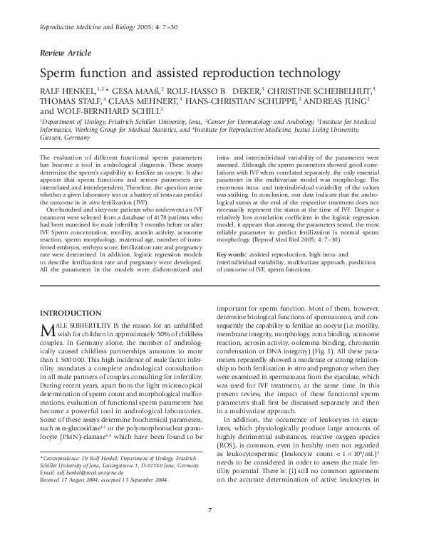
 Ralf Henkel