Neurotoxicologyand Teratology,Vol. 16, No. 1, pp. 11-15, 1994
Copyright©1994ElsevierScienceLtd
Printedin the USA.All rightsreserved
Pergamon
0892-0362/94 $6.00 + .00
Nerve Conduction Velocity Decreaseand
Synaptic Transmission Alterations in
Caffeine-Treated Rats
ANGEL RAYA, ANA MARIA CUERVO, FERNANDO MACIAN,
FRANCISCO JAVIER ROMERO AND JOAQUfN ROMA ~
Experimental Toxicology & Neurotoxicology Unit, Department o f Physiology, School o f Medicine
and Dentistry, University of Valencia, A vda. Blasco Ibd~ez 17, E-46010 Valencia, Spain
Received 19 D e c e m b e r 1992; Accepted 13 July 1993
RAYA, A, A. M. CUERVO, F. MACIAN, F. J. ROMERO AND J. ROMA. Nerveconduction velocitydecreaseand
synaptic transmission alterationsin caffeine-treatedrats. NEUROTOXICOL TERATOL 16(1) 11-15, 1994.--The action of
caffeine on peripheral neuromuscular function was studied by means of in vivo determinations of electrophysiological
parameters, i.e., amplitude of extracellularly recorded muscle action potentials and nerve conduction velocity in the dorsal
skeletal muscle and caudal nerve of the rat tail, respectively. Repeated exposure of the rats was carried out by adding caffeine
to the drinking water for 10 days. Here we report the novel finding that motor nerve conduction velocity showed a significant
decrease in caffeine-treated animals, whereas no change was observed in the amplitude of indirectly evoked extraceUular
muscle action potentials. The physiological recovery of the amplitude of the compound muscle action potential observed in
nonintoxicated rats after high-frequency stimulation (10 Hz) was not observed in intoxicated animals and is also discussed.
Caffeine
Neuromuscular toxicity
Peripheral neuromuscular function
Muscle action potential
Synaptic fatigue
Nerve conduction velocity
distribution in nervous tissue have been proposed (1,8,10,14),
that could explain the facilitating action of caffeine.
This report studied the effects of chronic caffeine administration on peripheral neuromuscular function. In the first part
of the study those effects were defined in terms of variation of
two electrophysiological parameters, motor nerve conduction
velocity (MNCV) and extracellular-recorded muscle action potential (MAP) amplitude, when MAPs were obtained by single
electrical stimuli. In the second part, the effects of caffeine
on MAP amplitude, when varying the stimulation frequency
were determined to test the previously reported frequencydependent facilitating action of caffeine (19).
CAFFEINE (1,3,7-trimethylxanthine) is a well known and extensively used drug with effects on motor activity. It has been
widely used as a pharmacological tool in the study of excitation-contraction coupling in muscle physiology. The early descriptions that this xanthine could potentiate muscular activity
(3) and induce contractures (4) gave rise to numerous studies
to elucidate the main action mechanism of this drug on muscular function.
A large number of reports studying caffeine's effects on
skeletal and smooth muscle contraction have been published.
However, the effects of caffeine on nerve conduction velocity,
neuromuscular transmission, and muscle action potentials are
less defined. It has been reported that caffeine facilitates neurotransmitter (NT) release in the synapse during nerve stimulation (5,6). This effect has been attributed to (a) modifications
in NT release probability (2); (b) changes in the size of the
NT available pool (6); or (c) alterations of NT mobilization on
presynapse, which has been reported to be frequency-dependent (19). Moreover, several models of intracellular calcium re-
METHOD
Animals
Forty male Wistar rats (weight and age controlled) were
used. At the beginning of the experiments they had a body
weight (Mean + SD) of 226.6 + 32.1 g and aged 83 + 8
Requests for reprints should be addressed to J. Rom~i,Experimental Toxicology & Neurotoxicology Unit, Department of Physiology, School
of Medicine and Dentistry, University of Valencia, Avda. Blasco Ib~flez 17, E-46010 Valencia, Spain.
11
�12
RAYA ET AL.
days. Twenty-five animals were divided in 3 groups, caffeineintoxicated (10 rats), pair-fed (10 rats), and control (5 rats)
and were used in the first part of the study. The other 15
animals were divided in three groups (5 in each as described
above) and were used in the second part of the study.
ad lib during 10 days. The mean dose of caffeine that each
animal of the treated group received was 597.4 + 28.6 mg
(n = 15), determined as the amount contained in the volume
consumed. Care was taken to use a daily fresh prepared caffeine solution. Electrophysiological recordings were carried
out in the three groups after the I0 days treatment.
Treatment of A nimais
Eiectrophysiologicai Studies
The animals were fed a standard diet (Letica, Hospitalet,
Spain). Caffeine (Merck, Darmstadt, Germany) was added to
the drinking water in gradually increasing amounts: 2 g/l the
first 3 days, 4 g/l the next 3 days, and finally 8 g/l the last 4
days, following a previously described procedure (7). Sucrose
50 g/1 was added to the caffeine solutions to mask the bitter
taste. The weight of the animals was monitored daily throughout the experiment. Moreover, water and food consumed by
the caffeine-treated group was quantitated once a day while
replenishing the reservoires of the cages. Pair-fed rats were
given those same amounts of chow and sucrose solution but
without caffeine. The control group was fed the standard diet
®
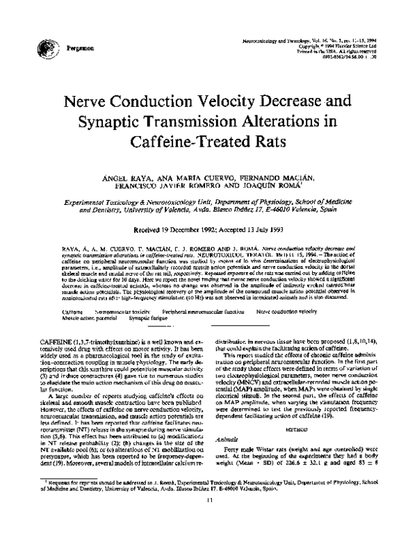
The method used allows in vivo determinations of electrophysiological parameters on the neuromuscular tall preparation on nonanesthetized rats (15). This method has shown to
be adequate for the study of the peripheral neuromuscular
toxicity (13,16). Rats were conveniently restrained by fixing them to a platform with elastic ribbons. The f'med tall
was placed in a thermostated paraffin bath, to maintain the
tall temperature constant at 270C (physiological temperature of the rat tall), and actual inner temperature of the tall
was monitored by a needle thermopar inserted along the tall
(Fig. IA). Foam rubber was placed between platform and
Proximal
stimulation
electrode
\
Recording
electrode
Base of the
rat's tail
/
i
i
i
i
i
l
Needle
thermopar
Ground
Distal
stimulation
electrode
~!~ Distance 2
60 mm
Distance 1
( ~
j
E
r-ms
MAP2
Latency 1
-
1
FIG. 1. (A) Diagram of experimental technique for obtaining extracellular-recorded muscle action potentials (MAPs), showing electrodes placement on the rat tail. Each stimulation electrode consisted of two
needles (+ = anode, - = cathode). (B) Typical plots of MAPs obtained by electric stimulation from proximal (MAP 1, lower trace) and distal electrode (MAP 2, middle trace). The intramuscular nerve action
potential (INAP) indicated by the arrow in the middle trace is amplified in the upper trace. Latencies were
defined as the intervals between time of stimulation and onset of MAPs. The point where the extrapolated
baseline intersects with a straight line drawn on the ascending trace through the 10%0 and 90% of the
maximal amplitude value, determines the onset of a MAP. Motor nerve conduction velocity was calculated
as (Distance l-Distance 2)/(Latency l-Latency 2).
�CAFFEINE-INDUCED NEUROMUSCULAR TOXICITY
animals where appropriate to avoid pain or discomfort to the
animals.
Evoked muscle action potentials were obtained using a
standard technique (12) as follows. Square cathodal stimuli
have been used throughout the experiments. The pulse length
was 0.1 ms and its height was adjusted to 30070 over the minimum voltage required to evoke maximal response (supramaximal stimuli) for each experiment. These stimuli did not produce any signs of discomfort and were delivered by two pairs
of electrodes (2 needles each) inserted in two positions, proximal and distal of the base of the tail and both at 5 mm depth.
The extracellular compound muscle action potentials obtained, proximal (MAPI) and distal (MAP2), were recorded
by one pair of recording electrodes inserted distally at 2 mm
depth in the dorsal tall musculature, displayed on a singlebeam oscilloscope screen and photographed or alternatively
digitized. Distance between recording and proximal stimulation electrodes was about 80 mm and around 20 mm from
recording to distal stimulation electrodes (see Fig. 1A). In the
first series of experiments the electrophysiological parameters
studied were the amplitude and the latency of appearance of
compound MAPs. MNCV was calculated by the indirect
method (latency differences; see Fig. IB). In the second series
of experiments, stimuli were delivered at 10 Hz frequency
from the distal electrode for 1 min. Afterward, single stimuli
were delivered, 1 every min during 15 min to study the recovery of evoked M A P amplitude.
13
Motor nerve conduction
velocity (m/s)
30
20
10
0
CONTROL
PAIR-FED
CAFFEINE
FIG. 2. Effect of chronic caffeine administration on motor nerve
conduction velocity (MNCV). Extracellular-recorded muscle action
potentials were obtained and MNCV was calculated as described in
Fig. 1. Results are expressed in m/s and are the Means + SD, F(2,
22) = 9.29, p < 0.01; *p < 0.01, significantly different from pairfed and control values.
Statistical Analysis
Results are expressed as Mean + SD. Kolmogorov:Smirnov test and analysis of variance (ANOVA) were used to evaluate the results. When A N O V A was significant, the significance between groups was assessed by means of unpaired
Student's t test; p values less than 0.01 were considered to be
significant.
RESULTS AND DISCUSSION
Our results show a decrease in body weight after treatment
(10 days) in both caffeine-treated (33.2 + 7.3%) and pair-fed
rats (18.1 + 8.3%). This decrease is statistically significant,
F(2, 37) = 67.82, p < 0.01, when compared to the control
group (17 < 0.01), which showed a 12.7 +_ 2.8% increase in
body weight. Therefore, the electrophysiological findings we
report cannot be attributed to weight loss.
The study of MNCV revealed a significant decrease of this
parameter in caffeine-treated rats (20.1 + 1.9 m/s) when
compared to both control (26.3 + 3.4 m/s) or pair-fed (26.3
+ 4.6 m/s) groups (see Fig. 2). There was no significant difference between MNCV values in control and pair-fed groups.
The ionic changes in nerve fiber caused by this drug described
in many in vitro studies could account for the MNCV decrease
found in our study. Calcium appears to be the main ion
involved in these changes. This ion may act on different
ionic channels regulating membrane excitability (9,17). Previous reports show that caffeine could increase calcium
concentration in nerve fibers (10), this fact and the ability
of calcium to activate potassium channels (18) could be responsible for the membrane hyperpolarization and consequently for the reduced excitability that might lead to MNCV
decrease.
No significant difference in the value of M A P amplitude
was found between caffeine-treated ( M A P I , 2.7 + 1.9 mV;
MAP2, 3.7 + 2.2 mV), pair-fed (MAP1, 3.2 + 1.2 mV;
MAP2, 4.1 + 2.1 mV) and control groups ( M A P I , 3.1 +
0.9 mY; MAP2, 4.5 +_ 1.8 mV) when MAPs were evoked by
single stimuli, F(2, 22) = 0.31, p > 0.5, for M A P 1 and, F(2,
22) = 0.19, p > 0.5 for MAP2. Although these results seem
to contradict previous in vitro findings showing that caffeine
facilitates NT release at the neuromuscular junction (2), it
must be taken into account that we study compound action potentials, while those in vitro studies record excitatory postsynaptic potentials (EPSPs). The increase in the
amplitude of EPSPs may become inappreciable when recording compound muscle action potentials, which are constantly
and invariably generated once EPSPs rise above a certain
threshold.
Our technique is useful to test synaptic fatigue by means
of high-frequency stimulation. The experiments carried out at
10 Hz frequency stimulation for 1 min in control animals,
show a decrease of MAPs amplitude probably due to a reduction in the number of muscle fibers depolarized. This is basically the result of the progresive exhaustion of the NT available pool in the neuromuscular synapse, i.e., synaptic fatigue.
After the high-frequency stimulation is finished, this NT pool
is restored, and M A P amplitude returns to the initial values
in control rats (see Fig. 3). Interestingly, while in the control
and pair-fed groups the subsequent physiological recovery of
MAPs amplitude occurs inmediately after high-frequency
stimulation is finished, caffeine-treated animals do not exhibit
such a recovery, at least within the first 15 min after highfrequency stimulation (see Fig. 3). Statistical analysis reveals significant differences of percentual MAPs amplitude
between caffeine-treated (38.0 + 13.9070) and control (69.5
+ 2.0°70) groups 1 min after high-frequency stimulation is
finished, and after 3 min when comparing caffeine-treated
(36.5 _+ 16.2070) and pair-fed (75.6 + 12.6070) animals. These
differences remain statistically significant until the end of the
experiment.
A possible explanation for this impairment of synaptic
transmission recovery after high-frequency stimulation could
�14
RAYA ET AL.
Amplitude of muscle
action potential (%)
120
,I/
100
8O
60
20
0
0
10
20
30
40
50
60
Time (s)
4
8
12
16
Time (min)
10 Hz S T I M U L A T I O N
D U R I N G 60 SECONDS
SINGLE STIMULI
EVERY MINUTE
FIG. 3. Caffeine-induced suppression of the recovery of muscle action potential (MAP) amplitude after highfrequency stimulation. Stimulation at 10 Hz frequency for 60 s was followed by single stimuli every minute for 15
rain. Note that MAP amplitude values in caffeine-treated animals (O) after high-frequency stimulation tend to
block whereas both control (O) and pair-fed (D) ones return to initial values. Results are expressed as percentage
of initial values and are the Mean 4= SD, F(2, 162) = 271.38, p < 0.01. p < 0.01, significantly different from
control values. *p < 0.01, significantly different from control and pair- fed values.
also be ascribed to calcium m o v e m e n t s . C a f f e i n e increases
stimulus-mediated calcium release f r o m the s m o o t h endoplasmic reticulum o f presynaptic nerve endings (11). M o r e o v e r ,
previous reports have p r o p o s e d t h a t caffeine m a y reduce the
stimulus-evoked calcium influx f r o m the extracellular comp a r t m e n t in a n e u r o m u s c u l a r p r e p a r a t i o n (14). T h e latter aut h o r hypothesized t h a t the large increase in cytosolic calcium
caused b y the caffeine-mediated release f r o m the s m o o t h end o p l a s m i c reticulum w o u l d be controlled b y m i t o c h o n d r i a l
sequestration (14). If such a decrease in the intracellular cytosolic calcium p o o l would occur, the s u b s e q u e n t effect o n N T
release could explain the fact t h a t caffeine-treated a n i m a l s are
u n a b l e to recover its M A P a m p l i t u d e a n d could also explain
its t r e n d to n e u r o m u s c u l a r blockade after a high-frequency
stimulation. W h e t h e r acetylcholine release is affected or n o t
in this experimental m o d e l is currently being investigated in
our laboratory.
ACKNOWLEDGEMENTS
We are thankful for the advice and suggestions of G. T. Sdez and
the late Dr. A. Jordd. We also thank B. Sofia for his helpful scientific
criticism and to C. Avellaneda for his expert technical assistance. This
work was partially supported by Grant No. 92/0403 from the FISS
and PM92/0146 from the DGICYT (Spain). AR is a research fellow
of the Conselleria de Cultura, Educaci6 i Ci~ncia de la Generalitat
Valenciana. A.M.C. and F.M. are research fellows of the Ministerio
de Educaci6n y Ciencia.
REFERENCES
1. Christesen, B. N.; Martin, A. R. The end-plate potential in mammalian muscle. J. Physiol. 210:933-945; 1970.
2. Elmqvist, D.; Feldman, D. S. Calcium dependence of spontaneous acetylcholine release at mammalian motor nerve terminals. J.
Physiol. 181:487-497; 1965.
3. Foltz, E.; Ivy, A. C.; Barborka, C. J. The use of double work
periods in the study of fatigue an the influence of caffeine on
recovery. Am. J. Physiol. 136:79-86; 1942.
4. Goffart, M.; Ritchie, J. M. The effect of adrenaline on the contraction of mammalian skeletal muscle. J. Physiol. 116:357-371;
1952.
5. Golberg, A. L.; Singer, J. J. Evidence for a role of cyclic AMP
in neuromuscular transmission. Proc. Natl. Acad. Sci. USA 64:
134-140; 1969.
6. Hoffmann, W. W. Caffeine effects on transmitter depletion and
mobilization at motor nerve terminals. Am. J. Physiol. 50:21092128; 1969.
7. Jordd, A.; Portol~s, M.; Guasch, R.; Bernal, D.; Sdez, G. T.
Effect of caffeine on urea biosynthesis and some related processes, ketone bodies, ATP and liver amino acids. Biochem.
Pharmacol. 38:2727-2732; 1989.
8. Lee, W.; Anwyl, R.; Rowan, M. Caffeine inhibits post-tetanie
potentiation but does not alter long-term potentiation in the rat
hippocampal slice. Brain Res. 426:250-256; 1987.
�CAFFEINE-INDUCED
NEUROMUSCULAR
TOXICITY
9. Meech, R. W. Calcium dependent potassium in nervous tissues.
Ann. Rev. Biophys. Bioeng. 7:1-18; 1978.
10. Neering, I. R.; McBurney, R. R. Role for microsomal Ca 2+ storage in mammalian neurones? Nature 309:158-160; 1984.
11. Ohta, Y.; Kuba, K. Inhibitory action of Ca 2+ on spontaneous
transmitter release at motor nerve terminals in a high K solution.
Pflfigers Arch. 386:29-34; 1980.
12. Petajan, J. H. Changes in rat ventral caudal nerve conduction
velocity during cold exposure. Am. J. Physiol. 214:130-132;
1968.
13. Raya, A.; Gallego, J.; Hermenegildo, C.; Puertas, F. J.; Romero, F. J.; Felipo, V.; M~ana, M. D.; Grisolia, S.; Rom~, J.
Prevention of the acute neurotoxic effects of phenytoin on rat
peripheral nerve by H7, an inhibitor of protein kinase C. Toxicology 75:249-256; 1992.
14. R0ed, S. Caffeine-induced blockade of neuromuscular transmis-
15
15.
16.
17.
18.
19.
sion and its reversal by dantrolene sodium. J. Pharmacol. 83:8390; 1982.
Rormi, J.; Soria, B. Isonicotinic acid hydrazide: Early effects on
peripheral nerve conduction velocity. Experientia 40:378-380; 1984.
Rom~t, J.; Cuervo, A. M.; Maci~n, F.; Raya, A.; GaUego, J.;
Llopis, J. E.; Romero, F. J. Temperature dependence of the toxic
effects of phenytoin on peripheral neuromuscular function of the
rat tall. Neurotoxicol. Teratol. 12:627-631; 1990.
Schwarz, W.; Passow, H. Calcium activated potassium channels
in erythrocytes and excitable cells. Ann. Rev. Physiol. 45:359364; 1983.
Smith, S. J.; McDermott, A. B.; Weight, F. F. Detection of
intracellular transits in sympathetic neurones using arsenazo Ill.
Nature 304:350-352; 1983.
Wilson, D. F. Effects of caffeine on neuromuscular transmission
in the rat. Am. J. Physiol. 225:862-865; 1973.
�
 J. Roma